Article

Aqueous Extracts of Fermented Macrofungi Cultivated in Oilseed Cakes as a Carbon Source for Probiotic Bacteria and Potential Antibacterial Activity

by
Joice Raísa Barbosa Cunha
1,2,
Daiana Wischral
1,
Rubén Darío Romero Peláez
1,3,
Pérola De Oliveira Magalhães
4,
Marina Borges Guimarães
1,4,
Maria Aparecida de Jesus
5,
Ceci Sales-Campos
5,
Thais Demarchi Mendes
1,
Eustáquio Souza Dias
2,
Simone Mendonça
1 and
Félix Gonçalves de Siqueira
1,*
1
Embrapa Agroenergia, Distrito Federal, Brasília 70770-901, Brazil
2
Programa de Pós-Graduação em Microbiologia Agrícola, Universidade Federal de Lavras (UFLA), Lavras 37200-000, Brazil
3
Instituto de Ciências Biológicas, Universidade Federal de Brasília, Brasília 70910-900, Brazil
4
Departamento de Farmácia, Faculdade de Ciências da Saúde, Universidade Federal de Brasília, Brasília 70910-900, Brazil
5
Instituto Nacional de Pesquisas da Amazônia (INPA), Manaus 69067-375, Brazil
*
Author to whom correspondence should be addressed.
Metabolites 2023, 13(7), 854; https://doi.org/10.3390/metabo13070854
Submission received: 16 May 2023 / Revised: 10 July 2023 / Accepted: 12 July 2023 / Published: 18 July 2023
(This article belongs to the Section Plant Metabolism)

Abstract

:
Plant biomass colonized by macrofungi can contain molecules with bioactive properties with applications to human/animal health. This work aimed to verify antibacterial activities from aqueous extracts from oil seed cakes of Jatropha curcas (JSC) and cottonseed (CSC), fermented by macrofungi for probiotic bacteria cultivation. Coriolopsis sp., Tyromyces sp., Panus lecomtei, and Pleurotus pulmonarius were cultivated in solid and submerged media. The aqueous extract of unfermented JSC was more efficient than glucose for the growth of all probiotic bacteria. Extracts from four macrofungi fermented in CSC favored Lactobacillus acidophilus growth. In solid fermentation, macrofungi extracts cultivated in JSC favored Bifidobacterium lactis growth. All fungi extracts showed more significant growth than carbohydrates among the four probiotic bacteria evaluated. Regarding antimicrobial activities, no fungal extract or bacterial supernatant showed a more significant inhibition halo for enteropathogenic bacteria than ampicillin (control). Extracts from P. lecomtei and Coriolopsis sp. in CSC showed inhibition halos for Salmonella enterica. Supernatants from L. acidophilus, B. lactis, and Lactobacillus rhamnosus resulted in more significant inhibition of Staphylococcus aureus than the control, which indicates possible antimicrobial activity. Unfermented JSC supernatant showed better results for bacterial growth, while supernatants and aqueous extracts from CSC fermentation can be used for probiotic bacteria culture.

1. Introduction

Mushrooms are known to be rich in antioxidant compounds with anti-inflammatory and anticancer activities [1] and complex carbohydrates that behave like intestinal fibers and can help in the growth of probiotic microorganisms and the production of short-chain fatty acids that favor these microorganisms over enteropathogens [2].
The most commonly bioactive compounds found in mushrooms include polysaccharides [3,4], proteins [5], terpenes [6,7], unsaturated fatty acids [8], and phenolic compounds [9,10]. In mushrooms, these compounds provide antioxidant, antimicrobial, antitumor, anti-inflammatory, antidiabetic, and hypocholesterolemic activities, among other important biological activities [10,11].
More than 400 species of bacteria are in the intestine, forming a complex and essential microbiota mainly composed of the genera Bifidobacteria and Lactobacillus [12], Gram-positive bacteria that ferment carbohydrates into acetic and lactic acids, respectively [13]. Some intestinal bacteria are homofermentative because they ferment glucose only into lactic acid, and others are heterofermentative because they ferment hexoses and pentoses via phosphoketolase, obtaining acetic acid, CO2, and ethanol [14]. Probiotics can improve feed conversion to nutrients, regulate the immune response, inhibit pathogen growth, stimulate beneficial microbiota, and form biofilms to block infections [15].
These Bifidobacteria and Lactobacillus are known as probiotics, as they suppress the growth of enteropathogenic bacteria, improve food absorption and gastrointestinal tract function, and are also capable of modulating the immune system [16]. Prebiotics are non-digestible food components that selectively stimulate the growth of these probiotic bacteria that, in addition to modulating the intestinal microbiota, attenuate health conditions such as diabetes, obesity, and cancer [17].
In addition to probiotic bacteria, other bacteria may be present in the gastrointestinal tract, and some may be pathogenic, causing harm to hosts. Bacteria such as Salmonella sp., Campylobacter, Escherichia coli, Listeria monocytogenes, Clostridium botulinum, Microsporidia, and Campylobacter jejuni are frequently associated with intestinal infections [18,19]. However, the inappropriate use of antibiotics over the years has led to resistant bacteria, which has been an increasing threat to health. Researchers from all around the world are now focused on finding alternative antimicrobials, and the chemical composition of some mushrooms suggests that they may have antiviral and antibacterial potential [20,21].
The present work aimed to evaluate the prebiotic (carbon sources of probiotic bacteria) and antibacterial potential of supernatants from submerged fermentation (SmF) and aqueous extracts from solid-state fermentation (SSF) of residues from cakes of seed oil cotton and Jatropha by Coriolopsis sp. INPA1646, Tyromyces sp. INPA1696, P. lecomtei BRM 044603, and P. pulmonarius BRM 055674.

2. Materials and Methods

2.1. Obtaining Supernatants and Extracts

Coriolopsis sp. INPA1646, Tyromyces sp. INPA1696, P. lecomtei BRM 044603, and P. pulmonarius BRM 055674 belong to the INPA Macrofungi Collection (Manaus/AM) and the Microorganisms and Microalgae Collection applied to the biorefinery of Embrapa Agroenergia—CMMABio (Braslia/DF). The strains were reactivated on plates with PDA (Potato Dextrose Agar) and incubated at 28 °C for 15 days.
Submerged fermentation (SmF) was performed in flasks with 5.0 g of each dry biomass and 100 mL of distilled water, and solid-state fermentation in flasks with 40 g of JSC or 20 g of dry CSC, with moisture adjusted to approximately 70%. After sterilization, the liquid medium flasks were inoculated with 10 mycelial plugs (8 mm in diameter) of each fungus and incubated at 28 °C and 180 rpm for 14 days. The solid culture flasks with 10 plugs in the JSC flasks and 5 in the CSC flasks were incubated at 28 °C for 14 days. Controls consisted of sterilized biomass under the same conditions without fungal inoculation.
After incubation, submerged cultures were vacuum filtered to obtain supernatants, and the solid-state fermented cultures were dried at 60 °C, crushed, and subjected to aqueous extraction under pressure and temperature in an ASE—Accelerated Solvent Extractor (Dionex ASE 350). Supernatants and aqueous extracts were frozen and lyophilized.

2.2. Determination of Prebiotic Activity

The potential prebiotic activity (carbon sources of probiotic bacteria) was evaluated by the ability of supernatants and aqueous extracts to stimulate the growth of Lactobacillus acidophilus, Lactobacillus plantarum, Lactobacillus rhamnosus, and Bifidobacterium lactis strains in MRS (−C) medium (10.0 g/L of peptone + 10.0 g/L of meat extract + 5.0 g/L of yeast extract + 1.0 g/L of polysorbate 80 + 2.0 g/L of ammonium citrate + 5.0 g/L of sodium acetate + 0.1 g/L of magnesium sulfate + 0.05 g/L of manganese sulfate + 2.0 g/L of monopotassium phosphate) for 48 h at 37 °C. The strains, provided by the Central Public Health Laboratory of the Federal District (Lacen-UnB), were reactivated in MRS Broth, and diluted in 0.9% saline solution to obtain a 106 CFU/mL concentration according to the McFarland standard.
The supernatants and aqueous extracts were diluted in sterile distilled water at a concentration of 40 mg/mL, filtered through a 0.22 µm membrane, and added to MRS Broth without glucose and twice the concentration of the other components (2×MRS-C) and 0.4% L-cysteine in a 1:1 (v:v) ratio. Three positive controls were performed: glucose (Sigma-Aldrich, Merck Millipore, Germany), fructooligosaccharides (FOS) at 90% purity (Newnutrition, Ribeirão Preto, SP, Brazil), and inulin from chicory (FiberBem, The Netherlands) at 20 mg/mL. The negative control consisted of sterile distilled water.
Bacterial growth was monitored in 2 mL of medium with 5 µL of inoculum and then transferred to two 96-well microplates (Elisa plates), 200 µL per well, in triplicate, and sterile controls consisting of wells with MRS medium without inoculum. The plates were sealed with highly translucent 2.0 polyethylene Platemax sealing membranes for PCR transport and storage (Axygen® Corning Inc., Corning, NY, USA) and incubated at 37 °C and 180 rpm for 48 h, with absorbption readings at 600 nm (OD 600) every hour for the first 12 h and then every 2 h for 48 h in total.
Viable cells were quantified in 100 µL of each treatment and collected at 12, 24, and 48 h of incubation at 37 °C for serial dilution and plating by spreading on plates of complete MRS medium with 0.2% L-cysteine, and colonies were counted after 48 h of incubation. The growth of total cells was represented by the change in OD and the growth of viable cells in Petri dishes.
Elisa plates, with the remaining 800 µL of the medium, were also incubated in a shaker at 180 rpm and 37 °C for 48 h to obtain the supernatants by centrifugation at 10,000 rpm for 15 min, which were also tested in the antimicrobial activity assays.

2.3. Determination of Antimicrobial Activity

Escherichia coli (ATCC 25922), Salmonella enterica subsp. Enterica serovar Typhimurium (ATCC 14028), and Staphylococcus aureus (ATCC 27154, enterotoxin-producing strain) from the Natural Products Laboratory of the University of Brasilia (UnB) were reactivated in Mueller–Hinton Agar at 37 °C for 48 h. Bacteria were grown for 24 h at 37 °C and 180 rpm in Mueller–Hinton broth and inoculated by spreading on Mueller–Hinton Agar plates for an antibiogram. Supernatants and dry aqueous extracts were diluted in sterile distilled water at a concentration of 100 mg/mL and filtered through a 0.22 µm membrane. Autoclaved paper discs were prepared with 20 µL of the samples, their dilutions (1:10, 1:100, and 1:1000), and the supernatants from the above-mentioned prebiotic bacteria growth. After room temperature drying, the discs were placed on the plates with the microorganisms and incubated at 35 °C for 24 h to assess the formation of inhibition halos. Ampicillin discs were used as a positive control (10 μg) [22].

2.4. Statistical Analysis

Experiments were performed in triplicate, and the results were submitted to the Shapiro–Wilk normality test and Tukey’s test with a significance level of 5% (p < 0.05) with SISVAR 5.6 software [23].

3. Results

3.1. Prebiotic Activity

3.1.1. Lactobacillus acidophilus

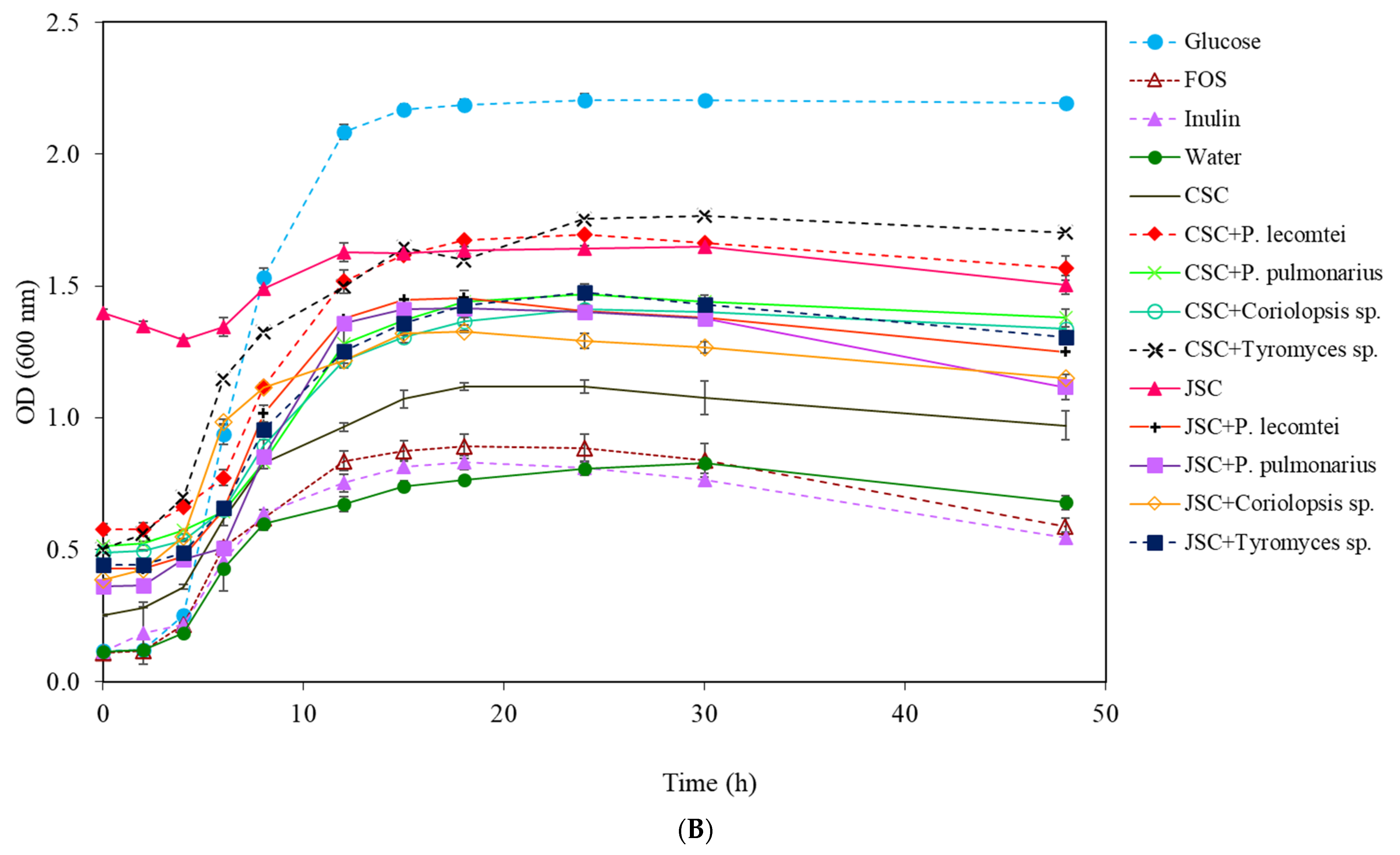
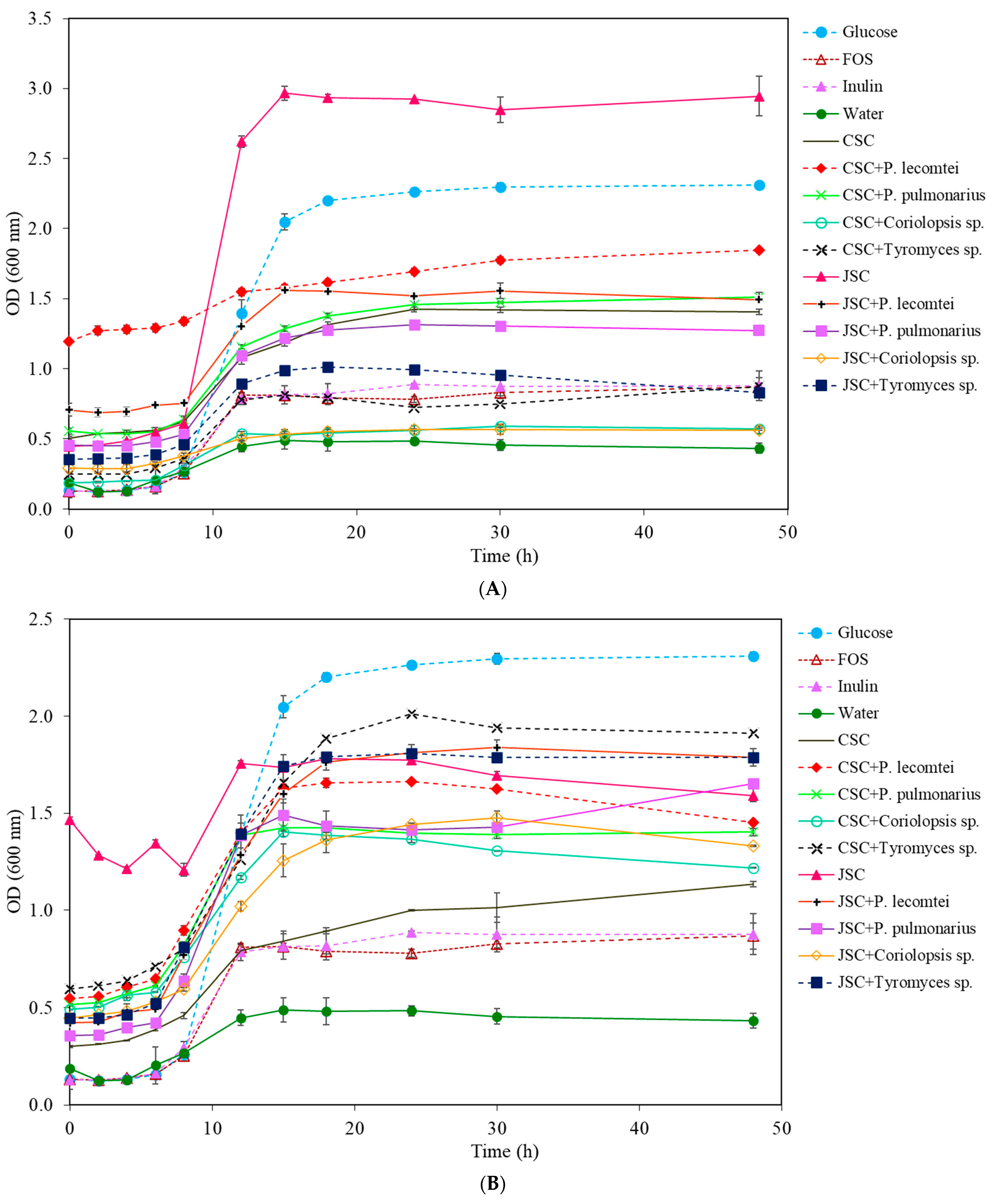
The highest increase of total cells of L. acidophilus was observed in MRS medium (−C) with 20 mg/mL of supernatant SmF-JSC incubated for 14 days at 28 °C without fungus (Figure 1A), a variation of 2.139 ± 0.022 A (Table 1), followed by medium containing glucose (2.081 ± 0.017 A). The positive controls, FOS, and inulin, provided similar results to the negative control (water), just as Rodrigues et al. (2016) observed in Lactobacillus sp. [24]. In the treatments of JSC fermented by fungi, the highest growth of L. acidophilus was obtained in the medium of P. pulmonarius BRM 055674 supernatant with 1.022 ± 0.015 A after 48 h of incubation, followed by the supernatants of Tyromyces sp. INPA1696 (0.960 ± 0.058 A), P. lecomtei BRM 044603 (0.849 ± 0.053 A), and Coriolopsis sp. INPA1646 (0.774 ± 0.028 A).
After 14 days of cultivation, supernatants of SSF fermentation by P. lecomtei BRM 044603, Tyromyces sp. INPA1696, and P. pulmonarius BRM 055674 resulted in more significant growth of L. acidophilus than unfermented SmF supernatant, probably due to the release of fermentable sugars (Figure 1B). Likewise, the SSF supernatant fermented by Coriolopsis sp. INPA1646 showed more significant total cell growth (0.922 ± 0.009 A) than the unfermented CSC supernatant (0.630 ± 0.026 A) and controls; however, its growth was lower when compared to the glucose treatment.
The variation in OD of total L. acidophilus cell growth at the end of 48 h of incubation allows us to conclude that the best culture medium was MRS (−C) with 20 mg/mL of JSC supernatant incubated for 14 days at 28 °C without fungal fermentation, having better results than the conventional MRS medium (with 20 mg/mL of glucose).
The growth results of L. acidophilus in the aqueous extracts from the solid-state fermentation of CSC were overall lower than those observed using the supernatants from the submerged fermentation, as seen in Table 1, which shows the variations in optical densities before and after 48 h of incubation. In this group, the highest growth of L. acidophilus was observed in the aqueous extract of SSF fermented by Tyromyces sp. INPA1696, followed by the CSC fermented by P. lecomtei BRM 044603. The growth of L. acidophilus in the aqueous extract of JSC without fungus was the lowest observed among all samples tested. The opposite was observed with the JSC supernatant; this suggests that the treatment and extraction, with agitation and 28 °C after 14 days, are more efficient than the aqueous extraction, under pressure and temperature in ASE, to obtain L. acidophilus culture medium, probably due to the release of fermentable sugars.
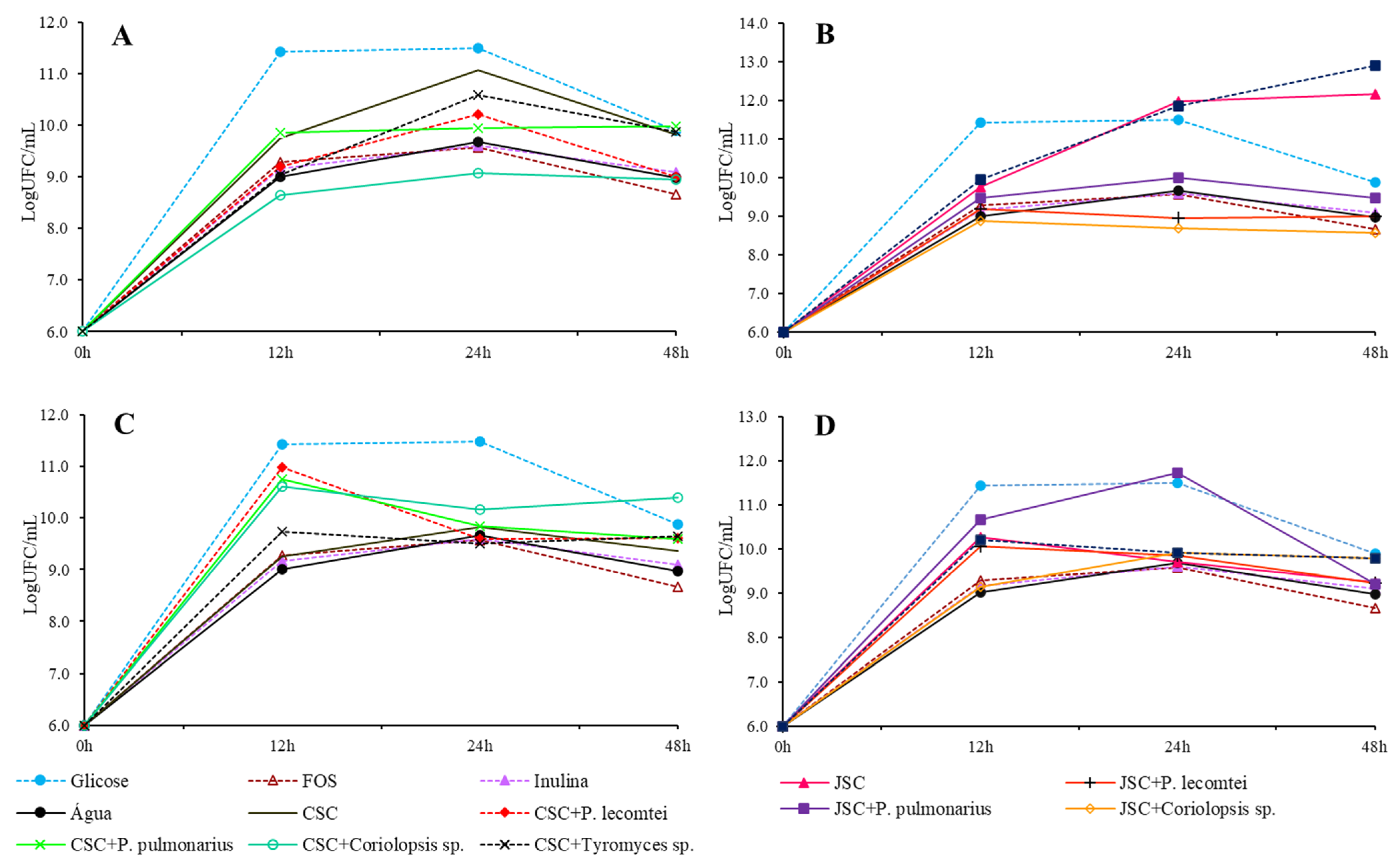
The peak of total cells in the medium does not always occur in the 48th hour of incubation. However, maintaining cell viability for long periods of time is crucial to consider as a prebiotic activity. The highest concentration of viable L. acidophilus cells is observed at the 12th hour of incubation in the medium containing the unfermented JSC supernatant (Figure 2B). Among the submerged CSC fermentation supernatants (Figure 2A), the highest concentration of viable cells was observed in the fermented medium with Tyromyces spp. INPA1696 after 24 h of incubation, P. pulmonarius BRM 055674, and Coriolopsis sp. INPA1646 after 48 h of incubation. For the solid CSC fermentation aqueous extracts (Figure 2C), the highest concentration occurs with the extract of P. lecomtei BRM 044603 at 24 h of incubation. In JSC treatments (Figure 2B,D), none surpasses the concentration in the medium with unfermented JSC supernatant after 12 h of incubation. However, the extract of solid fermentation by P. lecomtei BRM 044603 equals this concentration in 12 h of cultivation.

3.1.2. Bifidobacterium lactis

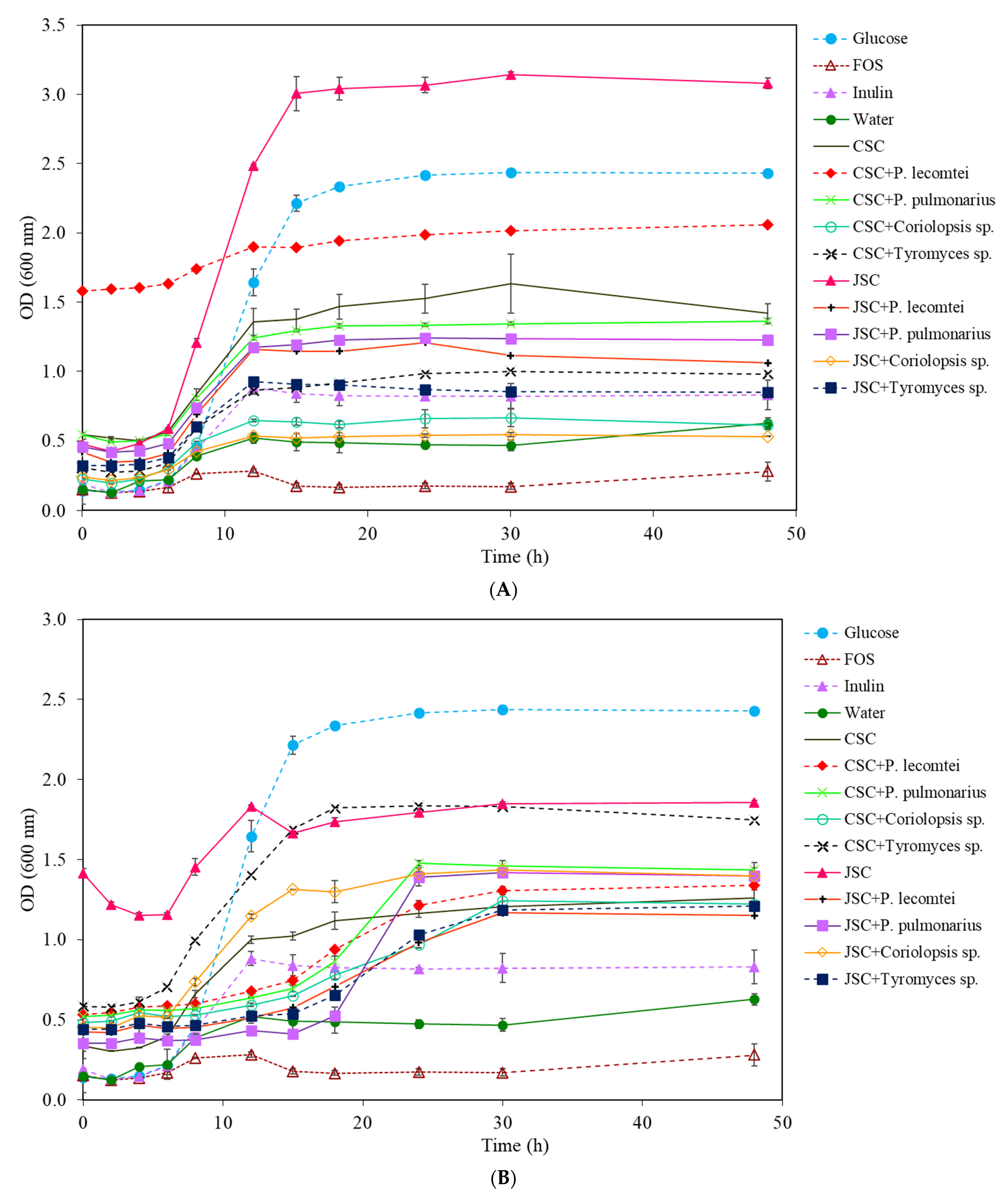
In B. lactis cultures, the commercial prebiotics FOS and inulin did not show satisfactory growth. The peak of growth in the medium with glucose was observed in the 15th hour of incubation (Figure 3A). The most significant growth of whole-cell B. lactis was also observed in the supernatant medium with JSC without fungal inoculation, with an increase of 2.501 ± 0.140 A after 48 h of incubation, followed by the glucose treatment with 2.122 ± 0.086 A.
In cultures with supernatants obtained from CSC, the most significant cell growth after 48 h of incubation was observed in the treatment fermented by P. pulmonarius BRM 055674, with an increase in OD of 0.949 ± 0.059 A, followed by the CSC supernatant without fungus, with an OD of 0.907 ± 0.012 A (Table 2). The supernatant from CSC fermentation by P. lecomtei (BRM 044603) showed an OD variation of 0.654 ± 0.003 A. However, the dark color of the assay may interfere with the absorbance reading and make it difficult to quantify the increase in total cells. The supernatant from the fermentation by Tyromyces sp. INPA1696 behaved similarly to the positive controls FOS and inulin in the B. lactis growth; the treatment with Coriolopsis sp. INPA1646 was similar to the negative control (water), with an OD of 0.379 ± 0.007 A (Figure 3B).
On the other hand, in cultures with aqueous extracts obtained from CSC, the treatment with Tyromyces sp. INPA1696 showed an OD variation of 1.318 ± 0.087 A after 48 h of incubation (Table 2). The treatment with CSC extract without fungus was similar to that observed in the media with CSC extracts fermented by P. pulmonarius BRM 055674 and P. lecomtei BRM 044603, while the absorbance in the medium with CSC extract fermented by Coriolopsis sp. INPA1646 was only 0.728 ± 0.005 A.
In cultures with supernatants obtained with JSC, treatments fermented by P. lecomtei BRM 044603 and P. pulmonarius BRM 055674 showed similar results in stimulating the growth of B. lactis and were higher when compared to the positive controls FOS and inulin. In cultivations with aqueous extracts, the treatments of JSC fermented by P. lecomtei BRM 044603, P. pulmonarius BRM 055674, and Tyromyces sp. INPA1696 showed greater growth stimulation of B. lactis (Table 2).
The highest concentrations of viable cells occurred after 24 h of incubation in the medium with glucose (conventional MRS), followed by the medium containing the unfermented JSC supernatant. However, if shorter incubation times were considered (e.g., 12 h), the media with the supernatants and biomass extracts had higher concentrations of viable cells (Figure 4).

3.1.3. Lactobacillus rhamnosus

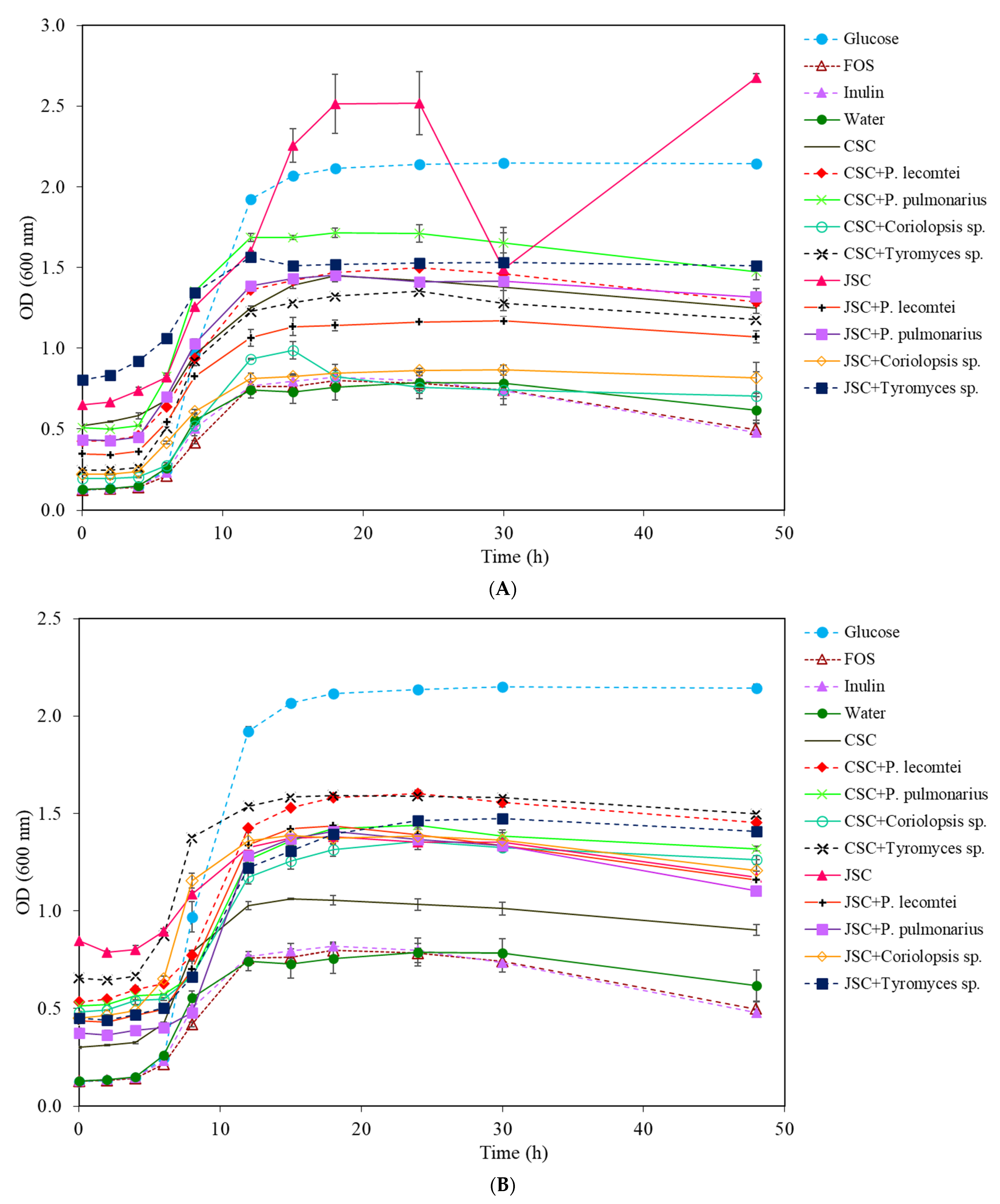
The growth of L. rhamnosus was strongly stimulated using JSC supernatant without fungus, reaching the exponential phase in a shorter time (6–12 h) (Figure 5A), with an increase in OD of 2.570 ± 0.057 A at the end of 48 h of incubation, higher than the control with glucose (OD 2.279 ± 0.025 A). Among the JSC supernatants tested, treatment with Coriolopsis sp. INPA1646 showed lower growth, with an increase in OD of 0.302 ± 0.032 A at the end of 48 h of incubation.
The results of this bacterium growth in the aqueous extracts obtained from solid-state fermentation with macrofungi did not have the same pattern as those observed in the supernatants (Figure 5B), with the lowest growth in the JSC extract without fungus (0.397 ± 0.091 A) and more significant growth in the extract of CSC fermented by Tyromices sp. INPA1696 (1.199 ± 0.091 A), followed by JSC extract fermented by P. pulmonarius BRM 055674 (1.042 ± 0.049 A).
In the cultures with CSC supernatants, growth of L. rhamnosus showed more significant variation in the treatment without fungus, with an OD of 0.881 ± 0.059 A. Among the fermented treatments, the highest growth value was observed in the treatment with P. pulmonarius BRM 055674 (0.825 ± 0.002 A), followed by P. lecomtei BRM 044603 (0.481 ± 0.003 A) and Coriolopsis sp. INPA1646 (0.404 ± 0.081 A) (Table 3). In aqueous extracts of CSC, the treatments fermented by Tyromyces sp. INPA1696 and P. pulmonarius BRM 055674 showed more significant growth of L. rhamnosus after 48 h than the aqueous extract of CSC without fungus. The highest concentration and viable cells were found after 24 h of incubation in the medium with 20 mg/mL of unfermented JSC supernatant and in the medium with glucose (conventional MRS) with 24 h of incubation as well (Figure 6).

3.1.4. Lactobacillus plantarum

The growth of the bacteria L. plantarum had a significant stimulus, indicated by the variation of OD, using MRS media with glucose (2.014 ± 0.018 A) and the supernatant JSC without fungus (2.026 ± 0.025 A), indicating that this medium has potential for prebiotic activity (Figure 7A). When the growth rates were compared in extracts from solid-state fermentation, the medium with glucose also stood out, with the most significant increase in OD, followed by JSC fermented by Tyromices sp. INPA1696 and CSC fermented by P. lecomtei BRM 044603, with OD variations of 0.954 ± 0.001 A and 0.922 ± 0.026 A, respectively (Figure 7B). All other fermented solids in both biomasses resulted in similar total cell growth of L. plantarum after 48 h of incubation.
Among the treatments with fermented cakes, the highest growth was observed in the supernatants obtained from submerged fermentation of JSC by P. pulmonarius BRM 055674, while the lower results of total cell growth were observed in the supernatants obtained from Coriolopsis sp. INPA1646 fermentations (Table 4). The highest concentrations of viable cells were observed after 48 h of incubation in MRS media with 20 mg/mL of supernatants from submerged fermentation of JSC by Tyromices sp. INPA1696 and P. pulmonarius BRM 055674. It is important to note that after 48 h of incubation in these media, the concentration of viable cells seems to continue to rise, maintaining cell viability longer (Figure 8).

3.2. Antimicrobial Activity

Table 5 shows all the results of the antibiogram performed with supernatants (SmF) and aqueous extracts (SSF) lyophilized and diluted to concentrations of 100, 10, and 1 mg/mL. Strains were considered sensitive to aqueous extracts or supernatants when they presented inhibition halos greater than 10 mm and resistant when there was no halo formation or when it was smaller than 10 mm, as suggested by some authors [25,26,27]. Only the aqueous extracts from solid-state fermentation of CSC by P. lecomtei BRM 044603 and Coriolopsis sp. INPA1646 showed a halo of inhibition against S. enterica at a 100 mg/mL concentration. The optimization of the cultivation time to evaluate the kinetics of supernatants on Salmonella should be performed in a further study.
In addition to the aqueous extracts and supernatants from the CSC and JSC fermentations, the probiotic bacteria growth supernatants were also tested in an antibiogram assay for inhibition of enteropathogenic strains of E. coli ATCC25922, S. aureus ATCC27154, and S. enterica ATCC14028. During their growth, in addition to lactic acid and acetic acid, probiotic bacteria produce short-chain fatty acids and other extracellular metabolites that can inhibit the growth of some enterobacteria [28].
The S. aureus strain is more sensitive than the other two bacteria tested, as it showed inhibition halos for several L. acidophilus supernatants in the media with aqueous extracts of CSC fermented by Tyromices sp. INPA1696 and non-fermented JSC (Table 6). These results suggest that L. acidophilus releases some metabolic and antimicrobial activity in these strains of enteropathogens during its growth.
The growth supernatants of B. lactis showed less inhibition of the enteropathogens than those of L. acidophilus. Table 6 shows the inhibition of supernatants of B. lactis, where it can be seen that only the supernatant of the growth of the probiotic with the supernatants (SmF) of JSC fermented by P. lecomtei BRM 044603 and Coriolopsis sp. INPA1646, CSC extracts without fungus, CSC fermented by Tyromices sp. INPA1696, JSC without fungus, and JSC fermented by Coriolopsis sp. INPA1646 can inhibit S. aureus, and only CSC extract fermented by Tyromices sp. INPA1696 and JSC without fungus were able to inhibit S. enterica.
The growth supernatants of L. rhamnosus have even less antibacterial activity on the strains tested, inhibiting only S. aureus and only when grown in the MRS with the CSC submerged fermentation supernatant (SmF) by Coriolopsis sp. INPA1646, and in the JSC supernatant without fungus. In L. plantarum supernatants, inhibition halos were not observed in any of the tested bacteria; because of that, the results of these supernatants do not appear in Table 6. Additionally, if any of the tested supernatants presented inhibition halos for E. coli, then just the results of S. aureus and S. enterica were shown in Table 6.

4. Discussion

Coriolopsis sp., Tyromyces sp., P. lecomtei, and P. pulmonarius were selected for this work because they showed the degradation activity of toxic compounds of plants such as gossypol and phorbol esters when grown by SSF in JSC (Jatropha seed cake) and CSC (cottonseed cake).
In general, supernatants from submerged fermentations are better for the growth of L. acidophilus than the aqueous extracts tested, and the effect of fermentation with macrofungi P. lecomtei BRM 044603 and Tyromyces sp. INPA1696 increases the efficiency of extracts in stimulating the growth of L. acidophilus. In B. lactis cultures, our work corroborates Candela et al. (2010), when the commercial prebiotics FOS and inulin showed unsatisfactory growth [29]. However, Rodrigues et al. (2016) reported the growth of Bifidobacterium sp. higher in FOS than the negative control, with more significant growth after 24 h of incubation [23].
In fermented JSC, macrofungi’s growth reduces the efficiency of its supernatants for the growth of this bacteria by up to 70% under the evaluated cultivation conditions. Longer fermentation times can generate more fungal mass structures, such as chitin, beta-glucans, and mycosterols, among other chemical compounds, that can positively influence probiotic growth, such as Ganoderma lucidum polysaccharides, which increase the abundance of Bifidobacteria, Lactobacillus, Roseburia, Lachnospiraceae, and Pleurotus eryngii, increasing the proliferation of bacteria from the families Porphyromonadaceae, Rikenellaceae, Bacteroidaceae, and Lactobacillaceae [30]. Nonetheless, the 14th day fermentation supernatants showed less stimulus to L. plantarum, probably because the fungi used the readily assimilated carbohydrates in the biomass for their growth.
Other studies also pointed out the prebiotic potential of Pleurotus extracts. Synytsya et al. (2008) evaluated aqueous and alkaline extracts from P. ostreatus and P. eryngii for prebiotic activity by stimulating four Lactobacillus ssp., three Bifidobacterium ssp., and two Enterococcus faecium strains [31]. They observed that the aqueous extracts of both fungi stimulated Lactobacillus ssp., mainly P. eryngii. Bifidobacterium ssp. behaved differently, with one strain growing more with P. ostreatus extracts, another growing only with P. eryngii extracts, and a third one not stimulated by any extract. Enterococcus faecium strains grew less than all other strains in any of the extracts and showed better growth in alkaline extracts [31].
Ethanolic extracts of P. ostreatus (MTCC142) cultivated in solid and submerged states in banana crop residues showed antimicrobial activity against Gram-positive and Gram-negative bacteria and antifungal activity in vitro. Extracts from solid-state fermentation showed higher antibacterial activity against Bacillus megaterium, Enterobacter, and P. aeruginosa than those from submerged fermentation, more significant activity against Micrococcus luteus than standard antibiotics, and activity against Enterobacter and P. aeruginosa similar to that of chloramphenicol [32].
Some studies indicate that the antimicrobial activity of mushrooms occurs mainly against Gram-positive bacteria [18,33,34], which do not present the external lipid bilayer as in the Gram-negative ones, which may indicate that compounds with antimicrobial activity are water-soluble ions and are unable to cross a lipid bilayer.
The metabolites produced by mushrooms and other fungi provide their extracts’ prebiotic and antimicrobial activities, as was observed in Clericuzio et al. (2021) [35]. In this work, metabolites were obtained by extraction using 90% acetone and 10% 2-propanol (v/v) from the fruiting bodies of 16 species of Basidiomycetes. Extracts were tested against pathogenic microorganisms by the disk diffusion technique. The results indicated that 12 of these species showed inhibition activity of P. aeruginosa ATCC 27853, mainly Cortinarius nanceiensis. However, none showed growth inhibition activity for S. aureus NCTC 6571, K. pneumoniae ATCC 13883, C. albicans ATCC 14053, or C. glabrata ATCC 15126. In our work, inhibition halos were observed in S. aureus with extracts of CSC fermented by P. lecomtei BRM 044603 and by Coriolopsis sp. INPA1646, indicating the great antimicrobial potential of these two extracts for pathogenic bacteria.
Some fungal exopolysaccharides obtained from aqueous extracts of Cerrena, Ganoderma, Lenzies, and Polyporus exhibited antimicrobial properties against Gram-positive and Gram-negative bacteria [2,36]. Demir and Yamac (2008) evaluated the antimicrobial activity of basidiocarp, submerged-growing mycelium, and exopolysaccharides from several basidiomycetes strains. Their results indicated that 91% of the 94 extracts showed activity against at least one tested microorganism, mainly S. aureus, M. luteus, and E. faecium [37].
In another study, the cultivation of Coriolus versicolor (Trametes versicolor (L.: Fr.)) for only 8 days in a bioreactor allowed the methanolic extraction of bioactive compounds with antimicrobial potential and high inhibitory activity for all Gram-positive bacteria tested (Duvnjak et al., 2016), mainly Bacillus spizizeni, Staphylococcus epidermidis, Yersinia enterocolitica, and six other Gram-negative bacterial strains from a total of ten tested. Extracellular polysaccharides were precipitated in the fermentation broth with ethanol and showed antibacterial activity against Enterococcus faecalis and S. aureus [38]. In this work, only aqueous extracts of CSC fermented by Coriolopsis sp. INPA1646 and P. lecomtei BRM 044603 inhibited S. enterica at the maximum concentration tested.
After extractions with ethyl acetate, acetone, and water, Chepkirui et al. (2018) isolated six compounds from the medicinal mushroom Sanghuangporus sp. three showed antimicrobial activity against Micrococcus luteus, and two of them showed non-selective antibacterial and antifungal activities [39]. Mahamat et al. (2018) performed the disc diffusion test and observed significant activity of Termitomyces clypeatus aqueous extract against E. coli ATCC 25922 (2.6 to 6.6 mm), Enterobacter aerogenes ATCC 13048 (1.8 to 2.8 mm), S. aureus MRSA (55 to 10.5 mm), Salmonella typhi Ty2 ATCC 700931 (0 to 5.5 mm), C. albicans ATCC10231 (3.9 to 8.5 mm), and C. glabrata ATCC 66032 (2.9 to 10.0 mm) [40], better results than those observed in this work for E. coli and S. aureus.
Although some supernatants have promoted significant inhibition halos in the antibiogram assays, mainly against S. aureus, it was impossible to perform MIC (minimum inhibitory concentration) and MBC (minimum bactericidal concentration) assays. The exact concentration of these lactic fermentation supernatants is unknown, as they are the medium MRS, supplemented or not, with the extracellular metabolites of the tested probiotic bacteria. Optimizing macrofungal cultivation to obtain supernatants and crude extracts as carbon sources for probiotic bacteria is a step to be studied further to seek better results against enteropathogenic bacteria.

5. Conclusions

Submerged or solid-state fermentation methods and obtaining liquid extracts can alter probiotic bacteria’s growth and inhibit enteropathogenic bacteria. Unfermented JSC supernatant showed better results for the growth of four bacterial strains than other treatments, indicating that Jatropha cake, in nature, contains metabolites or carbohydrate benefits for probiotic bacteria. Supernatants and aqueous extracts obtained from CSC fermentation can be used in culture media for probiotic bacteria, mainly L. acidophilus, and antimicrobials obtained from solid-state fermentation with P. lecomtei BRM 044603 and Coriolopsis sp. INPA1646. The chemical characteristics of these extracts can clarify the mechanism of probiotic and antimicrobial biological activities, so it is necessary to explore secretory analysis in future studies and evaluate the best conditions and combinations of these extracts. In the future, the best extract needs to have clarified mechanisms of antimicrobial activity through metabolomic analyses, in vivo trials, and evaluation of its effect on enteropathogens from the rumen.

Author Contributions

Conceptualization, J.R.B.C. and F.G.d.S.; formal analysis, J.R.B.C., R.D.R.P., and M.B.G.; funding acquisition, E.S.D., S.M. and F.G.d.S.; investigation, J.R.B.C. and M.B.G.; methodology, J.R.B.C., P.D.O.M. and S.M.; project administration, S.M.; resources, P.D.O.M., C.S.-C., M.A.d.J. and F.G.d.S.; supervision, S.M., T.D.M. and F.G.d.S.; validation, S.M.; visualization, J.R.B.C.; writing—original draft, J.R.B.C.; writing—review and editing, D.W. and E.S.D. All authors have read and agreed to the published version of the manuscript.

Funding

This research was funded by Fundação de Amparo a Pesquisa do Distrito Federal (FAPDF) (0193.001720/2017), JRBC (88882.349763/2019-01), RDRP (88882.384408/2019-01), and MBG (88882.384049/2019-01) and supported by a fellowship from Coordenação de Aperfeiçoamento de Pessoal de Nível Superior (CAPES).

Institutional Review Board Statement

Not applicable.

Informed Consent Statement

Not applicable.

Data Availability Statement

The data presented in this study are available in article.

Acknowledgments

We thank the Programa de pós-graduação em Microbiologia Agricola at the Universidade Federal de Lavras for the opportunity and Embrapa Agroenergia for the structure and support provided during the execution of the experiments.

Conflicts of Interest

The authors declare no conflict of interest.

References

  1. Adebayo, E.A.; Oloke, J.K.; Ayandele, A.A.; Adegunlola, C.O. Phytochemical, antioxidant and antimicrobial assay of mushroom metabolite from Pleurotus pulmonarius–LAU 09 (JF736658). J. Microbiol. Biotechnol. Res. 2012, 2, 366–374. [Google Scholar]
  2. Aida, F.M.N.A.; Shuhaimi, M.; Yazid, M.; Maaruf, A.G. Mushroom as a potential source of prebiotics: A review. Trends Food Sci. Technol. 2009, 20, 567–575. [Google Scholar] [CrossRef]
  3. Ruthes, A.C.; Smiderle, F.R.; Iacomini, M. Mushroom heteropolysaccharides: A review on their sources, structure and biological effects. Carbohydr. Polym. 2016, 136, 358–375. [Google Scholar] [CrossRef]
  4. Cheung, P.C. Mini-review on edible mushrooms as source of dietary fiber: Preparation and health benefits. Food Sci. Hum. Wellness 2013, 2, 162–166. [Google Scholar] [CrossRef] [Green Version]
  5. El Enshasy, H.A.; Hattikaul, R. Mushroom immunomodulators: Unique molecules with unlimited applications. Trends Biotechnol. 2013, 31, 668–677. [Google Scholar] [CrossRef] [PubMed]
  6. Wang, Y.; Bao, L.; Liu, D.; Yang, X.; Li, S.; Gao, H.; Yao, X.; Wen, H.; Liu, H. Two new sesquiterpenes and six norsesquiterpenes from the solid culture of the edible mushroom Flammulina velutipes. Tetrahedron 2012, 68, 3012–3018. [Google Scholar] [CrossRef]
  7. Lee, I.; Ahn, B.; Choi, J.; Hattori, M.; Min, B.; Bae, K. Selective cholinesterase inhibition by lanostane triterpenes from fruiting bodies of Ganoderma lucidum. Bioorg. Med. Chem. Lett. 2011, 21, 6603–6607. [Google Scholar] [CrossRef]
  8. Tel-Cayan, G.; Ozturk, M.; Duru, M.E.; Turkoglu, A. Fatty acid profiles in wild mushroom species from Anatolia. Chem. Nat. Compd. 2017, 53, 351–353. [Google Scholar] [CrossRef]
  9. Heleno, S.A.; Martins, A.; Queiroz, M.J.R.P.; Ferreira, I.C.F.R. Bioactivity of phenolic acids: Metabolites versus parent compounds: A review. Food Chem. 2015, 173, 501–513. [Google Scholar] [CrossRef] [Green Version]
  10. Ma, G.; Yang, W.; Zhao, L.; Pei, F.; Fang, D.; Hu, Q. A critical review on the health promoting effects of mushrooms nutraceuticals. Food Sci. Hum. Wellness 2018, 7, 125–133. [Google Scholar] [CrossRef]
  11. Rathore, H.; Prasad, S.; Sharm, S. Mushroom nutraceuticals for improved nutrition and better human health: A review. PharmaNutrition 2017, 5, 35–46. [Google Scholar] [CrossRef]
  12. Azmi, A.F.M.N.; Mustafa, S.; Hashim, D.M.; Manap, Y.A. Prebiotic activity of polysaccharides extracted from Gigantochloa levis (Buluh beting) shoots. Molecules 2012, 17, 1635–1651. [Google Scholar] [CrossRef] [PubMed]
  13. Kleerebezem, M.; Vaughan, E.E. Probiotic and gut lactobacilli and bifidobacteria: Molecular approaches to study diversity and activity. Annu. Rev. Microb. 2009, 63, 269–290. [Google Scholar] [CrossRef] [PubMed]
  14. Anjum, N.; Maqsood, S.; Masud, T.; Ahmad, A.; Sohail, A.; Momin, A. Lactobacillus acidophilus: Characterization of the species and application in food production. Crit. Rev. Food Sci. Nutr. 2014, 54, 1241–1251. [Google Scholar] [CrossRef]
  15. Tran, C.; Cock, I.E.; Chen, X.; Feng, Y. Antimicrobial Bacillus: Metabolites and Their Mode of Action. Antibiotics 2022, 11, 88. [Google Scholar] [CrossRef]
  16. Mitsuoka, T. Intestinal flora and human health. Asia Pac. J. Clin. Nutr. 1996, 5, 2–9. [Google Scholar] [CrossRef] [Green Version]
  17. Jayachandran, M.; Xiao, J.; Xu, B. A Critical Review on Health Promoting Benefits of Edible Mushrooms through Gut Microbiota. Int. J. Mol. Sci. 2017, 18, 1934. [Google Scholar] [CrossRef] [Green Version]
  18. Alves, M.J.; Ferreira, I.C.F.R.; Dias, J.; Teixeira, V.; Martins, A.; Pintado, M. A review on antimicrobial activity of mushroom (Basidiomycetes) extracts and isolated compounds. Planta Med. 2012, 78, 1707–1718. [Google Scholar] [CrossRef]
  19. Escobar, C.G.; Silva, T.; Costa, B.; Oliveira, M.; Correia, P.; Ferreira, G.C.; Costa, I.; Júlio, C.; Rodrigues, J.; Marques, J.M.A.; et al. Gastroenterite aguda em crianças internadas na área de Lisboa. Acta Pediatr. Port. 2013, 44, 155–162. [Google Scholar]
  20. Angelini, P.; Girometta, C.; Tirillini, B.; Moretti, S.; Covino, S.; Cipriani, M.; D’Ellena, E.; Angeles, G.; Federici, E.; Savino, E.; et al. A comparative study of the antimicrobial and antioxidant activities of Inonotus hispidus fruit and their mycelia extracts. Int. J. Food Prop. 2019, 22, 768–783. [Google Scholar] [CrossRef] [Green Version]
  21. WHO. World Health Organization—Antibiotic Resistance. Available online: http://www.who.int/news-room/fact-sheets/detail/antibiotic-resistance (accessed on 17 July 2021).
  22. Modarresi-Chahardehi, A.; Ibrahim, D.; Fariza-Sulaiman, S.; Mousavi, L. Screening antimicrobial activity of various extracts of Urtica dioica. Rev. Biol. Trop. 2012, 60, 1567–1576. [Google Scholar] [CrossRef] [PubMed] [Green Version]
  23. Ferreira, D.F. Análise estatística por meio do SISVAR (Sistema para Análise de Variancia) para Windows versão 4.0. In Proceedings of the 45th Reunião Anual da Região Brasileira da Sociedade Internacional de Biometria, São Carlos, SP, Brazil, July 2000; pp. 255–258. [Google Scholar]
  24. Rodrigues, D.; Walton, G.; Sousa, S.; Rocha-Santos, T.A.P.; Duarte, A.C.; Freitas, A.C.; Gomes, A.M.P. In vitro fermentation and prebiotic potential of selected extracts from seaweeds and mushrooms. LWT-Food Sci. Technol. 2016, 73, 131–139. [Google Scholar] [CrossRef]
  25. Silva, J.G.; Souza, I.A.; Higino, J.S.; Siqueira-Junior, J.P.; Pereira, J.V.; Pereira, M.S.V. Atividade antimicrobiana do extrato de Anacardium occidentale Linn. em amostras multiresistentes de Staphylococcus aureus. Rev. Bras. Farmacogn. 2007, 17, 572–577. [Google Scholar] [CrossRef]
  26. Oliveira, I.S.; Lima, J.C.S.; Silva, R.M.; Martins, D.T.O. Triagem da atividade antibacteriana in vitro do látex e extratos de Croton urucurana Baillon. Rev. Bras. Farmacogn. 2008, 18, 587–593. [Google Scholar] [CrossRef]
  27. Aguiar, J.S.; Costa, M.C.C.D.; Nascimento, S.C.; Sena, K.X.F.R. Antimicrobial activity of Lippia alba (Mill.) NE Brown (Verbenaceae). Rev. Bras. Farmacogn. 2008, 18, 436–440. [Google Scholar] [CrossRef] [Green Version]
  28. Douglas, L.C.; Sanders, M.E. Probiotics and prebiotics in dietetics practice. J. Am. Diet. Assoc. 2008, 108, 510–521. [Google Scholar] [CrossRef]
  29. Candela, M.; Centanni, M.; Fiori, J.; Biagi, E.; Turroni, S.; Orrico, C.; Bergmann, S.; Hammerschmidt, S.; Brigidi, P. DnaK from Bifidobacterium animalis subsp. lactis is a surfaceexposed human plasminogen receptor upregulated in response to bile salts. Microbiology 2010, 146, 1609–1618. [Google Scholar] [CrossRef] [Green Version]
  30. Meng, X.; Liang, H.; Luo, L. Antitumor polysaccharides from mushrooms: A review on the structural characteristics, antitumor mechanisms and immunomodulating activities. Carbohydr. Res. 2016, 424, 30–41. [Google Scholar] [CrossRef]
  31. Synytsya, A.; Mickova, K.; Synytsya, A.; Jablonsky, I.; Spevacek, J.; Erban, V.; Kováříkovád, E.; Čopíková, J. Glucans from fruit bodies of cultivated mushrooms Pleurotus ostreatus and Pleurotus eryngii: Structure and potential prebiotic activity. Carbohydr. Polym. 2008, 76, 548–556. [Google Scholar] [CrossRef]
  32. Kunjadia, P.D.; Nagee, A.; Pandya, P.Y.; Mukhopadhyaya, P.N.; Sanghvi, G.V.; Dave, G.S. Medicinal and Antimicrobial Role of the Oyster Culinary-Medicinal Mushroom Pleurotus ostreatus (Higher Basidiomycetes) Cultivated on Banana Agrowastes in India. Int. J. Med. Mushrooms 2014, 16, 227–238. [Google Scholar] [CrossRef]
  33. Venturini, M.E.; Rivera, C.S.; Gonzalez, C.; Blanco, D. Antimicrobial activity of extracts of edible wild and cultivated mushrooms against foodborne bacterial strains. J. Food Prot. 2008, 71, 1701–1706. [Google Scholar] [CrossRef] [PubMed]
  34. Shen, H.-S.; Shao, S.; Chen, J.-C.; Zhou, T. Antimicrobials from Mushrooms for Assuring Food Safety. CRFSFS 2017, 16, 316–329. [Google Scholar] [CrossRef] [PubMed]
  35. Clericuzio, M.; Bivona, M.; Gamalero, E.; Bona, E.; Novello, G.; Massa, N.; Dovana, F.; Marengo, E.; Robotti, E. A Systematic Study of the Antibacterial Activity of Basidiomycota Crude Extracts. Antibiotics 2021, 10, 1424. [Google Scholar] [CrossRef] [PubMed]
  36. Osińska-Jaroszuk, M.; Jarosz-Wilkołazka, A.; Jaroszuk-Ściseł, J.; Szałapata, K.; Nowak, A.; Jaszek, M.; Ozimek, E.; Majewska, M. Extracellular polysaccharides from Ascomycota and Basidiomycota: Production conditions, biochemical characteristics, and biological properties. World J. Microbiol. Biotechnol. 2015, 31, 1823–1844. [Google Scholar] [CrossRef] [Green Version]
  37. Demir, M.S.; Yamaç, M. Antimicrobial activities of basidiocarp, submerged mycelium and exopolysaccharide of some native basidiomycetes strains. J. Appl. Biol. Sci. 2008, 2, 89–93. [Google Scholar]
  38. Duvnjak, D.; Pantić, M.; Pavlović, V.; Nedović, V.; Lević, S.; Matijašević, D.; Sknepnek, A.; Nikšić, M. Advances in batch culture fermented Coriolus versicolor medicinal mushroom for the production of antibacterial compounds. IFSET 2016, 34, 1–8. [Google Scholar] [CrossRef]
  39. Chepkirui, C.; Cheng, T.; Matasyoh, J.; Decock, C.; Stadler, M. An unprecedented spiro [furan-2,1′-indene]-3-one derivative and other nematicidal and antimicrobial metabolites from Sanghuangporus sp. (Hymenochaetaceae, Basidiomycota) collected in Kenya. Phytochem. Lett. 2018, 25, 141–146. [Google Scholar] [CrossRef]
  40. Mahamat, O.; André-Ledoux, N.; Chrisopher, T.; Mbifu, A.A.; Albert, K. Assessment of antimicrobial and immunomodulatory activities of termite associated fungi, Termitomyces clypeatus R. Heim (Lyophyllaceae, Basidiomycota). Clin. Phytosci. 2018, 4, 28. [Google Scholar] [CrossRef] [Green Version]
Figure 1. Growth curves of L. acidophilus at 20 mg/mL for 48 h at 37 °C: (A) supernatants (carbon source—prebiotics) from submerged fermentations by macrofungi in cottonseed cake (SmF-CSC) and jatropha seed cake (SmF-JSC); (B) supernatants from solid-state fermentation by macrofungi in cottonseed cake (SSF-CSC) and jatropha seed cake (SSF-JSC).
Metabolites 13 00854 g001aMetabolites 13 00854 g001b
Figure 2. Growth of L. acidophilus in LogUFC/mL with 0, 12, 24, and 48 h of incubation in MRS-C medium with 20 mg/mL of CSC submerged fermentation supernatants: (A), submerged fermentation supernatants of JSC (B), solid-state fermentation aqueous extracts of CSC (C), and solid-state fermentation extracts of JSC (D).
Metabolites 13 00854 g002
Figure 3. Growth curves of B. lactis at 20 mg/mL for 48 h at 37 °C: (A) supernatants from submerged fermentations by macrofungi in cottonseed cake (SmF-CSC) and jatropha seed cake (SmF-JSC); (B) supernatants from solid-state fermentation by macrofungi in cottonseed cake (SSF-CSC) and jatropha seed cake (SSF-JSC).
Metabolites 13 00854 g003
Figure 4. Growth of B. lactis in LogUFC/mL with 0, 12, 24, and 48 h of incubation in MRS-C medium with 20 mg/mL of CSC submerged fermentation supernatants (A), submerged fermentation supernatants of JSC (B), solid-state fermentation aqueous extracts of CSC (C), and solid-state fermentation extracts of JSC (D).
Metabolites 13 00854 g004
Figure 5. Growth curves of L. rhamnosus at 20 mg/mL for 48 h at 37 °C: (A) supernatants from submerged fermentations by macrofungi in cottonseed cake (SmF-CSC) and jatropha seed cake (SmF-JSC); (B) solid-state fermentation by macrofungi in cottonseed cake (SSF-CSC) and jatropha seed cake (SSF-JSC).
Metabolites 13 00854 g005
Figure 6. Growth of L. rhamnosus in LogUFC/mL with 0, 12, 24, and 48 h of incubation in MRS-C medium with 20 mg/mL of CSC submerged fermentation supernatants (A), submerged fermentation supernatants of JSC (B), solid-state fermentation aqueous extracts of CSC (C), and solid-state fermentation extracts of JSC (D).
Metabolites 13 00854 g006
Figure 7. Growth curves of L. plantarum at 20 mg/mL for 48 h at 37 °C: (A) supernatants from submerged fermentations by macrofungi in cottonseed cake (SmF-CSC) and jatropha seed cake (SmF-JSC); (B) supernatants from solid-state fermentation by macrofungi in cottonseed cake (SSF-CSC) and jatropha seed cake (SSF-JSC).
Metabolites 13 00854 g007
Figure 8. Growth of L. plantarum in LogUFC /mL with 0, 12, 24, and 48 h of incubation in MRS-C medium with 20 mg/mL of CSC submerged fermentation supernatants: (A), submerged fermentation supernatants of JSC (B), solid-state fermentation aqueous extracts of CSC (C), and solid-state fermentation extracts of JSC (D).
Metabolites 13 00854 g008
Table 1. Total cell growth of L. acidophilus (OD 600) in 48 h of incubation (mean ± SD) in MRS medium with 20 mg/mL of the different supernatants from submerged cultures and aqueous extracts from solid-state fermentation using macrofungi grown in JSC and CSC.
Type of FermentationFermented BiomassVariation of OD (Tf-Ti *)
Control (C−)Water0.564 ± 0.028 j
Controls (C+)Glucose2.081 ± 0.017 b
FOS0.476 ± 0.031 k
Inulin0.432 ± 0.026 k
Supernatants of submerged fermentationsCSC0.630 ± 0.026 i
CSC + P. lecomtei1.383 ± 0.028 c
CSC + P. pulmonarius1.139 ± 0.018 d
CSC + Coriolopsis sp.0.922 ± 0.009 e
CSC + Tyromyces sp.1.217 ± 0.005 d
Aqueous extracts of solid-state fermentationsCSC0.720 ± 0.056 h
CSC + P. lecomtei0.991 ± 0.026 e
CSC + P. pulmonarius0.865 ± 0.026 f
CSC + Coriolopsis sp.0.851 ± 0.007 f
CSC + Tyromyces sp.1.203 ± 0.058 d
Supernatants of submerged fermentationsJSC2.139 ± 0.022 a
JSC + P. lecomtei0.849 ± 0.053 f
JSC + P. pulmonarius1.022 ± 0.015 e
JSC + Coriolopsis sp.0.774 ± 0.028 h
JSC + Tyromyces sp.0.960 ± 0.058 e
Aqueous extracts of solid-state fermentationsJSC0.107 ± 0.046 l
JSC + P. lecomtei0.816 ± 0.001 g
JSC + P. pulmonarius0.748 ± 0.062 g
JSC + Coriolopsis sp.0.764 ± 0.008 g
JSC + Tyromyces sp.0.861 ± 0.019 f
* Tf-Ti = difference between final absorbance (after 48 h of incubation) and initial absorbance. Different letters indicate statistically significant differences using Tukey’s test (p < 0.05).
Table 2. Growth of total B. lactis cells (OD 600) in 48 h of incubation (mean ± SD) in MRS medium with 20 mg/mL of different supernatants from submerged cultures and aqueous extracts from solid-state fermentation using macrofungi grown in JSC and CSC.
Type of FermentationFermented BiomassVariation of OD (Tf-Ti *)
Control (C−)Water0.303 ± 0.041 i
Controls (C+)Glucose2.122 ± 0.086 b
FOS0.735 ± 0.077 e
Inulin0.745 ± 0.120 e
Supernatants of submerged fermentationCSC0.907 ± 0.012 d
CSC + P. lecomtei0.654 ± 0.003 e
CSC + P. pulmonarius0.949 ± 0.059 d
CSC + Coriolopsis sp.0.379 ± 0.007 h
CSC + Tyromyces sp.0.627 ± 0.021 f
Aqueous extracts of solid-state fermentationCSC0.833 ± 0.020 e
CSC + P. lecomtei0.906 ± 0.001 d
CSC + P. pulmonarius0.888 ± 0.015 d
CSC + Coriolopsis sp.0.728 ± 0.005 e
CSC + Tyromyces sp.1.318 ± 0.087 c
Supernatants of submerged fermentationJSC2.501 ± 0.140 a
JSC + P. lecomtei0.845 ± 0.052 d
JSC + P. pulmonarius0.817 ± 0.020 d
JSC + Coriolopsis sp.0.272 ± 0.027 i
JSC + Tyromyces sp.0.478 ± 0.067 g
Aqueous extracts of solid-state fermentationJSC0.126 ± 0.020 j
JSC + P. lecomtei1.366 ± 0.034 c
JSC + P. pulmonarius1.295 ± 0.017 c
JSC + Coriolopsis sp.0.906 ± 0.140 d
JSC + Tyromyces sp.1.342 ± 0.022 c
* Tf-Ti = difference between final absorbance (after 48 h of incubation) and initial absorbance. Different letters indicate statistically significant differences using Tukey’s test (p < 0.05).
Table 3. Total cell growth of L. rhamnosus (OD 600) in 48 h of incubation (mean ± SD) in MRS medium with 20 mg/mL of the different supernatants from submerged cultures and aqueous extracts from solid state fermentations using macrofungi grown in JSC and CSC.
Type of FermentationFermented BiomassVariation of OD (Tf-Ti *)
Control (C−)Water0.438 ± 0.113 h
Controls (C+)Glucose2.279 ± 0.025 b
FOS0.137 ± 0.126 i
Inulin0.677 ± 0.046 f
Supernatants of submerged fermentationCSC0.881 ± 0.059 ef
CSC + P. lecomtei0.481 ± 0.003 fg
CSC + P. pulmonarius0.825 ± 0.002 e
CSC + Coriolopsis sp.0.404 ± 0.081 fg
CSC + Tyromyces sp.0.636 ± 0.038 f
Aqueous extracts of solid-state fermentationCSC0.913 ± 0.089 cd
CSC + P. lecomtei0.802 ± 0.003 f
CSC + P. pulmonarius0.965 ± 0.104 de
CSC + Coriolopsis sp.0.825 ± 0.137 ef
CSC + Tyromyces sp.1.199 ± 0.091 c
Supernatants of submerged fermentationJSC2.570 ± 0.057 a
JSC + P. lecomtei0.641 ± 0.025 f
JSC + P. pulmonarius0.766 ± 0.005 e
JSC + Coriolopsis sp.0.302 ± 0.032 h
JSC + Tyromyces sp.0.512 ± 0.042 g
Aqueous extracts of solid-state fermentationJSC0.397 ± 0.091 h
JSC + P. lecomtei0.725 ± 0.029 f
JSC + P. pulmonarius1.042 ± 0.049 d
JSC + Coriolopsis sp.0.922 ± 0.051 e
JSC + Tyromyces sp.0.920 ± 0.026 e
* Tf-Ti = difference between final absorbance (after 48 h of incubation) and initial absorbance. Different letters indicate statistically significant differences using Tukey’s test (p < 0.05).
Table 4. Total cell growth of L. plantarum (OD 600) in 48 h of incubation (mean ± SD) in MRS medium with 20 mg/mL of different submerged culture supernatants and aqueous extracts of macrofungal solid-state fermentations of JSC and CSC.
Type of FermentationFermented BiomassVariation of OD (Tf-Ti *)
Control (C−)Water0.462 ± 0.105 f
Controls (C+)Glucose2.014 ± 0.018 a
FOS0.370 ± 0.041 f
Inulin0.351 ± 0.017 f
Supernatants of submerged fermentationCSC0.728 ± 0.023 d
CSC + P. lecomtei0.862 ± 0.018 c
CSC + P. pulmonarius0.965 ± 0.048 b
CSC + Coriolopsis sp.0.578 ± 0.107 e
CSC + Tyromyces sp.0.931 ± 0.011 b
Aqueous extracts of solid-state fermentationCSC0.601 ± 0.033 e
CSC + P. lecomtei0.922 ± 0.026 b
CSC + P. pulmonarius0.805 ± 0.016 d
CSC + Coriolopsis sp.0.779 ± 0.022 d
CSC + Tyromyces sp.0.776 ± 0.058 d
Supernatants of submerged fermentationJSC2.026 ± 0.025 a
JSC + P. lecomtei0.723 ± 0.039 d
JSC + P. pulmonarius0.881 ± 0.054 bc
JSC + Coriolopsis sp.0.593 ± 0.095 e
JSC + Tyromyces sp.0.706 ± 0.033 d
Aqueous extracts of solid-state fermentationJSC0.329 ± 0.030 f
JSC + P. lecomtei0.724 ± 0.017 d
JSC + P. pulmonarius0.727 ± 0.019 d
JSC + Coriolopsis sp.0.729 ± 0.044 d
JSC + Tyromyces sp.0.954 ± 0.001 b
* Tf-Ti = difference between final absorbance (after 48 h of incubation) and initial absorbance. Different letters indicate statistically significant differences using Tukey’s test (p < 0.05).
Table 5. Results of antibiogram tests to evaluate inhibition halos of E. coli, S. aureus, and S. enterica strains when treated with submerged fermentation supernatants (SmF) and aqueous extracts from solid state fermentations (SSF) of macrofungi in CSC and JSC.
TreatmentInhibition Halo Diameter
E. coliS. aureusS. enterica
100
mg/mL
10
mg/mL
1
mg/mL
100
mg/mL
10
mg/mL
1
mg/mL
100
mg/mL
10
mg/mL
1
mg/mL
Water---------
CSC (SmF)---------
CSC + BRM 044603 (SmF)---------
CSC + BRM 055674 (SmF)---------
CSC + INPA1646 (SmF)---------
CSC + INPA1696 (SmF)---------
JSC (SmF)---------
JSC + BRM 044603 (SmF)---------
JSC + BRM 055674 (SmF)---------
JSC + INPA1646 (SmF)---------
JSC + INPA1696 (SmF)---------
CSC (SSF)---------
CSC + BRM 044603 (SSF)------12 mm--
CSC + BRM 055674 (SSF)---------
CSC + INPA1646 (SSF)------7.3 mm--
CSC + INPA1696 (SSF)------ --
JSC (SSF)---------
JSC + BRM 044603 (SSF)---------
JSC + BRM 055674 (SSF)---------
JSC + INPA1646 (SSF)---------
JSC + INPA1696 (SSF)---------
Antibiotic (C+)21 mm37.5 mm36 mm
Table 6. Results of antibiogram assays to assess halos of inhibition of E. coli, S. aureus, and S. enterica strains by probiotic bacteria growth supernatants in the samples tested in the prebiotic activity assays.
L. acidophilus SupernatantsB. lactis SupernatantsL. rhamnosus Supernatants
E. coliS. aureusS. entericaE. coliS. aureusS. entericaE. coliS. aureusS. enterica
Glucose-9.5 mm-------
FOS-9 mm-------
Inulin-7 mm-------
Water---------
CSC (SmF)---------
CSC + BRM 044603 (SmF)---------
CSC + BRM 055674 (SmF)---------
CSC +
INPA1646 (SmF)
-8 mm-----13 mm-
CSC +
INPA1696 (SmF)
-7 mm-------
JSC (SmF)-10 mm-----10 mm-
JSC + BRM 044603 (SmF)-8 mm--7 mm----
JSC + BRM 055674 (SmF)-10 mm-------
JSC + INPA1646 (SmF)-10 mm--10.5 mm----
JSC + INPA1696 (SmF)---------
CSC (SSF)-11 mm--9.5 mm----
CSC + BRM 044603 (SSF)---------
CSC + BRM 055674 (SSF)---------
CSC +
INPA1646 (SSF)
---------
CSC +
INPA1696 (SSF)
--14 mm-11.5 mm14 mm---
JSC (SSF)-9 mm13 mm-8 mm13 mm---
JSC + BRM 044603 (SSF)---------
JSC + BRM 055674 (SSF)---------
JSC + INPA1646 (SSF)-10 mm--12 mm----
JSS +
INPA1696 (SSF)
---------
Antibiotic (C+)21 mm37.5 mm36 mm21 mm37.5 mm36 mm21 mm37.5 mm36 mm
Disclaimer/Publisher’s Note: The statements, opinions and data contained in all publications are solely those of the individual author(s) and contributor(s) and not of MDPI and/or the editor(s). MDPI and/or the editor(s) disclaim responsibility for any injury to people or property resulting from any ideas, methods, instructions or products referred to in the content.