Article

A Metabolomic Study of Epichloë Endophytes for Screening Antifungal Metabolites

by
Krishni Fernando
1,2,
Priyanka Reddy
1,
Kathryn M. Guthridge
1,
German C. Spangenberg
1,2 and
Simone J. Rochfort
1,2,*
1
AgriBio, Centre for AgriBioscience, Agriculture Victoria, Bundoora, VIC 3083, Australia
2
School of Applied Systems Biology, La Trobe University, Bundoora, VIC 3083, Australia
*
Author to whom correspondence should be addressed.
Metabolites 2022, 12(1), 37; https://doi.org/10.3390/metabo12010037
Submission received: 28 November 2021 / Revised: 23 December 2021 / Accepted: 29 December 2021 / Published: 4 January 2022
(This article belongs to the Special Issue Metabolomics 2021 Online)

Abstract

:
Epichloë endophytes, fungal endosymbionts of Pooidae grasses, are commonly utilized in forage and turf industries because they produce beneficial metabolites that enhance resistance against environmental stressors such as insect feeding and disease caused by phytopathogen infection. In pastoral agriculture, phytopathogenic diseases impact both pasture quality and animal production. Recently, bioactive endophyte strains have been reported to secrete compounds that significantly inhibit the growth of phytopathogenic fungi in vitro. A screen of previously described Epichloë-produced antifeedant and toxic alkaloids determined that the antifungal bioactivity observed is not due to the production of these known metabolites, and so there is a need for methods to identify new bioactive metabolites. The process described here is applicable more generally for the identification of antifungals in new endophytes. This study aims to characterize the fungicidal potential of novel, ‘animal friendly’ Epichloë endophyte strains NEA12 and NEA23 that exhibit strong antifungal activity using an in vitro assay. Bioassay-guided fractionation, followed by metabolite analysis, identified 61 metabolites that, either singly or in combination, are responsible for the observed bioactivity. Analysis of the perennial ryegrass-endophyte symbiota confirmed that NEA12 and NEA23 produce the prospective antifungal metabolites in symbiotic association and thus are candidates for compounds that promote disease resistance in planta. The “known unknown” suite of antifungal metabolites identified in this study are potential biomarkers for the selection of strains that enhance pasture and turf production through better disease control.

Graphical Abstract

1. Introduction

Perennial ryegrass and tall fescue, widely utilized for pasture and turf, are the key source of feed for grazing animals across agricultural industries globally. Pasture yield, quality, and performance directly affect animal health performance, product quality, and industry profitability. Therefore, pasture species and varieties are extensively studied for their yield properties and performance traits to identify the best performing pasture grasses suitable for a vast range of climatic regions throughout the world. Yield, growth parameters, digestibility, nutrient concentrations, abiotic and biotic stress tolerance are some of the characteristics widely studied [1,2,3,4]. For decades genetic research has been carried out to develop pasture cultivars that are better performing in target climates [5,6,7].
Epichloë sp., of the family Clavicipitaceae, are fungal symbionts of cool-season grasses from the Poaceae sub-family Pooideae [8,9]. Epichloë sp. endophytes improve host grass performance by providing the benefits of plant growth promotion, abiotic stress tolerance, pest tolerance, and disease resistance to the host plant by the production of functional secondary metabolites [10,11,12,13,14,15,16]. Due to the importance of these alkaloidal metabolites in animal welfare and pest protection, comprehensive investigations of their biological activity [17,18], mode of action [19,20], and quantitative levels in grasses infected with Epichloë sp. endophytes have been reported in the literature [21,22]. The genes and biosynthetic pathways for lolitrem B [23,24,25], epoxy janthitrems [26], ergovaline [27] peramine [28,29], and the lolines [23,30] have also been described and extensively characterized.
Some Epichloë sp. endophytes produce alkaloid toxins that are undesirable in an agricultural scenario as they are detrimental to grazing mammals [17,31]. For example, in response to environmental conditions such as high temperature, lolitrem B produced by some Epichloë festucae var. lolii strains in association with perennial ryegrass can cause ryegrass staggers in cattle and sheep and ergovaline produced by Epichloë sp. in association with tall fescue or perennial ryegrass can cause fescue toxicosis to cattle, sheep, and horses [19,32,33]. However, alkaloids such as ergovaline, peramine, and the lolines also provide pest protection to the host plant, which is beneficial in both forage and turf industries as well as in ecological settings [21,34,35].
Epichloë sp. endophytes are also capable of improving disease resistance of host grasses, mediated by secondary metabolites possessing antimicrobial activity [18,36,37]. Pasture and turf grasses are threatened by many pathogens that cause devastating and costly losses; some examples include Puccinia coronata (crown rust), Puccinia graminis (stem end rust), Pyricularia grisea (grey leaf spot), Ceratobasidium cereale (yellow patch), Drechslera sp. (blights and blotches), and Fusarium sp. (fusarium patch) [10,38,39,40,41,42,43,44,45]. Thus, improved disease resistance is a desirable trait for both pasture and turf industries. Epichloë endophytes that do not produce anti-mammalian toxins have recently been identified as potential candidates for improving host plant disease resistance [45].
Traditional methods of evaluating endophyte-mediated disease resistance involve in vitro detached leaf assays [42,46], in planta glasshouse trials [46,47,48], and field trials [49,50,51]. Endophyte-mediated disease resistance has been demonstrated by the use of in planta assays and field trials [42,44,47,48,49,50,52,53,54,55]. Tian et al., (2008) described a reduction in lesion size on detached leaves and pot trials of endophyte-infected perennial ryegrass plants when infected with the pathogens Alternaria alternata, Biolaris sorokiniana, Curvularia lunata, and Fusarium avenaceum [46]. Li et al., (2018), using pot trials, showed that Epichloë festucae var. lolii reduced disease incidence in perennial ryegrass plants when infected by Bipolaris sorokiniana [48]. However, glasshouse and field trials are particularly complex, time-consuming, and costly to set up. In addition, experimental designs involve varying environmental and ecological conditions that could strongly influence the outcome of the study.
Information regarding strain is not often provided in these studies; however, it is most likely that an Epichloë festucae var. lolii strain, SE (Standard Endophyte; also referred to as wild type or common endophyte, CE), was investigated rather than the animal-friendly strains utilized in pastoral agriculture. Fernando et al., (2020) showed that the SE strain was indeed bioactive using in vitro bioassays; however, SE also produces toxins detrimental to animal welfare and is therefore not suitable for use in pastures [45,56]. Thus, to better exploit endophyte-mediated disease resistance, improve pasture and turf quality and reduce the impact of phytopathogen disease on animal welfare, animal-friendly Epichloë sp. endophyte strains should be investigated.
Metabolite analysis and identification of antifungal metabolites in vitro and in planta allow for better prediction of an Epichloë sp. strains performance in disease control before investing in glasshouse and field trials. A previous study by Fernando et al., (2020) [45] showed that two animal-safe Epichloë sp. endophyte strains, NEA12 and NEA23, exhibited in vitro antifungal activity against the grass pathogens Ceratobasidium sp., Fusarium sp., and Drechslera sp. The differential bioactivity observed between the two strains examined also indicated variation in the production of bioactive metabolites and their composition. Importantly, the in vitro antifungal phenotypes observed by Fernando et al., (2020) were consistent; observed in independent isolates, across duplicate assays, and under two different culture environment conditions; solid plate-based dual culture assays and agar well diffusion assays using extracts of liquid media supernatant. A second study by Fernando et al., (2021) established that the known antimammalian and insecticidal alkaloids are not responsible for the antifungal activity observed in bioactive Epichloë endophyte strains [57]. However, the bioactive compounds should be discoverable using bioassay-guided fractionation and isolation.
The bioassay-guided isolation of compounds is a common method used in natural products chemistry to characterize bioactive compounds produced by plants or microbes [58]. Plants and microbes tend to produce a vast range of bioactive compounds, from small molecules such as fatty acids and terpenoids to large proteins. Though their chemical properties can be evaluated by many analytical techniques (UV, mass-spectrometry), their biological activity can only be studied using bioassays [59,60]. In bioassay-guided extraction, each step of extraction, fractionation, and purification are tested for the target activity, using a suitable bioassay to identify the extracts/fractions containing the bioactive compounds. This process is important to identify and isolate the compounds responsible for the biological activity.
This study investigates antifungal activity mediated by endophyte-derived metabolites by conducting an untargeted metabolite analysis of two bioactive Epichloë sp. endophyte strains, NEA12 and NEA23, first identified in an in vitro dual culture bioassay screen [45]. This approach is led by the bioassay-guided extraction of antifungal metabolites and ‘mining’ in vitro and in planta metabolomes for endophyte-derived bioactive candidates. The annotated ‘known unknown’ metabolites from bioactive extracts could be utilized as biomarkers to characterize the antifungal activity of in vitro or in planta extracts of Epichloë sp. endophytes and thus assist in the selection of superior endophyte × cultivar combinations.

2. Results

2.1. Extraction of Antifungal Metabolites

Media supernatant from Epichloë sp. endophyte strains, NEA12 and NEA23, grown in in vitro culture was extracted in 80% methanol yielding two media extracts (NEA12 MS and NEA23 MS). Seven crude fractions (CF) were then obtained from each MS extract as described in the isolation scheme (Figure 1) using high-performance liquid chromatography (HPLC) for separation and liquid chromatography—mass spectrometry (LCMS) for metabolite profile analysis. Subsequently, all MS and CF extracts were subjected to agar well diffusion assays against the phytopathogen Ceratobasidium sp. (VPRI 22537) to assess antifungal activity. Bioactivity was visually characterized by three pathogen growth parameters: growth area, mycelial density, and direction of growth. Image analysis using Image-J software allowed quantification of the pathogen growth area.
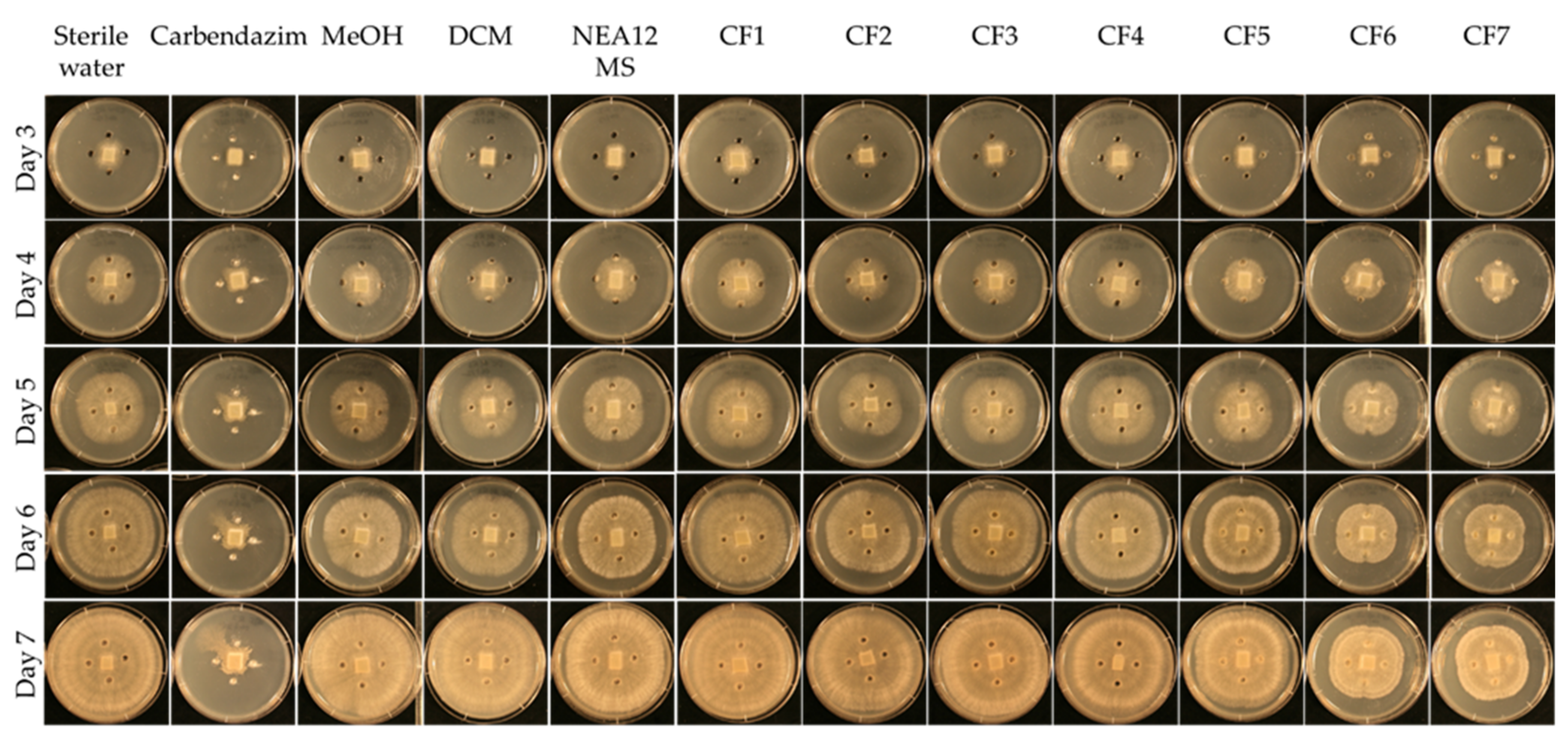
In the assay, the NEA12 MS-derived fractions CF6 and CF7 displayed strong activity against Ceratobasidium sp. (pathogen growth area < 20 cm2, Figure 2 and Figure 3a). Strong bioactivity was characterized by a smaller growth area and lower mycelial density compared to the negative controls. This was evident throughout the duration of the bioassay. CF5 moderately inhibited the growth of the pathogen compared to the controls (pathogen growth area 21–32 cm2). CF1, CF2, CF3, and CF4 didn’t display activity against Ceratobasidium sp.
A one-way ANOVA analysis compared the effect of twelve treatments (degrees of freedom = 11), eight antifungal treatments (CF1–CF7 and NEA12 MS), and four controls on pathogen growth area, a measure of antifungal activity. The ANOVA results showed that at least two treatments are significantly different in antifungal activity at 99% confidence (p < 0.001) (Table 1). Tukey’s Post-Hoc test grouped the CFs 1–7, NEA12 MS, and controls to four groups at 99% based on the statistically significant mean difference of the average pathogen growth area (Figure 3a). Tukey simultaneous tests for differences of means for pathogen growth area on day 6 confirmed inhibitory activity of CF5 (p < 0.001,), CF6 (p < 0.001), and CF7 (p < 0.001) compared to the respective solvent controls. CF1 (p = 0.998), CF2 (p = 0.971), CF3 (p = 1.000), and CF4 (p = 0.216) did not display significant inhibitory activity compared to the respective negative controls. NEA12 MS showed moderate activity, and the activity was significant (p < 0.001) compared to its control. Carbendazim (1000 ppm), a systemic benzimidazole fungicide commonly used in laboratory assays and used in this study as a positive control, significantly inhibited phytopathogen growth (p < 0.001) compared to the 80% methanol control. Using these observations as a guide, the bioactive crude fractions, CF5, CF6, and CF7, were selected for further analysis.
In the presence of NEA23 MS and derived crude extracts CF8–CF14, the pathogen was also observed growing away from the agar wells, evident by the ‘X’ like growth pattern. NEA23 MS and fractions CF11, CF12, CF13, and CF14 showed strong activity against the pathogen, as was evident by the smaller growth area and lower mycelial density of the pathogen compared to the negative controls (pathogen growth area < 20 cm2; Figure 3b and Figure 4). CF8, CF9, and CF10 displayed moderate activity compared to the negative controls (pathogen growth area 21–32 cm2).
A one-way ANOVA analysis compared the effect of twelve treatments (degree of freedom = 11), eight antifungal treatments (CF8–CF14 and NEA23 MS) and four controls on pathogen growth area which indicates the antifungal activity. The ANOVA results showed that at least two treatments are significantly different in antifungal activity at 99% confidence (p < 0.01) (Table 1). Tukey’s Post-Hoc test grouped the CFs 8–14, NEA23 MS and controls to six groups at 99% confidence based on the statistically significant mean difference of average pathogen growth area (Figure 3b). Tukey simultaneous tests for differences of means for pathogen growth area on day 6 confirmed inhibitory activity by CF11 (p < 0.001), CF12 (p < 0.001), CF13 (p < 0.001) and CF14 (p < 0.001) and NEA23 MS (p < 0.001) compared to the respective solvent controls (Figure 3b). The inhibitory activity of CF8 (p < 0.001), CF9 (p < 0.001), and CF10 (p < 0.001) was also significant compared to the 80% methanol control. The antifungal compound carbendazim displayed strong inhibitory activity (p < 0.001) compared to the 80% methanol control. Using these observations as a guide, the highly active crude fractions, CF12, CF13, and CF14 were selected for further analysis.
According to bioassay results, the strongly bioactive crude fractions NEA12 MS (CF6, CF7) and NEA23 MS (CF13, CF14) eluted with non-polar solvent systems (methanol: DCM 1:1 v/v and 100% DCM). The moderately bioactive crude fractions, having activity similar to that observed by the original MS extracts, eluted with 100% methanol (NEA12—CF5; NEA23—CF11 and CF12), while CFs in water (100%, CF1 and CF8; 80% CF2 and CF9; and 50% CF3 and CF10) exhibited a lower antifungal activity than that observed by the original MS extracts.

2.2. Metabolite Profiling

Further fractionation and purification of crude fractions (CF5, CF6, CF7, CF12, CF13, CF14) by HPLC using C18 reverse phase column chromatography provided refined fractions (RF). RF1–RF7 were derived from NEA12 MS CF5, CF6, CF7, and RF8–RF10 were derived from NEA23 MS CF12, CF13, CF14 (Figure 1).
The LCMS analysis was then used to detect metabolites in refined fractions (NEA12 RF1–7, NEA23 RF8–10) and determine if they are consistently present in both in vitro culture (NEA12 ME and NEA23 ME) and in planta (NEA12 PE and NEA23 PE). In planta extracts (NEA12 PE, n = 48 and NEA23 PE, n = 30) were obtained from the pseudo stems of individual genotypes of mature perennial ryegrass-endophyte symbiota. The media supernatant from replicate in vitro cultures (NEA12 ME, n = 27 and NEA21 ME, n = 28) were also individually extracted and analyzed to determine the reproducibility of the metabolites produced.
The LCMS spectral data obtained for in vitro (ME), in planta (PE), and refined fractions (RF) was analyzed using Refiner and Analyst modules of Genedata Expressionist® to annotate metabolites and compare metabolite profiles. To ensure the metabolites in an in vitro extract metabolome were free of PDB media derived metabolites, sterile media was also extracted, analyzed in parallel by LCMS, and metabolites detected in the media alone were filtered out of the in vitro extract metabolomes.
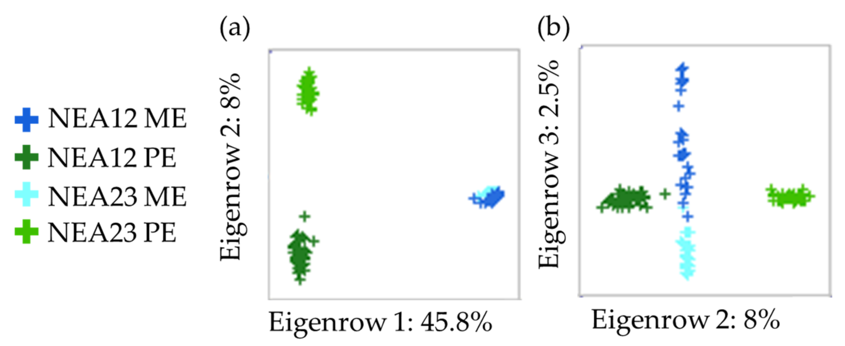
As expected, the metabolomes of PE and ME are distinct since the in planta and in vitro environments differ, and the former also contains plant-only metabolites that are not found in culture. Principal component analysis (PCA) revealed the separation of PE populations from ME population samples on PC1 (45.8% of the total variation; Figure 5a). Within each population, individual samples are tightly clustered, indicating that the metabolome is reproducible. Importantly, as shown in Figure 5b, there is a small but observable variation between the two different endophyte strains in the same environment—in planta and in vitro. The two PE populations (NEA12 PE and NEA23 PE) form distinct clusters on PC2 (8.0% of the total variation), while the two ME populations (NEA12 ME, NEA23 ME) separate out on PC3 (2.5% of the total variation).
Venn diagrams were generated to compare the metabolome of endophytes in different growth environments (in vitro, ME, and in planta, PE) and identify the presence of antifungal metabolites in refined fractions (RF) (Figure 6). It is first interesting to note that approximately 30% of the metabolites produced by the endophyte strain in vitro are also present in planta (NEA12, 3460/11,182, 30.3%; NEA23, 3435/11,354 30.9%); these metabolites are common to both environments (Figure 6a,b). The presence of an endophyte also impacts the symbiotum metabolome, as shown in Figure 6c, where more than 80% (19,829) of metabolites are in common (NEA12, 19,829/24,020, 82.6%; NEA23, 19,829/23,175, 85.6%), while approximately 16% may be attributed in part due to the presence of different endophyte strains (NEA12, 4191/24,020, 17.4%; NEA23, 3346/23,175, 14.4%).
Of the metabolites identified in the NEA12 refined fractions (RF1–7), 82% (29/35) are present in both NEA12 ME and NEA12 PE samples (Figure 6a). For NEA23, 82% (41/50) of metabolites identified in the refined fractions (RF8–10) are present in both the NEA23 ME and NEA23 PE samples (Figure 6b).
The compounds detected in the RF samples that were not detected in both PE and ME (NEA12, 1/35; NEA23, 1/50) are likely impurities introduced from the extraction process. These two compounds eluted very early in the LCMS profile, suggesting they are likely unrelated to the non-polar refined fractions.
There are also metabolites only detected in in vitro populations (NEA12 ME and NEA12 RF, 5/35; NEA23 ME and NEA23 RF, 2/50). The presence of these compounds likely reflects the different growth environments; in vitro culture compared to the natural environment of the endophyte in symbiotic association. Some metabolites were not observed in the ME samples but were detected in planta (NEA23 PE–6 of 50).
In refined fractions RF1–10, a total of 68 metabolites were detected (Figure 6c). Of these 68 metabolites, 59 were detected in both NEA12 PE and NEA23 PE, two were exclusive to NEA12 PE, and seven were exclusive to the RF fractions. Therefore, the majority of detected prospective antifungal metabolites synthesized in vitro (61/68, 89.7%) are also produced by the endophyte in symbiotic association with perennial ryegrass.
Of the 68 metabolites detected in RFs, 63 were present in at least one in vitro culture, NEA12 ME and/or NEA23 ME (Supplementary Table S1). The majority of these, 80% (52/63), were observed at a frequency greater than 80% in at least one in vitro secretome (NEA12 ME ≥ 22/27; NEA23 ME ≥ 23/28). From the detected metabolites 14/63 are significantly different (Q < 0.001) in relative abundance between the two in vitro metabolome populations and exhibited greater than 2-fold differential abundance (directed effect size (DES) ≥ 2). The remaining 49 metabolites either did not vary significantly in average relative abundance (Q > 0.001) between populations or exhibited less than 2-fold differential abundance (DES < 2). The DES of significant metabolites was obtained by calculating the ratio of average relative abundances of NEA12 ME: NEA23 ME for each metabolite.
Table 2 describes the properties of the 61 prospective antifungal metabolites synthesized in vitro, enriched for in refined fractions, and their presence in the NEA12 PE and NEA23 PE symbiota metabolomes. The distribution of these 61 metabolites in refined fractions RF1-RF10 was different from one another (Supplementary Table S2).
Considering the presence of each metabolite, 75% (45/61) were observed in the plant metabolome at a frequency of greater than 80% in at least one in planta population (NEA12 PE ≥ 38/48 symbiota; NEA23 PE ≥ 24/30 symbiota). Only 5/61 were observed at a frequency of less than 20% in both populations (NEA12 PE ≤ 9/48 and NEA23 PE ≤ 6/30). The remaining 16% (10/61) were observed more frequently in one symbiotum metabolome, most often NEA12 PE (9/10 metabolites).
The differences in the relative abundance of metabolites between the two PE metabolomes are described using a Q-value; if the Q-value < 0.01, the relative abundances are significantly different. Variation was further described by looking at the DES of metabolites that were significantly different among the two populations.
Among the 45 metabolites present in at least one symbiotum metabolome population at a frequency greater than 80%, 31 are significantly different (Q < 0.01) in relative abundance. Here, 7/31 are more abundant (DES > 2) in the NEA12 PE metabolome, and 6/31 are more abundant (DES > 2) in the NEA23 PE metabolome.
Of the metabolites observed more frequently in the NEA12 PE plant metabolome, 4 metabolites are found almost exclusively in the NEA12 PE metabolome (M8, M13, M32, and M43) and a statistical analysis was not possible; 5 metabolites are not significantly different in terms of relative abundance between in planta populations; and one metabolite (M29) is significantly different (Q < 0.001) in relative abundance and the DES indicates M29 (11.6) is more abundant in NEA23 PE. The one metabolite observed more frequently in the NEA23 PE metabolome (M21) is more abundant in the NEA12 PE metabolome (Q = 0.024, DES 1:2.386).
A total of 13/61 metabolites were significantly different (Q < 0.01) in relative abundance between the two symbiota metabolome populations and exhibited a greater than 2-fold differential abundance (DES): M5 (1:3.927), M18 (1:7.060), M19 (2.49), M21 (1:2.386), M23 (3.93), M24 (5.117), M25 (1:2.198), M29 (11.857), M34 (1:4.525), M39 (1:9.063), M42 (1:3.271), M48 (2.038) and M52 (2.477). The remaining 48 metabolites either did not vary significantly in average relative abundance (Q > 0.01) between two symbiota populations or exhibited a less than 2-fold differential abundance (DES < 2).
Considering the abundance of the metabolites described in Table 2, in most instances the in vitro (RF) samples are enriched for potential bioactive metabolites when compared to the average abundance of the corresponding metabolite in planta (PE) (Supplementary Figure S1). Crude extraction of the media supernatant followed by bioassay-guided extraction, from a starting point of mycelia cultured in 10 L of media supernatant, has resulted in refined extracts that are enriched for a suite of metabolites that are candidates for in vitro bioactivity. The presence of these 61 metabolites in planta (PE) and refined fractions (RF) further confirms that perennial ryegrass-endophyte symbiota produce these metabolites (Supplementary Figure S1).
Together this data describe a suite of potentially bioactive ‘known unknown’ Epichloë sp.-derived metabolites identified using bioassay-guided extraction and establish that most of the metabolites in the in vitro Epichloë sp. secretome are also produced in perennial ryegrass-NEA12 and perennial ryegrass-NEA23 symbiota.

3. Discussion

Epichloë sp. endophytes associated with pasture and turf grasses improve the disease resistance of their host grasses. Endophyte-mediated disease resistance is an outcome of complex host-pathogen-endophyte interactions resulting in antimicrobial compound production, secretion, and distribution throughout the host plant [61]. Though this process is not well understood, some studies have identified antifungal metabolites from sexual (pathogenic) Epichloë species [18,36,62,63,64,65], in particular Epichloë festucae [10,44,66], and characterized their effects on phytopathogens. Most studies confirm the antifungal activity using in vitro assays of the endophyte culture itself, culture filtrates, or isolated compounds [10,18,36,45,46,66,67]. In contrast, the investigation into the compounds responsible for endophyte-mediated disease resistance in asexual, vertically transmitted, Epichloë species that are utilized in perennial ryegrass and tall fescue are limited.
In Fernando et al., (2020) [45], an in vitro dual culture screen of asexual Epichloë sp. endophyte strains from perennial ryegrass and tall fescue identified bioactive strains that suppressed the growth of phytopathogens. A further investigation of four strains—SE, NEA12, NEA21, and NEA23—revealed the differential bioactivity of the strains against grass pathogens Ceratobasidium sp., Drechsler sp., and Fusarium sp. and identified the endophyte secretome as having higher bioactivity, compared to the mycelia, in liquid culture. A second study reported that the ‘known knowns’—Epichloë sp. related antimammalian and insecticidal alkaloids—are not responsible for the observed antifungal bioactivity [57]. Here, pure forms of endophyte-related alkaloids—namely peramine, n-acetylloline, n-formylloline, lolitrem B, janthitrem A, paxilline, terpendole E, terpendole C, ergovaline, ergotamine, ergocornine, ergocryptine, ergotaminine—were tested against Ceratobasidium sp., Drechslera sp. and Fusarium sp. in agar well diffusion assays [57]. Thus, the opportunity now arises to investigate novel Epichloë sp. for their potential in endophyte-mediated disease resistance and identify metabolite biomarkers for bioactive animal-friendly strains.
For the application and a better understanding of endophyte-mediated disease resistance, studying antifungal metabolite presence and abundance in planta is important. In this study, antifungal metabolites responsible, either singly or in combination, for in vitro bioactivity were identified using bioassay-guided extraction of the Epichloë sp. endophyte secretome, and their presence and abundance in Epichloë sp.-perennial grass symbiota was explored. The secretome—media supernatant (MS) extracts—of two bioactive endophyte strains, NEA12 and NEA23, were fractionated using solvent systems with different polarities to obtain seven crude fractions (CF) from each endophyte strain. These MS extracts and the derived CFs were then tested against the pathogen Ceratobasidium sp. in agar well diffusion assays to determine their in vitro antifungal activity. Agar well diffusion assays can be easily replicated and generate accurately quantifiable results.
The NEA12 secretome showed moderate activity, and NEA23 showed strong activity. These observations were consistent with previous findings [45]. NEA12 MS extracts in agar-well diffusion assays exhibited moderate inhibition of Ceratobasidium sp.; while the ‘X’ like growth pattern of Ceratobasidium sp. was observed in the presence of NEA23 MS, indicating significant growth inhibition [45]. The differential antifungal activity of the two Epichloë sp. strains against the same pathogen may be due to differences in the presence and/or abundance of antifungal metabolite in each secretome. It is noteworthy that the two studies were four months apart, an indication that the antifungal metabolites produced by these endophyte strains are stable at −80 °C and at room temperature (22 °C) for the duration of the assay (7 d).
Crude fractions in 1:1 methanol: DCM and 100% DCM (CF6, CF7, CF13, and CF14) exhibited significantly stronger antifungal activity compared to the original MS extracts and other CFs, thus confirming enrichment of antifungal metabolites from the endophyte secretome in these fractions. Thus, the antifungal metabolites are most likely not highly polar compounds as they have eluted with organic solvent systems.
A Metabolome analysis may entail either a targeted analysis of a certain class of metabolite, or a total metabolite profiling of a given sample, or population of samples. Preliminary screening of the total metabolome provides a view on overall metabolite production; whereas, targeted bioassay-guided isolation and characterization provides a more concise detail about metabolites and their potential uses as antifungal compounds [60].
Bioassay-guided extraction/isolation processes are often used to isolate bioactive metabolites from plants and microorganisms [10,58,68,69,70]. Yue et al. isolated six antifungal metabolites, indole-3-acetic acid, indole-3-ethanol, methylindole-3-carboxylate, indole-3-carboxaldehyde, diacetamide, and cyclonerodiol from Epichloë sp., in in vitro cultures using a bioassay-guided extraction process which inhibited the growth of Cryphonectria parasitica, Lactisaria fusiformis, Magnaporthe poae and Rhizoctonia solani [10].
Strong bioactive crude fractions (CF5–7 and CF11–14) were selected for further characterization. With further fractionation, refined fractions were obtained from both endophyte strains. Complex crude fractions were subjected to another round of chromatography to obtain cleaner refined fractions, which facilitates better identification in terms of abundance and intensity. While further purification is necessary to fully characterize the antifungal metabolites in these refined fractions, LCMS analysis can accurately annotate the metabolites, providing level 5 metabolite identification based on their unique mass, m/z and retention time (RT) [71,72]. To enhance the accuracy of the refining process, low tolerance thresholds were used (RT alignment ± 0.1 min, isotope clustering RT ± 0.02 min, m/z ± 0.05 Dalton). Thus, information on chemical properties and elution times can be used to detect the presence of the same metabolites in any matrix, using suitable extraction, and LCMS analysis methods. The exact methods adopted will depend on the chemical characteristics of the active metabolites e.g., polarity, and in some instances, more efficient purifications could be developed e.g., extraction of aqueous fractions using ethyl acetate or dicholormethane.
Annotated metabolite data of refined fractions, secretome extracts of in vitro cultures (ME), and extracts from populations of endophyte-perennial ryegrass symbiota (PE) of NEA12 and NEA23 were used to determine the presence of metabolites from the refined fractions in replicated samples of the endophyte secretome in vitro and in planta.
Tight clustering patterns were observed and show the homogenous nature of the metabolome within in vitro and in planta populations. The tight clustering patterns in PCA plots clearly distinguish the in vitro and in planta populations, as expected the two growth environments are different. Importantly, a small yet observable separation of the two endophyte strains was noted by the separate clustering of individuals from each population within the two growth environments. This separation represents the difference in metabolomes due to the genetic differences between the two endophyte strains, NEA12 and NEA23.
Most of the metabolites in refined fractions derived from the NEA12 and NEA23 secretomes are also present in the in vitro and in planta populations examined, providing strong evidence for their constitutive and reproducible production. A small portion of RF metabolites (NEA12; 5/35, NEA23; 2/50) were produced only in in vitro culture. These metabolites may be produced exclusively in the in vitro environment or present at trace concentrations in planta (and therefore below threshold levels set in this study). Alternatively, they may be due to the introduction of contaminates during the purification process or induced in response to environmental conditions, such as a stress response, that the Epichloë sp.-perennial ryegrass symbiota, maintained under optimal conditions in the glasshouse, were not exposed to [73,74,75,76].
In this study, about 23,500 features were detected in Epichloë sp.-perennial ryegrass symbiota. The two symbiota populations shared about 80% of their metabolome, while about 16% may be attributed to the presence of different endophyte strains. A recent untargeted metabolomic study in endophyte-free perennial ryegrass plants detected about 17,500 features in ESI+ mode after applying filters for accuracy [77]. There is some evidence that Epichloë sp. endophyte infection, apart from having different ‘known known’ alkaloid profiles, significantly influences the host plant metabolome [78,79,80]. However, recent studies do not usually distinguish the endophyte metabolome from the plant metabolome in symbiosis [61,81]. By identifying biomarkers from in vitro endophyte extracts, this study has provided a strategy to unravel endophyte-derived bioprotective metabolites from the symbiotia.
The metabolites conferring antifungal bioactivity in in vitro assays, enriched for using bioassay-guided extraction, and present in perennial ryegrass-endophyte symbiota potentially have a role in conferring disease resistance. Considering the potentially bioactive metabolites detected in refined fractions, the vast majority, 87% (59/68), are also present in both symbiota populations; just two metabolites are exclusive to the NEA12-perennial ryegrass population. This outcome indicates that there is a suite of prospective antifungal metabolites common to the two Epichloë sp. strains investigated, and they are also expressed in planta.
The two bioactive strains in this study are from different lineages and classified as different species. NEA23, a FaTG-3 (Festuca arundinacea taxonomic group-3) isolated from tall fescue [82,83,84] and NEA12 a Lolium perenne taxonomic group-3 (LpTG-3) strain isolated from perennial ryegrass [26,85,86]. The two strains exhibit different ‘known known’ alkaloid profiles (NEA12, epoxy janthitrems; NEA23, peramine, n-acetylloline and n-formylloline) [82,85]. Given the noted genetic and alkaloid profile differences and observed similarity in the metabolite profiles in this study, an in-depth study on the bioprotectives of these two strains can unravel the potential of using biomarkers to identify superior endophyte strains.
A suite of common potentially bioactive endophyte-derived metabolites that are synthesized in planta enables/allows for their utilization as biomarkers for bioprotection. In this study, 75% (45/61) of the metabolites were observed in the plant metabolome at a frequency of greater than 80% in at least one symbiotum population. Metabolites that are exclusive to one symbiota population, as observed for NEA12, may be strain-specific biomarkers and potentially associated with differential bioactivity [45].
Moreover, of interest is the relative abundance of the metabolites in planta; only 21% (13/61) of significantly different metabolites vary greater than 2-fold in relative abundance between the two symbiota metabolome populations. The size of the symbiota populations (NEA12 n = 48, NEA23 n = 30) is large enough to minimize the impact of variation between individual symbiota due to host-endophyte genetic interaction (noting that while the endophytes are clonal, the ryegrass is not) and determine differences if they are present [87,88]. Further investigation is required to determine the effect of the endophyte strain on antifungal metabolite abundance in planta.
Metabolites present in vitro and absent in planta may be present in endophyte-perennial ryegrass symbiota at trace levels, below the threshold set for detection in this study. It is also plausible that those metabolites present only in vitro were not identified in symbiota because they are stress-induced [89]. It is possible that the abundance of these in vitro-only metabolites (and endophyte-derived metabolites in symbiota) may change in response to challenge by disease-causing pathogens [89,90]. The environment in which endophyte mycelia is grown in culture, out of its natural environment, and grown in culture for extended periods can activate metabolic pathways and stimulate the production of bioactive compounds; thus, it is likely that some metabolites produced in vitro may not be produced by plants growing in optimal environmental conditions [73,74,75,76]. It would be interesting to further investigate these prospective antifungal metabolites as they could be candidates for bioprotection/abiotic stress tolerance if they are induced by stress. In this study, there is some evidence for the presence of inducible metabolites in the data. A small proportion of metabolites were observed infrequently in symbiota and may be induced in response to environmental stimuli. The refined fractions (RF) may pick up some impurities during the isolation procedure.
For compound discovery using bioassay-guided isolation, investigating multiple endophyte strains and symbiota populations allows for bioprospecting for novel bioprotective compounds and identification of biomarkers for phytopathogen disease control. The presence of a predictive suite of annotated metabolites, categorized as “known unknowns” [91], in other grass-endophyte symbiota and/or in vitro cultures may provide a way to accurately predict disease resistance conferred by novel Epichloë sp. endophyte strains prior to establishing field trials. The robust analysis methods used have allowed the analysis of a larger number of samples in a shorter amount of time and confirmed the consistent presence of detected metabolites in symbiotic associations. This is important as the production of secondary metabolites can be dependent on biotic and abiotic effects. Without consistent production of these metabolites, it is not possible to conclude or predict that endophytes may display the expected bioactivity under field conditions. Such compounds are better suited for further characterization and purification and are also more important in routine testing toolkits before commercially introducing novel endophyte associations. Furthermore, the pipeline for bioassay-guided antifungal metabolite isolation, annotation, and analysis can be used in future studies to screen for antifungal activity of other Epichloë sp. endophyte strains. Finally, the highly reproducible nature of metabolite biosynthesis both in in vitro culture as well as in planta indicates that the methodology can be replicated and developed further to isolate and purify antifungal metabolites from Epichloë sp. endophyte strains efficiently. In this study, compounds from refined fractions were annotated based on their unique mass, m/z and retention time (RT), a more accurate characterization is possible with further purification and structure analysis.

4. Materials and Methods

4.1. Plant Material

All plant material was obtained from a glasshouse maintained (natural day lengths and a mean temperature of 22 °C) collection at the Agriculture Victoria, Bundoora, Victoria, Australia. Prior to harvesting for metabolomics analysis, the presence and identity of endophyte strains was confirmed by the extraction of DNA using Qiagen MagAttract DNA (Qiagen, Hilden, Germany) and SNP-based diagnostic testing, KASP analysis (Kompetitive Allele Specific PCR) (KASP—, LGC Genomics, Teddington, UK).

4.2. Fungal Cultures

4.2.1. Endophyte Cultures

Epichloë sp. endophyte strains were isolated by Fernando et al., (2020) from pre-inoculated perennial ryegrass plants maintained at the Agriculture Victoria, Bundoora, Victoria, Australia [45]. Isolates were maintained in culture and stored as solid cultures on potato dextrose agar (PDA) (Sigma-Aldrich, Castle Hill, NSW, Australia) at 22 °C in the dark [85] and sub-cultured every two months. The identity of the endophyte strain was confirmed by KASP analysis prior to the study, as described by Fernando et al., (2020) [45].

4.2.2. Pathogen Cultures

A culture of Ceratobasidium sp. (VPRI 22537), the causative phytopathogen of yellow patch disease in perennial ryegrass and sharp eyespot in Triticum aestivum (wheat), was obtained from the National Collection of Fungi, Bundoora Herbarium, Victoria. The pathogen was stored as a solid culture on PDA (Sigma-Aldrich, Castle Hill, NSW, Australia) at 22 °C in the dark and sub-cultured every two months to maintain stocks. The genus of the pathogen was confirmed by ITS sequence analysis as described by Fernando et al., (2020) [45].

4.3. Chemicals

All extraction and mobile-phase solvents were HPLC grade; methanol (99.9% pure; Fisher chemicals, Fair Lawn, NJ, USA), acetonitrile with 0.1% formic acid (≥99.9% pure; Sigma-Aldrich CHROMASOLV®, Castle Hill, NSW, Australia, HPLC grade), dichloromethane (DCM) (≥99.9% pure; Burdick & Jackson, HPLC grade), water with 0.1% formic acid (Fisher Chemical, Fair Lawn, NJ, USA), and ammonium formate (99.0% pure; Sigma-Aldrich, St. Louis, MO, USA). C18 powder (Grace Davidson Discovery Sciences, CA, USA) was used for the adsorption of extracts. For the positive control, a 1000 ppm solution of antifungal compound carbendazim (97% pure; sourced from Sigma-Aldrich, Castle Hill, NSW, Australia) was prepared in a 4:1 ration of (v/v) methanol: H2O.

4.4. Extraction and Fractionation of In Vitro Culture

4.4.1. Crude Extracts

Highly active crude media supernatant (MS) extracts from liquid cultures of Epichloë sp. endophyte strains NEA12 and NEA23, described by Fernando et al., (2020), were used for further fractionation and isolation of antifungal metabolites [45]. MS extracts collected from two-week-old NEA12 and NEA23 PDB cultures are termed NEA12 MS and NEA23 MS, respectively.

4.4.2. Solid Phase Fractionation of Crude Extracts

Aqueous methanolic media supernatant extract was dried in the rotary evaporator and weighed (Table 3). Highly active crude media supernatant (MS) extracts were adsorbed to C18 (C18 Silica gel spherical, Sigma-Aldrich, St. Louis, MO, USA) in a 1:10 v/w ratio and dried in the rotary evaporator (Heidolph Laborota 4000, Heidolph Instruments, Schwabach, Germany) at 40 °C to obtain a fine yellow powder. The extract adsorbed to C18 was tightly packed in a 1 L preparative column for crude fractionation in the HPLC system (Dionex Ultimate 3000 (Dionex, Sunnyvale, CA, USA)). Fractionation was carried out sequentially using seven solvent systems (Table 3), where the column was flushed with one column (1 L) volume of each solvent system to obtain seven crude fractions. The obtained crude fractions (CF1–CF14) were dried in the rotary evaporator at 40 °C and weighed for dry weight. Fractions were then resuspended in appropriate solvents to give a concentration of 1 g/mL. From each crude fraction, 60 µL was aliquoted into HPLC vials separately for further analysis using LCMS.

4.4.3. Agar Well Diffusion Assay for Crude Fractions

Agar well diffusion assays were conducted for crude fractions on PDA plates according to the method described by Fernando et al. 2020 [45]. A small plug of Ceratobasidium sp. mycelia (5 × 5 mm) was transferred onto the center of the PDA plate, and 40 µL of the extract was placed in each of the four equidistant wells (4 mm diameter). Bioassay plates were prepared in replicate (n = 5). Plates were incubated at 22 °C for eight days and observations were taken daily from day three. Control assays included sterile distilled water, 4:1, v/v methanol: H2O, DCM, respective MS (NEA12 or NEA23) extract and carbendazim (1 mg/mL) (97%) (methyl benzimidazol-2-ylcarbamate; Sigma-Aldrich, Castle Hill, NSW, Australia). The growth of the fungal pathogen Ceratobasidium sp., was observed daily for up to 8 days. The measurements of the pathogen growth were analyzed using ImageJ 1× 5 mm) was transferred onto the center of the PDA plate, and 40 µL of the extract was placed in each o software (NIH, Bethesda, MA, USA) [92] and expressed as area (cm2). A one-way-ANOVA statistical analysis was performed on day 6 using Minitab® 19 Statistical Software Minitab (LLC, State College, PA, USA, a generated Tukey’s Post-Hoc test was used to separate group treatments with statistical significance, and Tukey’s simultaneous tests for differences of means at 99% confidence level was used to express the significant differences of the bioactivity among crude fractions.

4.4.4. Fractionation Using Semi Preparative High-Performance Liquid Chromatography

Moderate to strongly bioactive crude fractions (CF) were further purified by fractionation. Fractions were dried, weighed, and resuspended in 4:1, v/v methanol: H2O to obtain a concentration of 1 g/mL. A Dionex Ultimate 3000 solvent delivery system (Dionex, Sunnyvale, CA, USA) was equipped with a binary pump, photodiode array detector (PDA 3000), attached to a Rheodyne Model 7725 injector with a 1 mL injector loop and operated using Chromeleon version 6.8 software (Dionex, Sunnyvale, CA, USA). MiliQ water methanol/acetonitrile (Sigma-Aldrich CHROMASOLV®, Castle Hill, NSW, Australia, HPLC grade) was used as mobile phases A and B. Using a C18 semi prep column Thermo Scientific Hypersil gold 5 µm C18 (2) 100 Å, 150 × 10 mm column, crude fractions were separately subjected to further fractionation to obtain refined fractions.

4.5. Media Supernatant Extraction of Endophyte Cultures

4.5.1. Preparation of Liquid Cultures

Liquid PDB (Potato Dextrose Broth) (Sigma-Aldrich, Castle Hill, NSW, Australia) cultures were prepared by using a sterile scalpel blade to cut a small section (1 × 1 cm) of Epichloë sp. mycelia at the periphery and place it into a sterile 1.7 mL Eppendorf tube containing 500 µL PDB. A sterile plastic pestle was used to gently grind the mycelia and agar. Another 500 µL PDB medium was added to the Eppendorf tube containing the ground endophyte. The ground mycelia, in 15 µL aliquots, was distributed into 100 mL culture vessels containing 30 mL of PDB. A total of 28 liquid culture replicates were prepared for each endophyte strain. Culture vessels were incubated in the dark at 22 °C on a shaker at 150 rpm (Ratek OM11, Adelaide, SA, Australia) for period of 14 days.

4.5.2. Extraction

Two-week-old liquid cultures were transferred to sterile 50 mL falcon tubes and centrifuged at 3836× g for 15 min at 4 °C (SIGMA Laborzentrifugen 4-16KS, Osterode, Germany). The media supernatant was then separated from the cell pellet, frozen, and lyophilized (Freeze dryer ALPHA, Christ, Germany). Samples were weighed to 20 mg ± 2 mg and extracted using methanol: H2O (v/v, 4:1, 1:3 w/v). For extraction, samples were vortexed (5 min) (Ratek multi-tube vortex mixer, MTV1, Boronia, VIC, Australia) and sonicated (10 min) (SoniClean, 250TD, Thebarton, SA, Australia) with the solvent. A 60 µL aliquot of the extracts were transferred into HPLC vials ready for LCMS analysis. Replicated (n = 28) small-scale extracts collected from two-week-old NEA12 and NEA23 PDB cultures are termed NEA12 ME and NEA23 ME, respectively. Separately, PDB media only controls were prepared; 30 mL sterile PDB media samples (n = 18) were freeze-dried and extracted (PDB ME).

4.6. Plant Material Extraction

Pseudostems from mature plants were harvested into collection tubes and freeze-dried for 48 h before being grinded to a fine powder (28 Hz for 2 min, Geno Grinder, NJ, USA). The freeze-dried, ground plant material (20 mg ± 0.2 mg) was extracted twice with methanol: water (80:20, v:v, 1 mL). Extracts were combined, dried, and reconstituted in methanol: water (80:20, v:v, 200 µL). Replicated extracts collected from mature pseudostems of perennial ryegrass-NEA12 (n = 48) and perennial ryegrass-NEA23 (n = 30) symbiota are termed NEA12 PE and NEA23 PE, respectively.

4.7. LCMS Data Acquisition and Analysis

All extracts were analyzed on a Vanquish Ultra-High Performance Liquid Chromatography (UHPLC) system (Thermo Fisher Scientific, Bremen, Germany) with a binary pump, autosampler, and temperature-controlled column compartment coupled with a Thermo Fisher Q Exactive Plus mass spectrometer (QE MS) (Waltham, MA, USA; Thermo, Bremen, Germany). Extracts were separated using a Thermo Scientific Hypersil Gold 1.9 μm, 150 × 2.1 mm column (Thermo Fisher Scientific, Waltham, MA, USA). Chromatographic separation was performed by gradient elution using water with 0.1% formic acid (Sigma-Aldrich CHROMASOLV®, Castle Hill, NSW, Australia, Castle Hill, NSW, Australia, HPLC grade) as Solvent A and acetonitrile with 0.1% formic acid (Sigma-Aldrich CHROMASOLV®, Castle Hill, NSW, Australia, HPLC grade ≥ 99.9%) as Solvent B at a flow rate of 0.3 mL/min. Initial conditions were 98% A, which was then progressed to linear gradient to 100% B over 11 min, and this was maintained for 4 min before returning to the initial gradient conditions. The injection volume was 3 μL.
The MS detector was operated in FT positive and negative mode using a full-scan with a mass range of m/z 80–1200. The ESI drying gas (N2) was set at a flow rate of 7 L/min at 350 °C, and the nebulizer gas (N2) pressure was set at 45 psig. Capillary, fragmentor, and skimmer voltage was set at 3500 V, 175 V, and 65 V, respectively. Prior to data acquisition, the system was calibrated with Pierce® LTQ Velos ESI Positive and Negative Ion Calibration Solution (Thermo Fisher Scientific™). Mass spectrometry data were acquired using Thermo Xcalibur V. 2.1 (Thermo Fischer Scientific Inc., San Jose, CA, USA) and data was analyzed using Thermo Xcalibur Qual Browser V. 2.1.
Acquired data were further analyzed using Refiner MS and Analyst (Genedata, Basel) [11,67]. Using Refiner MS module of Genedata Expressionist® spectral data of the media, plant extracts (PE), media supernatant extracts (ME), and refined fractions (RF) were separately processed: (1) Selection of positive mode data; (2) blank subtraction by Blank Sample Intensity Factor 1 to eliminate metabolites from the blanks; (3) chemical noise subtraction by a retention time (RT) window of 151 scans at 50% quantile and different intensity threshold for each type of extracts (intensity thresholds; RF: 10,000,000; PE and ME: 5000); (4) intensity thresholding using a clipping method and an intensity threshold of 100; (5) RT structure removal by removing signals that did not extend to at least six scans; (6) chromatogram RT alignment using a pairwise alignment-based tree and a maximum RT shift of 0.1 min and; (7) saved all data files as snapshots for three extract types separately.
Metabolite annotation was carried out together for extracts: (1) loading data from saved snapshot files; (2) chromatogram RT alignment using a pairwise alignment-based tree and a maximum RT shift of 0.1 min; (3) chromatogram peak detection using a five scans summation window, a minimum peak size of 0.1 min, a maximum merge distance of 0.005 Da, a boundary merge strategy, a maximum gap/peak ratio of 7% with moving average smoothing over three scans for peak RT splitting; (4) chromatogram isotope clustering using RT of 0.02 min, m/z tolerance of 0.05 Dalton, maximum missing peaks of 1 and a first allowed gap position of 3, with a peptide isotope shaping envelope fitting method where the maximum charge was 1; (5) singleton filter for removal of peaks that do not belong to a cluster (Supplementary Figure S2). Features were exported to the Analyst module of Genedata Expressionist® (Supplementary Figure S3). Using Genedata analyst all data was analyzed to create Venn diagrams and compare metabolites in extracts from endophytes grown in vitro (ME) and in planta (PE) with refined fractions. Features from sterile PDB extracts were filtered out from in vitro (ME) extract features to eliminate metabolites from the media. For the plant extracts of two endophyte strains, a t-test was used to generate Benjamini-Hochberg adjusted p values (Q value) to identify the significance of relative abundance of metabolites. The directed effect size was calculated by obtaining the ratio of average relative abundance of desired extracts.

5. Conclusions

Natural product isolation and structure elucidation can be time-consuming and labor-intensive, particularly if the bioactive metabolite is present in very small amounts. The workflow developed in this study allows for testing of endophyte bioactivity while ensuring that the metabolite is expressed in planta and is so useful for field deployment. This method also provides a shortlist of potential bioactives (in this case 61 metabolites out of more than 20,000 in planta) that can be used to explore endophyte-mediated disease resistance in populations of perennial ryegrass-endophyte symbiota. Metabolites present in vitro and absent in planta are also of interest as they may be induced in response to environmental stimuli. Further investigation is needed to determine the presence and abundance of these metabolites in symbiota in response to challenges by disease-causing pathogens. Purification, structural characterization, and confirmation of the bioactivity of isolated compounds will allow for the identification of metabolites associated with endophyte-mediated disease control/resistance in pasture and turf grasses.

Supplementary Materials

The following are available online at https://www.mdpi.com/article/10.3390/metabo12010037/s1, Table S1: Metabolites present in refined fractions extracted from NEA12 MS and NEA23 MS that are also present in the metabolomes of in vitro cultures (NEA12 ME and NEA23 ME); Table S2: Metabolite distribution in refined fractions derived from bioactive crude fractions; Figure S1: Relative abundance of metabolites (a) M1-M15, (b) M16-M30, (c) M31-M45 and (d) M46-M61 present in NEA12 (NEA12_PE) and NEA23 (NEA23_PE) plant extract metabolomes and in refined bioactive fractions of NEA12 (RF1-RF7) and NEA23 (RF8-RF10). Error bars represent + standard error; Figure S2: LCMS data imported to Refiner MS and processed for data refining using suitable parameters (a) positive ion data of plant extracts, (b) processed plant extract data after blank subtraction, chromatogram noise subtraction, intensity thresholding, RT structure removal and chromatogram RT alignment, (c) workflow for data processing; Figure S3: Refined data imported from (a) plant extracts (b) media supernatant extracts (c) refined fractions to be annotated together in Refiner MS. (d) Annotated data after chromatogram RT alignment, chromatogram peak detection, chromatogram isotope clustering followed by singleton filter and (e) workflow for data processing.

Author Contributions

Conceptualization: K.F., P.R., K.M.G. and S.J.R.; methodology: K.F. and P.R.; data analysis, K.F.; manuscript writing—original draft preparation, K.F.; writing—editing and review: K.F., P.R., S.J.R., K.M.G. and G.C.S.; supervision and project administration: K.M.G., P.R., S.J.R. and G.C.S. All authors have read and agreed to the published version of the manuscript.

Funding

The research was funded by Agriculture Victoria and DairyBio.

Institutional Review Board Statement

Not applicable.

Informed Consent Statement

Not applicable.

Data Availability Statement

Data is available from authors upon reasonable request.

Acknowledgments

The authors thank the following people for their contribution: Jacqueline Edwards for providing Cerabasidium sp. pathogen culture from the National Collection of Fungi. Emma J. Ludlow and Inoka K. Hettiarachchige for endophyte diagnostics and providing endophyte positive plant samples for LCMS analysis. Simone Vassiliadis for processing plant samples in preparation for LCMS analysis.

Conflicts of Interest

The authors declare no conflict of interest.

References

  1. Wilkins, P. Breeding perennial ryegrass for agriculture. Euphytica 1991, 52, 201–214. [Google Scholar] [CrossRef]
  2. Waller, R.A.; Sale, P.W.G. Persistence and productivity of perennial ryegrass in sheep pastures in south-western Victoria: A review. Aus. J. Experi. Agricul. 2001, 41, 117–144. [Google Scholar] [CrossRef]
  3. Vargas, J.M. Management of Turfgrass Diseases; CRC Press: New York, NY, USA, 2018. [Google Scholar]
  4. Dairy Australia. Forage Value Index. Available online: https://www.dairyaustralia.com.au/feed-and-nutrition/growing-feed-for-the-herd/growing-pastures/forage-value-index#.YXXbevlBxPZ (accessed on 25 October 2021).
  5. Cogan, N.O.I.; Smith, K.F.; Yamada, T.; Francki, M.G.; Vecchies, A.C.; Jones, E.S.; Spangenberg, G.C.; Forster, J.W. QTL analysis and comparative genomics of herbage quality traits in perennial ryegrass (Lolium perenne L.). Theore. App. Gen. 2005, 110, 364–380. [Google Scholar] [CrossRef] [Green Version]
  6. Pearson, A.; Cogan, N.O.; Baillie, R.C.; Hand, M.L.; Bandaranayake, C.K.; Erb, S.; Wang, J.; Kearney, G.A.; Gendall, A.R.; Smith, K.F. Identification of QTLs for morphological traits influencing waterlogging tolerance in perennial ryegrass (Lolium perenne L.). Theore. App. Gen. 2011, 122, 609–622. [Google Scholar] [CrossRef]
  7. Yamada, T.; Jones, E.; Cogan, N.; Vecchies, A.; Nomura, T.; Hisano, H.; Shimamoto, Y.; Smith, K.; Hayward, M.; Forster, J. QTL analysis of morphological, developmental, and winter hardiness-associated traits in perennial ryegrass. Crop Sci. 2004, 44, 925–935. [Google Scholar]
  8. Leuchtmann, A.; Bacon, C.W.; Schardl, C.L.; White, J.F., Jr.; Tadych, M. Nomenclatural realignment of Neotyphodium species with genus Epichloë. Mycologia 2014, 106, 202–215. [Google Scholar] [CrossRef]
  9. Schardl, C.L. The Epichloë, symbionts of the grass subfamily Poöideae. Ann. Miss. Bot. Gard. 2010, 97, 646–665. [Google Scholar] [CrossRef]
  10. Yue, Q.; Miller, C.J.; White, J.F.; Richardson, M.D. Isolation and characterization of fungal inhibitors from Epichloë festucae. J. Agri. Food Chem. 2000, 48, 4687–4692. [Google Scholar] [CrossRef]
  11. Schardl, C.L.; Balestrini, R.; Florea, S.; Zhang, D.; Scott, B. Epichloë endophytes: Clavicipitaceous symbionts of grasses. In Plant Relationships; Springer: Berlin/Heidelberg, Germany, 2009; pp. 275–306. [Google Scholar]
  12. Vázquez-de-Aldana, B.R.; Romo, M.; García-Ciudad, A.; Petisco, C.; García-Criado, B. Infection with the fungal endophyte Epichloë festucae may alter the allelopathic potential of red fescue. Ann. App. Biol. 2011, 159, 281–290. [Google Scholar] [CrossRef]
  13. Karpyn Esqueda, M.; Yen, A.L.; Rochfort, S.; Guthridge, K.M.; Powell, K.S.; Edwards, J.; Spangenberg, G.C. A review of perennial ryegrass endophytes and their potential use in the management of African black beetle in perennial grazing systems in Australia. Front Plant Sci. 2017, 8, 3. [Google Scholar] [CrossRef] [Green Version]
  14. Xia, C.; Li, N.; Zhang, Y.; Li, C.; Zhang, X.; Nan, Z. Role of Epichloë endophytes in defense responses of cool-season grasses to pathogens: A review. Plant Dis. 2018, 102, 2061–2073. [Google Scholar] [CrossRef] [Green Version]
  15. Singh, L.P.; Gill, S.S.; Tuteja, N. Unraveling the role of fungal symbionts in plant abiotic stress tolerance. Plant Signal. Behav. 2011, 6, 175–191. [Google Scholar] [CrossRef] [Green Version]
  16. Ruppert, K.G.; Matthew, C.; McKenzie, C.M.; Popay, A.J. Impact of Epichloë endophytes on adult Argentine stem weevil damage to perennial ryegrass seedlings. Entomol. Exp. Appl. 2017, 163, 328–337. [Google Scholar] [CrossRef]
  17. Reddy, P.; Hemsworth, J.; Guthridge, K.M.; Vinh, A.; Vassiliadis, S.; Ezernieks, V.; Spangenberg, G.C.; Rochfort, S.J. Ergot alkaloid mycotoxins: Physiological effects, metabolism and distribution of the residual toxin in mice. Sci. Rep. 2020, 10, 9714. [Google Scholar] [CrossRef]
  18. Siegel, M.R.; Latch, G.C. Expression of antifungal activity in agar culture by isolates of grass endophytes. Mycologia 1991, 83, 529–537. [Google Scholar] [CrossRef]
  19. Gallagher, R.; Campbell, A.; Hawkes, A.; Holland, P.; McGaveston, D.; Pansier, E.; Harvey, I. Ryegrass staggers: The presence of lolitrem neurotoxins in perennial ryegrass seed. N. Z. Vet. J. 1982, 30, 183–184. [Google Scholar] [CrossRef]
  20. Tor-Agbidye, J. Correlation of endophyte toxins (ergovaline and lolitrem B) with clinical disease: Fescue foot and perennial ryegrass staggers. Vet. Hum. Tox. 1993, 43, 140–146. [Google Scholar]
  21. Leuchtmann, A.; Schmidt, D.; Bush, L.P. Different levels of protective alkaloids in grasses with stroma-forming and seed-transmitted Epichloë/Neotyphodium endophytes. J. Chem. Ecol. 2000, 26, 1025–1036. [Google Scholar] [CrossRef]
  22. Guerre, P. Ergot alkaloids produced by endophytic fungi of the genus Epichloë. Toxins 2015, 7, 773–790. [Google Scholar] [CrossRef] [Green Version]
  23. Young, C.A.; Felitti, S.; Shields, K.; Spangenberg, G.; Johnson, R.D.; Bryan, G.T.; Saikia, S.; Scott, B. A complex gene cluster for indole-diterpene biosynthesis in the grass endophyte Neotyphodium lolii. Fung. Genet. Biol. 2006, 43, 679–693. [Google Scholar] [CrossRef]
  24. Young, C.A.; Bryant, M.K.; Christensen, M.J.; Tapper, B.A.; Bryan, G.T.; Scott, B. Molecular cloning and genetic analysis of a symbiosis-expressed gene cluster for lolitrem biosynthesis from a mutualistic endophyte of perennial ryegrass. Mol. Genet. Genom. 2005, 274, 13–29. [Google Scholar] [CrossRef]
  25. Saikia, S.; Takemoto, D.; Tapper, B.A.; Lane, G.A.; Fraser, K.; Scott, B. Functional analysis of an indole-diterpene gene cluster for lolitrem B biosynthesis in the grass endosymbiont Epichloë festucae. FEBS Lett. 2012, 586, 2563–2569. [Google Scholar] [CrossRef] [Green Version]
  26. Ludlow, E.J.; Vassiliadis, S.; Ekanayake, P.N.; Hettiarachchige, I.K.; Reddy, P.; Sawbridge, T.I.; Rochfort, S.J.; Spangenberg, G.C.; Guthridge, K.M. Analysis of the indole diterpene gene cluster for biosynthesis of the epoxy-janthitrems in Epichloë endophytes. Microorganisms 2019, 7, 560. [Google Scholar] [CrossRef] [Green Version]
  27. Fleetwood, D.J.; Scott, B.; Lane, G.A.; Tanaka, A.; Johnson, R.D. A complex ergovaline gene cluster in Epichloë endophytes of grasses. App. Environ. Microb. 2007, 73, 2571–2579. [Google Scholar] [CrossRef] [Green Version]
  28. Tanaka, A.; Tapper, B.A.; Popay, A.; Parker, E.J.; Scott, B. A symbiosis expressed non-ribosomal peptide synthetase from a mutualistic fungal endophyte of perennial ryegrass confers protection to the symbiotum from insect herbivory. Mol. Microbiol. 2005, 57, 1036–1050. [Google Scholar] [CrossRef]
  29. Schardl, C.L.; Young, C.A.; Hesse, U.; Amyotte, S.G.; Andreeva, K.; Calie, P.J.; Fleetwood, D.J.; Haws, D.C.; Moore, N.; Oeser, B.; et al. Plant-symbiotic fungi as chemical engineers: Multi-genome analysis of the clavicipitaceae reveals dynamics of alkaloid loci. PLoS Genet. 2013, 9, e1003323. [Google Scholar] [CrossRef] [Green Version]
  30. Young, C.A.; Tapper, B.A.; May, K.; Moon, C.D.; Schardl, C.L.; Scott, B. Indole-diterpene biosynthetic capability of Epichloë endophytes as predicted by ltm gene analysis. App. Enviro. Microb. 2009, 75, 2200–2211. [Google Scholar] [CrossRef] [Green Version]
  31. Reddy, P.; Guthridge, K.; Vassiliadis, S.; Hemsworth, J.; Hettiarachchige, I.; Spangenberg, G.; Rochfort, S. Tremorgenic mycotoxins: Structure diversity and biological activity. Toxins 2019, 11, 302. [Google Scholar] [CrossRef] [Green Version]
  32. Gallagher, R.T.; Hawkes, A.D.; Steyn, P.S.; Vleggaar, R. Tremorgenic neurotoxins from perennial ryegrass causing ryegrass staggers disorder of livestock: Structure elucidation of lolitrem B. J. Chem. Soc. Chem. Comm. 1984, 614–616. [Google Scholar] [CrossRef]
  33. Paterson, J.; Forcherio, C.; Larson, B.; Samford, M.; Kerley, M. The effects of fescue toxicosis on beef cattle productivity. J. Anim. Sci. 1995, 73, 889–898. [Google Scholar] [CrossRef]
  34. Rowan, D.D. Lolitrems, peramine and paxilline: Mycotoxins of the ryegrass/endophyte interaction. Agric. Ecosyst. Environ. 1993, 44, 103–122. [Google Scholar] [CrossRef]
  35. Koulman, A.; Lane, G.A.; Christensen, M.J.; Fraser, K.; Tapper, B.A. Peramine and other fungal alkaloids are exuded in the guttation fluid of endophyte-infected grasses. Phytochemistry 2007, 68, 355–360. [Google Scholar] [CrossRef]
  36. Christensen, M. Antifungal activity in grasses infected with Acremonium and Epichloë endophytes. Australas. Pl. Pathol. 1996, 25, 186–191. [Google Scholar] [CrossRef]
  37. Holzmann-Wirth, A.; Dapprich, P.; Eierdanz, S.; Heerz, D.; Paul, V. Anti-fungal substances extracted from Neotyphodium endophytes. In Proceedings of the 3rd International Conference on Harmful and Beneficial Microorganisms in Grassland, Pasture and Turf, Paderborn, Germany, 26–29 September 2000; pp. 65–69. [Google Scholar]
  38. Koshino, H.; Yoshihara, T.; Okuno, M.; Sakamura, S.; Tajimi, A.; Shimanuki, T. Gamahonolides A, B, and gamahorin, novel antifungal compounds from stromata of Epichloë typhina on Phleum pratense. Biosci. Biotechnol. Biochem. 1992, 56, 1096–1099. [Google Scholar] [CrossRef]
  39. Steinebrunner, F.; Schiestl, F.P.; Leuchtmann, A. Ecological role of volatiles produced by Epichloë: Differences in antifungal toxicity. FEMS Microbiol. Ecol. 2008, 64, 307–316. [Google Scholar] [CrossRef] [Green Version]
  40. Mousa, W.K.; Raizada, M.N. The diversity of anti-microbial secondary metabolites produced by fungal endophytes: An interdisciplinary perspective. Front. Microbiol. 2013, 4, 65. [Google Scholar] [CrossRef] [Green Version]
  41. Song, Q.Y.; Nan, Z.B.; Gao, K.; Song, H.; Tian, P.; Zhang, X.X.; Li, C.J.; Xu, W.B.; Li, X.Z. Antifungal, phytotoxic, and cytotoxic activities of metabolites from Epichloë bromicola, a fungus obtained from Elymus tangutorum grass. J. Agric. Food Chem. 2015, 63, 8787–8792. [Google Scholar] [CrossRef]
  42. Zhou, L.; Zhang, X.; Li, C.; Christensen, M.J.; Nan, Z. Antifungal activity and phytochemical investigation of the asexual endophyte of Epichloë sp. from Festuca sinensis. Sci. China Life Sci. 2015, 58, 821–826. [Google Scholar] [CrossRef] [Green Version]
  43. Niones, J.T.; Takemoto, D. Viba, a homologue of a transcription factor for fungal heterokaryon incompatibility, is involved in antifungal compound production in the plant-symbiotic fungus Epichloë festucae. Eukaryotic Cell 2015, 14, 13–24. [Google Scholar] [CrossRef] [Green Version]
  44. Tian, Z.; Wang, R.; Ambrose, K.V.; Clarke, B.B.; Belanger, F.C. The Epichloë festucae antifungal protein has activity against the plant pathogen Sclerotinia homoeocarpa, the causal agent of dollar spot disease. Sci. Rep. 2017, 7, 5643. [Google Scholar] [CrossRef] [Green Version]
  45. Fernando, K.; Reddy, P.; Hettiarachchige, I.K.; Spangenberg, G.C.; Rochfort, S.J.; Guthridge, K.M. Novel antifungal activity of Lolium-associated Epichloë endophytes. Microorganisms 2020, 8, 955. [Google Scholar] [CrossRef]
  46. Tian, P.; Nan, Z.; Li, C.; Spangenberg, G. Effect of the endophyte Neotyphodium lolii on susceptibility and host physiological response of perennial ryegrass to fungal pathogens. Eur. J. Plant Pathol. 2008, 122, 593–602. [Google Scholar] [CrossRef]
  47. Clarke, B.B.; White, J.F.; Hurley, R.H.; Torres, M.S.; Sun, S.; Huff, D.R. Endophyte-mediated suppression of dollar spot disease in fine fescues. Plant Dis. 2006, 90, 994–998. [Google Scholar] [CrossRef] [Green Version]
  48. Li, F.; Guo, Y.e.; Christensen, M.J.; Gao, P.; Li, Y.; Duan, T. An arbuscular mycorrhizal fungus and Epichloë festucae var. lolii reduce Bipolaris sorokiniana disease incidence and improve perennial ryegrass growth. Mycorrhiza 2018, 28, 159–169. [Google Scholar] [CrossRef]
  49. Wiewióra, B.; Żurek, G.; Żurek, M. Endophyte-mediated disease resistance in wild populations of perennial ryegrass (Lolium perenne). Fungal Ecol. 2015, 15, 1–8. [Google Scholar] [CrossRef]
  50. Welty, R.; Barker, R.; Azevedo, M. Response of field-grown tall fescue infected by Acremonium coenophialum to Puccinia graminis subsp. Graminicola. Plant Dis. 1993, 77, 574–575. [Google Scholar] [CrossRef]
  51. Pańka, D.; Jeske, M.; Troczyński, M. Effect of Neotyphodium uncinatum endophyte on meadow fescue yielding, health status and ergovaline production in host-plants. J. Plant Prot. Res. 2011, 51, 362–370. [Google Scholar] [CrossRef]
  52. Clarke, R.; Eagling, D. Effects of pathogens on perennial pasture grasses. N. Z. J. Agric. Res. 1994, 37, 319–327. [Google Scholar] [CrossRef] [Green Version]
  53. Gwinn, K.; Gavin, A. Relationship between endophyte infestation level of tall fescue seed lots and Rhizoctonia zeae seedling disease. Plant Dis. 1992, 76, 911–914. [Google Scholar] [CrossRef]
  54. Burpee, L.L.; Bouton, J.H. Effect of eradication of the endophyte Acremonium coenophialum on epidemics of Rhizoctonia blight in tall fescue. Plant Dis. 1993, 77, 157–159. [Google Scholar] [CrossRef]
  55. Bonos, S.A.; Wilson, M.M.; Meyer, W.A.; Reed Funk, C. Suppression of red thread in fine fescues through endophyte-mediated resistance. Appl. Turfgrass Sci. 2005, 2, 1–7. [Google Scholar] [CrossRef]
  56. Vassiliadis, S.; Elkins, A.C.; Reddy, P.; Guthridge, K.M.; Spangenberg, G.C.; Rochfort, S.J. A simple LC–MS method for the quantitation of alkaloids in endophyte-infected perennial ryegrass. Toxins 2019, 11, 649. [Google Scholar] [CrossRef] [Green Version]
  57. Fernando, K.; Reddy, P.; Vassiliadis, S.; Spangenberg, G.C.; Rochfort, S.J.; Guthridge, K.M. The known antimammalian and insecticidal alkaloids are not responsible for the antifungal activity of Epichloë endophytes. Plants 2021, 10, 2486. [Google Scholar] [CrossRef]
  58. Malviya, N.; Malviya, S. Bioassay guided fractionation-an emerging technique influence the isolation, identification and characterization of lead phytomolecules. Int. J. Hosp. Pharm. 2017, 2. [Google Scholar] [CrossRef]
  59. Balouiri, M.; Sadiki, M.; Ibnsouda, S.K. Methods for in vitro evaluating antimicrobial activity: A review. J. Pharm. Anal. 2016, 6, 71–79. [Google Scholar] [CrossRef] [Green Version]
  60. Stromstedt, A.A.; Felth, J.; Bohlin, L. Bioassays in natural product research–strategies and methods in the search for anti-inflammatory and antimicrobial activity. Phytochem. Anal. 2014, 25, 13–28. [Google Scholar] [CrossRef]
  61. Kusari, S.; Hertweck, C.; Spiteller, M. Chemical ecology of endophytic fungi: Origins of secondary metabolites. Chem. Biol. 2012, 19, 792–798. [Google Scholar] [CrossRef] [Green Version]
  62. Christensen, M.J.; Latch, G.C.M.; Tapper, B.A. Variation within isolates of Acremonium endophytes from perennial ryegrasses. Mycol. Res. 1991, 95, 918–923. [Google Scholar] [CrossRef]
  63. Hiroyuki, K.; Satoshi, T.; Shun-ichi, T.; Yoshihara, T.; Sakamura, S.; Shimanuki, T.; Sato, T.; Tajimi, A. New fungitoxic sesquiterpenoids, chokols A-G, from stromata of Epichloë typhina and the absolute configuration of Chokol E. Agric. Biol. Chem. 1989, 53, 789–796. [Google Scholar] [CrossRef]
  64. Koshino, H.; Togiya, S.; Yoshihara, T.; Sakamura, S.; Shimanuki, T.; Sato, T.; Tajimi, A. Four fungitoxic C-18 hydroxy unsaturated fatty acids from stromata of Epichloë typhina. Tetrahedron Lett. 1987, 28, 73–76. [Google Scholar] [CrossRef]
  65. Yoshihara, T.; Togiya, S.; Koshino, H.; Sakamura, S.; Shimanuki, T.; Sato, T.; Tajimi, A. Three fungitoxic cyclopentanoid sesquiterpenes from stromata of Epichloë typhina. Tetrahedron Lett. 1985, 26, 5551–5554. [Google Scholar] [CrossRef]
  66. Niones, J.T.; Takemoto, D. An isolate of Epichloë festucae, an endophytic fungus of temperate grasses, has growth inhibitory activity against selected grass pathogens. J. Gen. Plant Pathol. 2014, 80, 337–347. [Google Scholar] [CrossRef]
  67. Purev, E.; Kondo, T.; Takemoto, D.; Niones, J.T.; Ojika, M. Identification of ε-poly-l-lysine as an antimicrobial product from an Epichloë endophyte and isolation of fungal ε-PL synthetase gene. Molecules 2020, 25, 1032. [Google Scholar] [CrossRef] [Green Version]
  68. Saravana Kumar, P.; Yuvaraj, P.; Gabrial Paulraj, M.; Ignacimuthu, S.; Abdullah Al-Dhabi, N. Bio-prospecting of soil Streptomyces and its bioassay-guided isolation of microbial derived auxin with antifungal properties. J. Mycol. Med. 2018, 28, 462–468. [Google Scholar] [CrossRef]
  69. Nothias, L.-F.L.; Nothias-Esposito, M.L.; da Silva, R.; Wang, M.; Protsyuk, I.; Zhang, Z.; Sarvepalli, A.; Leyssen, P.; Touboul, D.; Costa, J. Bioactivity-based molecular networking for the discovery of drug leads in natural product bioassay-guided fractionation. J. Nat. Prod. 2018, 81, 758–767. [Google Scholar] [CrossRef] [Green Version]
  70. Weller, M.G. A unifying review of bioassay-guided fractionation, effect-directed analysis and related techniques. Sensors 2012, 12, 9181–9209. [Google Scholar] [CrossRef]
  71. Schrimpe-Rutledge, A.C.; Codreanu, S.G.; Sherrod, S.D.; McLean, J.A. Untargeted metabolomics strategies—challenges and emerging directions. J. Am. Soc. Mass Spectrom. 2016, 27, 1897–1905. [Google Scholar] [CrossRef] [Green Version]
  72. Schymanski, E.L.; Jeon, J.; Gulde, R.; Fenner, K.; Ruff, M.; Singer, H.P.; Hollender, J. Identifying small molecules via high resolution mass spectrometry: Communicating confidence. Environ. Sci. Technol. 2014, 48, 2097–2098. [Google Scholar] [CrossRef]
  73. Da Silva Lima, G.; da Rocha, A.M.; dos Santos, G.F.; D’Silva, A.F.; Marriel, I.E.; Takahashi, J.A. Metabolic response of Aspergillus sydowii to OSMAC modulation produces acetylcholinesterase inhibitors. Phytochem. Lett. 2018, 24, 39–45. [Google Scholar] [CrossRef]
  74. Hewage, R.T.; Aree, T.; Mahidol, C.; Ruchirawat, S.; Kittakoop, P. One strain-many compounds (OSMAC) method for production of polyketides, azaphilones, and an isochromanone using the endophytic fungus Dothideomycete sp. Phytochemistry 2014, 108, 87–94. [Google Scholar] [CrossRef]
  75. Bayram, Ö.; Krappmann, S.; Ni, M.; Bok, J.W.; Helmstaedt, K.; Valerius, O.; Braus-Stromeyer, S.; Kwon, N.-J.; Keller, N.P.; Yu, J.-H. VelB/VeA/LaeA complex coordinates light signal with fungal development and secondary metabolism. Science 2008, 320, 1504–1506. [Google Scholar] [CrossRef]
  76. Nielsen, K.F.; Holm, G.; Uttrup, L.; Nielsen, P. Mould growth on building materials under low water activities. Influence of humidity and temperature on fungal growth and secondary metabolism. Internat. Biodeter. Biodegrad. 2004, 54, 325–336. [Google Scholar] [CrossRef]
  77. Cao, M.; Fraser, K.; Jones, C.; Stewart, A.; Lyons, T.; Faville, M.; Barrett, B. Untargeted Metabotyping Lolium perenne Reveals Population-Level Variation in Plant Flavonoids and Alkaloids. Front. Plant Sci. 2017, 8, 133. [Google Scholar] [CrossRef] [Green Version]
  78. Rasmussen, S.; Parsons, A.J.; Fraser, K.; Xue, H.; Newman, J.A. Metabolic profiles of Lolium perenne are differentially affected by nitrogen supply, carbohydrate content, and fungal endophyte infection. Plant Physiol. 2008, 146, 1440–1453. [Google Scholar] [CrossRef] [Green Version]
  79. Rasmussen, S.; Parsons, A.J.; Newman, J.A. Metabolomics analysis of the Lolium perenneNeotyphodium lolii symbiosis: More than just alkaloids? Phytochem. Rev. 2009, 8, 535–550. [Google Scholar] [CrossRef] [Green Version]
  80. Rasmussen, S.; Parsons, A.J.; Popay, A.; Xue, H.; Newman, J.A. Plant-endophyte-herbivore interactions: More than just alkaloids? Plant Signal. Behav. 2008, 3, 974–977. [Google Scholar] [CrossRef] [Green Version]
  81. Venugopalan, A.; Srivastava, S. Endophytes as in vitro production platforms of high value plant secondary metabolites. Biotechnol. Adv. 2015, 33, 873–887. [Google Scholar] [CrossRef]
  82. Ekanayake, P.N.; Kaur, J.; Tian, P.; Rochfort, S.J.; Guthridge, K.M.; Sawbridge, T.I.; Spangenberg, G.C.; Forster, J.W. Genomic and metabolic characterisation of alkaloid biosynthesis by asexual Epichloë fungal endophytes of tall fescue pasture grasses. Genome 2017, 60, 496–509. [Google Scholar] [CrossRef]
  83. Ekanayake, P.; Rabinovich, M.; Guthridge, K.; Spangenberg, G.; Forster, J. Phylogenomics of fescue grass-derived fungal endophytes based on selected nuclear genes and the mitochondrial gene complement. BMC Evol. Biol. 2013, 13, 270. [Google Scholar] [CrossRef] [Green Version]
  84. Ekanayake, P.N.; Hand, M.L.; Spangenberg, G.C.; Forster, J.W.; Guthridge, K.M. Genetic diversity and host specificity of fungal endophyte taxa in Fescue pasture grasses. Crop Sci. 2012, 52, 2243. [Google Scholar] [CrossRef]
  85. Kaur, J.; Ekanayake, P.N.; Tian, P.; van Zijll de Jong, E.; Dobrowolski, M.P.; Rochfort, S.J.; Mann, R.C.; Smith, K.F.; Forster, J.W.; Guthridge, K.M.; et al. Discovery and characterisation of novel asexual Epichloë endophytes from perennial ryegrass (Lolium perenne L.). Crop Past. Sci. 2015, 66, 1058. [Google Scholar] [CrossRef]
  86. Hettiarachchige, I.K.; Ekanayake, P.N.; Mann, R.C.; Guthridge, K.M.; Sawbridge, T.I.; Spangenberg, G.C.; Forster, J.W. Phylogenomics of asexual Epichloë fungal endophytes forming associations with perennial ryegrass. BMC Evol. Biol. 2015, 15, 72. [Google Scholar] [CrossRef] [Green Version]
  87. Pérez-Llorca, M.; Fenollosa, E.; Salguero-Gómez, R.; Munné-Bosch, S. What is the minimal optimal sample size for plant ecophysiological studies? Plant Physiol. 2018, 178, 953–955. [Google Scholar] [CrossRef] [Green Version]
  88. Guthridge, K.; Dupal, M.; Kölliker, R.; Jones, E.; Smith, K.; Forster, J. AFLP analysis of genetic diversity within and between populations of perennial ryegrass (Lolium perenne L.). Euphytica 2001, 122, 191–201. [Google Scholar] [CrossRef]
  89. De Oliveira, G.P.; de Almeida Martins, B.; Lima, M.T.N.S.; Takahashi, J.A. Modulation of Fungal Metabolome by Biotic Stress. In Advancing Frontiers in Mycology & Mycotechnology; Springer: Berlin/Heidelberg, Germany, 2019; pp. 599–626. [Google Scholar]
  90. Pusztahelyi, T.; Holb, I.J.; Pócsi, I. Secondary metabolites in fungus-plant interactions. Front. Plant Sci. 2015, 6, 573. [Google Scholar] [CrossRef] [Green Version]
  91. Wishart, D.S. Computational strategies for metabolite identification in metabolomics. Bioanalysis 2009, 1, 1579–1596. [Google Scholar] [CrossRef]
  92. Schneider, C.A.; Rasband, W.S.; Eliceiri, K.W. NIH Image to ImageJ: 25 years of image analysis. Nat. Methods 2012, 9, 671–675. [Google Scholar] [CrossRef]
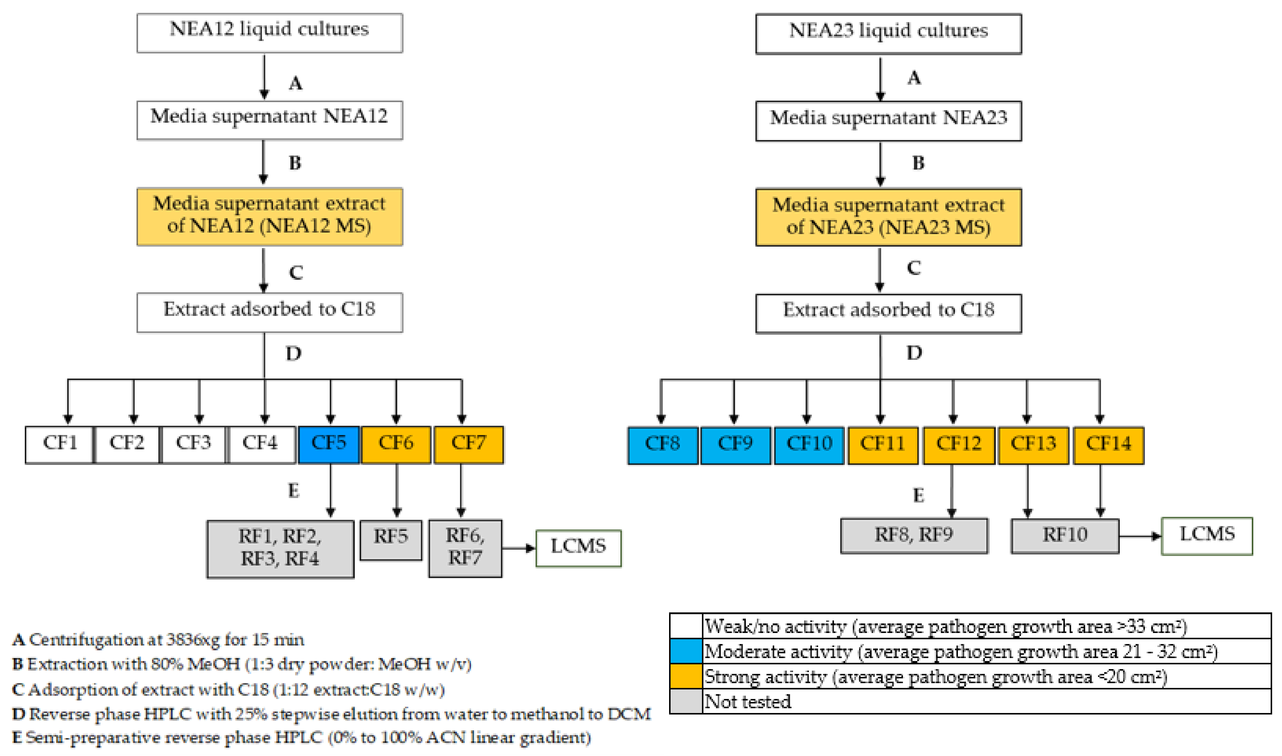
Figure 1. Step-wise bioassay-guided fractionation scheme for obtaining refined fractions of antifungal metabolites.
Metabolites 12 00037 g001
Figure 2. Agar-well diffusion assay for pathogen Ceratobasidium sp. in the presence of NEA12 MS derived crude fractions (CF). From left to right: Sterile water; antifungal compound carbendazim (1000 ppm); 80% methanol; 100% DCM; NEA12 MS; CF1; CF2; CF3; CF4; CF5; CF6 and CF7. All bioassay treatments and controls were prepared in replicates of five (n = 5). Temporal variation is described in the rows from day three to seven. The images are a typical representation of the five replicates.
Metabolites 12 00037 g002
Figure 3. Growth inhibition of Ceratobasidium sp. pathogen by Epichloë spp. in vitro culture extracts at day 6 (a) NEA12 MS and derived crude fractions (CF1, CF2, CF3, CF4, CF5, CF6, CF7); (b) NEA23 MS and derived crude fractions (CF8, CF9, CF10, CF11, CF12, CF13, CF14). Controls, sterile distilled water (SDW); carbendazim (1000 ppm); 80% methanol (MeOH), 100% dichloromethane (DCM). Image analysis measured growth area (cm2) of the pathogen in the agar well diffusion assay. All data are mean ± standard error, n = 5. Significance was determined by one-way ANOVA and Tukey simultaneous tests for differences of means; p < 0.01 indicates significant inhibition. Means that do not share a letter are significantly different.
Metabolites 12 00037 g003
Figure 4. Agar-well diffusion assay for pathogen Ceratobasidium sp. in the presence of NEA23 MS derived crude fractions (CF). From left to right: sterile water; antifungal compound carbendazim (1000 ppm); 80% methanol; 100% DCM; NEA23 MS; CF8; CF9; CF10; CF11; CF12; CF13; and CF14. All bioassay treatments and controls were prepared in replicates of five (n = 5). Temporal variation is described in the rows from day three to seven. The images are of a typical representation of the five replicates.
Metabolites 12 00037 g004
Figure 5. Principal component analysis scores plot (a) PC1 and PC2; (b) PC2 and PC3 of ESI+ UHPLC-HRMS data of NEA12 in planta (NEA12 PE, n = 48), NEA12 in vitro (NEA12 ME, n = 27), NEA23 in planta (NEA23 PE, n = 30), NEA23 in vitro and (NEA23 ME, n = 28).
Metabolites 12 00037 g005
Figure 6. Venn diagram showing the metabolite distribution of (a) NEA12 in planta (NEA12 PE, n = 48), NEA12 in vitro (NEA23 ME, n = 27) and NEA12 RF (RF1-RF7); (b) NEA23 in planta (NEA23 PE, n = 30), NEA23 in vitro (NEA23 ME, n = 28) and NEA23 RF (RF8-RF10); (c) NEA12 in planta (NEA12 PE, n = 48), NEA23 in planta (NEA23 PE, n = 30), and RF (RF1-RF10) by ESI+ MS.
Metabolites 12 00037 g006
Table 1. One-way ANOVA analysis results for pathogen growth.
Endophyte StrainSourceDF 1Seq SS 2ContributionAdj SS 3Adj MS 4F-Valuep-Value
Factor117054.196.63%7054.1641.282122.33<0.001
NEA12Error47246.43.37%246.45.242
Total587300.5100.00%
Factor117948.097.47%7948.0722.550168.00<0.001
NEA23Error48206.42.53%206.44.301
Total598154.5100.00%
1 Degrees of freedom, 2 Sequential sums of squares, 3 Adjusted sequence sum of squares and 4 Adjusted mean squares.
Table 2. Metabolites present in refined fractions extracted from NEA12 MS and NEA23 MS that are also present in the metabolomes of perennial ryegrass-endophyte symbiota (NEA12 PE and NEA23 PE).
Metabolite
No.
Massm/zRTPresence/Absence in Refined FractionsPresence in Planta (Number of Symbiota) 1Q-Value 2Directed Effect Size
(NEA23 PE: NEA12 PE)
NEA23 RFsNEA12 RFsNEA23 PENEA12 PE
M1242.0412243.04852.91+9230.189NA
M2255.2558256.263111.19+30470.0041.59
M3364.1280365.13539.14++30480.891NA
M4365.3625366.369712.95+3048<0.0011.47
M5365.3652366.372512.84+3048<0.0011:3.92
M6365.3682366.375512.95+3048<0.0011.57
M7367.4173368.424513.50+3048<0.0011:1.68
M8407.3153408.32268.36+09NANA
M9426.2376427.244910.80+25400.191NA
M10426.2375427.244810.90+30450.749NA
M11452.3370453.34428.61+340.316NA
M12452.3344453.34178.70+330.661NA
M13452.3341453.34148.72+02NANA
M14457.3394458.34679.92+29480.919NA
M15462.2965463.30389.83+30480.792NA
M16466.3496467.35699.25+30470.248NA
M17483.3763484.38359.24++4470.363NA
M18496.3602497.36748.71++15390.0041:7.06
M19527.4024528.40969.24++2043<0.0012.49
M20532.3574533.36479.24++8480.772NA
M21546.3005547.307711.85+1790.0241:2.39
M22571.4285572.43589.24++5240.945NA
M23576.3837577.39099.23++338<0.0013.93
M24589.4180590.42539.89+3040<0.0015.12
M25590.4236591.430811.79+3048<0.0011:2.20
M26594.3594595.36669.82+3048<0.0011.31
M27594.3744595.38179.90+3048<0.0011.82
M28607.4645608.471811.83+3100.234NA
M29615.4546616.46189.24++317<0.00111.86
M30620.4100621.41739.24++7290.218NA
M31634.4644635.471711.85+42NANA
M32651.4907652.498011.85+112NANA
M33662.4203663.42769.80+3048<0.0011.71
M34662.4171663.424314.30++3048<0.0011:4.53
M35662.4447663.452014.38++3048<0.0011.25
M36663.4473664.454614.33++3048<0.0011:1.71
M37678.3797679.386911.94+30480.235NA
M38678.4871679.494411.95+27480.453NA
M39684.4323685.439514.23+2948<0.0011:9.06
M40684.4297685.436914.53+3048<0.0011.58
M41684.4313685.438514.58+3048<0.0011.64
M42685.4159686.423214.29+3048<0.0011:3.27
M43695.5170696.524311.87+19NANA
M44695.5172696.524511.96+5320.086NA
M45721.4687722.475911.97+24370.174NA
M46762.5085763.515814.59+30480.177NA
M47762.5091763.516414.68+3048<0.0011.74
M48762.5095763.516814.74+3047<0.0012.04
M49762.5071763.514414.38++3048<0.0011.23
M50762.5090763.516314.54+3048<0.0011.54
M51763.4952764.502414.64+3048<0.0011.80
M52763.5110764.518214.42++3048<0.0012.48
M53763.5109764.518214.33++3048<0.0011:1.90
M54787.5037788.510912.10+2639<0.0011:1.19
M55788.5186789.525911.90+30430.919NA
M56788.5210789.528311.94+29400.419NA
M57802.5324803.539712.41+30480.607NA
M58802.5319803.539212.46+3048<0.0011.24
M59802.5335803.540812.26+3048<0.0011:1.34
M60802.5342803.541512.33+30480.960NA
M61858.5970859.604312.59+36<0.0011.18
1In planta extracts are from pseudo stems harvested from glasshouse maintained perennial ryegrass-endophyte symbiota. NEA12 populations (n = 48): Bronsyn (n = 24) and Alto (n = 24); NEA23 populations (n = 30): Bronsyn (n = 15) and Trojan (n = 15). 2 Benjamini-Hochberg adjusted p values.
Table 3. Solvent systems used in fractionation of media supernatant (MS) extracts collected from Epichloë sp. strains NEA12 and NEA23 grown in PDB.
Crude MS Extract StrainCrude FractionWater (%) Methanol (%)DCM (%)Dry Weight/(g)
NEA12 MSCF11000-40.69
CF28020-10.17
CF35050-4.36
CF42080-22.01
CF50100014.23
CF6-50507.38
CF7-010010.41
NEA23 MSCF81000-32.87
CF98020-8.13
CF105050-7.83
CF112080-7.26
CF12010003.29
CF13-50502.30
CF14-01003.51
Publisher’s Note: MDPI stays neutral with regard to jurisdictional claims in published maps and institutional affiliations.