Article

Automated Volume Status Assessment Using Inferior Vena Cava Pulsatility


1
Mathematical Biology and Physiology, Department of Electronics and Telecommunications, Politecnico di Torino, 10129 Turin, Italy
2
Integrative Physiology Lab, Department of Neuroscience, Universitá di Torino, 10125 Turin, Italy
3
Department of Medical Sciences, Universitá di Torino, 10126 Turin, Italy
*
Author to whom correspondence should be addressed.
Electronics 2020, 9(10), 1671; https://doi.org/10.3390/electronics9101671

Submission received: 14 September 2020 / Revised: 6 October 2020 / Accepted: 7 October 2020 / Published: 13 October 2020
(This article belongs to the Special Issue Biomedical Image Processing and Classification)

Abstract

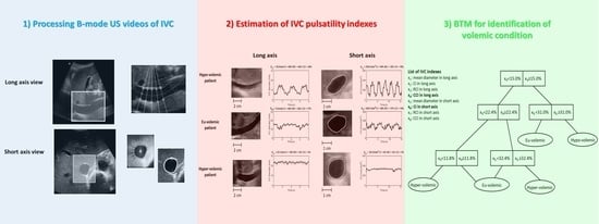
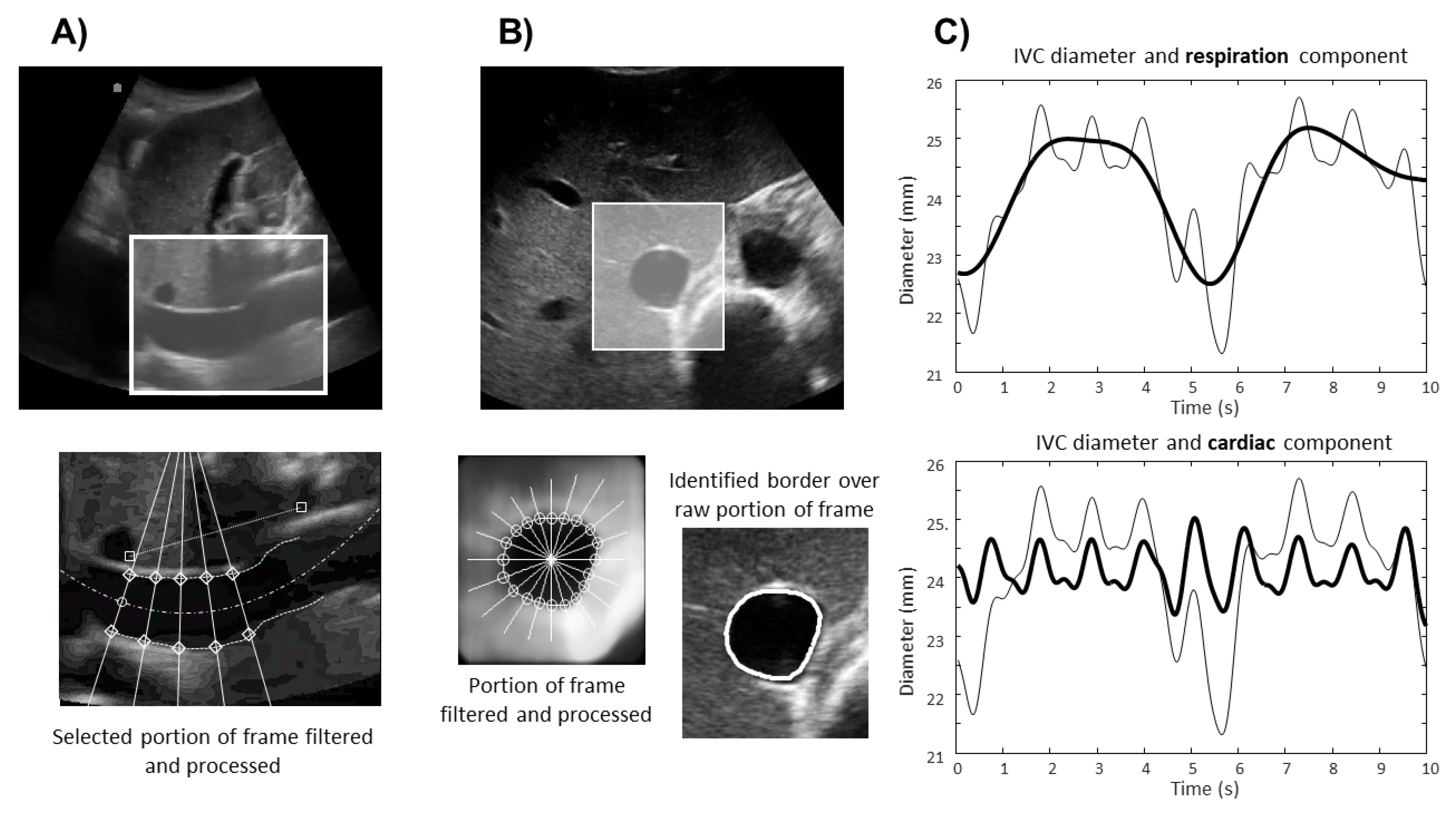
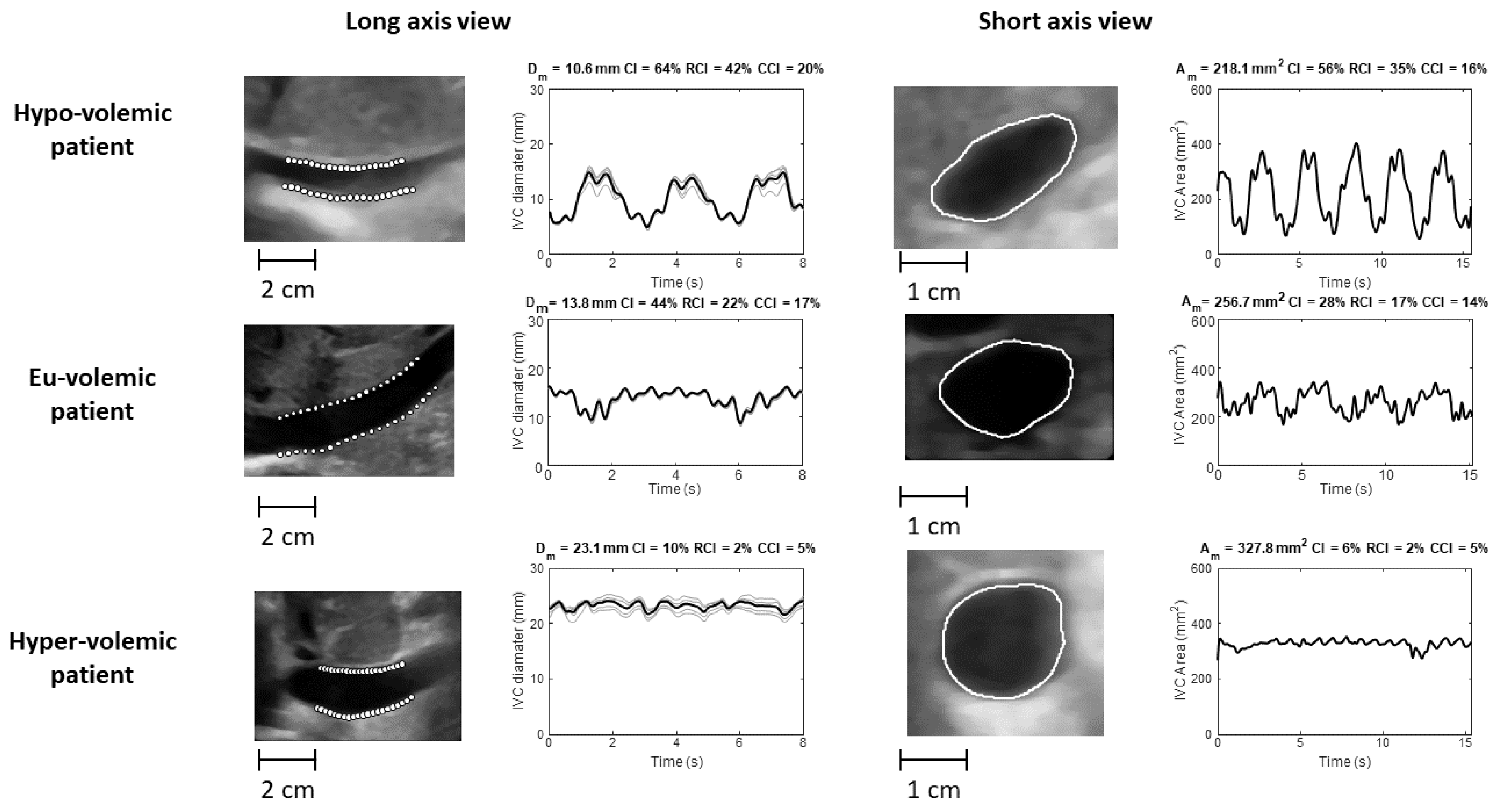
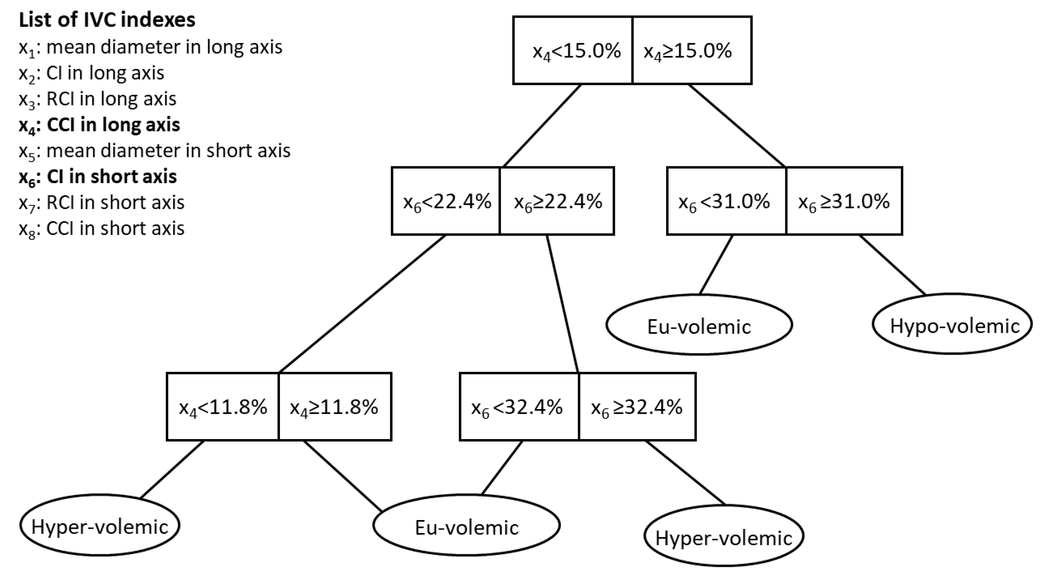
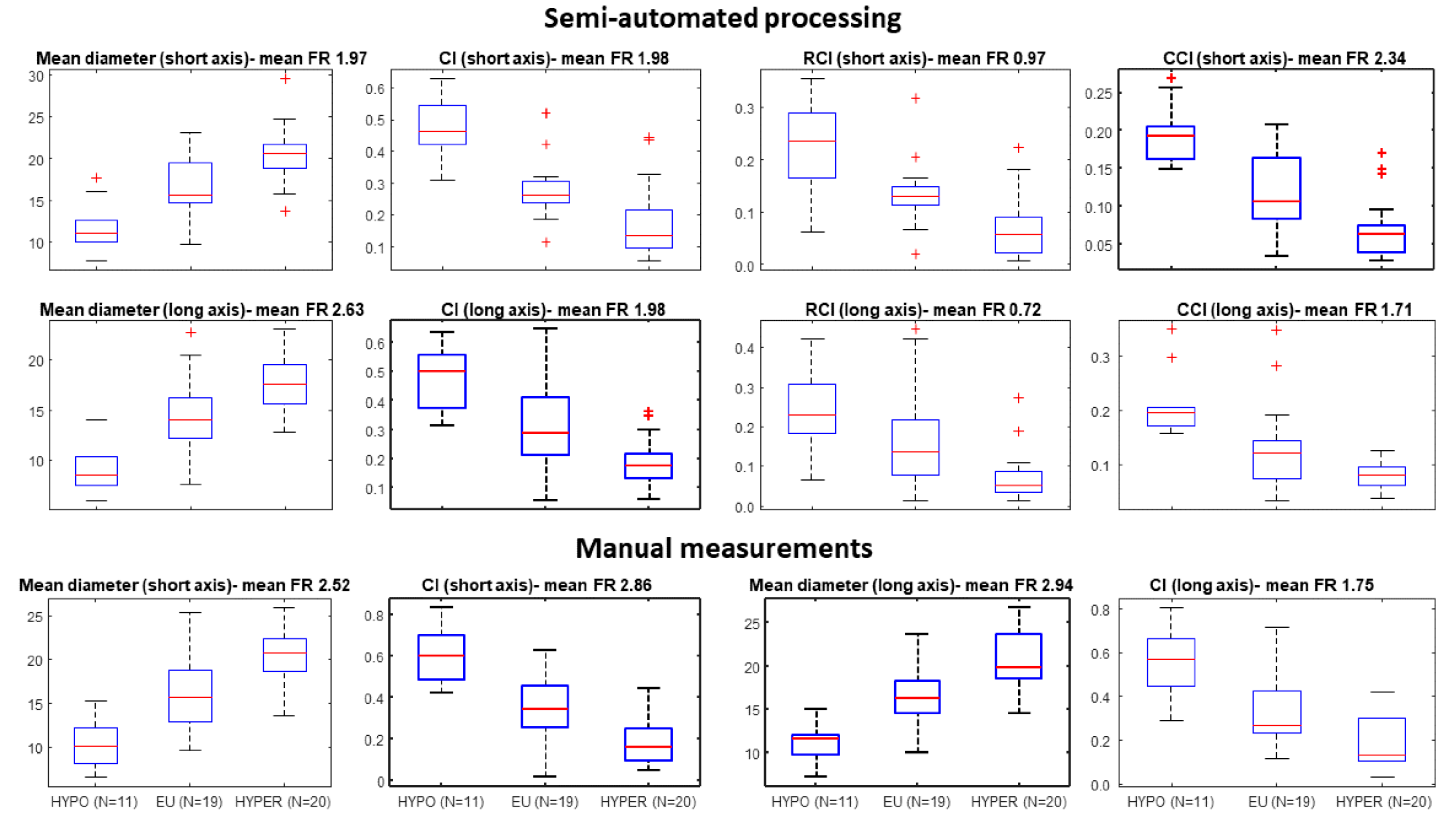
Assessment of volume status is important to correctly plan the treatment of patients admitted and managed by cardiology, emergency and internal medicine departments. Non-invasive assessment of volume status by echography of the inferior vena cava (IVC) is a promising possibility, but its clinical use is limited by poor reproducibility of current standard procedures. We have developed new algorithms to extract reliable information from non-invasive IVC monitoring by ultrasound (US) imaging. Both long and short axis US B-mode video-clips were taken from 50 patients, in either hypo-, eu-, or hyper-volemic conditions. The video-clips were processed to extract static and dynamic indexes characterizing the IVC behaviour. Different binary tree models (BTM) were developed to identify patient conditions on the basis of those indexes. The best classifier was a BTM using IVC pulsatility indexes as input features. Its accuracy (78.0% when tested with a leave-one-out approach) is superior to that achieved using indexes measured by the standard clinical method from M-mode US recordings. These results were obtained with patients in conditions of normal respiratory function and cardiac rhythm. Further studies are necessary to extend this approach to patients with more complex cardio-respiratory conditions.
Keywords: inferior vena cava; ultrasound imaging; binary tree model; pulsatility; fluid volume assessment inferior vena cava; ultrasound imaging; binary tree model; pulsatility; fluid volume assessment
Graphical Abstract