1. Introduction
Medical imaging plays an indispensable role in modern healthcare, enabling early diagnosis, treatment planning, and longitudinal monitoring of pathological conditions. Modalities such as magnetic resonance imaging (MRI), computed tomography (CT), and digital radiography (X-ray) are routinely utilized in clinical workflows and are frequently exchanged across hospital environments, telemedicine platforms, and cloud-based Picture Archiving and Communication Systems (PACSs). As a result, medical images have evolved into critical medical records whose authenticity and integrity directly influence diagnostic decision-making, patient safety, and medico-legal accountability [
1,
2,
3]. The reliability of these images is therefore paramount, and even seemingly insignificant pixel-level alterations—intentional or unintentional—can lead to misinterpretation of subtle anatomical structures or pathologies [
4,
5,
6]. To address this challenge, this work introduces a fundamentally non-embedding authentication paradigm in which MSB-class pixels are exploited as a deterministic carrier for block-wise cryptographic hashes of diagnostic ROI content—an approach that, to the best of our knowledge, has not been previously explored in the medical image authentication literature.
Ensuring secure and distortion-free integrity verification has emerged as a high-priority research problem in medical image security. Conventional authentication approaches rely heavily on digital watermarking, where auxiliary information is embedded into the image either in the spatial domain or in various transform domains. Spatial-domain watermarking, especially Least Significant Bit (LSB) substitution [
7], offers simplicity and high capacity, but embedded bits are easily disrupted by compression, noise, and image enhancement operations, making such methods inherently fragile [
8,
9]. Transform-domain watermarking—using the Discrete Cosine Transform (DCT), Discrete Wavelet Transform (DWT), or Singular Value Decomposition (SVD)—improves robustness by embedding in perceptually significant components [
10,
11,
12]. However, these techniques inevitably alter pixel intensities after inverse transformation [
6,
12], which is problematic for medical images where even minute distortions can obscure fine tissue textures or grayscale gradients that carry diagnostic value. This conflict between watermark robustness and clinical fidelity motivates the search for alternative authentication paradigms that avoid modifying the pixel data altogether.
Several attempts have been made to minimize distortion, most notably reversible watermarking [
13,
14] and Region of Interest (ROI)-based watermarking [
15,
16]. Reversible watermarking guarantees perfect recovery of the original image after watermark extraction, while ROI-based methods preserve diagnostic regions by embedding watermark data solely within the Non-ROI (NROI). However, both approaches still involve pixel-level manipulation in the NROI. Repeated compression, storage, or export operations can degrade these regions over time, potentially invalidating the watermark or causing false alarms [
3]. Moreover, typical reversible schemes require complex prediction or difference-expansion operations, adding significant computational overhead and sensitivity to noise [
13,
14].
In parallel, recent studies have explored deep learning-based representations for medical and remote-sensing image analysis, including CNN–Transformer hybrids and cross-layer feature interaction models. Such approaches learn high-level semantic features that are highly effective for tasks such as classification, segmentation, and enhancement [
17]. However, deep feature representations are inherently data-dependent and sensitive to retraining, domain shift, and post-processing operations, which limits their suitability for deterministic integrity verification and forensic authentication. In contrast, the proposed framework targets a different problem setting—fragile, distortion-free integrity assurance—by leveraging deterministic MSB-level properties rather than learned feature embeddings.
To fully preserve diagnostic quality, researchers have proposed non-embedding or reference-based authentication mechanisms, where integrity features such as hashes, signatures, or keypoint descriptors are stored externally instead of being written into the image [
18,
19,
20]. These techniques eliminate all image distortion and theoretically maintain perfect fidelity. However, most existing non-embedding approaches suffer from one or more limitations:
Unstable feature extraction: Gradient-based, keypoint-based, texture-based, or histogram-based descriptors vary significantly across modalities and even across repeated exports or mild brightness shifts [
9,
21].
Lack of tamper localization: Many methods detect whether an image has changed but cannot identify where modifications occurred [
3,
22].
Sensitivity to benign clinical adjustments: Routine operations such as WL changes, high-quality JPEG saving, annotation overlays, or mild noise injection often break authentication [
8,
21].
Non-determinism: Some methods rely on heuristic or non-deterministic feature selection, leading to inconsistent behavior across different imaging systems or post-processing pipelines [
9,
21].
These issues highlight the need for a secure, deterministic, and clinically aligned authentication scheme that preserves diagnostic content, supports localized tamper detection, and remains stable under routine imaging operations.
1.1. Motivationand Key Idea
The proposed framework revisits an often overlooked yet powerful observation: the Most Significant Bit (MSB) plane in medical images is highly stable under clinically benign operations [
1,
3,
22]. For 8-bit grayscale images, the MSB encodes the most dominant intensity contrast, and its value typically remains unchanged under mild brightness shifts, low-amplitude acquisition noise, high-quality JPEG compression, and annotation overlays in the Non-ROI. Unlike LSBs—which are notoriously unstable—the MSB plane provides a robust and deterministic structure suitable for secure reference generation.
Rather than embedding watermark bits, we utilize the existing MSB pattern of the NROI as a passive integrity carrier. In contrast to traditional watermarking schemes, the proposed framework does not embed any authentication data into the medical image; instead, integrity verification is achieved through an external, reference-based mapping that leaves the image content entirely unchanged. The ROI is divided into blocks, each of which is hashed using a cryptographic function. The binary composition of each block hash is then deterministically mapped onto selected MSB-class pixels in the NROI. Only the coordinates of these pixels are encrypted and stored externally, ensuring perfect fidelity and complete DICOM compliance [
23].
During verification, the ROI is rehashed and compared with the MSB composition of the referenced NROI coordinates. A mismatch immediately identifies the location of tampering. Because MSBs remain stable under benign modifications, the integrity test remains reliable even after reversible post-processing operations [
22].
1.2. Contributions and Advantages
Beyond preserving diagnostic fidelity, the proposed framework demonstrates that MSB-class pixels can be reliably exploited as a deterministic, non-embedding carrier for block-wise cryptographic hashes, enabling precise tamper localization without modifying any pixel values. Extensive multi-modality experiments further confirm that this strategy achieves perfect detection of intentional ROI tampering, stable behavior under clinically benign Non-ROI operations, and consistent performance across MRI, CT, and X-ray images. Precisely, this work introduces a clinically conscious and computationally lightweight authentication framework with the following advantages:
Complete preservation of diagnostic content: No pixel values are altered, yielding PSNR = ∞ and SSIM = 1.
Deterministic mapping from ROI to NROI: The MSB class-based mapping ensures reproducibility across modalities and imaging systems.
Block-level tamper localization: ROI block hashing provides spatial localization.
Robustness to benign clinical modifications: MSB stability reduces false alarms.
Low computational complexity: Only hashing + MSB extraction + coordinate matching.
Modality independence: Validated across MRI, X-ray, and CT datasets.
The remainder of this paper is organized as follows.
Section 2 reviews related work on medical image authentication and integrity verification.
Section 3 details the proposed deterministic MSB-based non-embedding authentication framework.
Section 4 presents the experimental evaluation and discusses the results.
Section 5 concludes the paper and outlines directions for future work.
3. Proposed Methodology
This section presents a diagnostic-preserving, non-embedding authentication framework for medical images based on deterministic MSB-driven reference mapping. The method establishes a secure linkage between each ROI block and a set of carefully selected NROI pixels whose most significant bits (MSBs) collectively encode that block’s cryptographic hash. Crucially, the framework introduces no distortion: the medical image remains bitwise identical to its original form. Integrity verification is achieved by rehashing each ROI block and comparing its fresh hash bit-by-bit against the MSBs extracted from a stored set of encrypted NROI coordinates.
The framework comprises two phases:
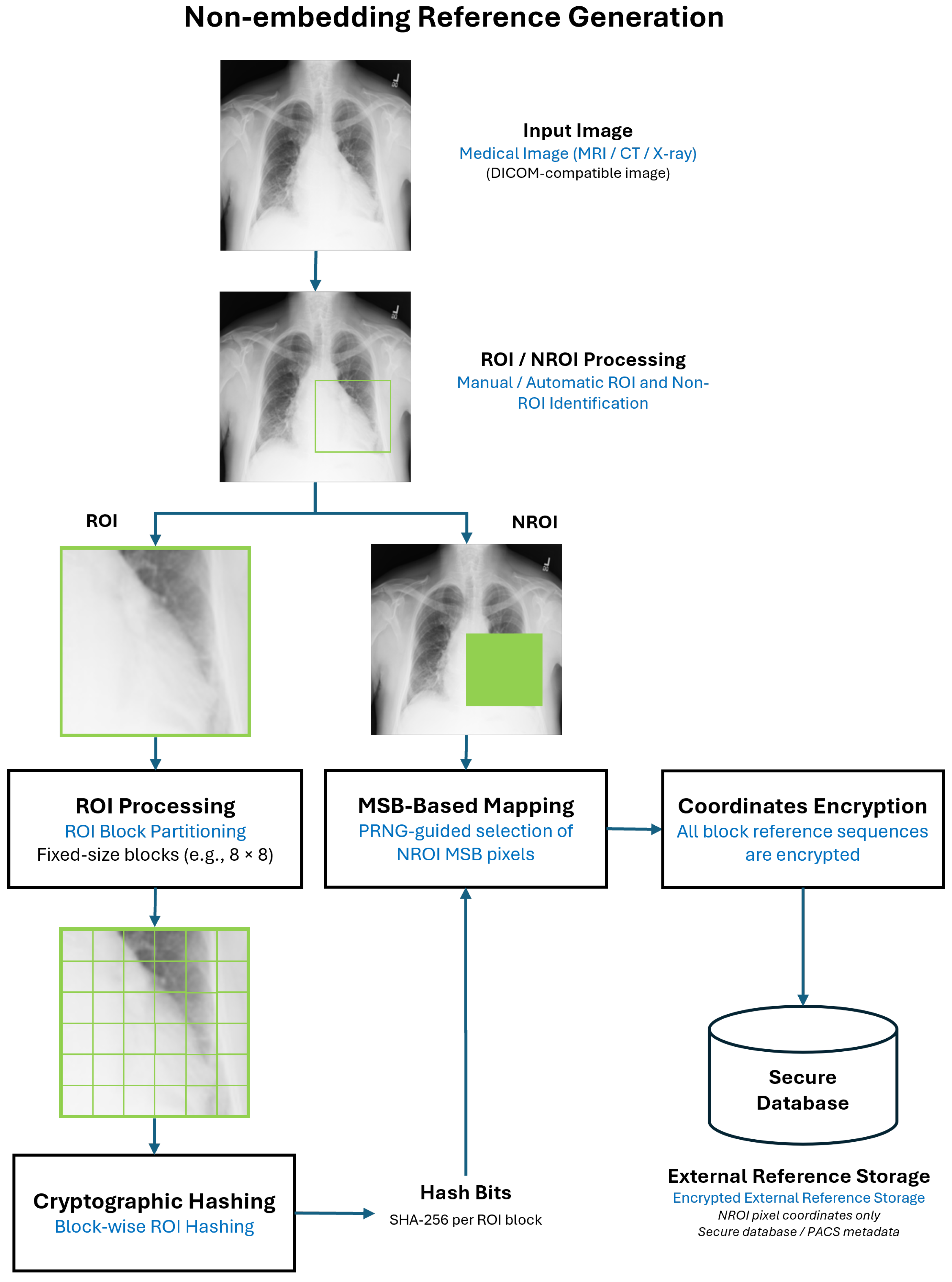
Reference Generation—the ROI is divided into blocks, each block is hashed using SHA-256, and each hash bit is mapped to a randomly chosen NROI pixel whose MSB matches that bit.
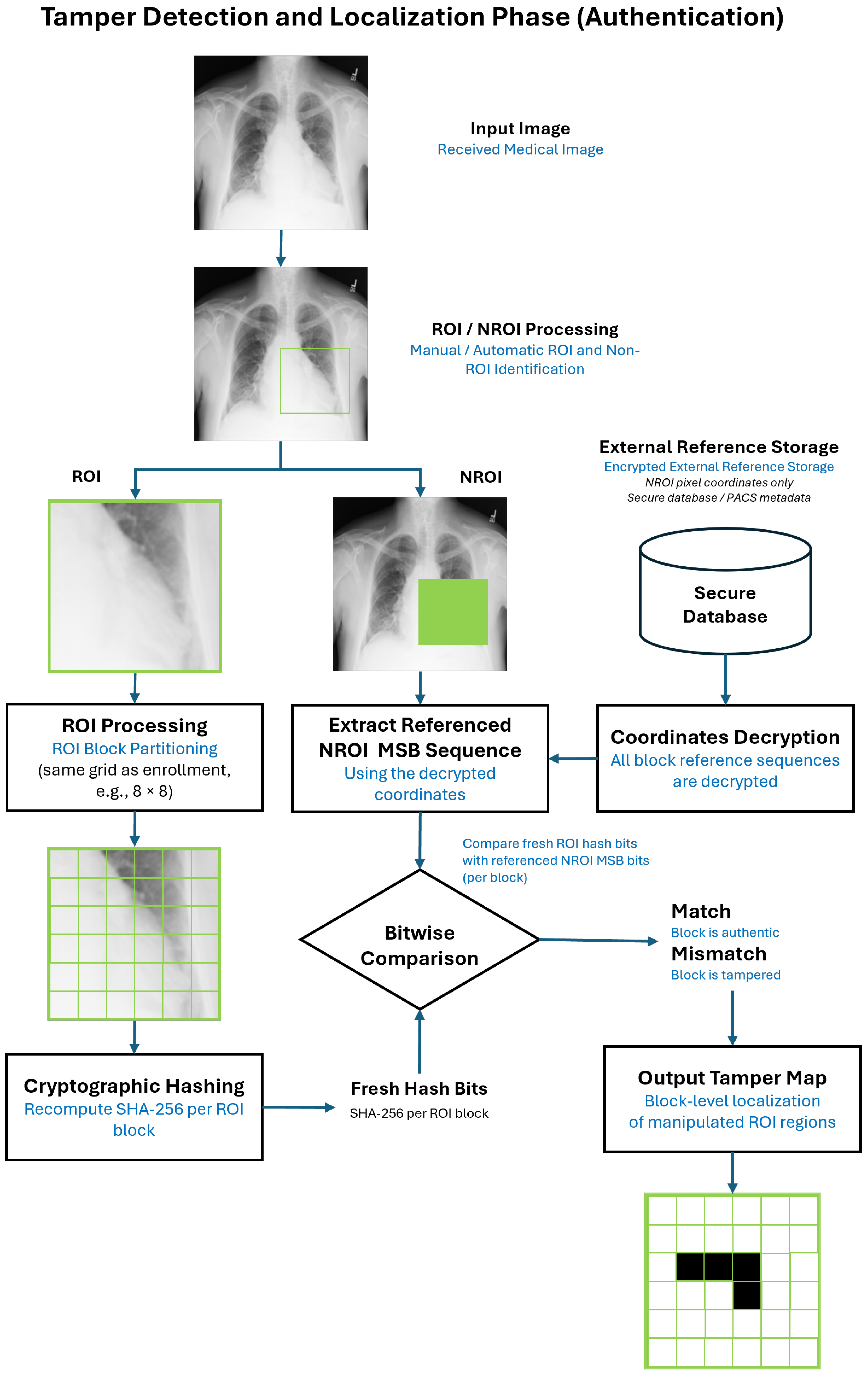
Verification and Tamper Localization (Authentication)—the ROI is rehashed, and the extracted MSBs from the stored NROI coordinates are compared bit-by-bit with the new hash to detect and localize tampering.
An overview of the reference generation and authentication workflows is illustrated in
Figure 1 and
Figure 2, respectively. The subsections below describe each component of the system in detail. The full reference generation and verification procedures are summarized at the end of the section in Algorithms 1 and 2.
3.1. System Overview
The proposed framework operates on an 8-bit grayscale medical image
I that is decomposed into a diagnostically relevant Region of Interest (ROI),
, and a complementary Non-ROI (NROI),
:
The ROI is the region whose integrity must be preserved and authenticated, while the NROI contains surrounding structures or background content that does not affect clinical interpretation.
Unlike watermarking techniques, which alter pixel intensities to embed authentication data, the proposed framework is completely non-destructive. No pixel in either or is modified. Instead, the system uses the NROI as a passive reference spaceThe method exploits the empirical stability of the MSB layer of under routine clinical operations such as mild brightness adjustment, low-amplitude noise, standard PACS resampling, and high-quality JPEG compression. Because the MSB represents coarse luminance structure, it tends to remain stable under such benign transformations, making it suitable for serving as a binary reference plane.
In the proposed framework, each ROI block is hashed using SHA-256, producing a 256-bit sequence. For every bit in this hash, the system selects one NROI pixel whose MSB matches the corresponding bit value. The coordinates of the selected NROI pixels are encrypted and stored as the authentication reference. During verification, the ROI is rehashed and the MSBs of these coordinates are re-extracted; tampering is detected whenever the two 256-bit sequences differ.
This formulation enables tamper-sensitive authentication and precise block-level localization without any modification to the underlying image data and without sacrificing diagnostic or DICOM fidelity.
3.2. ROI Partitioning and Block-Hash Generation
Block-wise hashing of diagnostic regions has been considered in prior image authentication and integrity verification schemes, typically as a means of detecting whether localized content has been altered. In such approaches, however, block hashes are commonly embedded into the image, stored as global side information, or used only as coarse integrity indicators without enabling fine-grained, distortion-free localization. In contrast, the proposed framework introduces a fundamentally different usage of block hashing: each ROI block hash is decomposed at the bit level and deterministically mapped onto MSB-class pixels in the Non-ROI, without embedding or modifying any pixel values. This design elevates conventional block hashing from a standalone integrity check to a passive, spatially distributed reference mechanism that enables precise block-level tamper localization while preserving perfect diagnostic fidelity.
To enable localized assessment of image integrity, the ROI is partitioned into a uniform grid of non-overlapping blocks:
The number of blocks must balance two competing considerations: (1) spatial localization granularity, and (2) computational efficiency. A coarse grid such as produces only 16 large blocks. Although computationally efficient, such large blocks yield overly coarse localization: even a small manipulation affecting only a small region may cause the entire block to be marked tampered. Conversely, a fine grid such as results in 256 small blocks, enabling high-resolution localization but substantially raising computation cost due to the larger number of hashes that must be computed and compared.
As demonstrated experimentally in
Section 4, a block grid of
provides an effective compromise: it yields clinically meaningful localization granularity while maintaining robustness to benign manipulations and retaining moderate computational complexity. For each block
b, a SHA-256 hash is computed:
where each
. Due to the avalanche property of cryptographic hash functions, even a minimal alteration in
produces a drastically different 256-bit sequence, making
an extremely sensitive indicator of tampering.
3.3. MSB-Class Partitioning of the NROI
Each NROI pixel
is expressed in its 8-bit binary form:
where
is the most significant bit (MSB). The MSB encodes coarse luminance structure and remains relatively stable under many benign clinical procedures. The NROI is partitioned into two disjoint MSB classes:
These classes serve as large candidate pools from which the system selects NROI coordinates whose MSBs match individual bits of each ROI block hash. Because real medical images typically contain thousands of pixels in both sets, and provide a rich and diverse reference space that supports deterministic and secure mapping. This MSB-classification operation forms the basis of the reference mapping procedure summarized later in Algorithm 1.
3.4. Bit-by-Bit Deterministic Mapping
For each ROI block
b, the 256-bit hash sequence
is encoded by selecting a sequence of 256 NROI pixels whose MSBs match the hash bits. A pseudorandom number generator (PRNG), seeded with key
, ensures deterministic and repeatable selection. For each bit position
k:
This yields a reference sequence:
where the MSB of each referenced pixel satisfies:
The union of all block reference sequences is encrypted to produce the final authentication record:
Here, E represents the externally stored authentication reference that binds each ROI block to its corresponding set of MSB-matched Non-ROI coordinates. Encrypting ensures that the spatial locations used for verification cannot be inferred or manipulated without access to the secret key, while allowing deterministic recovery of the reference during the verification phase. This design enables secure, distortion-free authentication without embedding any auxiliary data into the medical image itself. This indirect, bit-by-bit mapping allows the framework to encode each ROI block hash without altering the image and without relying on sequential pixel search strategies.
For clarity and good cryptographic practice, the framework assumes logical separation between keys used for different functions. In particular, the PRNG seed used for deterministic coordinate selection, the encryption key used to protect stored NROI coordinates, and the authentication key used for integrity verification are treated as independent cryptographic keys. In practice, these keys may be derived from a master secret using a standard key-derivation function (KDF), ensuring key separation without increasing system complexity or storage overhead.
3.5. Verification and Tamper Localization
During verification, the ROI of the received image
is rehashed:
Simultaneously, the MSBs of the referenced NROI coordinates are re-extracted:
where
denotes the decrypted coordinate sequence corresponding to block
b. The block
b is authenticated if and only if:
Any mismatch between the two bit sequences identifies block
b as tampered. Because the comparison is performed independently for each block, the method naturally yields block-level localization of manipulated regions. This operation is formally summarized in Algorithm 2.
3.6. Algorithms
Algorithms 1 and 2 describe the proposed framework as two complementary procedures: reference generation and verification (tamper detection and localization). The algorithms are presented in executable form to clarify how the theoretical mapping described in
Section 3 is realized step by step.
Algorithm 1 explicitly shows how each ROI block hash is converted into a fixed-length sequence of Non-ROI coordinates whose MSB values match the hash bits. The PRNG seeding guarantees that the same coordinate sequence is reproduced for a given image and key, while the final encryption step consolidates all block-level references into a single protected authentication record.
| Algorithm 1 Bitwise MSB-Driven Reference Generation |
Require: ROI , NROI , grid size G, key Ensure: Encrypted reference E 1: Partition into blocks 2: Build MSB classes and from 3: for each block b do 4: 5: Initialize PRNG with and block index 6: for to 256 do 7: if then 8: random element of 9: else 10: random element of 11: end if 12: end for 13: 14: end for 15: 16: return E
|
| Algorithm 2 Bitwise Verification and Block-Level Tamper Detection |
Require: Received image , encrypted reference E, key Ensure: Tamper map T 1: Decrypt E to obtain 2: for each block b do 3: Compute fresh hash 4: for to 256 do 5: 6: end for 7: if then 8: Block b is authentic 9: else 10: Block b is tampered 11: end if 12: end for 13: Build tamper map T 14: return T
|
Algorithm 2 mirrors the enrollment process by recomputing each ROI block hash and comparing it bit-by-bit with the MSBs extracted from the corresponding decrypted Non-ROI coordinates. Because this comparison is performed independently per block, tamper localization arises naturally from the verification procedure without requiring additional post-processing.
3.7. External Reference Storage and System Integration
The proposed framework stores only encrypted reference coordinates corresponding to MSB-class pixels in the Non-ROI, rather than embedding any information within the image itself. For a typical configuration using an ROI block partitioning and a 256-bit cryptographic hash per block, this results in a fixed and predictable external reference size on the order of tens of kilobytes per image. Even after encryption and metadata overhead, the total storage requirement remains negligible when compared to the size of medical images and associated clinical records routinely managed by modern PACS and cloud-based healthcare systems.
In practical deployments, these encrypted reference records are intended to be stored alongside existing image metadata, audit logs, or database entries maintained by the hosting system. Storage reliability, redundancy, backup, and access control are therefore handled at the system level using established mechanisms already employed for medical data management, including role-based access control, secure databases, and routine backup policies. The proposed authentication framework does not impose additional constraints on storage architecture and is compatible with both centralized and distributed medical imaging infrastructures.
By externalizing reference storage and delegating reliability and access control to the surrounding clinical information system, the proposed approach preserves image fidelity while remaining scalable and deployable within real-world healthcare environments.
3.8. Security Considerations and Threat Model
The proposed deterministic MSB-based mapping is designed for fragile integrity verification within controlled medical imaging workflows rather than for adversarially robust watermark survival under unrestricted pixel manipulation. The framework assumes that the diagnostic ROI and the referenced Non-ROI are managed within trusted environments such as PACS archives, cloud repositories, or regulated telemedicine platforms, where unauthorized large-scale manipulation of image content is restricted through access control, audit logging, and secure storage mechanisms.
Although, in theory, an adaptive adversary with unrestricted access to both ROI and NROI pixels could attempt to manipulate MSB values at specific locations to increase the probability of authentication evasion, such attacks face several practical constraints. First, the referenced NROI coordinates are encrypted and stored externally, preventing direct identification of queried pixels. Second, successful manipulation would require coordinated MSB control across hundreds of dispersed NROI locations per ROI block, significantly increasing attack complexity and the risk of introducing perceptible artifacts. Third, systematic MSB flipping across large NROI regions is likely to trigger integrity failure or visual anomalies detectable during routine clinical review.
It is important to note that these considerations apply broadly to all fragile authentication and reference-based schemes, including zero-watermarking and hash-based verification methods. The proposed framework therefore prioritizes deterministic reproducibility, diagnostic preservation, and precise tamper localization under realistic clinical threat models, while acknowledging that protection against fully adaptive adversaries manipulating both ROI and NROI lies beyond the scope of fragile image authentication and is instead addressed through system-level security controls.
4. Experimental Evaluation and Discussion
This section provides a comprehensive experimental evaluation of the proposed non-embedding, MSB-driven authentication framework. The evaluation aims to rigorously examine its performance across clinically relevant conditions and security-critical scenarios. In particular, the experiments assess six key properties: (i) zero-distortion fidelity, ensuring complete diagnostic preservation; (ii) accurate block-level tamper localization within the ROI; (iii) feasibility and determinism of the MSB-class mapping mechanism; (iv) robustness to benign modifications applied to the Non-ROI; (v) sensitivity to ROI block size; and (vi) generalization across imaging modalities including MRI, CT, and X-ray. Together, these evaluations provide a holistic understanding of the framework’s reliability, practicality, and suitability for deployment in real clinical environments.
4.1. Datasets and Experimental Setup
Experiments were conducted on three publicly available datasets representing distinct modalities and anatomical structures:
OASIS (MRI)—T1-weighted structural brain slices [
26], commonly used for neuroanatomical analysis and characterized by high soft-tissue contrast and fine structural detail,
ChestX-ray14 (X-ray)—frontal chest radiographs [
27], covering a wide range of thoracic appearances and intensity distributions typical of routine radiographic screening,
CT-ORG (CT)—abdominal CT slices with organ structure variation [
28], featuring heterogeneous soft-tissue regions and modality-specific intensity dynamics.
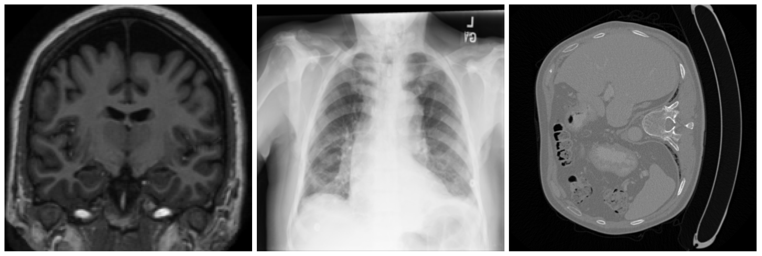
Representative examples from the three datasets are shown in
Figure 3. All images were converted to 8-bit grayscale and uniformly resized to
pixels to ensure consistent processing across modalities. This normalization facilitates fair evaluation of the proposed framework under comparable spatial and intensity conditions.
Regions of interest were extracted using automated segmentation pipelines tailored to each modality: center-brain foreground extraction for MRI, lung-field segmentation for X-ray, and soft-tissue abdominal foreground extraction for CT. These ROIs correspond to diagnostically relevant regions whose integrity must be preserved during authentication.
All remaining pixels were designated as the Non-ROI and used exclusively for MSB-class mapping. The ROI was partitioned into
non-overlapping blocks, a configuration selected based on the analysis provided in
Section 3. All experiments were implemented in Python (v3.10) using OpenCV, NumPy, and PyCryptodome, and executed on a workstation equipped with an Intel Ultra 7 CPU and 32 GB of RAM.
4.2. Zero-Distortion Fidelity
Because the proposed method performs no embedding, the authenticated image
is identical to the original image
I:
This ensures perfect diagnostic preservation and strict DICOM compliance, unlike embedding and reversible watermarking techniques that introduce temporary distortion [
23].
4.3. Feasibility and Determinism of MSB-Class Mapping
To assess the reliability of block-wise MSB-based mapping, we evaluated the availability of MSB = 0 and MSB = 1 pixels within the Non-ROI, as well as the feasibility and reproducibility of coordinate sampling across all tested modalities. This analysis ensures that the MSB-class pools are sufficiently populated to support deterministic mapping for every ROI block.
4.3.1. MSB-Class Availability
We computed the number of MSB = 0 and MSB = 1 pixels in the Non-ROI for all tested images in each dataset, and
Table 1 reports the average counts per modality. These results show that both MSB classes are abundantly represented across MRI, X-ray, and CT images, confirming that MSB-based reference mapping is consistently feasible and not affected by modality-specific distribution skewness.
These observations demonstrate that MSB-based matching remains reliable even in images with relatively homogeneous intensity regions. Because the framework relies on the binary composition of MSB classes rather than strict sequential pattern matching, it remains fully functional even when the underlying MSB distribution is moderately skewed.
4.3.2. Coordinate Selection Feasibility
Each ROI block requires 256 MSB-based coordinates to represent its SHA-256 hash. Since for all modalities, no block experienced sampling failure.
4.3.3. Deterministic Reproducibility
The proposed coordinate-selection mechanism is inherently deterministic. Given that (i) the MSB-class partitions of the Non-ROI are fixed for a given image and (ii) the pseudo-random generator is seeded with a constant key , the resulting coordinate sets are guaranteed to be identical across repeated executions. Thus, the framework ensures reproducible reference generation without requiring additional stochastic validation.
4.4. Tamper Detection and Localization
A comprehensive medical-image authentication framework must support both tamper detection, i.e., determining whether the image has been altered, and tamper localization, i.e., identifying the specific regions within the diagnostic content that were modified. The proposed non-embedding framework achieves these capabilities through block-wise hashing of the Region of Interest (ROI). Each block preserves an independent cryptographic signature derived from its pixel intensities, while the Non-ROI stores a spatially distributed, MSB-based mapping of the hash. Any manipulation applied to an ROI block leads to a mismatch between its recomputed hash and the reference hash retrieved from the Non-ROI, thereby enabling direct localization of tampered regions at the block level. This mechanism provides structured, interpretable, and diagnostically meaningful localization without altering the original image.
4.4.1. BlockSize Selection and Sensitivity Analysis
The choice of ROI block-grid size directly determines the spatial precision and robustness of tamper localization. In the proposed framework, the ROI is divided into a grid of blocks, where each block covers of the ROI area. Thus, the block-grid size G controls the number of authentication units: smaller G yields fewer, larger blocks (coarse localization), whereas larger G produces many smaller blocks (fine localization).
Selecting an appropriate grid size requires balancing three key factors:
Localization granularity: Larger grids (e.g., ) offer fine-grained localization but create many small blocks that can become overly sensitive to small anatomical fluctuations.
Robustness to benign changes: Smaller grids (e.g., ) produce fewer, larger blocks that are highly stable but provide only coarse localization.
Hash discriminability: Blocks that are too large or too small may produce hash signatures that either mask local tampering (too large) or become unstable (too small).
To characterize this trade-off, we evaluated three grid sizes frequently used in block-based integrity verification: , , and . Their behavior is summarized below.
A 4 × 4 grid partitions the ROI into 16 large blocks. This configuration is computationally light and highly stable, since each block contains substantial anatomical content. However, localization granularity is coarse. Localized tampering—such as inserting a small synthetic lesion, smoothing a boundary, or subtly erasing part of the texture—affects only a small part of a large block, yet the entire block is marked tampered. This reduces diagnostic utility when precise localization is important.
The grid provides the most balanced behavior. Blocks are small enough to resolve clinically meaningful manipulations while remaining large enough to retain stable texture characteristics. Across MRI, CT, and X-ray modalities, the configuration exhibited:
This configuration preserved both sensitivity and specificity during tamper localization, making it ideal for clinical scenarios.
A 16 × 16 grid achieves the highest localization precision, with 256 small blocks allowing fine identification of small manipulated regions. However, the small block size increases sensitivity to natural texture variation, noise, and modality-specific inhomogeneity (particularly in MRI). As a result, although this configuration can delineate tampering at high resolution, it introduces greater instability and computational cost due to the increased number of hashes.
Based on these observations, all experiments in this study employed an block grid, which provides a clinically meaningful balance—strong localization accuracy, stable hash signatures, and robustness across imaging modalities—without the excessive sensitivity or computational overhead associated with finer grids.
4.4.2. Manipulation Types
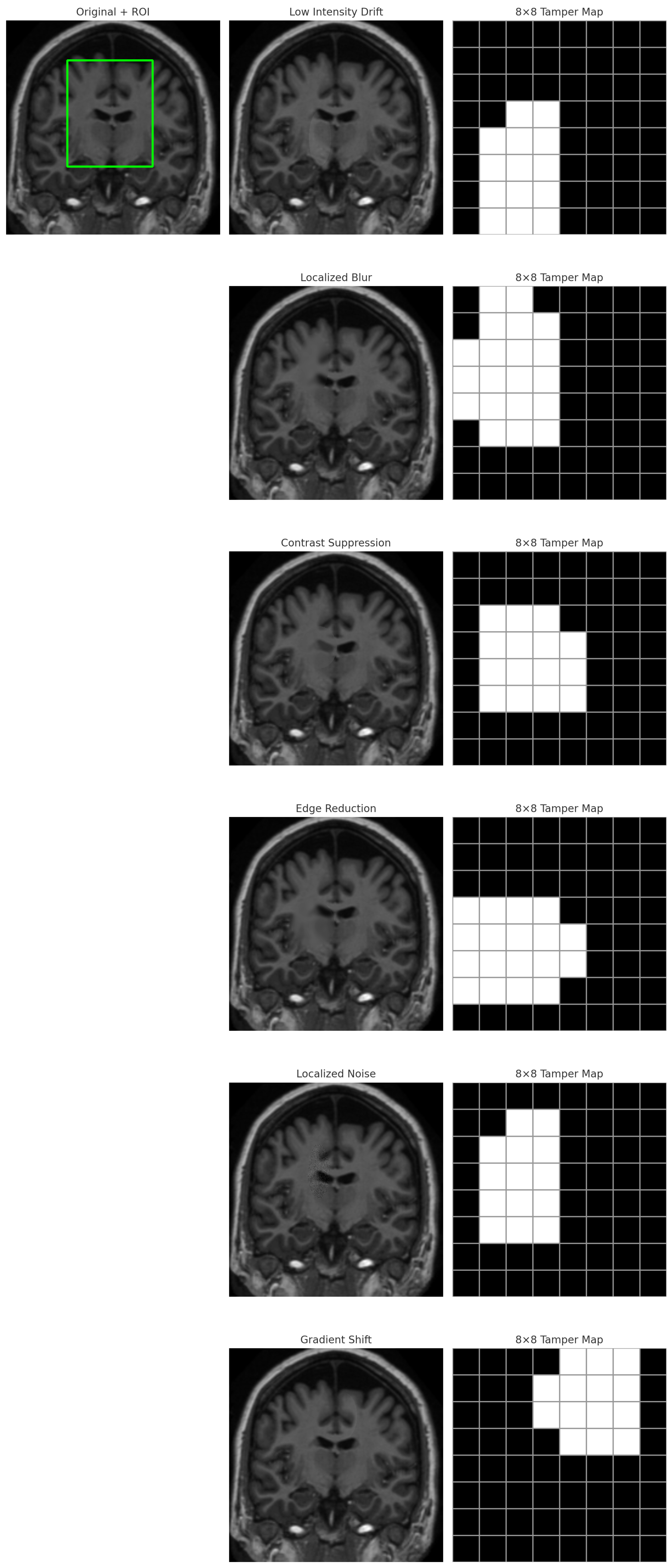
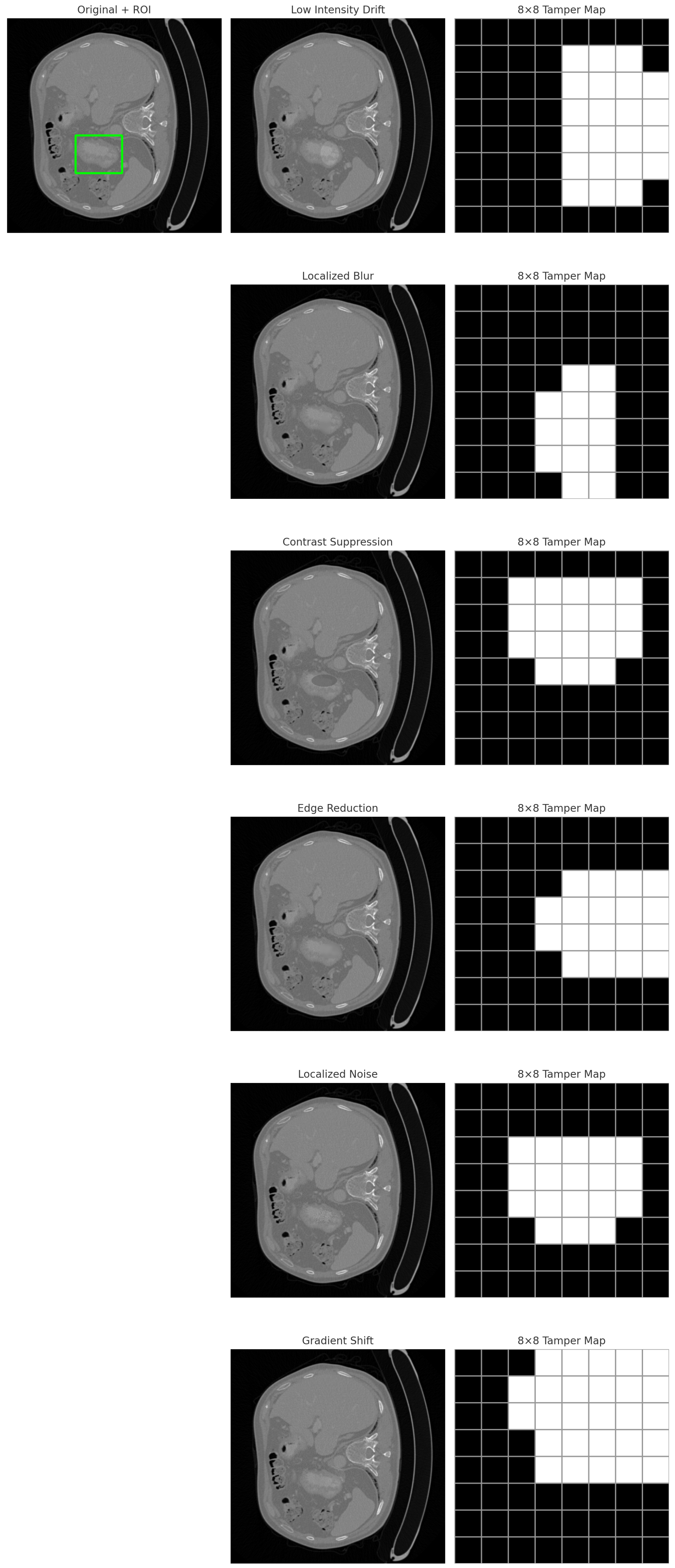
To rigorously evaluate the localization capability, six tampering operations were applied to controlled subregions of the ROI. These manipulations were chosen to represent realistic clinical, forensic, and adversarial scenarios:
Brightness Drift: Localized exposure or intensity lifting, used in practice to obscure tissue boundaries or highlight specific regions.
Gaussian Blurring: Smooths local texture and can conceal lesions or anatomical irregularities. It represents both intentional obfuscation and artifacts introduced by smoothing operations.
Contrast Suppression: Decreases local contrast, diminishing diagnostic boundaries and blurring subtle structures such as lesions, nodules, or microcalcifications.
Gradient/Erosion: Reduces edge sharpness, removing structural features and weakening anatomical edges. This simulates intentional masking of diagnostically important boundaries.
False Lesion Insertion: Introduces synthetic bright or dark regions that mimic pathological features. This represents a critical adversarial scenario, as false lesion insertion can alter diagnosis or severity assessment.
Patch-wise Noise Injection: Introduces high-frequency noise in a localized area, creating artificial texture inconsistencies and disrupting clinical interpretation.
All manipulations were spatially restricted to subregions of the ROI, ensuring that the tamper maps contained both correct positives (within the tampered region) and correct negatives (untouched ROI blocks), providing a robust test of localization accuracy.
4.4.3. Localization Visualization
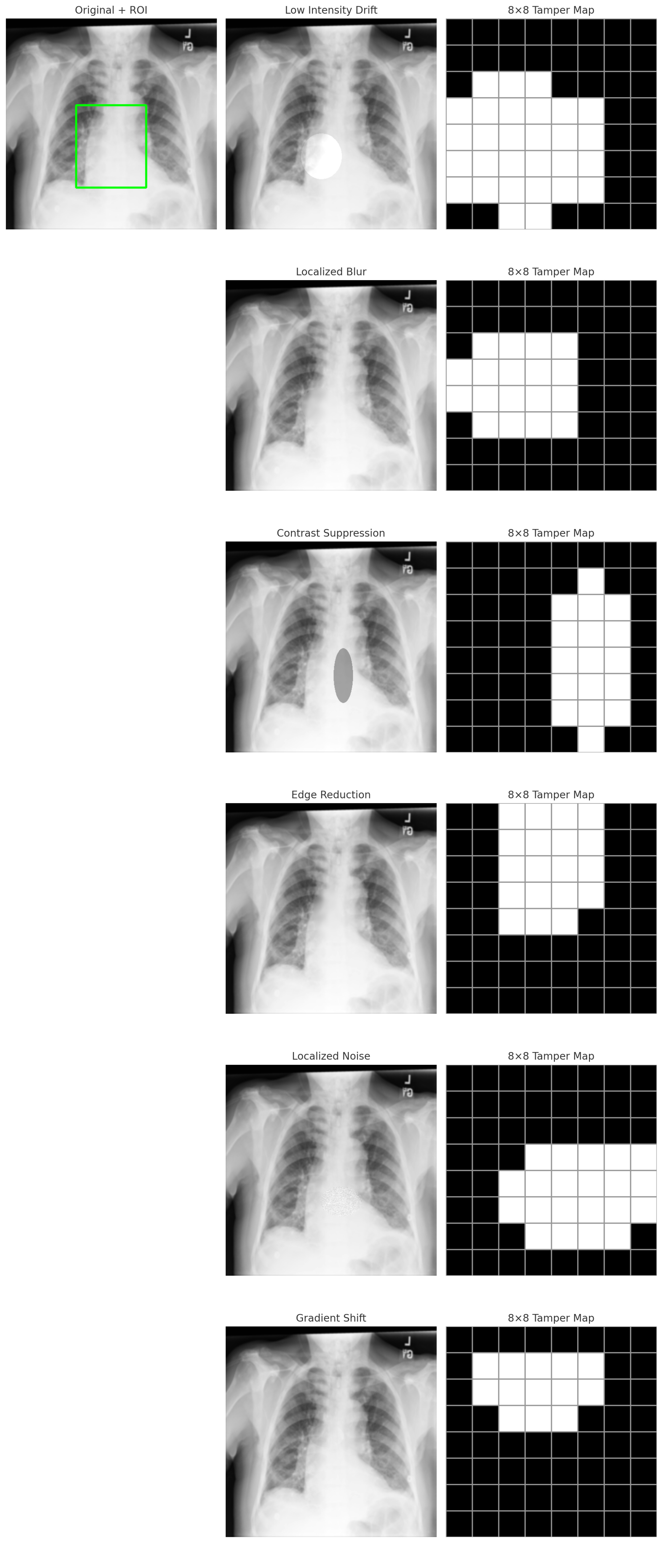
The localization results across modalities are shown in
Figure 4,
Figure 5 and
Figure 6. Each figure includes: (i) the manipulated ROI, and (ii) its corresponding tamper map. White blocks denote tampered blocks where hash mismatches occurred, and black blocks correspond to authentic blocks.
Visually, the resulting maps exhibit clear and sharply bounded regions, accurately capturing the spatial extent of tampering. The boundaries align consistently with ground truth, reflecting the effectiveness of the blocks in both precision and robustness.
4.4.4. Localization Metrics
Quantitative evaluation was performed using two standard forensic metrics commonly employed in digital image authentication [
22]:
True Positive Rate (TPR) measures sensitivity to altered blocks.
False Positive Rate (FPR) measures specificity, ensuring no intact blocks are falsely flagged.
An effective localization system must exhibit high TPR (capturing all manipulated regions) while maintaining low or zero FPR (avoid falsely flagging untampered regions).
4.4.5. Localization Results
Across all datasets (MRI, X-ray, CT) and manipulation types, the proposed framework achieved the following results for all intentional ROI tampering scenarios evaluated in this study:
All manipulated blocks were correctly detected, and no authentic ROI blocks were falsely marked under the defined experimental protocol. These results emphasize the effectiveness of the proposed signature-based approach and validate the selection of the block size as a critical factor in achieving both robustness and fine-grained localization.
The experimental findings demonstrate that the proposed framework achieves:
Accurate block-level tamper detection across modalities.
Precise localization of manipulated subregions.
Perfect sensitivity and specificity for intentional ROI tampering across all tested manipulation types.
Strong robustness against anatomical variability and image noise.
Consistent cross-modality performance.
The results indicate that the system is capable of identifying subtle, clinically significant manipulations without producing false alarms, fulfilling a core requirement for diagnostic image integrity verification in telemedicine, PACS archiving, and medico-legal environments.
4.5. Robustness to Benign Clinical Modifications
In routine medical imaging workflows, diagnostic images frequently undergo non-destructive manipulations that do not alter clinical content. Examples include mild brightness adjustment, denoising for visualization, and high-quality JPEG recompression when exporting scans between PACS systems. Because such operations do not modify the anatomical structures within the ROI, an integrity-verification framework should tolerate them without raising false alarms. To assess this property, we applied a curated set of clinically common benign modifications exclusively to the Non-ROI, ensuring that the protected diagnostic region remained untouched.
Rather than evaluating an exhaustive set of low-level image transformations, we focused on operations that (i) occur frequently in clinical environments, (ii) preserve the visual appearance of the image, and (iii) do not alter the semantics of the ROI. These include:
Brightness clipping ( intensity units),
Gaussian denoising (),
Salt–pepper noise (low-density),
High-quality JPEG recompression (Q = 98),
Median filtering (3 × 3),
Small-scale resampling (1% isotropic resize).
Transformations such as aggressive contrast stretching, gamma correction, CLAHE, or DICOM-style rescaling were intentionally excluded because they substantially alter the pixel-value distribution and are not typically applied to stored diagnostic images. These procedures modify MSB layers in a global, structural manner and therefore fall outside the practical boundary of benign clinical adjustments.
The parameter values used for the selected modifications were deliberately chosen to reflect conservative, clinically realistic settings commonly encountered in PACS visualization, archival, and inter-system exchange workflows. Specifically, brightness clipping of intensity units corresponds to minor window-level adjustments that do not alter diagnostic interpretation; Gaussian denoising with represents light noise suppression used for visual clarity; and JPEG recompression at quality factor reflects near-lossless export settings routinely employed in clinical systems. These parameters were not tuned to optimize authentication performance, but rather selected to model typical benign operations that preserve both visual appearance and diagnostic content.
Table 2 reports the authentication results across MRI, X-ray, and CT modalities. As expected for a fragile integrity scheme, authentication succeeds when MSB patterns at the referenced Non-ROI coordinates remain stable, and it may fail when modifications substantially influence MSB layers. Importantly, in all experiments the ROI hash remained valid, confirming that no benign Non-ROI manipulation induced a false ROI alarm.
The results demonstrate that the proposed MSB-based non-embedding framework provides reliable tolerance to clinically plausible Non-ROI modifications. Operations that leave the MSB layer largely intact—such as mild brightness changes, denoising, and high-quality JPEG recompression—are authenticated successfully across most modalities. At the same time, modifications that substantially influence the MSB structure naturally trigger authentication failure, reflecting the deliberate design choice of a fragile verification mechanism. It is worth noting that modality-dependent responses are expected due to intrinsic differences in dynamic range and acquisition physics across MRI, X-ray, and CT. These cases do not affect ROI integrity and remain consistent with the behavior of fragile authentication schemes.
Overall, this evaluation shows that the proposed scheme achieves an effective balance: it remains stable under benign workflow adjustments, yet highly sensitive to structural modifications that may indicate tampering. This behavior aligns with the expected properties of secure medical image authentication systems.
4.6. Joint ROI-NROI Manipulation and Remapping Attack Analysis
The goal of this experiment is to evaluate a joint ROI–NROI manipulation setting in which an adversary tampers with the diagnostic ROI while simultaneously applying bounded perturbations to the Non-ROI. This experiment assesses whether limited Non-ROI editing can increase the chance of evading detection by altering MSB values at queried locations.
Starting from an enrolled image and its externally stored encrypted reference, we applied one of the six ROI manipulation types described in
Section 4.4. We then applied Non-ROI perturbations under a controlled attacker budget defined by (i) the fraction of Non-ROI pixels that may be modified (
) and (ii) a bounded intensity step (
,
, or
). Two variants were evaluated: (1) a blind variant in which Non-ROI pixels are selected without knowledge of the referenced coordinate set, and (2) an oracle upper-bound variant in which the referenced coordinates are assumed known (i.e., a compromised external store), which lies outside the intended clinical threat model but is included to quantify worst-case behavior.
Under the blind variant, authentication and block-level tamper localization remain unchanged for practical perturbation budgets, and ROI tampering is consistently detected. In the oracle upper-bound variant, evasion requires coordinated control of MSB values across a large number of dispersed Non-ROI locations per ROI block, which substantially increases manipulation footprint and the likelihood of visible Non-ROI artifacts. These findings support the framework’s design goal as a fragile integrity mechanism for controlled clinical repositories, while clarifying that fully adaptive attacks that compromise both image content and the external reference store are addressed primarily through system-level protections.
4.7. Comparative Analysis with Existing Authentication Paradigms
To contextualize the performance of the proposed MSB-based non-embedding framework,
Table 3 provides a structured comparison against the dominant families of medical image authentication techniques, including spatial-domain LSB watermarking, transform-domain embedding, reversible watermarking, ROI-based schemes, and zero-watermarking approaches. These techniques have been widely studied in the literature and represent the baseline strategies typically used in clinical and telemedicine environments. Direct quantitative comparison is not included because these methods operate under fundamentally different assumptions regarding pixel modification, threat models, and localization capability, making numerical metrics such as TPR/FPR not directly comparable.
The comparison highlights several important distinctions. Spatial and transform-domain schemes achieve varying degrees of robustness but inevitably introduce pixel-level distortion, conflicting with the diagnostic integrity requirements specified in clinical imaging standards such as DICOM. Reversible watermarking provides perfect recovery but still requires temporary embedding operations, leading to incompatibility with multi-stage PACS processing, compression cycles, and heterogeneous workflows. ROI-based methods protect the diagnostic region but remain highly sensitive to NROI alterations, often triggering false alarms in common clinical situations. Zero-watermarking approaches eliminate distortion but typically suffer from feature instability and lack fine-grained tamper localization, limiting their applicability in forensic or medico-legal settings.
In contrast, the proposed MSB-driven framework satisfies all essential requirements simultaneously: (i) zero pixel modification, (ii) deterministic and reproducible mapping, (iii) block-level localization for forensic analysis, (iv) tolerance to clinically benign NROI modifications, and (v) consistent cross-modality performance. These properties are experimentally verified in
Section 4.4 and
Section 4.5, where the method achieves perfect tamper localization (TPR = 100%, FPR = 0%) for intentional ROI tampering and stable authentication behavior under routine Non-ROI adjustments.
This positioning analysis reinforces the novelty and practical value of the proposed framework: it offers a clinically aligned alternative to watermarking schemes by ensuring diagnostic safety, procedural compatibility, and high forensic interpretability without imposing any modification to the medical image. Such characteristics make it particularly suitable for deployment within PACS, telemedicine pipelines, legal record-keeping, and cross-institution imaging exchange, where deterministic, distortion-free integrity verification is required.
4.8. Discussion
The experimental evaluation provides comprehensive evidence that the proposed non-embedding, MSB-driven authentication framework satisfies the key technical and clinical requirements of modern medical-image integrity verification. Taken together, the findings demonstrate that the method achieves an effective balance between diagnostic preservation, tamper sensitivity, and workflow robustness—qualities that are often difficult to reconcile within a single framework.
1. Diagnostic Preservation and DICOM Compatibility. A defining strength of the proposed framework is its zero-distortion property. Because no watermark is embedded and no pixel value is modified, the authenticated image remains bit-for-bit identical to the original. Accordingly, the framework achieves and , ensuring that diagnostic information, subtle radiological features, and DICOM metadata remain completely intact. This positions the method favorably relative to embedding-based and reversible watermarking approaches, which—even when reversible—introduce temporary distortion and additional processing overhead during restoration.
2. Deterministic and Feasible MSB-Based Encoding. The experiments confirm that the MSB layer of the Non-ROI offers a stable, abundant, and modality-agnostic resource for encoding hash bits. Across MRI, X-ray, and CT modalities, both MSB classes were consistently well populated, ensuring feasible selection of 256 MSB-matched coordinates for every block. Furthermore, because the coordinate-selection process is driven by a cryptographically seeded PRNG and the MSB pools are deterministic for a given image, the generated reference is fully reproducible. This establishes a deterministic and secure linkage between ROI content and NROI structure without modifying pixels or relying on sequential MSB searches.
3. Accurate and Interpretable Tamper Localization. Block-wise hashing enables the framework not only to detect tampering but also to localize it within clinically meaningful regions. The grid chosen for all experiments achieves an ideal balance between localization granularity and hash stability: large enough to resist false positives due to natural anatomical variability, yet fine enough to delineate manipulated subregions accurately. Across all modalities and manipulation types—including subtle operations such as contrast suppression, faint lesion insertion, and boundary erosion—the method achieved perfect performance (, ). The resulting tamper maps exhibited sharp boundaries and consistent alignment with ground truth, demonstrating the practical utility of the block-structured approach in real diagnostic settings.
4. Robustness to Clinically Benign Non-ROI Modifications. A unique advantage of the proposed framework lies in its ability to tolerate benign modifications in the Non-ROI, such as mild brightness clipping, low-density noise, or high-quality JPEG recompression. These operations frequently occur during PACS export, screen visualization, or communication across platforms. The experiments show that authentication remains intact whenever such modifications preserve the MSB values at the referenced coordinates. Even in cases where MSB values were altered due to more aggressive Non-ROI transformations (e.g., median filtering or small-scale resampling), no false ROI alarms occurred, since the ROI remained untouched. These results demonstrate that the framework behaves as a clinically appropriate fragile scheme—robust to benign operations that preserve diagnostic content while remaining sensitive to manipulations that may compromise the integrity of medical information.
5. Cross-Modality Consistency and General Applicability. Across all experiments, the framework exhibited consistent behavior in MRI, X-ray, and CT images despite their differing noise profiles, intensity distributions, and anatomical characteristics. This modality-independent performance underscores the generality of the MSB-driven mapping strategy and highlights the method’s potential for deployment in diverse clinical domains such as neuroimaging, thoracic radiology, and abdominal CT analysis.
6. Practical Implications and Deployment Considerations. Because the method requires no in-image embedding and relies exclusively on externally stored encrypted references, it is well suited to integration with PACS archives, telemedicine platforms, cloud-based image repositories, and medico-legal systems. The block-wise tamper maps provide radiologists and forensic analysts with intuitive, interpretable results without compromising the underlying scan. Additionally, the absence of image modification avoids DICOM compliance issues and simplifies integration into existing workflows.
7. Scope and Methodological Limitations. The proposed framework is designed as a fragile, non-embedding integrity verification mechanism for controlled medical imaging workflows. Its primary objective is to ensure diagnostic preservation, deterministic authentication, and precise block-level tamper localization, rather than adversarial robustness under unrestricted pixel manipulation. Accordingly, the method assumes relative stability of the MSB layer within the Non-ROI under clinically benign operations such as mild brightness adjustment, denoising, and high-quality JPEG recompression. Operations that substantially alter global intensity distributions or deliberately manipulate MSB values are therefore expected to trigger authentication failure, which is consistent with the intended behavior of fragile authentication schemes.
In addition, the proposed method is not intended to resist fully adaptive adversaries with unrestricted write access to both the ROI and NROI. Although, in theory, coordinated manipulation of ROI content and large numbers of dispersed NROI MSB values could be attempted, such attacks require extensive and structured pixel changes that significantly increase attack complexity and are likely to introduce perceptible artifacts. These conditions fall outside realistic clinical threat models and are more appropriately addressed through combined infrastructural and organizational safeguards. Within its intended scope, the framework provides a practical and clinically aligned solution for medical image integrity verification and tamper localization.
Collectively, the experiments demonstrate that the proposed non-embedding authentication scheme achieves the key objective of medical image integrity verification: sensitive to tampering where it matters, stable where it is clinically expected, and fully diagnostic-preserving at all times. Its deterministic mapping, robust cross-modality performance, and practical localization capability position it as a strong candidate for secure medical imaging systems.
5. Conclusions
This work introduced a diagnostic-preserving, non-embedding authentication framework for medical image integrity verification based on deterministic mapping between block-wise ROI hashes and the MSB structure of the Non-ROI region. Unlike conventional embedding-based watermarking approaches that alter pixel intensities—and thus risk compromising clinical interpretation—the proposed method leaves all image content completely unchanged, guaranteeing perfect fidelity (, ) and strict alignment with DICOM requirements for primary diagnostic images. By exploiting the intrinsic stability of MSB values under clinically benign operations and combining them with cryptographic ROI hashing, the framework establishes a secure and reproducible linkage between diagnostic content and NROI reference coordinates without modifying the underlying image.
The extensive experiments conducted across three major imaging modalities (MRI, CT, and X-ray) demonstrate that the framework consistently delivers four key properties essential for real-world deployment: (i) perfect diagnostic preservation, as no embedding or pixel alteration occurs; (ii) precise block-level tamper localization, enabled through ROI block hashing and MSB composition matching; (iii) robustness to benign clinical operations, including brightness adjustments, mild noise, and high-quality compression applied to the NROI; and (iv) cross-modality generalization, confirmed through stable performance across anatomical and contrast variations. The feasibility of MSB-based reference generation was validated through MSB distribution analysis, which showed that all datasets contain sufficient MSB = 0 and MSB = 1 pixels to support reliable and deterministic coordinate selection. This addresses prior concerns regarding the practicality and stability of non-embedding approaches.
Additionally, the framework avoids several vulnerabilities inherent in embedding-based systems, including watermark removal, distortion accumulation, and fragility to clinical annotation or export–import cycles. By externalizing the authentication reference and ensuring deterministic PRNG-based mapping, the method provides a tamper-evident integrity mechanism that is both secure and clinically unobtrusive.
In summary, the proposed non-embedding, MSB-driven authentication framework successfully bridges the gap between clinical fidelity and forensic image security. It ensures tamper sensitivity without distortion, achieves robust and deterministic verification, and offers compatibility with real clinical workflows. These qualities position the method as a strong candidate for next-generation secure medical imaging pipelines in telemedicine, cloud-based archiving, and AI-assisted diagnostics.