Article

Protein Carbonylation as a Reliable Read-Out of Urban Pollution Damage/Protection of Hair Fibers

OxiProteomics SAS, 94000 Créteil, France
*
Author to whom correspondence should be addressed.
Cosmetics 2022, 9(5), 98; https://doi.org/10.3390/cosmetics9050098
Submission received: 22 July 2022 / Revised: 14 September 2022 / Accepted: 16 September 2022 / Published: 26 September 2022
(This article belongs to the Special Issue Scalp and Hair Health)

Abstract

:
(1) Background: Environmental factors, such as airborne pollutants and solar UV, induce oxidative damage to proteins and lipids on hair fibers, leading to decreased hair strength and shine, increased fiber porosity, brittleness, dryness, and stiffness. Traditional methods used for hair damage/protection/reparation assessment show limitations in sensitivity or specificity for evidencing the benefits to be gained from the protection/reparation of hair fibers against environmental stressors. (2) Methods: Ex vivo experimental models of hair fibers exposed to urban pollutants and UV irradiation were developed. Targeted proteomics approaches for the quantification of oxidatively damaged (carbonylated) proteins on hair fibers were optimized. (3) Results: A significant dose-dependent increase in carbonylation both in the cuticle and cortex proteins was observed upon exposure of hair fibers to particulate matter and UV-A radiation, at daily stress equivalent doses. Increased protein carbonylation on keratins and keratin-associated proteins led to loss of hair fiber structural integrity. The oxidative modification of proteins induced by urban pollution exposure led to hair cuticle structural damage revealed by an increased permeability. However, protein carbonylation was prevented in the presence of antioxidant compounds. (4) Conclusions: Protein carbonylation is an early event in hair fiber damage which can be used as a reliable biomarker for the efficacy of hair care interventions against environmental stressors.

1. Introduction

Hair fibers are anatomically constituted by three layers: cuticle, cortex, and in certain cases medulla. Flat and square-shaped cuticle cells are adhered tightly to the cortex cells proximally. A healthy cuticle is characterized by a smooth surface, allowing reflection of light and limiting friction between shafts [1]. Smooth cuticles result in glossier and soft hair. On the other hand, the cortex accounts for most of the hair shaft and is responsible for the great tensile strength of the fiber. It is localized around the medulla and is composed of elongated cortical cells packed tightly together and oriented parallel to the fiber direction. The organization of the cortex cells allows them to be stretched extensively [2].
The physico-chemical properties of hair fibers are the direct result of their structural elements arrangement, most significantly proteins which constitute almost 95% of hair fiber weight [3]. However, while hair grows and fibers age over time, both the internal structure of the cortex as well as the integrity of the external cuticle may become damaged due to cumulative environmental effects, such as exposition to daily solar ultraviolet light or urban pollutants [4]; as well as some weathering or chemical products used for decoloring-coloring, permanent waving, chemical relaxing (lanthionization) [5]. Since hair fibers are not metabolically active, they do not have any active protection/reparation mechanisms for counteracting oxidative damage. It is their intrinsic structure which oversees its preservation against external aggressions. Though, the maintenance of hair fiber structural intactness is essential for consumers’ perception of healthy and shiny hair.
As a common event, all stressors will induce physicochemical changes in the molecular components (proteins and lipids) of hair strands, in particular oxidation, leading to increased fiber surface porosity impacting later on weak hair, decreased shine, and flexibility. Hair fibers damage evaluation is mostly assessed by traditional methods such as single aminoacids analysis, tensile and thermal properties, swelling behavior and spectroscopic or microscopic imaging. In the case of minimal induced changes related to early events on hair fiber damage, the results provided by existing methods do not corroborate or are contradictory, making the damage evaluation of human hair a difficult approach [6].
Although biochemical analyses of human hair fibers oxidative damage have been mainly focused on the redox analysis of cystine (the oxidized dimer form of the amino acid cysteine) and in particular cysteic acid formation, it counts only for 9% of hair fiber amino acid composition [3,7]. The simplicity of the method for assessing cysteic acid in hair fibers could explain its popularity; however an important lack of sensibility is known for this method [8]. On the other hand, other residues such as proline (Pro), arginine (Arg), threonine (Thr), and lysine (Lys) in keratins and keratin-associated proteins are known to be targeted by other type of oxidative modification, known as carbonylation. Upon carbonylation, structures of amino acid side chains are modified, resulting in the introduction of new carbonyl groups [9,10]. Previous studies showed that protein carbonylation is associated with hair cuticle hydrophobicity changes and increased damage [11]. Importantly, hair shafts F-layer, at the surface of cuticles, is composed of covalently bound 18-methyleicosanoic acid (18-MEA) and by the presence of free fatty acids [12]. Previous studies have shown that aldehydes synthesized during lipid peroxidation due to reactions with unsaturated lipids and ROS, also initiate protein carbonylation. Protein carbonylation has also been shown to be induced by chemical insults such as perm treatment, bleach treatment, treatment with oxidative hair dyes, combing, heat treatment, and exposure to hypochlorous acid in swimming pools [4,13,14,15].
The objective of the present study was to develop a comprehensive analytical tool to address the early events of hair fibers damage induced by environmental stressors based on targeted proteomics analyses for protein carbonylation, and to evaluate the efficacy of cosmetics interventions to protect hair fibers from structural damage.

2. Materials and Methods

Human hair strands. Hair tresses (light brown hair from Caucasian donors) used in this study were provided by a commercial source and were free from any chemical treatment and physical damage. Before use, hair strands were washed for 1 h with Triton X-100 (0.1% v/v), rinsed with ultra-pure water (24 °C), and dried at room temperature. Hair fibers were kept at room temperature in the dark until analysis.
UV irradiation. Hair strands were gently cut into small pieces (1 cm) and disposed into small petri dishes, and irradiated with a UV-A LED source (365 nm) in an irradiation chamber. Three doses were independently applied: 42 J/cm2, 84 J/cm2, and 168 J/cm2 (as cumulative values).
Exposure to airborne pollutants followed by UV radiation. Hair strands were gently cut into small pieces (1 cm) and Particulate Matter HAPs from European Reference certified Material (CZ100) were applied (150 μg/cm2) followed by immediate exposure to UV radiation 84 J/cm2 as described above.
Scanning electron microscopy. Hair strands were deposited onto double sided scotch carbon disk and metalized with platinum using a sputter coating machine. Electron microscopy images were collected using a Zeiss GeminiSEM 500 microscope (1500X-EHT = 5 V, Jena, Germany).
In situ detection of carbonylated proteins. Hair fibers were embedded in cryoprotectant matrix (Cryomatrix OCT, Fisher Scientific, Illkirch, France). Cross-section of hair fibers (perpendicular of hair axis) of 5 µm were obtained (LEICA Crystat, Leica Biosystem, Nussloch, Germany). Carbonylated proteins were labeled in situ on hair strands with a specific fluorescent probe [16]. Hair images were collected by epi-fluorescence microscopy (LEICA DMi8–63X or Thermofisher EVOS M5000, Waltham, MA, USA) and integrated using ImageJ software [17] to obtain the carbonylation levels (intensity R.F.U. over surface) on the cuticle region or cortex, for each sample. The medulla region was excluded from the analysis. Identical conditions of acquisition time, exposure, focus, and resolution were performed for all samples within a series of analysis.
Carbonylated proteins analysis upon extraction. Hair strands were dipped into an extraction buffer optimized for this matrix. The proteins were extracted from 10 mg of hair samples by shaking into an extraction buffer containing chaotropic agents, detergents, and reductants. Protein extracts were clarified by centrifugation. The concentration of proteins was determined with the Bradford method using calibrated BSA as standard [18]. Carbonylated proteins were labeled using the above-mentioned fluorescent probe specific for carbonyls groups and proteins were resolved by high-resolution electrophoresis. Total proteins were post-stained with SyproRubyTM (Thermofisher) protein gel stain. Image acquisition for carbonylated and total proteins was performed using iBright (Thermofisher). Image processing and densitometric analysis of protein bands was performed using ImageJ [17]. The carbonylation levels (absolute quantification) were obtained by the densitometric quantification of the carbonylated protein signal normalized by the total protein signal, for each sample (carbonylation levels sample X = carbonylated prot. fluorescent signal sample X / total prot. fluorescent signal sample X).
In situ detection of melanin. Hair fibers were embedded in cryoprotectant matrix (Cryomatrix OCT). Cross-section of hair fibers (perpendicular of hair axis) of 5 µm were obtained (LEICA Crystat). In situ detection of melanin cluster was performed by brightfield microscopy (displayed in gray scale) or in full visible light microscopy (displayed in RGB (red, green, blue) colors) using EVOS M5000 Microscope (Thermofisher).
Hair fibers permeation studies—Fluorescein diffusion. Hair fibers were incubated in a aqueous fluorescein solution (0.1% (v/v), pH 5,6) at 37 °C for 3 h. The fibers were rinsed with ultra-pure water, then embedded in cryoprotectant matrix (Cryomatrix OCT). Hair images were collected by epi-fluorescence microscopy (Ex = 498 nm/Em = 517 nm; Thermofisher EVOS M5000) and were integrated using ImageJ software. Identical conditions of acquisition time, exposure, focus, and resolution were performed for all experimental groups within a series of analysis. The diffusion of the dye was evaluated reporting a % of diffusion value, obtained from the dye diffusion distances along the diameter (on the long axis) of hair fiber cross section.
Hair fibers anti-oxidant treatments. Hair strands were gently cut into small pieces (1 cm) and were incubated with agitation into a solution containing 0.5% of N-acetyl-cysteine (NAC) or 0.15% of nickel bis (hydroxy diphenyl methyl pyrrolidino methyl) pyridinediyl t-butylisocyano perchlorate, kindly provided by Ti-UNic Inc. (Tainan City, Taiwan) [19]. The solution of NAC was obtained by direct solubilization in ultra-pure water. The nickel bis (hydroxy diphenyl methyl pyrrolidino methyl) pyridinediyl T-butylisocyano perchlorate was solubilized in DMSO to a concentration of 4.5% (w/v) to generate an intermediate solution. The intermediate solution was then diluted in ultra-pure water (milliQ, Merck-Millipore, Guyancourt, France) to generate the 0.15% solution followed by filtration (0.22 μm) before the application on hair shafts.
Statistics. Statistical analyses were performed by GraphPad software [20] using t-test (unpaired, two-ways) for binary comparison or ANOVA followed by Dunnett’s post-hoc test for multi-comparisons.

3. Results and Discussion

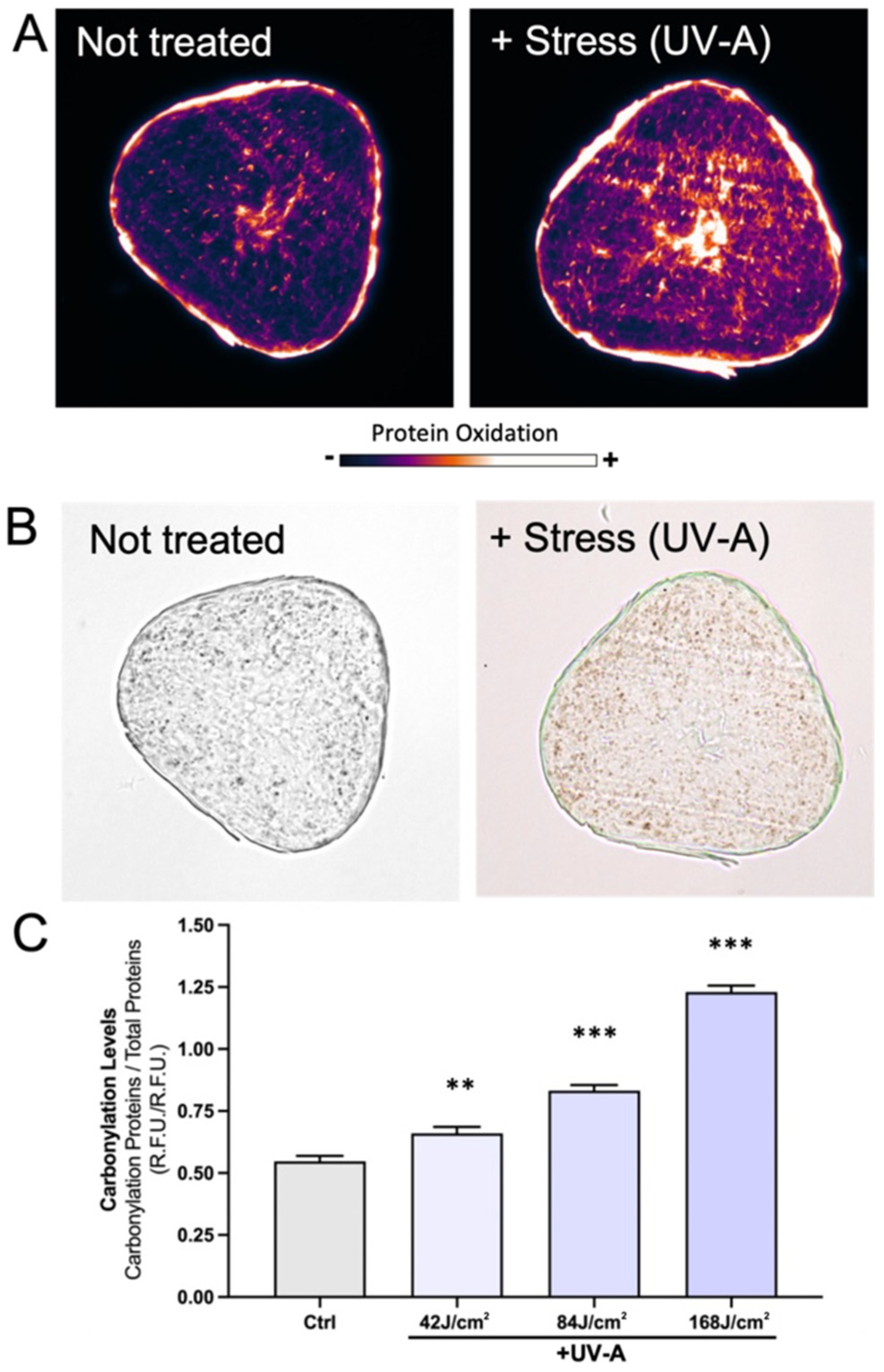
3.1. UV Irradiation Induces Protein Carbonylation in a Dose-Dependent Manner Both in the Cortex and Cuticle

In this study, we have developed a targeted proteomics approach for hair damage evaluation by detecting and quantifying carbonylated proteins on hair fibers. A specific functionalized fluorophore [16] has been developed to detect and quantify carbonylated protein in situ and upon protein extraction from hair fibers (Figure 1). UV-A irradiation was inflicted to hair strands ex vivo in order to evaluate its specific effects. To detect early damages inflicted by environmental stressors, the utilized UV-A doses are equivalent to the UV-A fraction cumulated in half-day, 1 or 2 days of sunlight exposure during an average European summer day [21]. As depicted in Figure 1A, carbonylation was detected both in cuticle and cortex proteins upon UV irradiation. Interestingly, the pattern of carbonylation is not associated with the distribution of melanin clusters, indicating that the obtained signal for carbonylation belongs to hair fibers structural proteins (Figure 1B).
After optimization and standardization of protein extraction methods from hair shafts, carbonylated proteins where then quantified. Protein extraction from hair shafts is complex due to their rearrangement in fibrillar structures. A specific protocol for extraction using detergents and chaotropic agents has been developed. Hair proteins carbonylation increases significantly in a dose-dependent manner upon UV irradiation (Figure 1C) validating the method also as a quantitative approach for hair damage assessment. The reproducibility was evaluated using quintuplicate independent determinations. In addition, this method is compatible with mass spectrometry approaches for the identification of the specific proteins and modified amino acids.

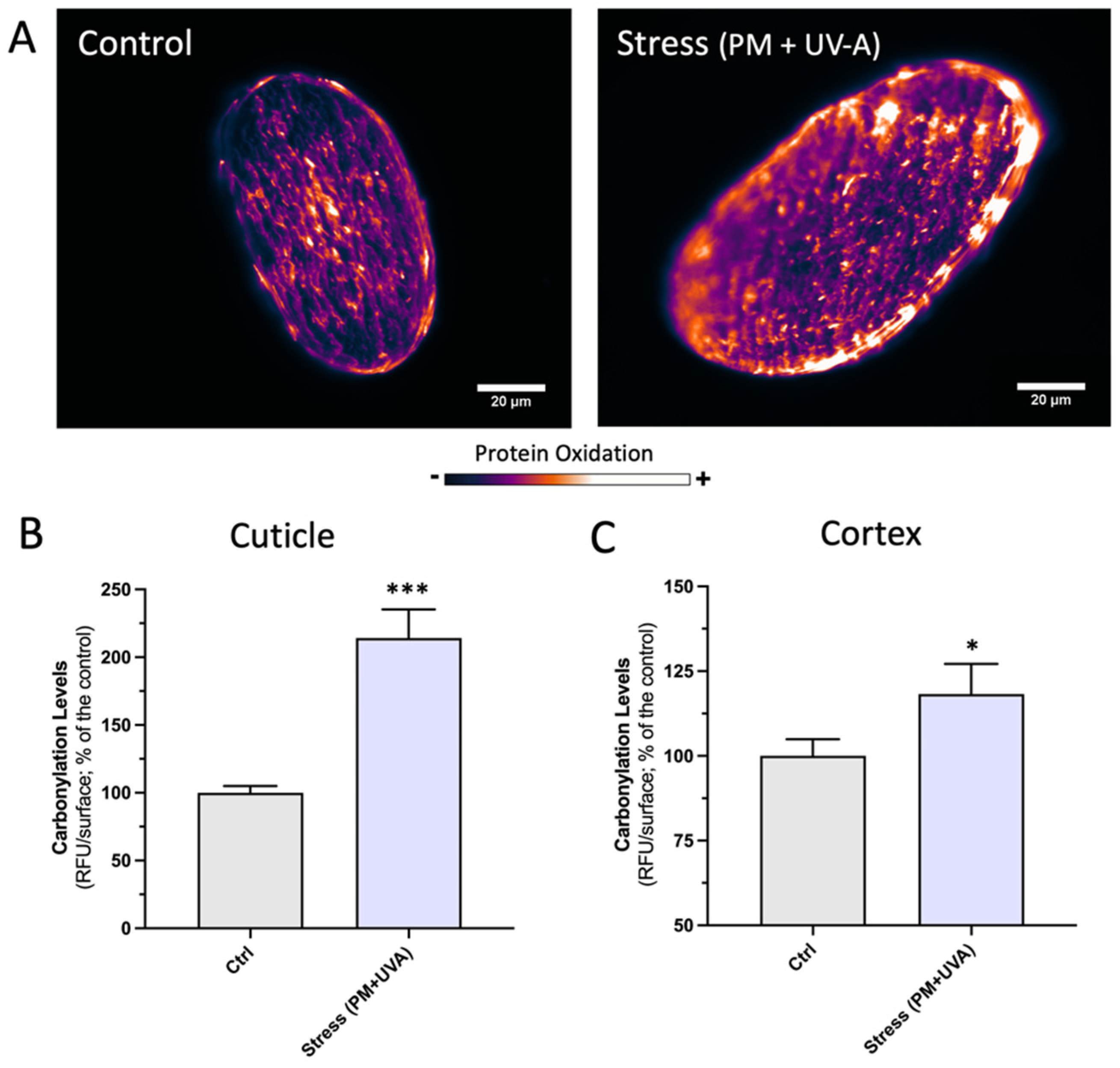
3.2. Airborne Pollutants Induces Protein Carbonylation on Hair Fibers

An innovative ex vivo model using hair fibers to assess the impact of airborne pollution has been settled-up by the application of referenced particulate matter (PM) containing PAHs, anthracene, pyrene, fluoranthene, and phenanthrene derivates among them.
The amount of particulate matter applied on ex vivo hair strands is equivalent to the amount of particulate matter accumulated on hair during 24 h in polluted cities, as shown by previous studies on the adhesion of PM on hair fibers in polluted cities [22]. In our model, the deposition of PM on ex vivo hair strands are also observed (Figure 2).
Since our goal is to approach real-life exposure conditions, the application of PM was followed by UV-A irradiation. Studying the combined impact of PM and UV light on hair fibers could therefore help to better understand the impact of urban pollution on hair shafts and to better evaluate the efficacy of interventions for hair fibers protection using cosmetics approaches. Ultraviolet solar radiation and air pollution are well-known aggressors to the human skin surface and deeper skin tissues, and more recently also to hair shafts [23]. However, hair damage at a molecular level has been traditionally evaluated by assessing the modification of single amino acids (cysteine or tryptophan) byproducts, such as cysteic acid [8]. In general, amount of cysteic acid within keratin fiber is estimated semi-quantitatively based on absorption peak intensity at 1040 cm−1 by IR spectroscopy, lacking sensitivity [24].
Previous studies have shown that UV absorption by PAHs present in PM leads to exited states and subsequent production of reactive oxygen species (ROS) that can alter DNA, proteins, and lipids [25,26,27,28]. The known photo-enhanced toxicity of different PAHs showed a correlation between PAH concentration and UV-induced photo damage, suggesting a major causative contribution of PAHs in the degradation process pointing toward a synergic effect of PM and UV-irradiation on hair fibers damage [23].
Increased levels of carbonylation has been observed both in the cuticle and cortex of hair fibers upon PM incubation and UV-irradiation (Figure 3). Interestingly, the pattern of carbonylation in the cuticle was observed to be more evident in specific regions, probably there where PM is deposited (Figure 3A). Quantitative analyses of carbonylation showed that the cuticle is severely damaged (Figure 3B), in accordance with previously published data on the impact of urban pollution on hair strands of people living in polluted cities [22].

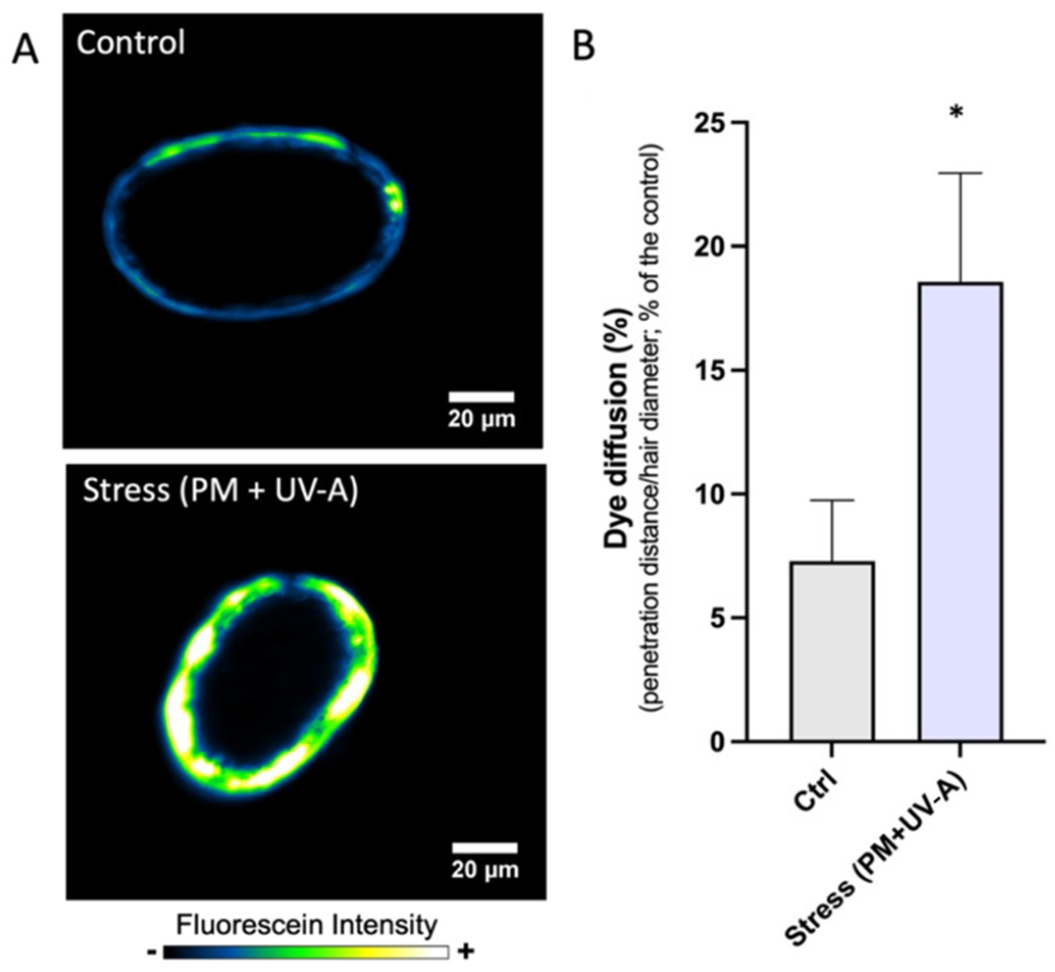
3.3. Protein Carbonylation Is Associated with Increased Hair Fiber Permeability

To study the structural consequences of increased protein carbonylation on hair fibers, permeation studies using a fluorescent dye were performed. Previous studies have shown that physical and chemical aggressions have significant effects on the structure of the hair fiber provoking changes in diffusion coefficients of a dye. It has been previously reported that diffusion rates of fluorescein into the cortex increases rapidly upon oxidative exposure of hair fibers. This increase in the rate of dye diffusion could result in both a breakdown (or damage) of the cuticle layers leading to a loss of barrier function, and an impairment of the structure in the cortex [29]. Localized oxidative damage resulting in decuticulation oh hair fiber has been associated to an increased penetration of a dye and decomposition of the regions in between the cuticle and cortex (cmc and the endocuticle).
In our hands, intact hair shafts showed a limited penetration of fluorescein dye (Figure 4A upper panel). However, increased diffusion of the dye through hair fibers was observed upon insult with urban pollutants (Figure 4A lower panel). Increased specific fluorescence signal, as well as in an increased distance of diffusion, when the fibers were exposed to the stress were observed (Figure 4B). Increased carbonylation in hair fibers yielded a statistically significant increase in the amount of dye that penetrated the hair fibers. Thus, the oxidative modification and damage of hair proteins induced by urban pollution exposure also presented a structural damage and enhanced dye diffusion in hair fibers. The penetration of the dye could be accelerated not by the decrease of cuticle layers, but by hair damage occurred during the oxidative decuticulation process [30]. Though looking for the prevention of protein oxidative modification (carbonylation) could also preserve the structure of hair fibers.

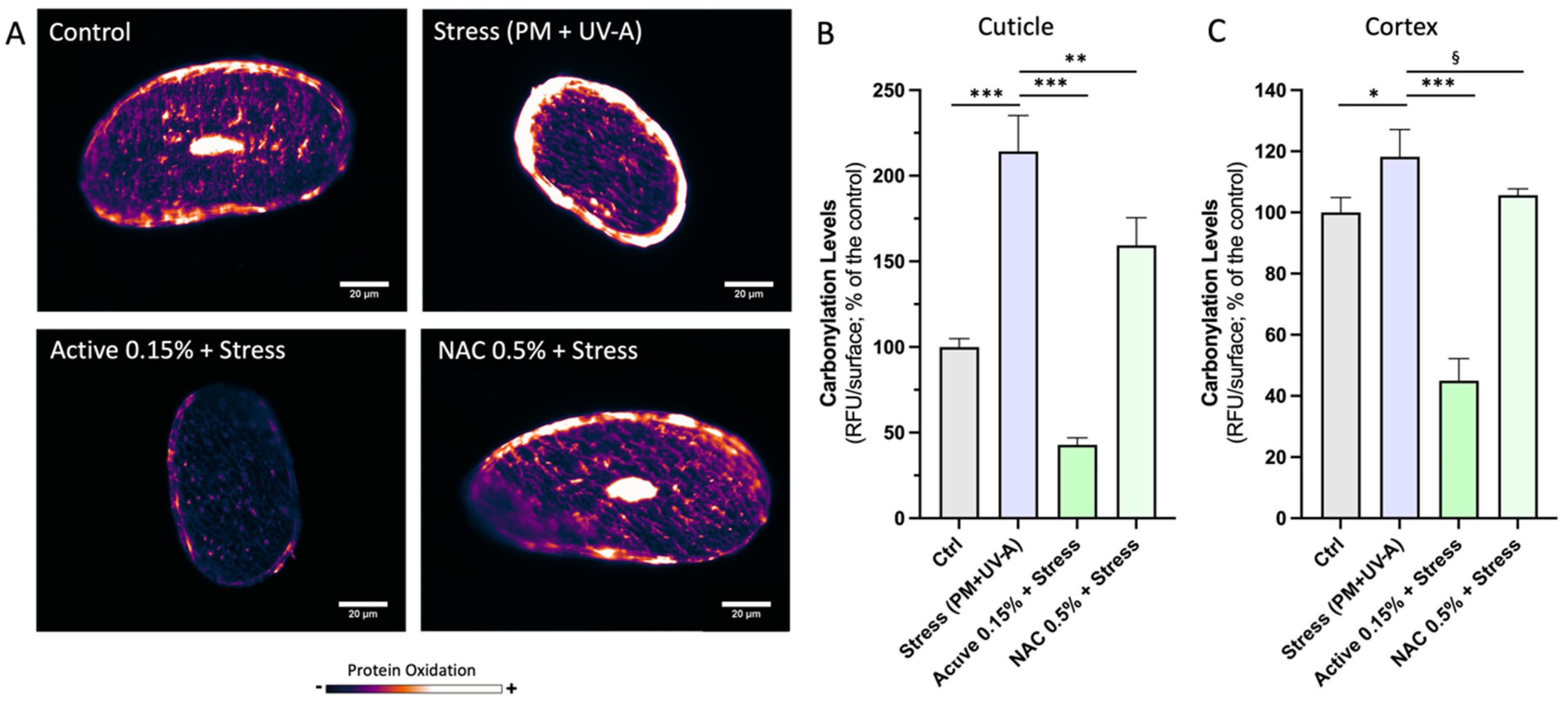
3.4. Prevention of Hair Fibers Carbonylation by Anti-Oxidants

The design cosmetics interventions aiming to prevent irreversible molecular damages such as protein carbonylation, could be a rational approach to preserve hair sheathing, smoothing, and therefore hair shine and softness. Thus, we have sought to identify a preventative strategy to protect hair fibers “scaffold” from oxidative damage. Previous studies have shown that treatments with antioxidants improved the mechanical properties of hair fibers and protected hair color after exposition to UV/VIS radiation [31]. In this study we evaluated the effectiveness of two different antioxidants to prevent protein carbonylation in our ex vivo model of urban pollution. We used N-acetyl cysteine (NAC), a well-known scavenger of oxygen radicals [32] and a nickel complex (NiBDPP) mimicking the active site of Ni-containing superoxide dismutase (NiSOD), which catalyze the disproportionation of the superoxide radical [33]. NAC is a free radical scavenger that replenishes glutathione and L-cysteine. It provides protection against the hydroxyl radical and the highly reactive lipid peroxide 4-hydroxynonenal. Superoxide dismutase (SOD) is known to be protective against oxidative stress-mediated skin dysfunction, here we explore the potential beneficial effects of a biometic NiSOD for hair care applications. Hair tresses subjected to UV radiation and PM were pre-treated with the two antioxidant solutions independently to assess the protection of hair properties. Both antioxidants prevented protein carbonylation in the cuticle and cortex from urban pollution-induced damage (Figure 5A). Quantitative analyses showed a significant effect in both cases (Figure 5B).

4. Conclusions

Taking together, our results suggest that since hair cannot be restored after deterioration, the protection of hair proteins from carbonylation could be an efficient approach for hair protection against airborne pollutants and UV radiation. The innovative ex vivo models and different methodologies assayed could help to evaluate the early damage to substantiate “anti-pollution” and “anti-oxidant” claims for hair care products.

Author Contributions

Conceptualization, M.A.B. and A.C.; methodology, A.C., A.S. and A.B.; software, A.C.; validation, A.C., A.S. and M.A.B.; formal analysis, A.C. and M.A.B.; investigation, A.C. and A.B.; resources, A.S.; data curation, A.C.; writing—original draft preparation, A.C.; writing—review and editing, M.A.B.; visualization, A.B.; supervision, M.A.B.; project administration, M.A.B.; funding acquisition, M.A.B. All authors have read and agreed to the published version of the manuscript.

Funding

This research received no external funding.

Institutional Review Board Statement

Not applicable.

Informed Consent Statement

Not applicable.

Data Availability Statement

Not applicable.

Conflicts of Interest

The authors declare no conflict of interest.

References

  1. Draelos, Z.K. Hair cosmetics. Dermatol. Clin. 1991, 9, 19–27. [Google Scholar] [CrossRef]
  2. Feughelman, M. Textbook of Morphology and properties of hair. In Hair and Hair Care; Johnson, D.H., Dekker, M., Eds.; Routledge: New York, NY, USA, 1997; pp. 1–12. [Google Scholar]
  3. Wolfram, L.J. Human hair: A unique physicochemical composite. J. Am. Acad. Dermatol. 2003, 48 (Suppl. S6), S106–S114. [Google Scholar] [CrossRef] [PubMed]
  4. Trüeb, R.M. The impact of oxidative stress on hair. Int. J. Cosmet. Sci. 2015, 37, 25–30. [Google Scholar] [CrossRef]
  5. Tolgyesi, E. Weathering of hair. Cosmet. Toilet. 1983, 98, 29–33. [Google Scholar]
  6. Mitu, A.M. Damage Assessment of Human HAIR by Electrophoretical Analysis of Hair Proteins. Ph.D. Dissertation, Bibliothek der RWTH Aachen, Aachen, Germany, 2004. [Google Scholar]
  7. Grosvenor, A.J.; Deb-Choudhury, S.; Middlewood, P.G.; Thomas, A.; Lee, E.; Vernon, J.A.; Woods, J.L.; Taylor, C.; Bell, F.I.; Clerens, S. The physical and chemical disruption of human hair after bleaching—Studies by transmission electron microscopy and redox proteomics. Int. J. Cosmet. Sci. 2018, 40, 536–548. [Google Scholar] [CrossRef] [PubMed]
  8. West, K.A.; Crabb, J.W. Textbook in Applications of Automatic PTC amino Acid Analysis; Angeletti, R.H., Ed.; Techniques in Protein Chemistry III; Academic Press: London, UK, 1992; pp. 233–242. ISBN 9780120587568. [Google Scholar] [CrossRef]
  9. Suzuki, Y.J.; Carini, M.; Butterfield, D.A. Protein carbonylation. Antioxid. Redox Signal. 2010, 12, 323–325. [Google Scholar] [CrossRef]
  10. Levine, R.L. Carbonyl modified proteins in cellular regulation, aging, and disease. Free Radic. Biol. Med. 2002, 32, 790–796. [Google Scholar] [CrossRef]
  11. Masaki, H.; Sinomiya, D.; Okano, Y.; Yoshida, M.; Iwabuchi, T. Impact of protein carbonylation on the chemical characteristics of the hair surface. Int. J. Cosmet. Sci. 2021, 43, 764–771. [Google Scholar] [CrossRef] [PubMed]
  12. Breakspear, S.; Smith, J.R.; Luengo, G. Effect of the covalently linked fatty acid 18-MEA on the nanotribology of hair’s outermost surface. J. Struct. Biol. 2005, 149, 235–242. [Google Scholar] [CrossRef] [PubMed]
  13. Dario, M.F.; Baby, A.R.; Velasco, M.V.R. Effects of solar radiation on hair and photoprotection. J. Photochem. Photobiol. B Biol. 2015, 153, 240–246. [Google Scholar] [CrossRef]
  14. Bloch, L.D.; Goshiyama, A.M.; Dario, M.F.; Escudeiro, C.C.; Sarruf, F.D.; Velasco, M.V.R.; Valente, N.Y.S. Chemical and physical treatments damage Caucasian and Afro-ethnic hair fibre: Analytical and image assays. J. Eur. Acad. Dermatol. Venereol. 2019, 33, 2158–2167. [Google Scholar] [CrossRef] [PubMed]
  15. Sebetić, K.; Sjerobabski Masnec, I.; Cavka, V.; Biljan, D.; Krolo, I. UV damage of the hair. Coll. Antropol. 2008, 2, 163–165. [Google Scholar]
  16. Baraibar, M.A.; Ladouce, R.; Friguet, B. Proteomic quantification and identification of carbonylated proteins upon oxidative stress and during cellular aging. J. Proteom. 2013, 30, 63–70. [Google Scholar] [CrossRef] [PubMed]
  17. Rasband, W.S. ImageJ. U.S. National Institutes of Health: Bethesda, MD, USA, 1997–2018. Available online: https://imagej.nih.gov/ij/ (accessed on 21 July 2022).
  18. Bradford, M.M. A rapid and sensitive method for the quantitation of microgram quantities of protein utilizing the principle of protein-dye binding. Anal. Biochem. 1976, 72, 248–254. [Google Scholar] [CrossRef]
  19. Lee, W.-Z.; Chiang, C.-W.; Lin, T.-H.; Kuo, T.-S. Ni Complex and Its Derivatives, Producing Method, and the Use Thereof as an Antioxidant. U.S. Patent 8642763, 12 July 2016. [Google Scholar]
  20. GraphPad Software, San Diego, CA, USA. Version 9. Available online: www.graphpad.com (accessed on 21 July 2022).
  21. STM G173-03:2012; Standard Tables for Reference Solar Spectral Irradiance: Direct Normal and Hemisferical on 37° Tilted Surface. ASTM International: West Conshohocken, PA, USA, 2012.
  22. Galliano, A.; Ye, C.; Su, F.; Wang, C.; Wang, Y.; Liu, C.; Wagle, A.; Guerin, M.; Flament, F.; Steel, A. Particulate matter adheres to human hair exposed to severe aerial pollution: Consequences for certain hair surface properties. Int. J. Cosmet. Sci. 2017, 39, 610–616. [Google Scholar] [CrossRef]
  23. Naudin, G. Human pollution exposure correlates with accelerated ultrastructural degradation of hair fibers. Proc. Natl. Acad. Sci. USA 2019, 10, 18410–18415. [Google Scholar] [CrossRef]
  24. Watanabe, K.; Nagami, K.; Suzuta, K.; Maeda, T.; Ito, L. Cysteic Acid Formation Behaviors in Bleached Hair of Southeast Asian Characterized by Infrared Spectroscopy. Adv. Life Sci. 2015, 5, 85–89. [Google Scholar] [CrossRef]
  25. Angerer, J.; Ewers, U.; Wilhelm, M. Human biomonitoring: State of the art. Int. J. Hyg. Environ. Health 2007, 210, 201–228. [Google Scholar] [CrossRef]
  26. Yu, H.; Xia, Q.; Yan, J.; Herreno-Saenz, D.; Wu, Y.S.; Tang, I.W.; Fu, P.P. Photoirradiation of polycyclic aromatic hydrocarbons with UVA light–A pathway leading to the generation of reactive oxygen species, lipid peroxidation, and DNA damage. Int. J. Environ. Res. Public Health 2006, 3, 348–354. [Google Scholar] [CrossRef]
  27. Butterfield, D.A.; Gu, L.; di Domenico, F.; Robinson, R.A. Mass spectrometry and redox proteomics: Applications in disease. Mass Spectrom. Rev. 2014, 33, 277–301. [Google Scholar] [CrossRef]
  28. Xia, Q.; Chiang, H.M.; Yin, J.J.; Chen, S.; Cai, L.; Yu, H.; Fu, P.P. UVA photoirradiation of benzo[a]pyrene metabolites: Induction of cytotoxicity, reactive oxygen species, and lipid peroxidation. Toxicol. Ind. Health 2015, 31, 898–910. [Google Scholar] [CrossRef]
  29. Tate, M.; Kamath, Y.; Ruetsch, S.; Weigmann, H. Quantification and prevention of hair damage. J. Soc. Cosmet. Chem. 1993, 44, 347–371. [Google Scholar]
  30. Takahashi, T. A highly resistant structure between the cuticle and the cortex of human hair. II. CARB, a penetration barrier; Toshie Takahashi. Int. J. Cosmet. Sci. 2019, 41, 28–35. [Google Scholar] [PubMed]
  31. Fernández, E.; Martínez-Teipel, B.; Armengol, R.; Barba, C.; Coderch, L. Efficacy of antioxidants in human hair. J. Photochem. Photobiol. B Biol. 2012, 117, 146–156. [Google Scholar] [CrossRef] [PubMed]
  32. Zhitkovich, A. N-Acetylcysteine: Antioxidant, Aldehyde Scavenger, and More. Chem. Res. Toxicol. 2019, 32, 1318–1319. [Google Scholar] [CrossRef]
  33. Shearer, J. Insight into the structure and mechanism of nickel-containing superoxide dismutase derived from peptide-based mimics. Acc. Chem. Res. 2014, 47, 2332–2341. [Google Scholar] [CrossRef]
Figure 1. Structural Proteins Carbonylation on hair fibers upon UV-A irradiation. (A) Epi-fluorescence imaging of carbonylation in hair fibers cross-sections. Carbonylation levels are presented as a gradient of colors (not treated on the left, stress exposed on right; from dark color for low levels to bright colors for high levels of carbonylation). (B) Melanin clusters visualization on the same samples. Non-treated or stressed hair fibers are detected by brightfield optical microscopy (in gray scale color, on the left) or in RGB color representation. (C) Protein carbonylation quantification upon protein extraction and resolution onto SDS-PAGE. Statistics: ANOVA and Dunnett’s post-hoc multi-comparison test versus Control (Ctrl), ** p < 0.01, *** p < 0.001.
Cosmetics 09 00098 g001
Figure 2. The adhesion of particulate matters on hair surface is detected and presented by image collection via scanning electron microscopy (SEM). (A) Not treated hair fiber; (B) hair fiber exposed to airborne pollutants (PM).
Cosmetics 09 00098 g002
Figure 3. (A) The visualization of in situ carbonylation levels on hair cross sections is presented in color range (dark color for low levels and bright colors for high levels of carbonylation. Significant increased levels of carbonylation upon stress (urban pollution) were observed on both cuticle (B) and cortex (C). Statistics: ANOVA and Dunnett’s post-hoc multi-comparison test versus Control (Ctrl), * p < 0.01, *** p < 0.001.
Cosmetics 09 00098 g003
Figure 4. Studies on hair fiber structural integrity. (A) Dye diffusion is presented as a color range of fluorescent (from a dark color for low intensity of dye to yellow color for high intensity levels of the dye). (B) The diffusion of the dye within the hair fiber is presented here as % of Permeability and obtained from the dye penetration distance along the long axis of hair fiber cross section. Statistics: t-test for binary comparison (unpaired, two-ways, alpha = 0.5; * p < 0.05.
Cosmetics 09 00098 g004
Figure 5. Protection of hair fibers carbonylation by anti-oxidants. (A) The in situ carbonylation levels on hair cross sections are visualized as a color range (from dark color for low levels to bright colors for high levels of carbonylation). The presence of the stress (PM + UV-A) induced significant increase of carbonylation in both (B) cuticle and (C) cortex regions. The presence of NiBDPP (active 0.15%) or N-acetyl cysteine (NAC 0.5%) preserved hair fibers from stress-induced carbonylation. Statistics ANOVA followed by Dunnett’s post-hoc test versus stress (two-ways; § p < 0.1, * p < 0.05, ** p < 0.01, *** p < 0.001).
Cosmetics 09 00098 g005
Publisher’s Note: MDPI stays neutral with regard to jurisdictional claims in published maps and institutional affiliations.

