Article

In Vitro and Ex Vivo Mechanistic Understanding and Clinical Evidence of a Novel Anti-Wrinkle Technology in Single-Arm, Monocentric, Open-Label Observational Studies

1
Research and Innovation, Department of Global Personal Care and Skin Health R&D, Colgate-Palmolive Company, 909 River Rd, Piscataway, NJ 08855, USA
2
Clinical Research, Department of R&D, Laboratoires Filorga, 2 rue de Lisbonne, 75008 Paris, France
3
Dermal Clinical Research, Department of Global Personal Care and Skin Health R&D, Colgate-Palmolive Company, 909 River Rd, Piscataway, NJ 08855, USA
*
Author to whom correspondence should be addressed.
Cosmetics 2022, 9(4), 80; https://doi.org/10.3390/cosmetics9040080
Submission received: 28 June 2022 / Revised: 26 July 2022 / Accepted: 27 July 2022 / Published: 2 August 2022

Abstract

:
Skin aging is a biological process leading to visible skin alterations. The mechanism of action, clinical efficacy and tolerance of a novel anti-wrinkle technology were evaluated in two skin care products formulated for different skin types. Two single-arm monocentric, open-label observational clinical studies, which were 56 days long, evaluated a cream-gel (n = 30) and a cream (n = 33) on the face and neck. Morphometric analyses of five types of wrinkles were performed at 0, 7, 28 and 56 days. Structural changes in extracellular matrix (ECM) including collagen, elastin and hyaluronic acid (HA) were visualized and quantified by histochemical imaging after daily treatment of skin explants for 6 days. Protein and gene expression related to barrier and hydration were analyzed using ELISA and qRT-PCR, respectively, in a reconstituted human skin model treated daily for 48 h. A decrease in wrinkle dimensions was found in the majority of parameters after 28 days of treatment. Collagen, elastin, HA, procollagen type I, hyaluronan synthases, HAS2 and HAS3 were all stimulated. Based on significant and consistent changes in our investigations, we conclude that the underlying mechanism of action of the novel anti-wrinkle technology could be the remodeling of dermal ECM, and both the test formulations were efficacious and well tolerated.

1. Introduction

During skin aging, face and neck skin are exposed to major clinical and morphological changes due to their specific physiological location [1]. The skin follows dynamic aging processes that are induced by combined intrinsic and extrinsic factors. Extrinsic aging is the result of exposure to environmental factors such as ultraviolet light which promoted rough wrinkles, while intrinsic aging is controlled by genetics that contribute to wrinkles [2,3]. It is well established that the extracellular matrix (ECM) of the dermis plays a key role in wrinkle generation that results from deregulated skin visco-elasticity and hydration levels [4,5]. In particular, collagen and elastin fibers are subjected to fragmentation and slower replacement during the aging process [6,7] and the hyaluronic acid (HA) reservoir decreases in the ECM during the aging processes. HA is mainly produced by hyaluronan synthases (HAS), for which roles have been reported in several biological functions including skin hydration and wound healing [8]. HAS are implicated in improving overall skin health by reducing signs of skin aging [9,10]. Aquaporin 3 (AQP3) which permeates both water and glycerol in epidermis is directly associated with the hydration of skin, with a relevant role in wound healing [11] and is often used as a marker of hydration in dermatological studies [12,13].
During skin aging, the combined aging factors lead to the clinical deterioration of the skin structure and its biomechanical properties translating into a wrinkling formation with potential psychological and social impacts [14]. Although dermatological procedures such as microinjection, facelift, and laser treatments are gold-standard procedures used to treat wrinkles, they are invasive interventions. The pain, cost and accessibility of these procedures could be the reason behind the decline in the number of these procedures [15]. Use of topical skin care products represent a pain-free, accessible and effective strategy to reduce established wrinkles and delay new wrinkle formation [16,17]. Nevertheless, there are a limited number of studies that characterize wrinkle reduction according to specific locations including the neck area using complementary morphometric parameters [18,19,20,21]. In addition, few studies provide comprehensive and dynamic insights using complementary clinical and experimental skin models. In our previous work, we investigated the collagen reorganization property of the anti-wrinkle technology present in the test formulations used in this study [22]. We gathered enough evidence of collagen reorganization at macro-, micro-, and nano scales following the use of the anti-wrinkle technology.
In the present study, we formulated two topical skin care formulations using the same active ingredients in the core technology, except with the difference of zinc salt of L-pyrrolidone carboxylic acid (Zinc-PCA) in the cream-gel to target oily skin. Our anti-wrinkle technology was formulated using a unique cocktail of natural extracts and synthetic peptides. Considering our objective was to target five types of aforementioned wrinkles, the selection of our active ingredients to formulate the core of our anti-wrinkle technology was inspired by aesthetic medicine techniques, and we adopted a multi-pronged approach to select ingredients previously reported to address wrinkles through different mechanisms of action. Organic kangaroo paw flower (Anigozanthos flavidus) botanical extract, rich in polyphenols and flavonoids, was incorporated for its collagen remodeling property that we investigated previously [22]. Anigozanthos flavidus was previously described in the composition of a line-targeting peptide serum that demonstrated clinical efficacy in as fast as 15 min when compared to placebo [23]. We incorporated Imperata cylindrica root extract, rich in potassium and 3-dimethylsulfopropionate, that provides long-term skin hydration and promotes anti-wrinkle effects [24]. Another plant extract we included in the formula was a lipid extract of sea fennel (Crithmum maritimum), known to contain totipotent stem cells [25,26] with properties such as dermal ECM synthesis [26], anti-inflammation [27] and anti-oxidation [28]. In the selection of synthetic peptides, we used a two-pronged approach to target wrinkles with peptides that stimulate collagen production (signal peptide), and the peptide that has a botox-like effect (Neurotransmitter inhibitor peptide). We used dipeptide diaminobutyroyl benzylamide diacetate (sequence: β-Ala-Pro-Dab-NHBn-2-Acetate), a reversible antagonist of the muscular nicotinic acetylcholine membrane’s receptors with neurotransmitter inhibition action that mimics Waglerin-1 protein [29]. In vivo, it demonstrated the ability to relax facial muscles and consequently decrease expression wrinkles [30,31]. In addition, a synergistic blend of two palmitoyl polypeptides (Pal-GHK/Palmitoyl Tripeptide-1 and Pal-GQPR/Palmitoyl Tetrapeptide-7), which is a new benchmark among signal peptides to boost collagen synthesis, was added to the formula [32]. These peptides are most popular among the peptides used in anti-aging cosmetic products, and their mechanism of action is well known [33].
The wrinkle reduction mechanism and underlying molecular changes were investigated using three skin models at the following three levels: in vivo (clinical), ex vivo (human skin explants/biopsies), and in vitro (reconstituted human skin model). Human subjects were recruited to participate in the clinical efficacy evaluation of the two formulations for 56 days. Human skin explants were used to characterize epidermal (filaggrin and Ki67 markers) and dermal (ECM markers) changes after daily treatment for 3 days and 6 days, respectively. A reconstituted human skin tissue model was selected to investigate protein and gene expression implicated in collagen synthesis and skin hydration, respectively, after two days of treatment.

2. Materials and Methods

2.1. Test Products

We formulated a cream (normal and dry skins) and a cream-gel (combination and oily skins) product for topical use, both of which share the same active ingredients in the core anti-wrinkle technology, except the difference of Zinc salt of L-Pyrrolidone Carboxylic Acid (Zinc-PCA) in the cream-gel to target oily skin. The proprietary anti-wrinkle technology consisted of plant extracts (Anigozanthos flavidus, Imperata cylindrica root and Crithmum maritimum), peptides and encapsulated hyaluronic acid. A cream-gel control formulation (negative control) was prepared without the core ingredients for the ex vivo and in vitro experiments. Ingredients of the two formulations are provided as Appendix A.

2.2. Human Volunteers and Clinical Study Design

The objective of the study was to assess the anti-wrinkle effect of the products after 7, 28 and 56 days of application on the face and neck. Efficacy was evaluated in relation to the improvement of face and neck wrinkles. During the study, the protocol was amended to extend the age from 60 to 65 years to improve the recruitment of subjects with neck wrinkles. Two independent observational open-label (unblinded) monocentric studies were performed to evaluate the topical daily use of cream-gel and cream finished formulations, individually in a 56-day (8 weeks) assessment in a single arm (Laboratoire COSderma, Bordeaux, France). The primary outcome was the evolution of wrinkles in the five target areas. The secondary outcome was the evaluation of skin tolerability. We hypothesized that at least 30 evaluable subjects per study to be a sufficient number for these observational studies.
Caucasian female subjects 35–65 years of age with Fitzpatrick skin types I-III were recruited according to a list of inclusion (Table 1) and exclusion criteria (Table 2). The subjects applied the product twice a day, morning and evening, on their whole face and neck for 56 days. n = 30 (cream-gel) and n = 33 (cream) subjects completed the study. Adverse event data were collected during the studies.

2.3. Wrinkle Morphometric Analysis and Quantitation

Standardized photographs were captured using standard 60 (diffuse) and cross polarized lighting at baseline (D0), as well as after 7 (D7), 28 (D28), and 56 (D56) days of product application. Subjects acclimated at 21 ± 1 ℃ and 45 ± 5% relative humidity prior to photography. Images were processed for morphometric analysis (length, surface, depth, volume) on the assigned facial (forehead, glabellar, crow’s feet, nasolabial folds) and neck areas (PhotoBench, Bordeaux, France). The wrinkle reduction was calculated using positive variation (%) of the means at D7, D28 and D56 compared to D0. Time courses of wrinkle reduction maps were generated using a proprietary method [35] that combined the 3D images and the clinical data of all participants to display the mean of the improvement in the form of continuous 3D color maps of the face (Newtone, Lyon, France).

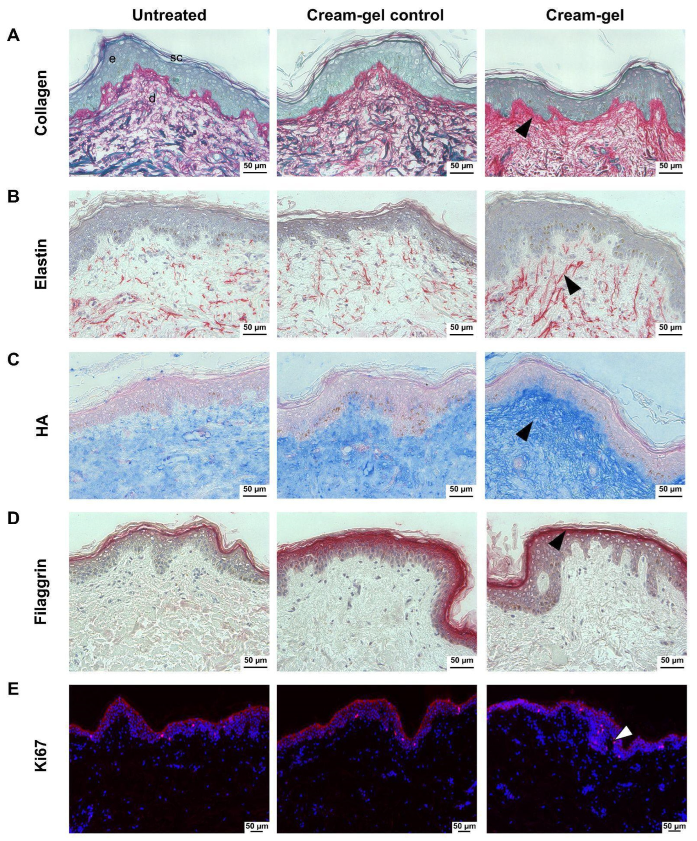
2.4. Human Skin Explant Experimentation and Histological Staining

The cream-gel or cream-gel control was uniformly applied once every day for 6 days on top of skin explants (n = 6 for each group) of 8 mm × 3 mm (diameter × thickness) from a 60 year-old female donor. On Day 7, skin samples were sectioned and stained with picrosirius red for histochemical imaging of total collagen or using a monoclonal antibody for immunohistochemical (IHC) imaging of elastin and filaggrin, or a biotinylated hyaluronic acid binding protein (HABP). Ki67 was revealed by immunofluorescence (IF) to assess epidermal cell proliferation. Filaggrin and Ki67 expression were imaged after 3 days of daily treatment, while dermal ECM components (collagen, elastin, and HA) were imaged after 6 days of daily treatment. Untreated skin served as the negative control (n = 6). ECM components (collagen, elastin and HA) were quantified in papillary dermis because the response of the papillary layer to the topical treatment was more dynamic. Ki67 and filaggrin were quantified in the epidermis layer.

2.5. Histological Image Processing and Marker Quantitation

The images were processed to obtain quantitative scores for collagen, elastin, HA, and filaggrin using the procedures reported in our previous work [36]. Briefly, two random sections from each skin sample (n = 6 skin samples for each group) were processed to obtain a total of 12 images or datapoints for each group. Markers (filaggrin and Ki67) were quantified in epidermis, while dermal ECM markers (collagen, elastin and HA) were quantified in papillary dermis, and quantified values were represented as scores. The Ki67 results are expressed in % of positive cells out of total cells, to indicate the proliferation score. All markers were imaged using bright field microscopy, except Ki67 that was imaged using fluorescence.

2.6. Reconstitutive Skin Tissue Model for ELISA and RT-PCR Analysis

To evaluate the concentrations of procollagen type I, a reconstituted human skin tissue model (EpiDermFT™, MatTek, Ashland, MA, USA) was used in the study. This cultured model consisted of a multilayered tissue forming a dermis constituted of fibroblasts in a collagen matrix and an epidermis constituted of keratinocytes. A total of 10 µL of the cream-gel (n = 5) or cream-gel control (n = 4) was applied to the surface of the skin once per day for two days. Untreated skin served as the negative control (n = 5). Procollagen I alpha 1 levels were measured from collected tissue culture media using ELISAs (R&D Systems, Minneapolis, MN, USA) following manufacturer’s protocols.
For RT-qPCR, RNA was extracted from collected tissues (RNeasy Plus Mini Kit, Qiagen, Germantown, MD, USA), and quantitated (NanoDrop, ThermoFisher Scientific, Waltham, MA, USA). cDNA synthesis was performed using the Maxima First Strand cDNA Synthesis kit (ThermoFisher). Gene expression analysis was performed using TaqmanTM array cards (QuantStudio 7 Flex, ThermoFisher). Gene expression of HAS2, HAS3, and AQP3 for the cream-gel was expressed relative to that of the cream-gel control.

3. Statistical Analysis

For clinical, histological and protein data, the mean values were presented as ± SEM (standard error of the mean). For RT-qPCR, mean data were presented with RQmin and RQmax for the 95% confidence level. Each wrinkle parameter of D7, D28, and D56 for subjects were compared to the baseline values (D0) using a univariate analysis of variance with two factors followed by a Dunnett test. Statistical significance for histological imaging was evaluated by conducting a one-way ANOVA followed by a t-test (B1SCLASSIC Software, SISSAD snc, Trieste, Italy). T-tests were used for a statistical analysis of the results derived from ELISAs and RT-qPCR. For RT-qPCR, Applied Biosystems™ Relative Quantitation Analysis Module (Life Technologies Corporation, Carlsbad- CA, USA) was used and the p-value was corrected with the Benjamini–Hochberg false discovery rate. A p-value < 0.05 was considered statistically significant.

4. Results

4.1. Demographics Data and Adverse Events

The two clinical studies were conducted between April and September 2021. Sample characteristics are indicated in Table 2. n = 30 and n = 33 subjects with mean ages of 57.1 (45–65) and 58.9 (46–65) years participated in the cream-gel and cream study, respectively. A total of 28 of the 33 subjects (84.8%) recruited had dry to dry combination skin type for cream testing, while 26 of 30 subjects (86.6%) had oily to oily combination skin type for cream-gel testing. One subject voluntarily withdrew consent in the cream-gel study before the study. Skin phototype II was the most represented in both studies. At the baseline of both clinical studies, the wrinkles presenting the longest length were located on the forehead (Table 3). Similarly, the glabellar wrinkles had the largest surface and volume in both studies. Higher depth values were observed for the nasolabial wrinkles for both the cream-gel and cream study. No serious adverse events were reported in the two clinical studies. One subject in each study reported mild pruritus on the neck or forehead. Both adverse events spontaneously resolved after permanent discontinuation of the products but subjects did not complete the study and dropped out for non-study related reasons or as a result of the investigator’s decision.

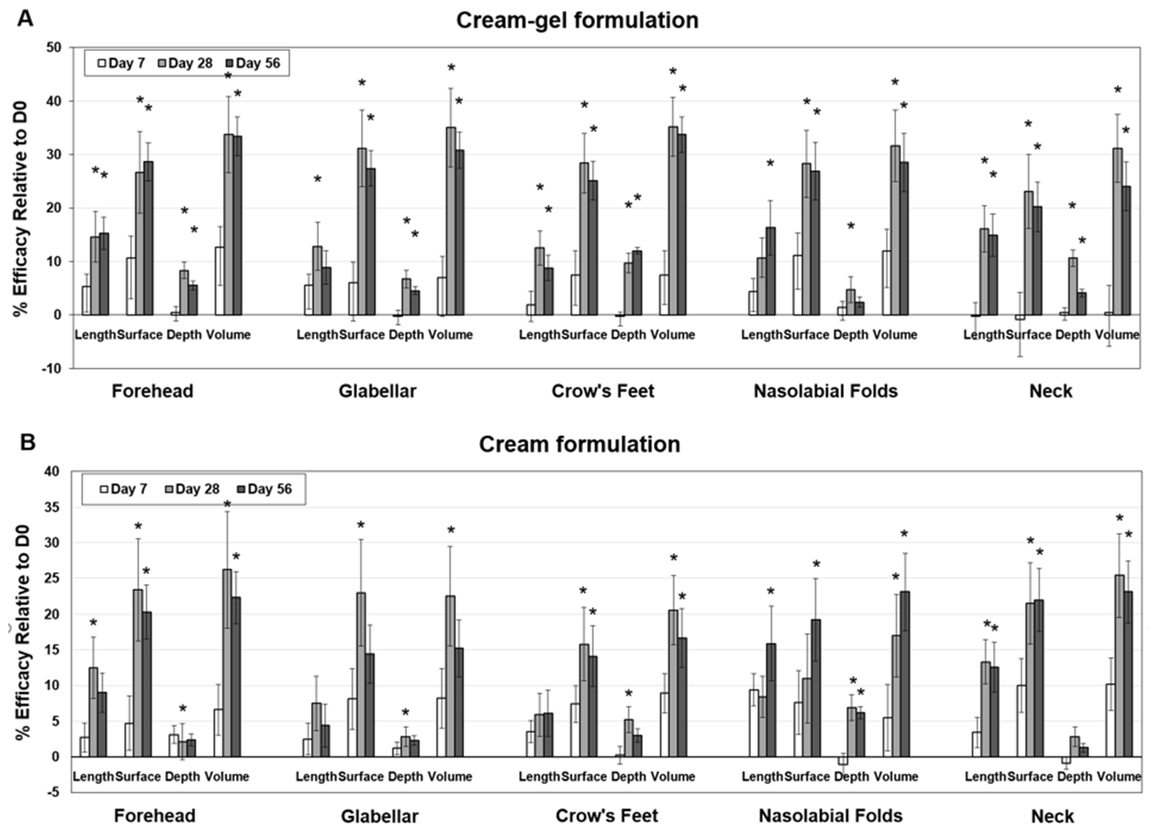
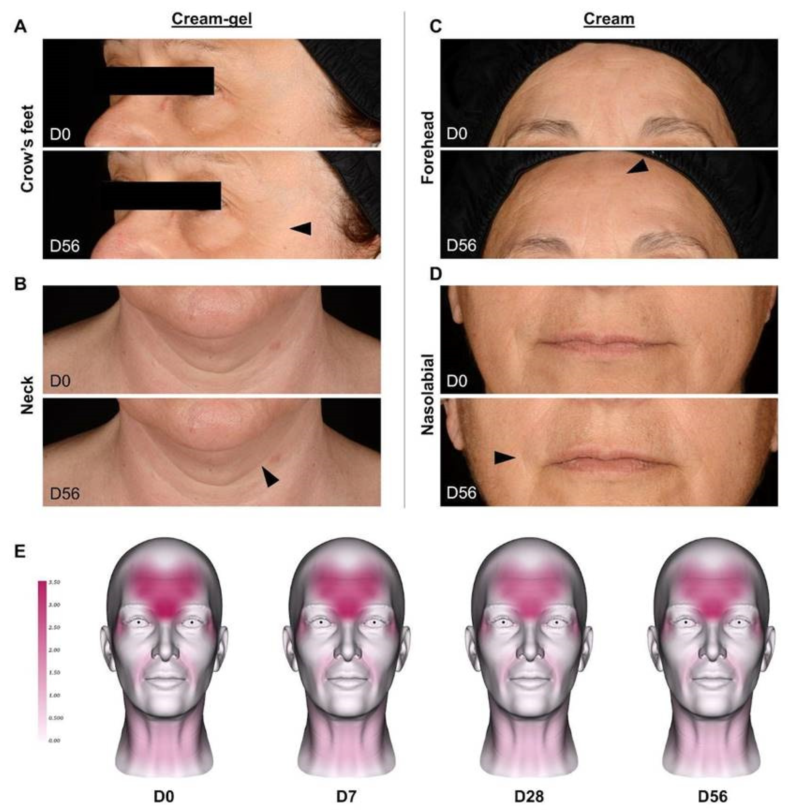
4.2. Clinical Reduction of Wrinkles after Cream-Gel Application

Twice daily topical application of the cream-gel formulation induced statistically significant wrinkle improvements in most parameters at D28 (Figure 1A). The improvements continued by D56. On D7, an earlier time point, there were no significant reductions. Indeed, the largest improvements above 20% relative efficacy were seen in the surface and volume parameters on D28 and D56 across all five wrinkle areas (forehead, glabellar, nasolabial, crow’s feet and neck). Forehead, crow’s feet (Figure 2A), and neck wrinkles (Figure 2B) were the areas that revealed the most consistent improvements in the various parameters as a result of the cream-gel. The smallest changes were observed for the depth parameter. A representative wrinkle map of the surface parameter of all subjects of the cream-gel study is reported in Figure 2E.

4.3. Clinical Reduction of Wrinkles after Cream Application

Similarly to the cream-gel study, the first significant clinical change in wrinkles were observed at D28 and D56 after cream application (Figure 1B). The largest improvements above 20% relative efficacy were seen in the surface and volume parameters on D28 and D56 for the forehead and neck wrinkles and on D28 for glabellar wrinkles. These same improvements above 20% were also seen in the volume parameter for crow’s feet wrinkles on D28 and on the nasolabial fold wrinkles on D56. Forehead (Figure 2C), nasolabial fold (Figure 2D) and neck wrinkles were the wrinkle areas that presented the most consistent improvements in the various parameters after cream treatment. Similarly to the cream-gel study, the smallest changes were observed for the depth parameter.

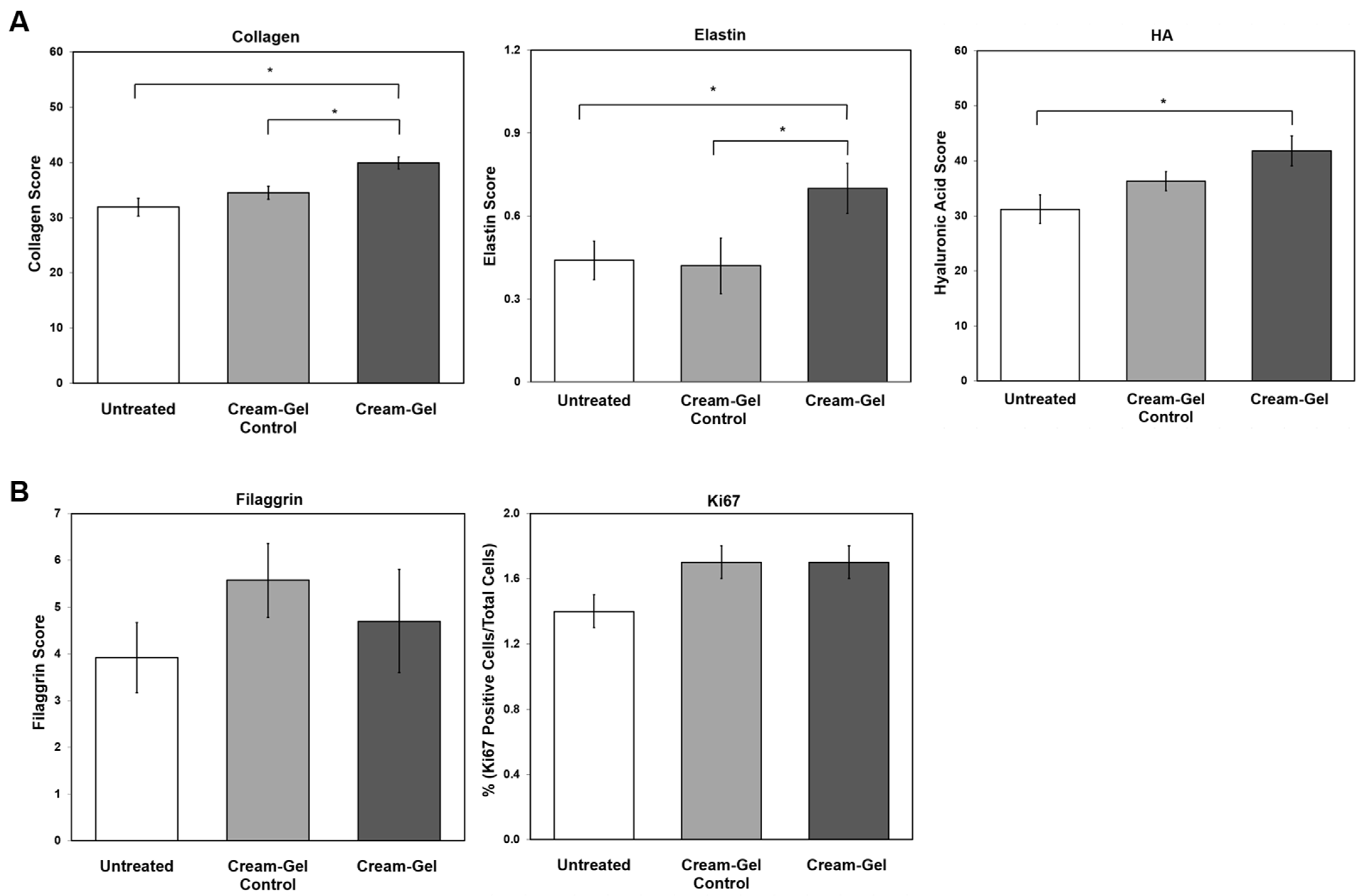
4.4. Histological Changes in Dermal ECM Components

In order to investigate the key structural changes involved in wrinkle reduction, we performed a histological analysis of the main constituents of the ECM (collagen, elastin and hyaluronic acid). Total collagen fibers of untreated skins were homogeneously distributed in the dermis (Figure 3A). The staining was particularly elevated in the papillary dermis near the dermo–epidermal junction. A dense network of longer collagen fibers was particularly concentrated in the papillary region of the dermis. As highlighted by the staining analysis (Figure 4A), there was a statistically significant increase in the total collagen score compared to control (+16%) or untreated samples (+25%). Elastin staining revealed longer vertical fibers throughout the dermis with a peculiar property of running towards the epidermis in the test samples than in control or in untreated skins (Figure 3B). Furthermore, the elastin score in tested skins statistically increased versus control (+67%) or untreated (+59%) samples (Figure 4A). We also performed a histological analysis of HA in the dermis. Increased staining with gradation was noticed in the cream-gel treated skins from the papillary area of the dermis to the deep layers of the epidermis (Figure 3C). This observation was evidenced by scoring elevated by 34% as compared to the untreated samples. There was no statistical difference between untreated and cream-gel control samples (Figure 4A). Similar to collagen, the expression of HA was more dominant in papillary dermis than in reticular dermis.

4.5. Histological Examination of the Epidermis

Although marginal improvements were found compared to untreated samples, the changes in the epidermal barrier (filaggrin marker, Figure 3D) and proliferation (Ki67 marker, Figure 3E) after treatment with cream-gel were not significant (Figure 4B). Considering the lack of difference between cream-gel and cream-gel control, this marginal improvement could be due to inactive ingredients present in the formulation.

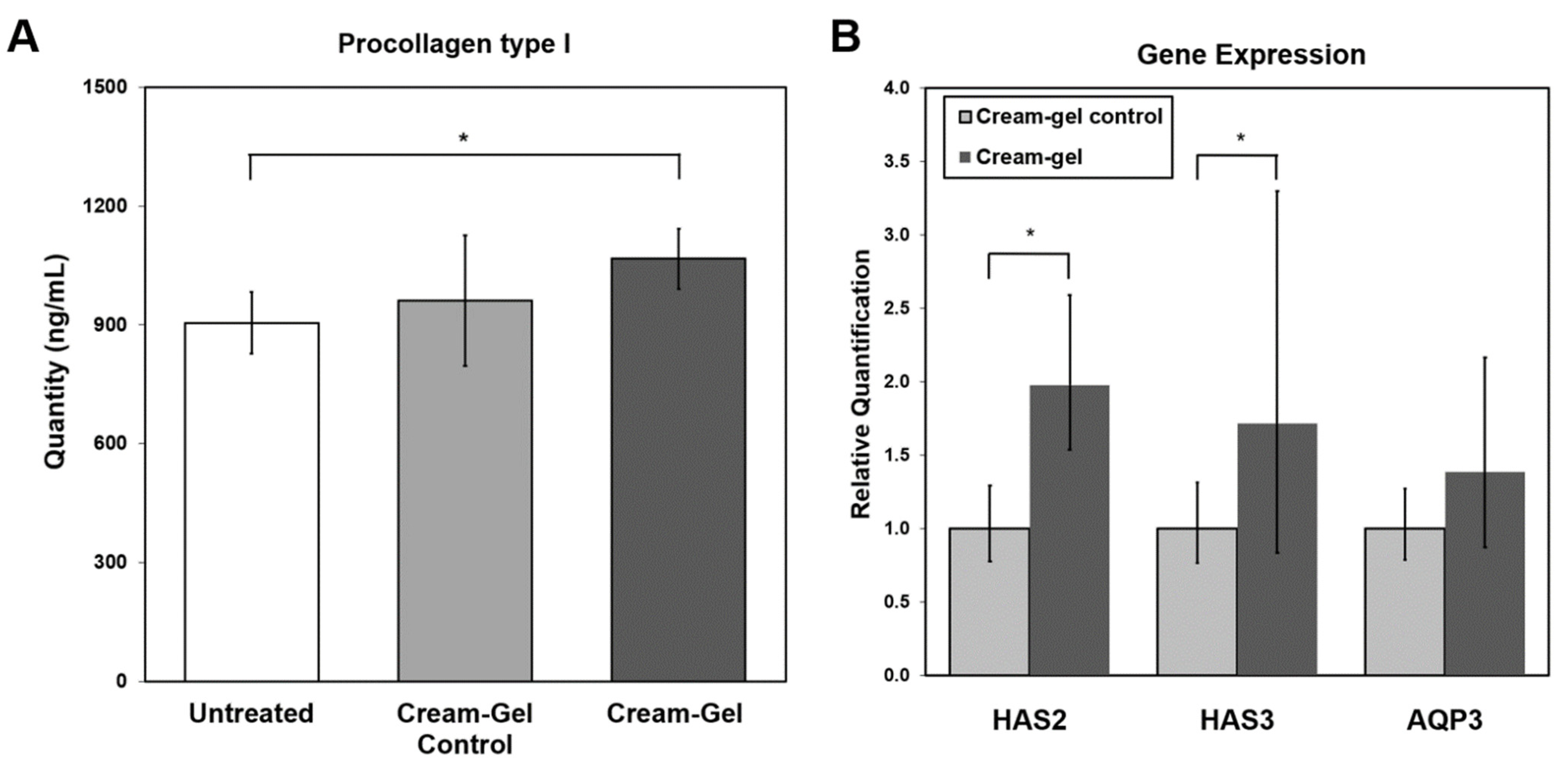
4.6. Protein Expression in Reconstitutive Skin Tissue Model

Using a reconstituted human skin tissue model treated by the cream-gel or cream-gel control, we quantified the protein levels of procollagen type I to investigate the collagen synthesis in the supernatant. The statistical analysis confirmed a significant increase in skin tissues treated with cream-gel after 48 h of topical treatment compared to untreated skins, but not compared to cream-gel control (Figure 5A).

4.7. Hydration-Related Gene Expression Changes in Reconstitutive Skin Tissue Model

Using the reconstituted human skin tissue model, we then explored the gene expression of key effectors in tissue hydration, hyaluronan synthases (HAS2, HAS3) and aquaporin (AQP3). After 48 h of treatment, HAS2 gene expression was statistically significantly increased by 2.0-fold and HAS3 by 1.7-fold as compared to the control samples (Figure 5B). There were no statistically significant differences in AQP3 gene expression between the two treatments.

5. Discussion

To investigate the anti-wrinkle effects of two skin care formulations (cream-gel and cream), we conducted a clinical study and characterized underlying histological and molecular changes using histochemical imaging and qPCR on skin explants and a full thickness skin model, respectively. We found evidence of clinical efficacy from both formulations showing significant attenuation of facial (forehead, glabellar, crow’s feet, nasolabial) and neck wrinkles after 28 days in human subjects as confirmed by morphometric analysis. Wrinkle analysis was conducted at three timepoints (D7, D28, D56) in complementary parameters (length, surface, depth, volume). Briefly, the largest decrease was observed in surface and volume parameters for all face and neck wrinkles at 28 and 56 days after twice-daily topical applications compared to baseline.
On careful observation of clinical efficacy data of cream and cream gel (Figure 1), we noticed that cream gel performed relatively better. This relative difference could be due to the effect of mattifying agents (zinc and polyacrylate crosspolymer-6) present in the cream-gel. The primary intended use of the polyacrylate crosspolymer-6 was to improve the thickness and texture of the cream-gel formulation; however, it might have created a layer of matrix gel on the skin surface with a masking effect to smooth and lift wrinkles. On the other hand, Zinc (present in cream-gel as Zinc salt of PCA) was hypothesized to play a more active anti-wrinkle role at the biological level. Zinc is an essential cofactor reported to regulate the function of metalloenzymes and transcription factors [37], including those responsible for the synthesis of collagen [38] and elastin [39]. This ingredient is widely used as a therapeutic agent alone or in combination in a number of dermatological conditions and also in anti-aging [37]. However, the cream and cream-gel were two independent clinical pilot studies performed on two different pools of subjects with different selection criteria appropriate to test the two formulations designed for different skin types/conditions.
In addition to the clinical work, we also observed a significant increase in the dermal ECM markers (collagen, elastin, HA) on day 7 after daily topical treatment of the human skin explant with the cream-gel.
First, we found that collagen quantity in skin tissues significantly increased in accordance with our in vitro findings of the overexpression of procollagen I after 48 h of treatment. Overall, these data suggest that the topical application of anti-wrinkle technology potentially induced the neosynthesis of collagen. In our previous study, we found evidence of collagen reorganization in skin explants after treatment with the aforementioned anti-wrinkle technology using a novel holistic imaging and image quantification approach at the macro-, micro- and nanoscale [22].
Second, we found a similar correlation between our in vitro and ex vivo results on HA, another component of ECM and related with skin hydration and aging. We found that at as early as 48 h, HAS2 and HAS3 gene expression increased in vitro as a result of treatment with the cream-gel product. Interestingly, another in vivo study found that after the application of fruit extract, an increase in HAS2 gene expression, hyaluronan levels and procollagen synthesis occurred. In cell line cultures, the overexpression of hyaluronan, HAS2 and procollagen genes by ginger extract was reported [10]. The increase in HAS2 and HAS3 gene expression, as well as the HA levels could be attributed to the test product formulation and possible induction of the production of new HA molecules into the extracellular space as observed in our ex vivo study and potentially increased water retention in the skin. HA chains of up to 4 MDa were generated by HAS2 while chains of less than 300 kDa were synthesized by HAS3 [40]. The use of HA in skin care formulas to increase tissue hydration and reduce wrinkles has been extensively studied [41,42,43] and is also reported to enhance delivery for drugs and cosmetics [44,45,46]. Additionally, we found a significant increase in elastin fibers after topical application of the test products on human skin explants. Regeneration of elastin fibers has been described in vivo using anti-wrinkle agents [39,47].
We did not identify a significant change in AQP3 gene expression, which is in contrast to another in vitro studies reporting the upregulation of AQP3 after moisture cream treatment [12]. On the contrary, AQP3 expression is known to increase in response to skin diseases including atopic eczema and agents such as retinoic acid [48,49]. Further investigation would be needed to determine the involvement of AQP3 in skin homeostasis (hydration) and diseases. The absence of a significant change in filaggrin at the histological level after treatment implied that anti-wrinkle test formulation did not compromise the skin barrier.

6. Conclusions

In summary, our in vitro, ex vivo and clinical investigations provided evidence of the strong efficacy and tolerance of the anti-wrinkle technology. Collectively, our investigations revealed the following: (1) significant wrinkle reduction after 28 and 56 days of daily application (clinical study) and (2) an underlying mechanistic understanding that anti-wrinkle technology shows evidence of action at the genomic level in as fast as 48 h (in vitro procollagen I and HAS genes over-expression) and on the structural level by day 7 (ex vivo overexpression of collagen, elastin and HA molecules). The technology was successfully formulated for all skin types (normal, dry, oily and combination) and showed good tolerance and efficacy. Our research revealed the implications of using innovative topical anti-wrinkle technology and that non-invasive wrinkle treatment options could be potentially integrated in aesthetic dermatologic procedures.

Author Contributions

J.N. designed, conducted, analyzed and interpreted the data from in vitro. V.B. designed, analyzed and interpreted the data from ex vivo. O.T. designed, conducted, interpreted and analyzed the data from clinical studies. J.M. supervised the ex vivo and J.W. in vitro research. I.D. supervised the clinical study. All authors contributed to draft preparation and substantively revised it. All authors have read and agreed to the published version of the manuscript.

Funding

The study was funded by Colgate-Palmolive Company and Filorga. This research received no external funding.

Institutional Review Board Statement

The study was conducted according to the guidelines of the Declaration of Helsinki, and approved by the Institutional Review Board (or Ethics Committee) of U.S. IRB, Inc. IRB Numbers U.S. IRB2021CPS/01 and U.S. IRB2021CPS/02).

Informed Consent Statement

Informed consent was obtained from all subjects involved in the study before participating in any study-related activities. Written informed consent was obtained from the patients to publish this paper.

Data Availability Statement

Data are available on request from the corresponding author.

Acknowledgments

The authors acknowledge the technical writing assistance of Mack Morrison, (Starfish Scientific Solutions, LLC, Lewes, DE, USA) and Nina Nguon (Laboratoires Filorga, Paris, France).

Conflicts of Interest

All authors are employees of Colgate-Palmolive Company, parent company of Filorga.

Appendix A

Appendix A.1. Cream-Gel Ingredient List

Aqua (water), glycerin, propylheptyl caprylate, silica, dicaprylyl carbonate, squalane, imperata cylindrica root extract, hydroxyethyl acrylate/sodium acryloyldimethyl taurate copolymer, zinc pca, nctf chronospheres, polyacrylate crosspolymer-6, phenoxyethanol, butylene glycol, sucrose palmitate, 1,2-hexanediol, parfum (fragrance), cetyl alcohol, stearalkonium hectorite, caprylic/capric triglyceride, glyceryl linoleate, tocopheryl acetate, propylene carbonate, polysorbate 60, sorbitan isostearate, biosaccharide gum-1, carbomer, sodium phytate, hydrogenated vegetable oil, adenosine, anigozanthos flavidus extract, benzyl alcohol, sodium lactate, sodium hydroxide, caprylyl glycol, dipeptide diaminobutyroyl benzylamide diacetate, glyceryl caprylate, levulinic acid, citric acid, t-butyl alcohol, polysorbate 20, potassium sorbate, sodium citrate, sodium hyaluronate, crithmum maritimum extract, p-anisic acid, tocopherol, acrylates/c10-30 alkyl acrylate crosspolymer, palmitoyl tripeptide-1, palmitoyl tetrapeptide-7.

Appendix A.2. Cream Ingredient List

Aqua (water, eau), glycerin, propylheptyl caprylate, butylene glycol, dicaprylyl carbonate, hydroxyethyl acrylate/sodium acryloyldimethyl taurate copolymer, squalane, cetearyl isononanoate, imperata cylindrica root extract, silica, sucrose palmitate, phenoxyethanol, parfum (fragrance), stearalkonium hectorite, 1,2-hexanediol, lauroyl lysine, sodium stearoyl glutamate, glyceryl linoleate, cetyl alcohol, caprylic/capric triglyceride, tocopheryl acetate, propylene carbonate, polysorbate 60, sorbitan isostearate, citric acid, prunus amygdalus dulcis (sweet almond) oil, sodium chloride, biosaccharide gum-1, carbomer, hydrogenated vegetable oil, adenosine, anigozanthos flavidus extract, benzyl alcohol, sodium lactate, caprylyl glycol, sodium hyaluronate, levulinic acid, glyceryl caprylate, potassium sorbate, polysorbate 20, sodium citrate, dipeptide diaminobutyroyl benzylamide diacetate, sodium hydroxide, crithmum maritimum extract, glucose, tocopherol, p-anisic acid, potassium chloride, calcium chloride, acrylates/c10-30 alkyl acrylate crosspolymer, magnesium sulfate, glutamine, sodium phosphate, ascorbic acid, sodium acetate, lysine hcl, arginine hcl, palmitoyl tripeptide-1, alanine, histidine hcl, valine, leucine, threonine, isoleucine, palmitoyl tetrapeptide-7, tryptophan, phenylalanine, tyrosine, glycine, polysorbate 80, serine, cystine, cyanocobalamin, glutathione, asparagine, aspartic acid, ornithine hcl, glutamic acid, nicotinamide adenine dinucleotide, biotin, proline, methionine, taurine, hydroxyproline, glucosamine hcl, coenzyme a, sodium glucuronate, thiamine diphosphate, retinyl acetate, inositol, niacin, niacinamide, pyridoxine hcl, calcium pantothenate, riboflavin, sodium tocopheryl phosphate, thiamine hcl, folic acid.

References

  1. Wolff, E.; Pal, L.; Altun, T.; Madankumar, R.; Freeman, R.; Amin, H.; Harman, M.; Santoro, N.; Taylor, H.S. Skin wrinkles and rigidity in early postmenopausal women vary by race/ethnicity: Baseline characteristics of the skin ancillary study of the KEEPS trial. Fertil. Steril. 2011, 95, 658–662. [Google Scholar]
  2. Makrantonaki, E.; Bekou, V.; Zouboulis, C.C. Genetics and skin aging. Dermato-Endocrinol. 2012, 4, 280–284. [Google Scholar]
  3. Quan, T.; Fisher, G.J. Role of age-associated alterations of the dermal extracellular matrix microenvironment in human skin aging: A mini-review. Gerontology 2015, 61, 427–434. [Google Scholar]
  4. Choi, J.W.; Kwon, S.H.; Huh, C.H.; Park, K.C.; Youn, S.W. The influences of skin visco-elasticity, hydration level and aging on the formation of wrinkles: A comprehensive and objective approach. Skin Res. Technol. 2013, 19, e349–e355. [Google Scholar]
  5. Uitto, J. The role of elastin and collagen in cutaneous aging: Intrinsic aging versus photoexposure. J. Drugs Dermatol. 2008, 7 (Suppl. S2), S12–S16. [Google Scholar]
  6. Chung, J.H.; Seo, J.Y.; Choi, H.R.; Lee, M.K.; Youn, C.S.; Rhie, G.E.; Cho, K.H.; Kim, K.H.; Park, K.C.; Eun, H.C. Modulation of skin collagen metabolism in aged and photoaged human skin in vivo. J. Investig. Dermatol. 2001, 117, 1218–1224. [Google Scholar]
  7. Antonicelli, F.; Bellon, G.; Debelle, L.; Hornebeck, W. Elastin-elastases and inflamm-aging. Curr Top. Dev. Biol 2007, 79, 99–155. [Google Scholar]
  8. Papakonstantinou, E.; Roth, M.; Karakiulakis, G. Hyaluronic acid: A key molecule in skin aging. Dermato-Endocrinol. 2012, 4, 253–258. [Google Scholar]
  9. Knott, A.; Reuschlein, K.; Mielke, H.; Wensorra, U.; Mummert, C.; Koop, U.; Kausch, M.; Kolbe, L.; Peters, N.; Stäb, F.; et al. Natural Arctium lappa fruit extract improves the clinical signs of aging skin. J. Cosmet. Dermatol. 2008, 7, 281–289. [Google Scholar]
  10. Park, S.J.; Lee, M.H.; Yun, J.M.; Kim, D.K.; Lee, Y.H. Effects of Zingiber mioga extract (FSH-ZM) on moisturizing, wrinkle improvement and whitening activity in skin cells. J. Korean Soc. Food Sci. Nutr. 2019, 48, 215–222. [Google Scholar]
  11. Boury-Jamot, M.; Daraspe, J.; Bonté, F.; Perrier, E.; Schnebert, S.; Dumas, M.; Verbavatz, J.M. Skin aquaporins: Function in hydration, wound healing, and skin epidermis homeostasis. In Aquaporins. Handbook of Experimental Pharmacology; Beitz, E., Ed.; Springer: Berlin/Heidelberg, Germany, 2009; Volume 190, pp. 205–217. [Google Scholar]
  12. McDaniel, D.H.; Dover, J.S.; Wortzman, M.; Nelson, D.B. In Vitro and in vivo evaluation of a moisture treatment cream containing three critical elements of natural skin moisturization. J Cosmet. Dermatol. 2020, 19, 1121–1128. [Google Scholar]
  13. Katekawa, E.; Caverzan, J.; Mussi, L.; Camargo-Junior, F.B.; Sufi, B.; Padovani, G.; Nazato, L.; Nogueira, C.; Magalhães, W.V.; Di Stasi, L.C. Novel topical skin hydration agent containing Anadenanthera colubrina polysaccharide-standardized herbal preparation. J. Cosmet. Dermatol. 2020, 19, 1691–1698. [Google Scholar]
  14. Farage, M.A.; Miller, K.W.; Berardesca, E.; Maibach, H.I. Psychological and social implications of aging skin: Normal aging and the effects of cutaneous disease. In Textbook of Aging Skin, 1st ed.; Farage, M.A., Miller, K.W., Maibach, H.I., Eds.; Springer: Berlin/Heidelberg, Germany, 2010; pp. 949–957. [Google Scholar]
  15. The Aesthetic Society. Aesthetic Plastic Surgery National Databank Statistics; The Aesthetic Society: Garden Grove, CA, USA, 2019; Available online: https://www.surgery.org/sites/default/files/Aesthetic-Society_Stats2019Book_FINAL.pdf (accessed on 21 March 2022).
  16. Draelos, Z.D. Revisiting the skin health and beauty pyramid: A clinically based guide to selecting topical skincare products. J. Drugs Dermatol. 2021, 20, 695–699. [Google Scholar]
  17. Nolan, K.A.; Marmur, E.S. Over-the-counter topical skincare products: A review of the literature. J. Drugs Dermatol. 2012, 11, 220–224. [Google Scholar]
  18. Meunier, M.; Chapuis, E.; Lapierre, L.; Auriol, P.; Paulus, C.; Elbaum, B.; Don Simoni, E.; Sandré, J.; Auriol, D.; Scandolera, A.; et al. Mannose-6-phosphate complex and improvement in biomechanical properties of the skin. J. Cosmet. Dermatol. 2021, 20, 1598–1610. [Google Scholar]
  19. Makino, E.T.; Kadoya, K.; Chung, R.; Jiang, L.; Mikati, M.; Mehta, R.C. Efficacy and tolerability of a novel topical treatment for neck: A randomized, double-blind, regimen-controlled study. J. Drugs Dermatol. 2021, 20, 184–191. [Google Scholar]
  20. Farris, P.K.; Edison, B.L.; Weinkauf, R.L.; Green, B.A. A novel, volumizing cosmetic formulation significantly improves the appearance of target Glabellar lines, nasolabial folds, and crow’s feet in a double-blind, vehicle-controlled clinical trial. J. Drugs Dermatol. 2014, 13, 41–46. [Google Scholar]
  21. Jeong, S.; Yoon, S.; Kim, S.; Jung, J.; Kor, M.; Shin, K.; Lim, C.; Han, H.S.; Lee, H.; Park, K.Y.; et al. Anti-wrinkle benefits of peptides complex stimulating skin basement membrane proteins expression. Int. J. Mol. Sci. 2019, 21, 73. [Google Scholar]
  22. Bhardwaj, V.; Fabijanic, K.I.; Cohen, A.; Mao, J.; Azadegan, C.; Pittet, J.C.; Bris, B.L. Holistic approach to visualize and quantify collagen organization at macro, micro, and nano-scale. Skin Res. Technol. 2022, 28, 419–426. [Google Scholar]
  23. Nguyen, T.Q.; Zahr, A.S.; Kononov, T.; Ablon, G. A randomized, double-blind, placebo-controlled clinical study investigating the efficacy and tolerability of a peptide serum targeting expression lines. J. Clin. Aesthet. Dermatol. 2021, 14, 5–14. [Google Scholar]
  24. Jung, Y.K.; Shin, D. Imperata cylindrica: A review of phytochemistry, pharmacology, and industrial applications. Molecule 2021, 26, 1454. [Google Scholar]
  25. Georgiev, V.; Slalov, A.; Vasileva, I.; Pavlov, A. Plant cell culture as emerging technology for production of active cosmetic ingredients. Eng. Life Sci. 2018, 18, 779–798. [Google Scholar]
  26. Hameury, S.; Borderie, L.; Monneuse, J.M.; Skorski, G.; Pradines, D. Prediction of skin anti-aging clinical benefits of an association of ingredients from marine and maritime origins: Ex vivo evaluation using a label-free quantitative proteomic and customized data processing approach. J. Cosmet. Dermatol. 2019, 18, 355–370. [Google Scholar]
  27. Giordano, R.; Saii, Z.; Fredsgaard, M.; Hulkko, L.S.S.; Poulsen, T.B.G.; Thomsen, M.E.; Henneberg, N.; Zucolotto, S.M.; Arendt-Nielsen, L.; Papenbrock, J.; et al. Pharmacological insights into halophyte bioactive extract action on anti-inflammatory, pain relief and antibiotics-type mechanisms. Molecules 2021, 26, 3140. [Google Scholar]
  28. Meot-Duros, L.; Magné, C. Antioxidant activity and phenol content of Crithmum maritimum L. leaves. Plant Physiol. Biochem. 2009, 47, 37–41. [Google Scholar]
  29. Balaev, A.N.; Okhmanovich, K.A.; Osipov, V.N. A shortened, protecting group free, synthesis of the anti-wrinkle venom analogue Syn-Ake® exploiting an optimized Hofmann-type rearrangement. Tetrahedron Lett. 2014, 55, 5745–5747. [Google Scholar]
  30. Fields, K.; Falla, T.J.; Rodan, K.; Bush, L. Bioactive peptides: Signaling the future. J. Cosmet. Dermatol. 2009, 8, 8–13. [Google Scholar]
  31. Gorouhi, F.; Maibach, H.I. Role of topical peptides in preventing or treating aged skin. Int. J. Cosmet. Sci. 2009, 31, 327–345. [Google Scholar]
  32. Schagen, S.K. Topical peptide treatments with effective anti-aging results. Cosmetics 2017, 4, 16. [Google Scholar]
  33. Ferreira, M.S.; Magalhães, M.C.; Sousa-Lobo, J.M.; Almeida, I.F. Trending anti-aging peptides. Cosmetics 2020, 7, 91. [Google Scholar]
  34. Bazin, R.; Doublet, E. Skin Aging Atlas. Caucasian Type; Editions Med’Com: Paris, France, 2007; Volume 1. [Google Scholar]
  35. Voegeli, R.; Rawlings, A.V.; Seroul, P.; Summers, B. A novel continuous colour mapping approach for visualization of facial skin hydration and transepidermal water loss for four ethnic groups. Int. J. Cosmet. Sci. 2015, 37, 595–605. [Google Scholar]
  36. Bhardwaj, V.; Sharma, K.; Maksimovic, S.; Fan, A.; Adams-Woodford, A.; Mao, J. Professional-grade TCA-lactic acid chemical peel: Elucidating mode of action to treat photoaging and hyperpigmentation. Front. Med. 2021, 8, 617068. [Google Scholar]
  37. Gupta, M.; Mahajan, V.K.; Mehta, K.S.; Chauhan, P.S. Zinc therapy in dermatology: A review. Dermatol. Res. Pract. 2014, 2014, 709152. [Google Scholar]
  38. Takino, Y.; Okura, F.; Kitazawa, M.; Iwasaki, K.; Tagami, H. Zinc l-pyrrolidone carboxylate inhibits the UVA-induced production of matrix metalloproteinase-1 by in vitro cultured skin fibroblasts, whereas it enhances their collagen synthesis. Int. J. Cosmet. Sci. 2012, 34, 23–28. [Google Scholar]
  39. Mahoney, M.G.; Brennan, D.; Starcher, B.; Faryniarz, J.; Ramirez, J.; Parr, L.; Uitto, J. Extracellular matrix in cutaneous ageing: The effects of 0.1% copper-zinc malonate-containing cream on elastin biosynthesis. Exp. Dermatol. 2009, 18, 205–211. [Google Scholar]
  40. Calve, S.; Isaac, J.; Gumucio, J.P.; Mendias, C.L. Hyaluronic acid, HAS1, and HAS2 are significantly upregulated during muscle hypertrophy. Am. J. Physiol. Cell Physiol. 2012, 303, C577–C588. [Google Scholar]
  41. Bukhari, S.N.A.; Roswandi, N.L.; Waqas, M.; Habib, H.; Hussain, F.; Khan, S.; Sohail, M.; Ramli, N.A.; Thu, H.E.; Hussain, Z. Hyaluronic acid, a promising skin rejuvenating biomedicine: A review of recent updates and pre-clinical and clinical investigations on cosmetic and nutricosmetic effects. Int. J. Biol. Macromol. 2018, 120 Pt B, 1682–1695. [Google Scholar]
  42. Caucanas, M.; Montastier, C.; Piérard, G.E.; Quatresooz, P. Dynamics of skin barrier repair following preconditioning by a biotechnology-driven extract from samphire (Crithmum maritimum) stem cells. J. Cosmet. Dermatol. 2011, 10, 288–293. [Google Scholar]
  43. Leite e Silva, V.R.; Schulman, M.A.; Ferelli, C.; Gimenis, J.M.; Ruas, G.W.; Baby, A.R.; Velasco, M.V.R.; Taqueda, M.E.; Kaneko, T.M. Hydrating effects of moisturizer active compounds incorporated into hydrogels: In Vivo assessment and comparison between devices. J. Cosmet. Dermatol. 2009, 8, 32–39. [Google Scholar]
  44. Pandey, M.; Choudhury, H.; Gunasegaran, T.A.P.; Nathan, S.S.; Md, S.; Gorain, B.; Tripathy, M.; Hussain, Z. Hyaluronic acid-modified betamethasone encapsulated polymeric nanoparticles: Fabrication, characterisation, in vitro release kinetics, and dermal targeting. Drug Deliv. Transl. Res. 2019, 9, 520–533. [Google Scholar]
  45. Avadhani, K.S.; Manikkath, J.; Tiwari, M.; Chandrasekhar, M.; Godavarthi, A.; Vidya, S.M.; Hariharapura, R.C.; Kalthur, G.; Udupa, N.; Mutalik, S. Skin delivery of epigallocatechin-3-gallate (EGCG) and hyaluronic acid loaded nano-transfersomes for antioxidant and anti-aging effects in UV radiation induced skin damage. Drug Deliv. 2017, 24, 61–74. [Google Scholar]
  46. Zhu, J.; Tang, X.; Jia, Y.; Ho, C.T.; Huang, Q. Applications and delivery mechanisms of hyaluronic acid used for topical/transdermal delivery—A review. Int. J. Pharm. 2020, 578, 119127. [Google Scholar]
  47. Rossetti, D.; Kielmanowicz, M.G.; Vigodman, S.; Hu, Y.P.; Chen, N.; Nkengne, A.; Oddos, T.; Fischer, D.; Seiberg, M.; Lin, C.B. A novel anti-ageing mechanism for retinol: Induction of dermal elastin synthesis and elastin fibre formation. Int. J. Cosmet Sci. 2011, 33, 62–69. [Google Scholar]
  48. Hara-Chikuma, M.; Verkman, A.S. Roles of aquaporin-3 in the epidermis. J. Investig. Dermatol. 2008, 128, 2145–2151. [Google Scholar]
  49. Olsson, M.; Broberg, A.; Jernås, M.; Carlsson, L.; Rudemo, M.; Suurküla, M.; Svensson, P.A.; Benson, M. Increased expression of aquaporin 3 in atopic eczema. Allergy 2006, 61, 1132–1137. [Google Scholar]
Figure 1. Quantification of wrinkle dimensions following topical treatment of products over 56 days in human subjects. Twice daily, subjects applied (A) a cream-gel or (B) a cream formulation. The data presented represent efficacy relative to D0 in % ± SEM in treated subjects. Statistical comparisons of mean values to D0 by a univariate analysis of variance with two factors followed by a Dunnett test (* p < 0.05).
Cosmetics 09 00080 g001
Figure 2. Evolution of the wrinkles at D0 (baseline) and D56 on the crow’s feet (A), neck (B), forehead (C) and nasolabial (D) regions of representative subjects. The black arrow indicates the clinical improvement of the wrinkle. (E) Time course of wrinkle reduction modeled by continuous 3D color density imaging, representative of the mean surface parameter of the cream-gel study (n = 30 subjects).
Cosmetics 09 00080 g002
Figure 3. Representative images of the histological changes in the human skin explant model exposed to topical cream-gel or control product. Histological sections of skin biopsies are representative of n = 6 samples in each group (untreated, cream-gel control, cream-gel). Skin is composed of a thin stratum corneum (sc), epidermis (e) and the dermis (d). (A) Collagen by picrosirius red staining, (B) Elastin, (C) HA (Hyaluronic acid) and (D) Filaggrin, and (E) Ki67 by IHC (ImmunoHistoChemistry). p < 0.05 determined by one-way ANOVA followed by t-test. Arrow indicates papillary dermis, where highest modulation is observed, and quantified to obtain a quantitative score. Scale bar for all images was 50 μm.
Cosmetics 09 00080 g003
Figure 4. Quantitation score graphs of (A) ECM constituents (collagen, elastin and HA) and (B) epidermal barrier and proliferation markers (filaggrin and Ki67). The data presented are mean ± SEM. Statistical comparisons with untreated samples were determined by one-way ANOVA followed by t-test (* p < 0.05).
Cosmetics 09 00080 g004
Figure 5. (A) Protein quantification of procollagen type I using ELISA in a reconstituted human skin tissue model after topical application of cream-gel product. The data presented are mean ± SEM. (B) Relative expression of related genes in skin hydration, HAS2, HAS3 and AQP3 in a reconstituted human skin tissue model after topical application of cream-gel product. The data presented are RQ with RQmin and RQmax for the 95% confidence level, obtained from n = 4 samples for cream-gel control, and n = 5 for cream-gel treated skins and the untreated. Statistical comparisons with control samples were determined by t-test. P-value was corrected by Benjamini–Hochberg false discovery rate (* p < 0.05).
Cosmetics 09 00080 g005
Table 1. Inclusion criteria of the observational clinical studies. Check Marks ✓ means yes, and X means no. The selection criteria were the same for both test formulations, except the difference of recruiting normal to dry skin for cream testing and combination oily to oily skin for cream-gel testing.
Inclusion CriteriaCream-GelCream
Caucasian
Female, age 35–60, extended to 65 (amended protocol)
Fitzpatrick skin type I to III
Combination oily to oily skin on face with no more than 5 subjects enrolled that are not of this type (e.g., dry combination)X
Normal to dry skin on faceX
50% of subjects with sensitive skin (self-assessed)
Presenting wrinkles and fine lines with grade ≥2 and ≤4 on the crow’s feet, forehead, nasolabial fold, neck and glabellar regions [34]
Table 2. Exclusion criteria of the observational clinical studies. Check Marks ✓ means yes, and X means no. The exclusion criteria were the same for testing both the formulations.
Exclusion CriteriaCream-GelCream
Known allergic or reactivity to cosmetic products
For subjects undergoing hormonal treatment, a change in treatment within the past 3 months or an expectation of a change in treatment
Any anti-allergy, antibiotic, anti-inflammatory, dermatological treatment or corticosteroid treatment within the past 2 weeks
Pregnant or breast-feeding. Not taking precautions to prevent pregnancy.
Application of skin care products to the face and neck within 24 h of the inclusion visit
Washing of the face with anything other than water on the inclusion visit
Application of anti-aging or anti-wrinkle products to the face and neck within 15 days of the inclusion visit
Having received injections on the face and neck within 6 months of the inclusion visit or the anticipation of having them during the study
Application of self-tanning products to the face and neck within 2 weeks of the inclusion visit
Cutaneous marks on the face and neck that could interfere with the visual assessment
Sun/Ultraviolet radiation exposure within 2 weeks of the inclusion visit or the expectation of such exposure during the study
Treatment with vitamin A or derivatives during 3 months prior to the inclusion visit
Retinoid based oral treatment within 6 months prior to the inclusion visit or topical treatment within 2 months of the inclusion visit
Carotene based treatment within 2 weeks of the inclusion visit
Previous aesthetic or dermatological surgery on the face and neck or the expectation of having such surgery during the study
Table 3. Subject demographics. Two independent observational open-label monocentric studies were performed to evaluate the cream-gel and cream individually in human subjects on the face and neck for 56 days. Baseline data were collected prior to treatment. Wrinkle parameters measured were length, surface, depth and volume.
Cream-Gel Study
n = 30
Cream Study
n = 33
Age
Mean age57.1 years (100%)58.9 years (100%)
Age range45–65 years (100%)46–65 years (100%)
Phototype (caucasians)
Phototype I0 (0%)1 (3.0%)
Phototype II20 (66.7%)23 (69.7%)
Phototype III10 (33.7%)9 (27.3%)
Sensitivity by self-assessment
Sensitive skin22 (73.3%)23 (69.7%)
Non-sensitive skin8 (26.7%)10 (30.3%)
Skin characteristics
Normal0 (0%)5 (15.2%)
Dry skin/Dry combination4 (13.3%)28 (84.8%)
Oil skin/Oily combination26 (86.6%)0 (0%)
Clinical assessment of wrinkles
Facial and neck wrinkles between grade 2 and 430 (100%)33 (100%)
Forehead wrinkles
Length4.69 ± 0.17 mm4.44 ± 0.17 mm
Surface2.82 ± 0.18 mm22.60 ± 0.18 mm2
Depth408.06 ± 9.18 mm351.19 ± 8.37 mm
Volume1.16 ± 0.08 mm30.93 ± 0.08 mm3
Glabellar wrinkles
Length3.12 ± 0.11 mm3.03 ± 0.08 mm
Surface3.04 ± 0.22 mm23.39 ± 0.24 mm2
Depth383.09 ± 8.99 mm397.44 ± 12.35 mm
Volume1.17 ± 0.09 mm31.29 ± 0.06 mm3
Crow’s feet wrinkles
Length2.95 ± 0.08 mm2.87 ± 0.08 mm
Surface1.95 ± 0.11 mm21.84 ± 0.11 mm2
Depth263.76 ± 6.07 mm261.36 ± 7.73 mm
Volume0.51 ± 0.03 mm30.47 ± 0.02 mm3
Nasolabial wrinkles
Length1.92 ± 0.07 mm1.86 ± 0.05 mm
Surface0.96 ± 0.07 mm20.88 ± 0.05 mm2
Depth740.00 ± 21.31 mm663.33 ± 21.42 mm
Volume0.69 ± 0.05 mm30.57 ± 0.02 mm3
Neck wrinkles
Length2.74 ± 0.12 mm2.74 ± 0.10 mm
Surface0.93 ± 0.07 mm20.97 ± 0.06 mm2
Depth454.59 ± 13.03 mm437.75 ± 11.39 mm
Volume0.41 ± 0.03 mm30.43 ± 0.04 mm3
Publisher’s Note: MDPI stays neutral with regard to jurisdictional claims in published maps and institutional affiliations.

