Article

Anti-Oxidant and Anti-Aging Activities of Callus Culture from Three Rice Varieties

1
School of Cosmetic Science, Mae Fah Luang University, 333, Moo.1, Thasud, Muang, Chiang Rai 57100, Thailand
2
Cosmetic and Beauty Innovations for Sustainable Development (CBIS) Research Group, Mae Fah Luang University, 333, Moo.1, Thasud, Muang, Chiang Rai 57100, Thailand
*
Author to whom correspondence should be addressed.
Cosmetics 2022, 9(4), 79; https://doi.org/10.3390/cosmetics9040079
Submission received: 23 April 2022 / Revised: 18 June 2022 / Accepted: 6 July 2022 / Published: 30 July 2022

Abstract

:
The aims of this study were to induce calli from the seeds of three rice varieties (Hommali 105, Munpu, and Niawdum) and investigate their anti-aging potential. First, rice seeds were cultured on a Murashige and Skoog medium (MS medium) supplemented with 2 mg/L of 2,4-Dichlorophenoxyacetic acid (2,4-D), 1 mg/L of 1-Naphthalene acetic acid (NAA), and 1 mg/L of 6-Benzylaminopurine (BAP). After three weeks, the calli were extracted with ethanol. Then, their phenolic contents were determined by spectrophotometer and the amino acids were identified by ultra-performance liquid chromatography (UPLC). Their cytotoxicity, anti-oxidant (potassium ferricyanide reducing power assay (PFRAP), DPPH radical scavenging assay (DPPH), lipid peroxidation inhibition (LPO), and superoxide dismutase activity (SOD)), and anti-aging (keratinocyte proliferation, anti-collagenase, anti-inflammation, and anti-tyrosinase) activities were also investigated. Munpu callus (385%) was obtained with a higher yield than Hommali (322%) and Niawdum (297%) calli. The results revealed that the phenolic and amino acid contents were enhanced in the calli. Moreover, the calli were rich in glutamic acid, alanine, and gamma aminobutyric acid (GABA). The callus extracts showed no cytotoxic effects at a concentration of equal to or lower than 0.25 mg/mL. The highest anti-oxidant activities (PFRAP (0.81 mg AAE/mL), DPPH (68.22%), LPO (52.21%), and SOD (67.16%)) was found in Munpu callus extract. This extract also had the highest keratinocyte proliferation (43.32%), anti-collagenase (53.83%), anti-inflammation (85.40%), and anti-tyrosinase (64.77%) activities. The experimental results suggest that the amounts of bioactive compounds and anti-aging activities of rice seeds can be enhanced by the induction of callus formation.

1. Introduction

During a lifetime, the aging process involves gradual and ongoing physiological changes that ultimately result in senescence. Skin aging occurs as the result of intrinsic aging (natural consequences and genetics) and extrinsic aging (skin’s response to external damage). Aging skin is related to many physiological changes, such as histology, biochemistry, neurosensory perception, barrier function, wound healing, and benign and cancerous diseases [1]. Hyperpigmentation, the loss of elasticity and laxity, fine lines and wrinkles, telangiectasia, uneven texture, increased pores, puffy eyes, and keratosis are some of the clinical indicators of skin aging [2]. Worldwide interest in assuaging the aging process has been increasing, and this has resulted in developing cosmetic products with antioxidants to restore skin elasticity and slow down the formation of skin wrinkles through scavenging free radicals occurring in skin cells. Plant extracts are popularly used in anti-aging cosmetics because of their wide variety of purposes, including radical scavenging, UV protection, and their possession of anti-oxidants, depigments, immunostimulants, moisturizers, etc. [3].
Plant cell culture technique is based on the propagation of plant tissue under sterile conditions to produce a whole new plant that is independent of seasonal and environmental restraints [4]. In this technique, plant cells generate clumps of undifferentiated cells known as calli, or plant stem cells with the genetic ability to form an entire mature plant, significant proliferation power, and high differentiation potential, allowing a new plant to be grown from a single plant callus [5]. Moreover, this technique is an alternative way to harvest certain classes of specific metabolites from plant calli. The accumulation of metabolites in plant calli is dependent on culture conditions such as elicitation, the optimization of media and culture environments, the feeding of nutrients and precursors, biotransformation, and immobilization methods. The cosmetic industry is interested in the use of plant calli as sources of ingredients such as phenolics, sugars, lipids, proteins, and other undefined fractions of compounds for replenishing skin aging [3,6]. Numerous studies on plant callus extracts have demonstrated their anti-oxidant and anti-aging efficacy by providing general rejuvenation, imparting skin protection abilities against UV damage [7], inhibiting inflammatory activity [8], protecting skin cells against heavy metal toxicity [9], and increasing collagen synthesis in fibroblasts and dermatopontin expression in keratinocytes [10].
Rice (Oryza sativa L.) contains γ-oryzanols, vitamin E homologues, phenolic acids, anthocyanins, and procyanidins [11,12,13,14], which produce beneficial health properties such as a reduction in atherosclerotic plaque formation [15], the inhibition of aldose reductase activity [13], a reduction in hyperlipidemia [16], and the suppression of cancer cell proliferation [17]. Moreover, skin health benefits and the safety of using powerful anti-oxidants from the chemical constituents of rice (γ-oryzanol, tocopherols, tocotrienols, and phenolic compounds) to obtain some interesting bioactivities involving anti-aging strategies have been demonstrated [18]. Pigmented rice appears as light brown, reddish, or black depending on the bioactive pigments present in the aleurone layer of the seed grain. Cyanidin-3-glucoside is the major bioactive compound in black rice [19] and procyanidin, catechin, and tannin have been reported as the main phenolics in red rice [20,21]. Usually, pigmented rice extract is superior to non-pigmented rice extract in its anti-oxidant activity and nutritional quality, due to the higher amounts of phenolics along with the pigment compounds [14,21,22]. In Thailand, several rice varieties are cultivated and consumed. Hommali rice is the most popular white rice due to its sweet characteristic odor, and it is recognized as the world’s best fragrant rice by The World Rice Conference. Munpu rice (red Hommali) and Niawdum rice are well-known red and black local rice varieties of Northern Thailand. They are interesting because of their robust anti-oxidant activity [22]. Calli derived from rice embryonic cells have previously been shown to exhibit anti-aging properties [5,23,24]. In our previous study, rice (Oryza sativa) callus was induced from rice seed and tested for its anti-aging abilities on 28 volunteers ranging in age from 30 to 55 years. Creams with or without rice callus extract were applied twice a day on the facial skin for 12 weeks. The treated group had a greater increase in skin lightening (38.57%), hydration (74.62%), and elasticity (88.41%) than the placebo group [23]. In addition, the plasma treatment of Japonica rice (variety Dongjin) stem cells can be beneficial in improving the antioxidant and cell functions in skin regeneration, such as cell viability, proliferation, and collagen synthesis in human fibroblasts [5]. The transgenic resveratrol rice DJ526 alleviated age-related symptoms in D. melanogaster, such as locomotor impairment, body weight gain, ocular degeneration, and neurodegeneration [24]. However, there has not been a comparison reported of the anti-aging efficiency of rice calli derived from different rice varieties. This research aimed to explore the anti-aging capabilities of the induced calli derived from the seeds of Thai non-pigmented (Hommali rice) and pigmented (Munpu and Niawdum rice) rice. The present work is the first report on a comparison study of the bioactive compounds, anti-oxidant activity (PFRAP, DPPH, LPO, and SOD) and anti-aging activity (keratinocyte proliferation, anti-collagenase, anti-inflammation, and anti-tyrosinase) of calli from non-pigmented and pigmented rice seeds.

2. Materials and Methods

2.1. Materials

In this study, healthy seeds of three rice (Oryza sativa) varieties, Hommali 105 (nonpigmented), Munpu (red rice), and Niawdum (black rice), were collected in Chiang Rai, Thailand. MS medium, 2,4-D, NAA, gallant gum, and BAP were purchased from Phyto Technology Laboratories, Lenexa, KS, USA. Sodium hypochlorite was derived from Clorox household bleach solution (5.25% NaOCl; Clorox, Oakland, CA, USA), which is widely used in plant-tissue culture laboratories. Ethanol, sulfuric acid, and trichloroacetic acid were acquired from Merck, Darmstadt, Germany. Catechin, 1,1-Diphenyl-2-picrylhydrazyl (DPPH), Folin–Ciocalteu reagent, gallic acid, lipopolysaccharide (LPS), phenylmethanesulfonyl fluoride, sodium carbonate, SOD assay Kit, Tris/HCl, triton x-100 and vanillin were purchased from Sigma-Aldrich Co., St. Louis, MO, USA. Ferric chloride and potassium ferricyanide were purchased from Fisher scientific, Waltham, MA, USA. Dimethyl sulfoxide (DMSO) and 3-(4,5-dimethylthiazol-2-yl)-2,5-diphenyltetrazolium bromide (MTT) were purchased from Bio Basic Inc., Toronto, Canada. Fetal bovine serum, medium 154, normal adult human primary epidermal keratinocyte cells (NHEK), penicillin streptomycin solution, phosphate buffered saline (PBS), and typsin/EDTA solution were purchased from Gibco, Grand Island, NY, USA.

2.2. Callus Induction

Excessive sterilization times and doses may lower callus development and its biological activity. A preliminary experiment was conducted to determine the lowest concentration of surface sterilizing agent and the shortest incubation time required to achieve complete sterilization. Dehusked Munpu rice seeds (30 g) were sterilized with Clorox at various concentrations (10, 20, 30, and 40%) for 30 min. Then, rice seeds were rinsed three times with autoclaved distilled water and dried on autoclaved tissue paper. The sterilized rice seeds were then cultured on medium for callus induction. Next, the optimal concentration was selected based on a high callus induction yield without contamination to determine the optimal incubation time. The rice seeds were immersed in 30% Clorox at various incubation times including 10, 20, 30, and 40 min. Afterward, they were rinsed three times with autoclaved distilled water and dried on autoclaved tissue paper. Later, rice seeds that had been surface sterilized were cultivated on a medium to induce calli. After 2 weeks of incubation, the contamination (%) and callus formation (%) were monitored and calculated using these equations.
Contamination (%) = (Number of contaminated seeds/Total number of cultivated seeds) × 100
Callus formation (%) = (Number of callus-forming seeds/Total number of cultivated seeds) × 100
After 3 weeks, only uncontaminated calli were collected, and their total phenolic content was determined. The optimal concentration and incubation period from which an uncontaminated high yield of callus induction with a high phenolic content was selected for further experimentation.
Dehusked rice seeds (Hommali 105, Munpu, and Niawdum) were sterilized with 30% sodium hyperchlorite solution for 30 min and then rinsed 3 times for 5 min with sterile distilled water and dried on sterile tissue paper for 20 min. Next, the sterilized seeds were planted on agar solidified basal MS medium enriched with sucrose (30 g/L), gallant gum (2 g/L), 2,4-D (2 mg/L), NAA (1 mg/L), and BAP (1 mg/L) at pH 5.8 on glass plate to induct calli. The rice calli were incubated at 25 ± 2 °C for a period of 3 weeks before being collected, extracted and analyzed for their antioxidant and anti-aging activities. Each rice variety was cultured on a petri dish, which was considered as 1 replication, and this experiment was performed 2 times and replicated 3 times per treatment. The growth percentage of callus was calculated based on the explant rice seed weight using the following equation.
Callus growth (%) = (weight of collected callus/weight of rice seed explant) × 100

2.3. Rice Seed and Callus Extraction

The harvested calli were rinsed thoroughly with distilled water and dried in a hot air oven at 40 °C for 2 days. It was then ground into powder and a portion (1 g) of each sample was extracted with 10 mL of ethanol in a sonication bath at 20 MHz at room temperature for 2 hours. The extract was filtered using Whatman filter paper no. 1 and kept at 4 °C until being analyzed.

2.4. Total Phenolic Content (TPC) Determination

Total phenolic content was analyzed using the Folin–Ciocalteu assay [25]. The extract (20 µL) was mixed with deionized water (50 µL), Folin–Ciocalteu reagent (20 µL) and 7% sodium carbonate (125 µL). Then, it was incubated at room temperature for 90 min. The absorbance was determined at 750 nm using a microplate reader (UVM 340, Biochrom, Holliston, MA, USA). The sample was analyzed and expressed as gallic acid equivalents (mg GAE/mL extract) by using regression equation to calculate the calibration curve of the standard solution of gallic acid (Y = 4.928X + 0.0013, R2 = 0.9990).

2.5. Amino Acids Identification

Amino acids were analyzed by using Waters Acquity Ultra-performance liquid chromatography (UPLC) system [26]. An Acquity UPLC AccQTag Ultra Column 2.1 mm × 100 mm (Waters Corporation, Milford, MA, USA) was used, and the column temperature was set at 55 °C. A Tunable UV detector was linked to Waters EmpowerTM 2 software (Waters Corporation, Milford, MA, USA) and using a detection wavelength of 260 nm. Before being injected, the samples were filtered using a 0.22 mm syringe filter (Millipore, MA, USA). A gradient mobile phase composed of 10 mM ammonium formate in water (A) and acetonitrile (B) was used in order to separate the samples. The gradient condition was 0–7.75 min, 0.1% B; 7.75–9.00 min, 20.0–24.5% B; 9.00–10.00 min, 0.1% B; and, finally, the column was reconditioned with 0.1 % B for 1 min. The injection volumes for all samples and standards were 1.0 µL, and the flow rate was set at 0.70 mL/min.

2.6. Potassium Ferricyanide Reducing Power Assay (PFRAP)

Reduction capability was determined according to the potassium ferricyanide reducing power method [27,28]. The extract mixture (25 µL) and 1% potassium ferricyanide (50 µL) were incubated at room temperature for 60 min. Then, 10% trichloroacetic acid (25 µL) and deionized water (75 µL) were added. Absorbance at 700 nm was measured using a microplate reader (UVM 340, Biochrom, Holliston, MA, USA) as absorbance 1 (A1). Next, 0.1% ferric chloride (25 µL) was added into the solution and the mixture absorbance at 700 nm was again measured as absorbance 2 (A2). The following equation was used to calculate the optical density of each sample:
Optical density = (A2 − A1)sample − (A2 − A1)control
Calibration curve of ascorbic acid (Y = 7.3975X + 0.0011; R2 = 0.9998) was used to determine the reducing power activity of each extract and expressed as ascorbic acid equivalents (mg AAE/mL extract).

2.7. DPPH Radical Scavenging Assay (DPPH)

DPPH free radicals scavenging activity of the extract was evaluated by the method of Rangkadilok et al. [29]. Reaction mixtures of extract (5 µL) and 0.1 mM DPPH solution (195 µL) were incubated at room temperature for 30 min. Absorbance at 515 nm was measured using a microplate reader (UVM 340, Biochrom, Holliston, MA, USA). DPPH scavenging activity was derived as follows:
DPPH scavenging activity (%) = [(Acontrol − Asample)/Acontrol] × 100
where, Acontrol is the absorbance of the control (without extract) and Asample is the absorbance in the presence of the test sample.

2.8. Lipid Peroxidation Inhibition (LPO)

The thiobarbituric acid-reactive substances method was used to assess the inhibition level of lipid peroxidation [30]. After mixing the extract (100 µL) with 50% linoleic acid (900 µL), the mixture was incubated at 95 °C for 20 min. Then 0.1% thiobarbituric acid in 10% trichloroacetic acid aqueous solution (1 mL) was added to the mixture and heated at 95 °C for 30 min. The microplate reader (UVM 340, Biochrom, Holliston, MA, USA) was used to measure the absorbance at 532 nm. The lipid peroxidation inhibition was determined by the following equation:
Lipid peroxidation inhibition (%) = [(Acontrol − Asample)/Acontrol] × 100
where, Acontrol is the absorbance of the control (without extract) and Asample is the absorbance in the presence of the test sample.

2.9. Superoxide Dismutase Activity (SOD)

Normal adult human primary epidermal keratinocyte cells (NHEK) were fed with 100 µL of the sample solution and incubated in a 5% CO2 humidified incubator at 37 °C for 24 h. Then, the medium was discarded and cells were rinsed with PBS. Cells were harvested using the trypsinization method and lysis buffer was used for cell lysis. After that, the cell solution underwent centrifugation for 15 min at a speed of 3000 rpm. The supernatants were measured for SOD activity by using the SOD assay Kit [28,31]. The mixture of supernatant (20 µL), WST working solution (200 µL), and enzyme working solution (20 µL) were incubated at 37 °C for 20 min, and the absorbance at 450 nm was measured using a microplate reader (Biochrom, Holliston, MA, USA). The SOD activity was calculated using the following equation:
SOD activity (%) = [(Acontrol − Asample)/Acontrol] × 100
where, Acontrol is the absorbance of the control (without extract) and Asample is the absorbance in the presence of the test sample.

2.10. Cytotoxicity

The cytotoxicity assessment was performed by MTT assay [21]. Cells were seeded in a 96-well plate and treated with various extract concentrations (0–2.0 mg/mL) in media for 24 h. Afterward, the culture medium was removed and 0.1 mg/mL of MTT solution (50 µL) was added to each well. After being incubated for 4 h, DMSO (100 µL) was added and the mixture was then incubated at ambient temperature for 30 min. The absorbance of each sample was determined at 570 nm using the microplate reader (UVM 340, Biochrom, Holliston, MA, USA). The formula used to compute the percentage of cell viability is the following:
Viable cell (%) = (Atreated group/Auntreated group) × 100
where Auntreated group is the absorbance of the control (without extract) and Atreated group is the absorbance in the presence of the test sample.
The cytotoxic activity was calculated and expressed as the 50% cytotoxic concentration (IC50) of extract.

2.11. Promoting Keratinocyte Proliferation

The effectiveness in promoting keratinocyte proliferation was established by the modified MTT method of Takahashi et al. [32]. After adding the extract, the NHEK cells were incubated at 37 °C which were humidified with 5% CO2 and cultured for 72 h. The medium was discarded and 0.1 mg/mL of filtered, sterilized MTT solution (50 µL) was added. The mixture was allowed to incubate for 4 h and then dimethylsulfoxide (100 µL) was added and incubated for 30 min. A microplate reader (UVM 340, Biochrom, Holliston, MA, USA) was used in order to get an accurate reading of the absorbance at 570 nm. The percentage of keratinocyte proliferation was calculated as:
Keratinocytes’ proliferation (%) = [(Asample − Acontrol)/Acontrol)] × 100
where, Acontrol is the absorbance of the control (without extract) and Asample is the absorbance in the presence of the test sample.

2.12. Anti-Collagenase Activity

Matrix metalloproteinase-1 (MMP-1) colorimetric drug discovery kit, which was designed to screen MMP-1 inhibitors using a thiopeptide as a chromogenic substrate, was used to determine collagenase inhibition [33]. Volumes of 20 µL of 153 mU/µL MMP-1 and 20 μL of 1.3 µM N-Isobutyl-N-(4-methoxyphenylsulfonyl) glycylhydroxamic acid were added to the sample. To allow interaction between the inhibitor and the enzyme, they were incubated at 37 °C for 60 min. After adding 100 µM substrate (10 μL) to initiate a reaction, the absorbance was measured at 412 nm. The collagenase inhibition percentage was calculated as:
Collagenase inhibition (%) = [(Acontrol − Asample)/Acontrol] × 100
where Acontrol is the absorbance of the control (without extract) and Asample is the absorbance in the presence of the test sample.

2.13. Anti-Inflammatory Activity

The nitric oxide production in the supernatant of the NHEK cells treated with lipopolysachharide (LPS), which is a well-documented response of human keratinocytes after inflammatory stimulation, was performed by using Griess reagent [28,34,35]. The NHEK cells were supplemented with 100 μL of the sample solution before being stimulated with 1 μg/mL LPS and then incubated at 37 °C in a 5% CO2 humidified incubator for 24 h. The culture medium (50 μL) was combined with 50 μL of sulfanilamide solution. Then, 50 μL of 0.1% N-1-napthylethylenediamine dihydrochloride aqueous solution was added and absorbance was measured at 540 nm. The quantity of nitrite in the sample was quantified using a standard curve of sodium nitrite. The inhibition percentage of nitric oxide was estimated using the following equation:
Inhibition of nitric oxide (%) = [(Acontrol − Asample)/Acontrol] × 100
where Acontrol is the absorbance of the control (without extract) and Asample is the absorbance in the presence of the test sample.

2.14. Anti-Tyrosinase Activity

A tyrosinase inhibitory activity assay was described by Saewan, et al. [36], in which L-DOPA was used as the substrate in a modified dopachrome method. In a 96-well plate, the sample (20 µL) was added to 0.1 M phosphate buffer (pH 6.8, 100 μL) and 1 mM L-DOPA in 0.1 M phosphate buffer (pH 6.8, 40 μL). The samples were incubated at 37 °C for 10 min and then, 200 unit/mL tyrosinase in 0.1 M phosphate buffer (pH 6.5, 40 μL) was added to the samples. The mixture was incubated at 37 °C for 15 min. Absorbance was measured at 475 nm. Kojic acid was used as positive controls. Tyrosinase inhibition was calculated as:
Tyrosinase inhibition (%) = [(Acontrol − Asample)/Acontrol] × 100
where Acontrol is the absorbance of the control (without extract) and Asample is the absorbance in the presence of the test sample.

2.15. Statistical Analysis

The results of each measurement were recorded three times. Using the SPSS application (SPSS Inc., Chicago, IL, USA), version 11.5 for Windows, a statistical analysis of the data was acquired. One Way Analysis of Variance (ANOVA) was utilized in order to conduct a comparative analysis on all of the data. The presence of lowercase letters in the data indicates the existence of statistically significant differences (p < 0.05) between the extract samples.

3. Results and Discussion

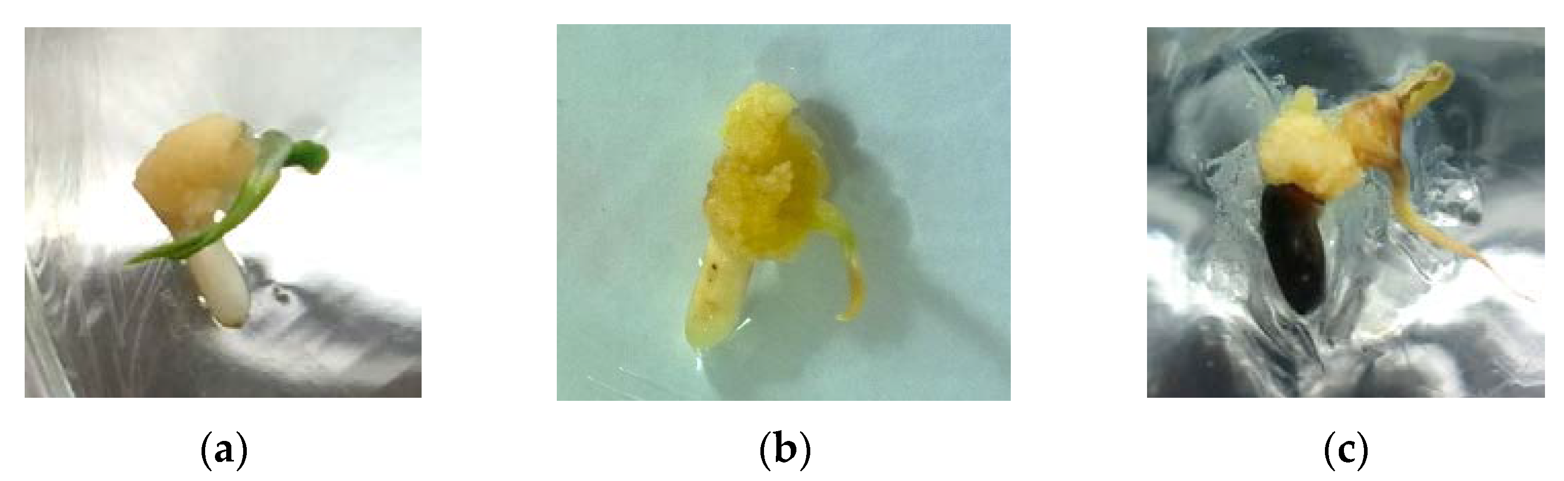
3.1. Callus Induction

The process of producing calli involves a considerable risk of infection because there is a large chance that the plant may be contaminated with environmental microorganisms. Surface sterilization is required as part of plant tissue culture techniques to decrease pathogen-related losses when callus germinates. Sodium hypochlorite, a potent oxidant with broad-spectrum antimicrobial action, is the primary substance to sterilize explant surfaces. The preliminary investigation consisted of Munpu rice seeds being sterilized with different concentrations of Clorox (10, 20, 30, and 40%) for 30 min with the purpose of determining the concentration that would provide a high germination yield. The outcome demonstrated that the contamination of the rice seeds decreased when the sterilizing agent was increased (Table 1). Contamination reduced from 25.71 to 2.14% when the sterilizing agent was raised from 10 to 20%. Complete surface sterilization was achieved with a sterilizing substance concentration of up to 30%. Surface sterilizing with 30% Clorox was shown to have the highest callus induction yield (97.86%). The lowest callus induction (73.57%) was noted to be 10% Clorox with most seeds having been infected by fungi (25.71%). It was found that increasing the sterilizing agent resulted in a reduction in total phenolic content (TPC) as shown in Figure 1a. Clorox had a noticeably higher TPC at 10% compared to other concentrations. The highest TPC and total disinfection were achieved with a Clorox concentration of 30%. Therefore, this concentration was employed for further experimentation on the optimal sterilizing time.
The Clorox incubation period for sterilizing the surface of the rice seeds was 10, 20, 30, and 40 min. The rice seed surface that was incubated for up to 30 min was thoroughly sterilized (Table 2). The lowest callus induction and highest fungal contamination rates were found at sterilizing times of 10 min. A higher yield of callus formation was achieved by extending the sterilization period. However, compared to 30 min, 40 min of incubation produced a lower yield and TPC (Figure 1b). These outcomes could be the result of Clorox’s ability to damage some plant cells in addition to destroying the cell membranes of microorganisms. Plant cells that have been harmed by an excess of either the Clorox concentration or a prolonged incubation produced fewer substances. The results indicated that 30% Clorox for 30 min provided complete surface sterilization (with no contamination) and had a high level of total phenolic content. Therefore, this condition was applied to sterilizing the surface of rice-seed to assess the anti-aging activity of different rice varieties.
After three days of placing the Hommali, Munpu, and Niawdum sterilized rice seeds on agar solidified MS medium supplemented with sugar, gellan gum, and hormones (2,4-D, NAA, and BAP), the callus began to form which was a sign of successful induction. At the end of week 3, large, yellow-colored compact texture calli were observed (Figure 2). Among the three varieties, Munpu callus was fluffier and softer than that of Hommali and Niawdum. The growth percentages of calli were 316 ± 8.79, 385 ± 10.13 and 297 ± 9.64% based on the weight of the original rice seeds for Hommali, Munpu, and Niawdum rice, respectively.

3.2. Bioactive Compounds

Anthocyanins, procyanidins, flavonoids, and phenolic acids have been reported as phenolic compounds in rice seed which provide broad biological activity [13]. In several plants, callus provided higher amounts of these bioactive compounds over the original tissue [37,38]. There are several induction conditions which can produce high amounts of phenolics in plants. Enhancing phenolics in cocoa (Theobroma and Herrania spices) was found by using the cell suspension culture technique [39]. Nitrogen and phosphorus deficiency contribute significantly to an increase in the amounts of anthocyanins in grape berry skin callus [40]. The ginger (Zingiber officinale Rosc.) rhizome callus that was treated with 100 mg/L yeast extract showed the most significant increase in the amount of phenolic and flavonoid compounds [41].
In this study, the phenolic quantity of the callus and seed extracts were determined by spectrophotometric techniques (Table 3). The total phenolic content was found in the range of 0.09–0.48 mg GAE/mL for rice seed extracts and 0.31–0.59 mg GAE/mL for the callus extracts. All rice callus cultures provided significantly (p < 0.05) higher phenolic compounds than their rice seeds. Among the rice callus extracts, Munpu had significantly (p < 0.05) higher phenolic content (0.59 mg GAE/mL), followed by Niawdum (0.42 mg GAE/mL) and Hommali (0.31 mg GAE/mL).
Amino acids are a major component of the natural moisturizing factor (NMF) which is necessary for proper stratum corneum hydration, barrier homeostasis, desquamation, and plasticity [42]. Many studies have confirmed the importance of amino acids, especially the role of arginine and glutamine in maintaining smooth and supple skin, flexible nails and healthy hair [43,44]. Amino acids in the rice callus and seed extracts were identified by UPLC (Table 4). The rice callus extracts had a significantly (p < 0.05) higher total amino acid than its seed extracts of about 6.0, 10.0, and 2.0 times for the Hommali, Munpu, and Niawdum rice, respectively. The enhancement of several amino acids consisting of arginine, glycine, glutamic acid, alanine, proline, and lysine were observed in the rice callus extracts. Results showed that glutamic acid in the callus extracts was 10 times higher than that of the seed extracts. Moreover, some amino acids such as histidine, serine, aspartic acid, threonine, tyrosine, and GABA were not found in the seed extracts, whereas they were detected in the callus extracts. Previous studies also reported that lysine, histidine, methionine, and tyrosine were higher in the callus inducing from root of rice var. Kinmaze than the normal tissue [45]. Although rice callus utilizes amino acid, especially arginine to maintain its growth, it still contains relatively high concentrations when compared to the original explant [46].
The investigation of bioactive compounds contained in the rice callus extracts indicated that cultivation enhanced the phenolics and amino acids.

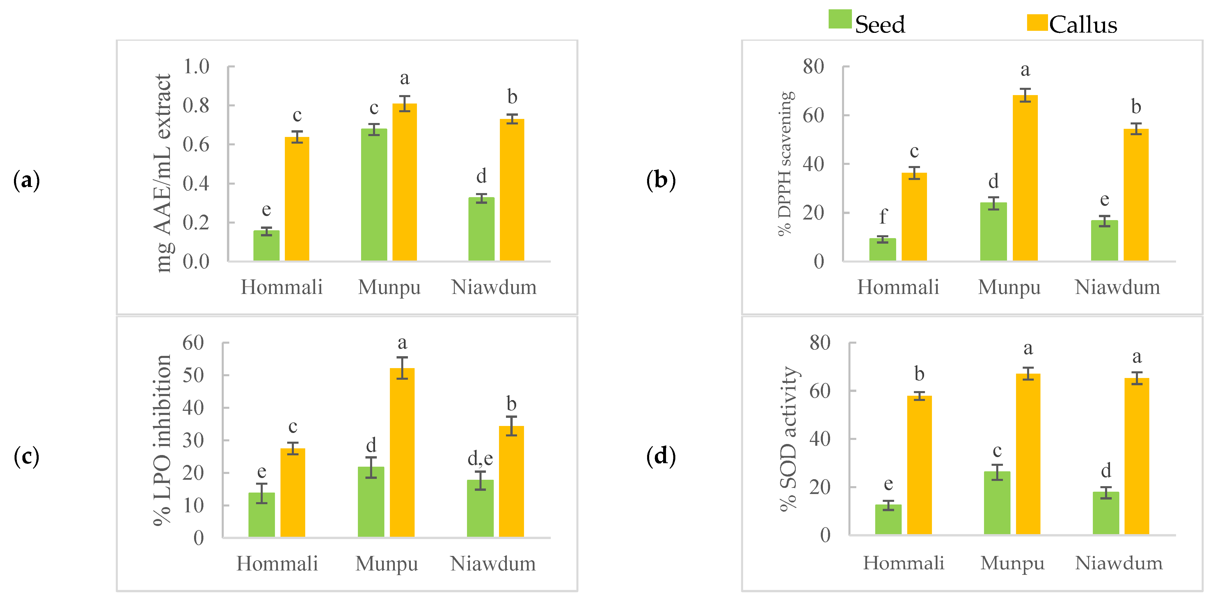
3.3. Anti-Oxidant Activities

The anti-oxidant activities of the extracts were evaluated with four different methods: PFRAP, DPPH, LPO, and SOD activities, and the results are shown in Figure 3. PFRAP assay measures the capacity in reduction of ferric ion (Fe3+)-ligand complex to ferrous (Fe2+) complex by antioxidants in the presence of a sample which donate an electron to the Fe3+/ferricyanide complex and then changed to Fe2+. This transformation can be detected as the presence of Prussian blue color at 700 nm [47,48]. In comparison to the native rice seeds, the callus extracts were more effective in PFRAP than that of in the seed extracts (Figure 3a). Moreover, the pigmented rice calli exhibit higher PFRAP than that of the non-pigmented rice. The highest PFRAP was found in Munpu callus extract (0.81 mg AAE/mL), followed by the Niawdum callus extract (0.73 mg AAE/mL), Munpu seed extract (0.68 mg AAE/mL), and Hommali callus extract (0.64 mg AAE/mL).
DPPH scavenging indicates the hydrogen-donating ability of the extract. When a solution of DPPH radical is mixed with that of an antioxidant compound (AH), which can donate a hydrogen atom (H) to DPPH, then this free radical transforms to the reduced form (DPPHH) losing its violet color [49]. In Figure 3b, Munpu callus extract showed 68.22% DPPH scavenging activity which was significantly higher than that of Niawdum and Hommali calli (54.40% and 36.30%, respectively). Furthermore, it was found that the DPPH levels significantly increased in the callus cultures when compared to their seed extracts. The optimum concentration range of ascorbic acid to plot the standard graph of % inhibition of the DPPH conditions was determined and the standard curve was Y = 317.63X − 0.9685 (R2 = 0.9989). All of the extracts displayed lower DPPH scavenging than 0.5 mg/mL of ascorbic acid (82.52%), but all the callus extracts were higher than 0.1 mg/mL of ascorbic acid (30.66%).
After lipid hydroperoxide decomposition, malondialdehyde (MDA) formed as a secondary product of oxidation. Lipid peroxidation inhibition assay measures the results of the product’s condensation between thiobarbituric acid (TBA) and MDA as a pink chromophore in an acidic environment [30]. As expected, the significantly highest LPO was Munpu callus (52.21%), followed by Niawdum callus (34.40%), and Hommali callus (27.51%) (Figure 3c). The callus culture of all rice varieties increased two times in LPO. The results were compared with those of butylated hydroxytoluene (BHT) as a reference antioxidant. Munpu callus extract had a higher LPO than 0.5 mg/mL of BHT (33.90%); Niawdum callus produced results that were comparable, while other extracts were lower than that of the standard compound.
SOD, a primary anti-oxidant enzyme, protects organisms from superoxide radicals (O2-) damaging effects by accelerating their dismutation into hydrogen peroxide (H2O2) and oxygen (O2) [50]. Promotion of SOD activity can enhance the prevention of the oxidative stress and photo-induced aging skin. SOD anti-oxidant activity of the rice callus extracts ranged from 57.89 to 67.16%, while the rice seed extracts ranged from 12.40 to 26.17% (Figure 3d). In comparison to the native rice seed extracts, the callus extracts had significantly effective antioxidant activities, and the pigmented rice callus extract exhibited significant higher antioxidant activities than that of the non-pigmented rice. Moreover, the extracts are comparable to ascorbic acid.
There were good correlation coefficients of TPC and antioxidant activities with R2 of 0.892, 0.770, 0.788, and 0.635 for PFRAP, DPPH, LPO, and SOD, respectively. These results indicated that phenolics were the major compound providing antioxidant activities resulting in higher antioxidant activities of the callus extract. This result is in accordance with several reports investigating the phytochemical content and biological potency of plant callus extracts. Song et al., [51] whose study of Ocimum species indicated that callus extracts showed significant increases in PFRAP due to a high accumulation of phenolic in the callus culture. Tadhani et al., [37] reported that Stevia rebaudiana callus showed higher DPPH than did the leaf explant. Coleus forskohlii callus extract (60 μg/mL) showed higher LPO antioxidant activity than that of its leaves [52]. Ślesak et al., [53] stated that Mesembryanthemum crystallinum callus culture increased in SOD activity. Furthermore, the callus’ culture conditions are crucial for controlling in vitro callus growth and the production of significant metabolites, such as light conditions favoring phenolic synthesis and SOD enzyme expression in Cnidium officinale callus [54]. Moreover, salt stress can increase antioxidant and lipid peroxidation levels in pepper callus tissues [55].

3.4. Anti-Aging Activities
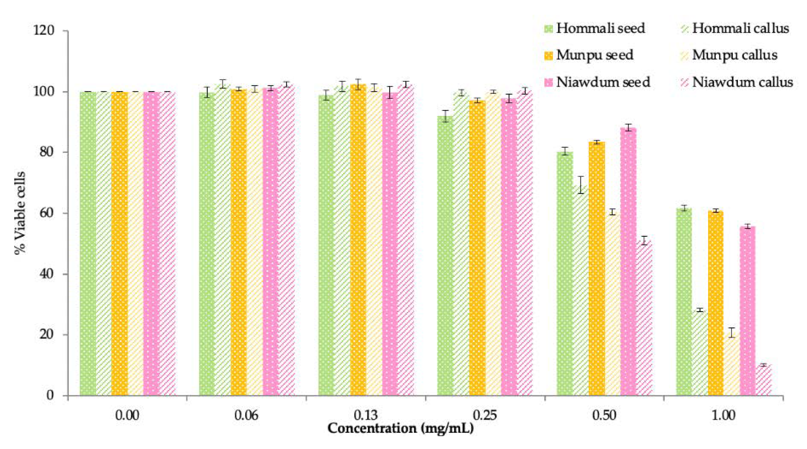
3.4.1. Cytotoxicity

Figure 4 shows the cytotoxicity in the dose-dependent extracts. Less than 0.25 mg/mL of all rice seed and callus extracts displayed more than a 90% cell survival. The cytotoxic activity of extracts was calculated and expressed as the IC50, the concentration causing 50% viable cell. The lowest IC50 value was Niawdum callus (0.51 mg/mL), followed by Munpu callus (0.63 mg/mL) and Hommali callus (0.73 mg/mL). While the IC50 values of the rice seed extract were 1.40 mg/mL, 1.11 mg/mL, and 1.27 mg/mL for the Hommali, Niawdum, and Munpu rice seed, respectively. Thus, higher IC50 values of the rice seed extracts indicated that they were less cytotoxic to skin cells than the callus extracts.

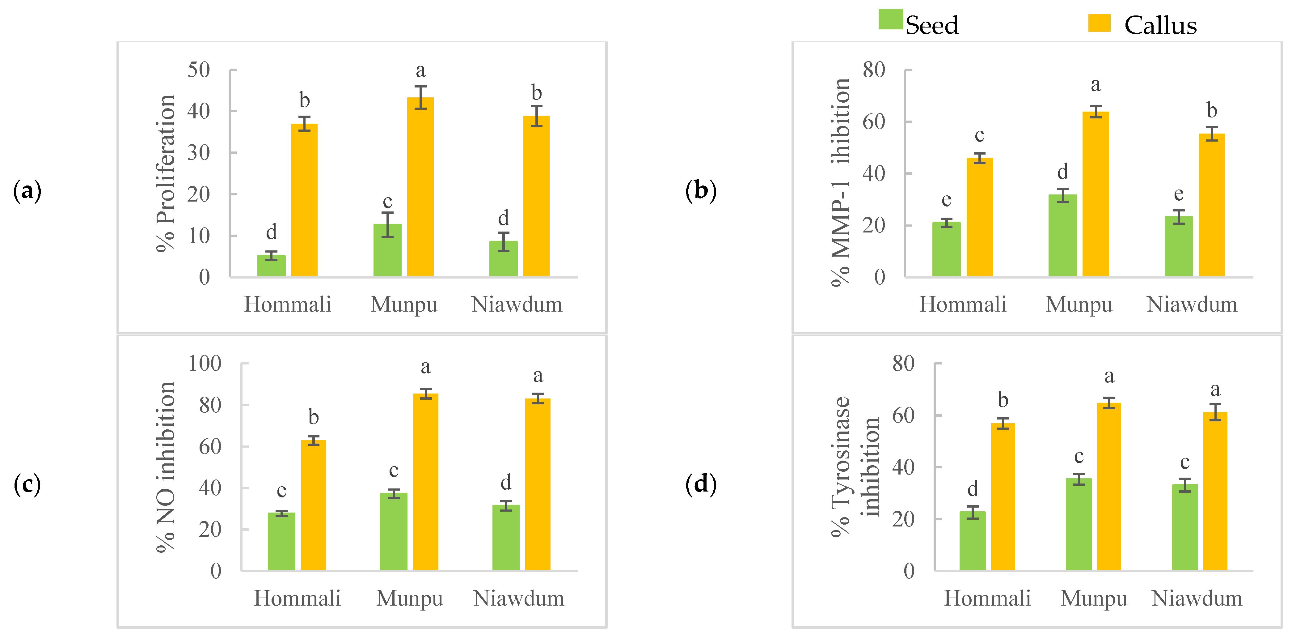
3.4.2. Promoting Keratinocytes’ Proliferation

Keratinocytes are the most common kind of cell found in the epidermis, and they play an essential part in the intricate process of wound healing [56]. The three phases that characterize these cells are the inflammatory, cell proliferation, and remodelling. These phases follow a certain order but overlap with one another [57]. Promoting keratinocytes’ proliferation is associated with the mechanisms that contribute to wound healing and anti-aging. The effect of the extracts on stimulating the growth of keratinocytes is seen in Figure 5a. The rice callus extracts showed excellent promotion of keratinocytes when compared with the rice seed extracts. The rice callus culture (37–43%) promoted about five times more keratinocytes than the original tissue (5–12%). The findings indicated that rice calli proliferate keratinocyte cells which can be employed as an active component in anti-aging cosmetics. Moreover, there was a strong correlation between the promotion of keratinocytes and TMC (r = 0.900) in which amino acids had a major effect on promoting cell activity.

3.4.3. Anti-Collagenase Activity

Anti-collagenase activity was determined by using thiopeptide as a chromogenic substrate for MMP-1 inhibitor. MMP-1 is a member of the matrix metalloproteinases that plays a role as collagenases to cleave interstitial collagens. The highest collagenase inhibition was found in Munpu callus (53.83 %), followed by Niawdum (45.32%) and Hommali (35.92%) calli as shown in Figure 5b. The rice callus extracts were observed to be approximately three times greater than their rice-seed extracts. This finding was comparable to the studies of Tito et al. [9] who reported that tomato callus extract can reduce MMP expression in keratinocytes. Furthermore, they also stated that tomato callus extract effected collagen stability and stimulated new type I and III collagen synthesis in fibroblasts. As a result of the accumulation of phenolic compounds, pigmented rice exhibited significantly greater levels of collagenase inhibition than that of non-pigmented rice. In addition, anti-collagenase activity had a strong correlation with TPC (r = 0.707) and a very strong one with TMC (r = 0.943), indicating amino acids had more impact on the anti-collagenase activity of the rice callus culture than that of the phenolic compounds. Based on these findings, it seems possible to use rice callus extracts in the creation of cosmetics that delay the signs of aging.

3.4.4. Anti-Inflammatory Activity

Under normal physiological states, nitric oxide (NO) acts as an anti-inflammatory agent, while under abnormal circumstances, the overproduction of NO is considered a pro-inflammatory mediator and an inflammatory inducer [58]. In this study, the extracts were evaluated for their anti-inflammatory activity in human keratinocytes which were activated by bacterial lipopolysaccharide (LPS) after which inhibition of NO was detected by using Griess reagent. Figure 5c displays the result of NO inhibition. The highest NO inhibition was observed in Munpu callus (85.40%), followed by Niawdum callus (83.13%), and Hommali callus (62.87%). NO inhibition levels of calli increased about 2.5 times more than their seeds. These findings agree with the research conducted by Daniela et al. [59], who found that callus cultures of edelweiss are a valuable source of anti-inflammatory substances. These substances have the potential to be useful in treating chronic inflammatory skin diseases as well as bacterial and atherogenic inflammation. In addition, the pigmented rice showed a markedly stronger NO inhibition than the non-pigmented rice. Moreover, the pigmented rice callus extracts appeared to be a potent anti-inflammatory agent. There is a strong correlation between the results of NO inhibition and TMC (r = 0.885), which reveals that the anti-inflammatory activity was related to amino acids. The results correspond with the previous report that amino acids, especially, cysteine, histidine, and glycine, have an anti-inflammatory effect [60,61]. According to the results, the rice callus extracts appear to have potent anti-inflammatory activity, especially, the pigmented rice callus extracts.

3.4.5. Anti-Tyrosinase Activity

Tyrosinase, a copper-containing enzyme, is a key enzyme in melanin production, which is important in the coloration of mammalian skin and hair [62]. The tyrosinase enzyme inhibitor is associated with reduction of melanogenesis, which is widely used as a depigmentation agent. Skin aging, wrinkling, roughness, and dryness occur along with certain hyperpigmentation such as age spots, solar lentigos, and melasma. In skin lightening cosmetic products, tyrosinase inhibitors, such as hydroquinone and kojic acid, are utilized as anti-pigmenting agents. However, synthetic compounds frequently induce skin irritation; thus, natural compounds are used as alternatives [63]. Callus extracts (56.85–64.77%) showed a significantly higher inhibition of tyrosinase activity than the seed extracts (22.59–35.35%) (Figure 5d). The result suggested that the rice callus extracts produced an active compound which responded to the tyrosinase inhibition. A strong correlation between the anti-tyrosinase activity and TMC was found (r = 0.887). These results suggest that the rice callus extracts contained an effective tyrosinase inhibitor and can be used as a skin whitening agent in cosmetic products.

4. Conclusions

According to the results of this study, the higher yield of rice calli were successfully produced when seeds were sterilized in 30% Clorox for 30 min and then grown on solid MS medium enriched with 2,4-D, NAA, and BAP. The investigation of bioactive compounds in the rice callus extract indicated that the condition for cultivation enhanced phenolics and amino acids. Antioxidant (PFRAP, DPPH, and SOD) and anti-aging (keratinocytes’ proliferation, anti-collagenase, anti-inflammatory, and anti-tyrosinase) activities of rice seeds were greatly enhanced by inducing callus formation. The rice callus extracts appeared to have potent antioxidant and anti-aging activities, especially Munpu rice. Therefore, the rice callus extracts, particularly the pigmented rice, are an alternative natural source with much potential for usage as an antioxidant and anti-aging active components in cosmetics, functional foods, and medications.

Author Contributions

Conceptualization, N.S.; Methodology, N.S. and W.V.; Validation, N.S. and W.V.; Formal Analysis, N.S. and W.V., Investigation, N.S. and W.V.; Resources, N.S.; Data Curation, N.S. and W.V.; Writing—Origination Draft Preparation, N.S. and W.V.; Writing—Review and Editing, N.S.; Visualization, W.V.; Supervision, N.S.; Project Administration, N.S.; Funding Acquisition, N.S. All authors have read and agreed to the published version of the manuscript.

Funding

This work was supported by research funding from the Agriculture Research Development Agency (Public Organization), grant number CRMP 560005021900.

Institutional Review Board Statement

Not applicable.

Informed Consent Statement

Not applicable.

Data Availability Statement

All the data are available in the manuscript.

Acknowledgments

The authors would like to acknowledge Cosmetic and Beauty Innovations for Sustainable Development (CBIS) Research Group and Mae Fah Luang University for providing scientific equipment and facilities for this work.

Conflicts of Interest

The authors declare no conflict of interest.

References

  1. Raschke, C.; Elsner, P. Skin Aging: A Brief Summary of Characteristic Changes. In Textbook of Aging Skin, 2nd ed.; Farage, M., Miller, K., Maibach, H., Eds.; Springer: Berlin/Heidelberg, Germany, 2010; pp. 37–43. [Google Scholar]
  2. Chiu, P.C.; Chan, C.C.; Lin, H.M.; Chiu, H.C. The clinical anti-aging effects of topical kinetin and niacinamide in Asians: A randomized, double-blind, placebo-controlled, split-face comparative trial. J. Cosmet. Dermatol. 2007, 6, 243–249. [Google Scholar] [CrossRef] [PubMed]
  3. Schürch, C.; Blum, P.; Zülli, F. Potential of plant cells in culture for cosmetic application. Phytochem. Rev. 2008, 7, 599–605. [Google Scholar] [CrossRef]
  4. Schmid, D.; Schürch, C.; Blum, P.; Belser, E.; Zülli, F. Plant stem cell extract for longevity of skin and hair. SOFW 2008, 134, 29–35. [Google Scholar]
  5. Ji, S.H.; Akter, M.; Ko, E.Y.; Choi, E.H.; Keum, Y.S.; Han, I. Enhancing antioxidant activities and anti-aging effect of rice stem cell extracts by plasma treatment. Appl. Sci. 2022, 12, 2903. [Google Scholar] [CrossRef]
  6. Draelos, Z.D. Plant stem cells and skin care. Cosmet. Dermatol. 2012, 25, 395–396. [Google Scholar]
  7. Schmid, D.; Zülli, F. Stimulating epidermal regeneration with plant-derived stem cells. C&T 2010, 125, 61–71. [Google Scholar]
  8. Barbulova, A.; Tito, A.; Carola, A.; Bimonte, M.; de Laurentis, F.; D’Ambrosio, P.; Apone, F.; Colucci, G.; Monoli, I.; Cucchiara, M. Raspberry stem cell extract to protect skin from inflammation and oxidative stress. C&T 2010, 125, 38–47. [Google Scholar]
  9. Tito, A.; Carola, A.; Bimonte, M.; Barbulova, A.; Arciello, S.; de Laurentiis, F.; Monoli, I.; Hill, J.; Gibertoni, S.; Colucci, G.; et al. A tomato stem cell extract, containing anti-oxidant compounds and metal chelating factors, protects skin cells from heavy metal-induced damages. Int. J. Cosmet. Sci. 2011, 33, 543–552. [Google Scholar] [CrossRef]
  10. Ludwig, P.; Bennett, S.; Gruber, J. Rice meristem stimulates epigenetic rejuvenation: New technologies. SA Pharm. Cosmet. Rev. 2011, 38, 34–37. [Google Scholar]
  11. Zawistowski, J.; Kopec, A.; Kitts, D.D. Effects of a black rice extract (Oryza sativa L. indica) on cholesterol levels and plasma lipid parameters in Wistar Kyoto rats. J. Funct. Foods 2009, 1, 50–56. [Google Scholar] [CrossRef]
  12. Chung, H.S.; Shin, J.C. Characterization of anti-oxidant alkaloids and phenolic acids from anthocyanin-pigmented rice (Oryza sativa cv. Heugjinjubyeo). Food Chem. 2007, 104, 1670–1677. [Google Scholar] [CrossRef]
  13. Yawadio, R.; Tanimori, S.; Morita, N. Identification of phenolic compounds isolated from pigmented rices and their aldose reductase inhibitory activities. Food Chem. 2007, 101, 1616–1625. [Google Scholar] [CrossRef]
  14. Finocchiaro, F.; Ferrari, B.; Gianinetti, A. A study of biodiversity of flavonoid content in the rice caryopsis evidencing simultaneous accumulation of anthocyanins and proanthocyanidins in a black-grained genotype. J. Cereal Sci. 2010, 51, 28–34. [Google Scholar] [CrossRef]
  15. Bhat, F.M.; Sommano, S.R.; Riar, C.S.; Seesuriyachan, P.; Chaiyaso, T.; Prom-u-Thai, C. Status of bioactive compounds from bran of pigmented traditional rice varieties and their scope in production of medicinal food with nutraceutical importance. Agronomy 2020, 10, 1817. [Google Scholar] [CrossRef]
  16. Guo, H.; Ling, W.; Wang, Q.; Liu, C.; Hu, Y.; Xia, M.; Feng, X.; Xia, X. Effect of anthocyanin-rich extract from black rice (Oryza sativa L. indica) on hyperlipidemia and insulin resistance in fructose-fed rats. Plant Foods Hum. Nutr. 2007, 62, 1–6. [Google Scholar] [CrossRef]
  17. Nam, S.H.; Choi, S.P.; Kang, M.Y.; Kozukue, N.; Friedman, M. Antioxidative, antimutagenic, and anticarcinogenic activities of rice bran extracts in chemical and cell assays. J. Agric. Food Chem. 2005, 53, 816–822. [Google Scholar] [CrossRef]
  18. Chulasiri, M. Anti-aging cosmeceuticals from pigmented rice. Asian J. Pharm. Sci. 2016, 11, 30. [Google Scholar] [CrossRef]
  19. Vargas, C.G.; da Silva Junior, J.D.; Rabelo, T.K.; Moreira, J.C.F.; Gelain, D.P.; Rodrigues, E.; Augusti, P.R.; Rios, A.d.O.; Flôres, S.H. Bioactive compounds and protective effect of red and black rice brans extracts in human neuron-like cells (SH-SY5Y). Food Res. Int. 2018, 113, 57–64. [Google Scholar] [CrossRef]
  20. Oki, T.; Masuda, M.; Kobayashi, M.; Nishiba, Y.; Furuta, S.; Suda, I.; Sato, T. Polymeric procyanidins as radical-scavenging components in red-hulled rice. J. Agric. Food Chem. 2002, 50, 7524–7529. [Google Scholar] [CrossRef]
  21. Vichit, W.; Saewan, N. Anti-oxidant activities and cytotoxicity of Thai pigmented rice. Int. J. Pharm. Pharm. 2015, 7, 329–334. [Google Scholar]
  22. Nam, S.H.; Choi, S.P.; Kang, M.Y.; Koh, H.J.; Kozukue, N.; Friedman, M. Antioxidative activities of bran extracts from twenty one pigmented rice cultivars. Food Chem. 2006, 94, 613–620. [Google Scholar] [CrossRef]
  23. Saewan, N.; Vichit, W.; Prinyarux, T. Anti-aging efficacy of thai red rice callus cosmetic product. J. Appl. Sci. 2018, 17, 63–72. [Google Scholar]
  24. Mousumee, K.; Soyeon, P.; Hyeon-Jin, K.; Kui-Jae, L.; Dea-Heon, K.; So-Hyeon, B.; Seong-Tshool, H. The Resveratrol Rice DJ526 Callus Significantly Increases the Lifespan of Drosophila (Resveratrol Rice DJ526 Callus for Longevity). Nutrients 2019, 11, 983. [Google Scholar]
  25. Gajula, D.; Verghese, M.; Boateng, J.; Walker, L.T.; Shackelford, T.; Mentreddy, S.R.; Cedric, S. Determination of total phenolics, flavonoids and anti-oxidant and chemopreventive potential of basil (Ocimum basilicum L. and Ocimum tenuiflorum L.). Int. J. Cancer Res. 2009, 5, 130–143. [Google Scholar] [CrossRef]
  26. Liming, W.; Jinhui, Z.; Xiaofeng, X.; Yi, L.; Jing, Z. Fast determination of 26 amino acids and their content changes in royal jelly during storage using ultra-performance liquid chromatography. J. Food Compost. Anal. 2009, 22, 242–249. [Google Scholar] [CrossRef]
  27. Kuda, T.; Yano, T. Changes of radical-scavenging capacity and ferrous reducing power in chub mackerel Scomber japonicus and Pacific saury Cololabis saira during 4 °C storage and retorting. LWT-Food Sci. Technol. 2009, 42, 1070–1075. [Google Scholar] [CrossRef]
  28. Vichit, W.; Saewan, N. Effect of germination on anti-oxidant, anti-inflammatory and keratinocyte proliferation of rice. Int. Food Res. J. 2016, 23, 2006–2015. [Google Scholar]
  29. Rangkadilok, N.; Sitthimonchai, S.; Worasuttayangkurn, L.; Mahidol, C.; Ruchirawat, M.; Satayavivad, J. Evaluation of free radical scavenging and antityrosinase activities of standardized longan fruit extract. Food Chem. Toxicol. 2007, 45, 328–336. [Google Scholar] [CrossRef]
  30. Miguel, M.G. Anti-oxidant and anti-inflammatory activities of essential oils: A short review. Molecules 2010, 15, 9252–9287. [Google Scholar] [CrossRef] [PubMed]
  31. Ma, C.M.; Sato, N.; Li, X.Y.; Nakamura, N.; Hattori, M. Flavan-3-ol contents, anti-oxidative and α-glucosidase inhibitory activities of Cynomorium songaricum. Food Chem. 2010, 118, 116–119. [Google Scholar] [CrossRef]
  32. Takahashi, H.; Tsuji, H.; Hashimoto, Y.; Ishida-Yamamoto, A.; Iizuka, H. Cell proliferation and cytokine induction by TNF-alpha of psoriatic keratinocytes are not different from normal keratinocytes in vitro. Indian J. Dermatol. 2009, 54, 237–239. [Google Scholar] [CrossRef] [PubMed]
  33. Karim, A.A.; Azlan, A.; Ismail, A.; Hashim, P.; Abd Gani, S.S.; Zainudin, B.H.; Abdullah, N.A. Phenolic composition, anti-oxidant, anti-wrinkles and tyrosinase inhibitory activities of cocoa pod extract. BMC Complement. Altern. Med. 2014, 14, 381. [Google Scholar] [CrossRef]
  34. Laskin, J.D.; Heck, D.E.; Laskin, D.L. Multifunctional role of nitric oxide in inflammation. Trends Endocrinol. Metab. 1994, 5, 377–382. [Google Scholar] [CrossRef]
  35. Piktel, E.; Wnorowska, U.; Cieśluk, M.; Deptula, P.; Pogoda, K.; Misztalewska-Turkowicz, I.; Paprocka, P.; Niemirowicz-Laskowska, K.; Wilczewska, A.Z.; Janmey, P.A.; et al. Inhibition of inflammatory response in human keratinocytes by magnetic nanoparticles functionalized with PBP10 peptide derived from the PIP2-binding site of human plasma gelsolin. J. Nanobiotechnol. 2019, 17, 22. [Google Scholar] [CrossRef] [PubMed]
  36. Saewan, N.; Koysomboon, S.; Chantrapromma, K. Anti-tyrosinase and anti-cancer activities of flavonoids from Blumea balsamifera DC. J. Med. Plant Res. 2011, 5, 8. [Google Scholar]
  37. Tadhani, M.B.; Patel, V.H.; Subhash, R. In vitro anti-oxidant activities of Stevia rebaudiana leaves and callus. J. Food Compost. Anal. 2007, 20, 323–329. [Google Scholar] [CrossRef]
  38. Hakkim, F.L.; Shankar, C.G.; Girija, S. Chemical composition and anti-oxidant property of holy basil (Ocimum sanctum L.) leaves, stems, and inflorescence and their in vitro callus cultures. J. Agric. Food Chem. 2007, 55, 9109–9117. [Google Scholar] [CrossRef]
  39. Venkatramesh, M.; Wagner, D.R.; Lall, S.; Lejard, F.Y.; Yoo, S.H. Production and Extraction of Procyanidins from Plant Cell Cultures. U.S. Patent No. WO2010/114567A1, 7 October 2010. [Google Scholar]
  40. Zheng, H.-Z.; Wei, H.; Guo, S.-H.; Yang, X.; Feng, M.-X.; Jin, X.-Q.; Fang, Y.-L.; Zhang, Z.-W.; Xu, T.-F.; Meng, J.-F. Nitrogen and phosphorus co-starvation inhibits anthocyanin synthesis in the callus of grape berry skin. Plant Cell Tissue Organ Cult. 2020, 142, 313–325. [Google Scholar] [CrossRef]
  41. Ali, A.M.A.; El-Nour, M.E.M.; Yagi, S.M. Total phenolic and flavonoid contents and antioxidant activity of ginger (Zingiber officinale Rosc.) rhizome, callus and callus treated with some elicitors. J. Genet. Eng. Biotechnol. 2018, 16, 677–682. [Google Scholar] [CrossRef]
  42. Robinson, M.; Visscher, M.; Laruffa, A.; Wickett, R. Natural moisturizing factors (NMF) in the stratum corneum (SC). I. Effects of lipid extraction and soaking. J. Cosmet. Sci. 2010, 61, 13–22. [Google Scholar]
  43. Saini, R.; Badole, S.; Zanwar, A. Arginine derived nitric oxide: Key to healthy skin. In Bioactive Dietary Factors and Plant Extracts in Dermatology; Watson, R.R., Zibadi, S., Eds.; Nutrition and Health; Humana Press: Totowa, NJ, USA, 2013; pp. 73–82. [Google Scholar]
  44. Chessa, M.A.; Iorizzo, M.; Richert, B.; López-Estebaranz, J.L.; Rigopoulos, D.; Tosti, A.; Gupta, A.K.; Di Chiacchio, N.; Di Chiacchio, N.G.; Rubin, A.I.; et al. Pathogenesis, clinical signs and treatment recommendations in brittle nails: A review. Derm. Ther. 2020, 10, 15–27. [Google Scholar] [CrossRef] [PubMed]
  45. Yatazawa, M.; Furuhashi, K.; Kurihara, N.; Ohnishi, Y. Amino acid composition of rice callus tissue grown with different kinds of nitrogen sources. Soil Sci. Plant Nutr. 1968, 14, 85–88. [Google Scholar] [CrossRef]
  46. Lee, K.K.; Furuhashi, K.; Yatazawa, M. Utilization of arginine dy rice callus tissue. Soil Sci. Plant Nutr. 1972, 18, 179–184. [Google Scholar] [CrossRef]
  47. Pulido, R.; Bravo, L.; Saura-Calixto, F. Anti-oxidant activity of dietary polyphenols as determined by a modified ferric reducing/anti-oxidant power assay. J. Agric. Food Chem. 2000, 48, 3396–3402. [Google Scholar] [CrossRef] [PubMed]
  48. Prabha, M.R.; Vasantha, K. Anti-oxidant, cytotoxicity and polyphenolic content of Calotropis procera (Ait.) R. Br. flowers. J. Appl. Pharm. Sci. 2011, 1, 136–140. [Google Scholar]
  49. Alam, M.N.; Bristi, N.J.; Rafiquzzaman, M. Review on in vivo and in vitro methods evaluation of anti-oxidant activity. Saudi Pharm. J. 2013, 21, 143–152. [Google Scholar] [CrossRef] [PubMed]
  50. Pieme, C.A.; Penlap, V.N.; Ngogang, J.; Costache, M. In vitro cytotoxicity and anti-oxidant activities of five medicinal plants of Malvaceae family from Cameroon. Environ. Toxicol. Pharmacol. 2010, 29, 223–228. [Google Scholar] [CrossRef]
  51. Song, H.; Kumar, P.; Arivazhagan, G.; Lee, S.; Yoon, H.M.; Kim, I.H.; Kwon, H.J.; Kim, J.M.; Hakkim, F.L. Anti-oxidant property of leaves and calluses extracts of in-vitro grown 5 different Ocimum species Plant Biotechnol. J. 2012, 39, 146–153. [Google Scholar]
  52. Sunitha, K.; Chary, K.B.; Nimgulkar, C.C.; Kumar, B.D.; Rao, D.M. Identification, quantification and anti-oxidant activity of secondary metabolites in leaf and callus extracts of Coleus forskohlii. Int. J. Pharma Bio Sci. 2013, 4, 1139–1149. [Google Scholar]
  53. Ślesak, I.; Libik, M.; Miszalski, Z. Superoxide dismutase activity in callus from the C3-CAM intermediate plant Mesembryanthemum crystallinum. PCTOC 2003, 75, 49–55. [Google Scholar] [CrossRef]
  54. Adil, M.; Ren, X.; Jeong, B.R. Light elicited growth, antioxidant enzymes activities and production of medicinal compounds in callus culture of Cnidium officinale Makino. J. Photochem. Photobiol. B Biol. 2019, 196, 111509. [Google Scholar] [CrossRef] [PubMed]
  55. Furtana, G.B.; Sönmez, K.; Ellialtıoğlu, Ş.Ş.; Tıpırdamaz, R. The effects of hydrogen peroxide and nitric oxide on ion contents and lipid peroxidation levels of pepper callus tissues under salt stress. Acta Hortic. 2019, 1257, 123–130. [Google Scholar] [CrossRef]
  56. Wang, Y.; Graves, D.T. Keratinocyte function in normal and diabetic wounds and modulation by FOXO1. J. Diabet. Res. 2020, 2020, 3714704. [Google Scholar] [CrossRef]
  57. Diegelmann, R.F.; Evans, M.C. Wound healing: An overview of acute, fibrotic and delayed healing. Front. Biosci. 2004, 9, 283–289. [Google Scholar] [CrossRef] [PubMed]
  58. Debnath, T.; Park, S.; Kim, D.; Jo, J.; Lim, B. Anti-oxidant and anti-inflammatory activities of Inonotus obliquus and germinated brown rice extracts. Molecules 2013, 18, 9293–9304. [Google Scholar] [CrossRef] [PubMed]
  59. Daniela, L.; Alla, P.; Maurelli, R.; Elena, D.; Giovanna, P.; Vladimir, K.; Roberto, D.T.; Chiara, D.L.; Saveria, P.; Liudmila, K. Anti-Inflammatory effects of concentrated ethanol extracts of edelweiss (Leontopodium alpinum Cass.) callus cultures towards human keratinocytes and endothelial cells. Mediat. Inflamm. 2012, 2012, 498373. [Google Scholar] [CrossRef] [PubMed]
  60. Hasegawa, S.; Ichiyama, T.; Sonaka, I.; Ohsaki, A.; Hirano, R.; Haneda, Y.; Fukano, R.; Hara, M.; Furukawa, S. Amino acids exhibit anti-inflammatory effects in human monocytic leukemia cell line, THP-1 cells. Inflamm. Res. 2011, 60, 1013–1019. [Google Scholar] [CrossRef] [PubMed]
  61. Hasegawa, S.; Ichiyama, T.; Sonaka, I.; Ohsaki, A.; Okada, S.; Wakiguchi, H.; Kudo, K.; Kittaka, S.; Hara, M.; Furukawa, S. Cysteine, histidine and glycine exhibit anti-inflammatory effects in human coronary arterial endothelial cells. Clin. Exp. Immunol. 2012, 167, 269–274. [Google Scholar] [CrossRef]
  62. Parvez, S.; Kang, M.; Chung, H.S.; Bae, H. Naturally occurring tyrosinase inhibitors: Mechanism and applications in skin health, cosmetics and agriculture industries. Phytother. Res. 2007, 21, 805–816. [Google Scholar] [CrossRef] [PubMed]
  63. Momtaz, S.; Mapunya, B.M.; Houghton, P.J.; Edgerly, C.; Hussein, A.; Naidoo, S.; Lall, N. Tyrosinase inhibition by extracts and constituents of Sideroxylon inerme L. stem bark, used in South Africa for skin lightening. J. Ethnopharmacol. 2008, 119, 507–512. [Google Scholar] [CrossRef]
Figure 1. Effect of Clorox concentration (a) and incubation time (b) on phenolic content. Data with different letters (a, b, and c) indicate significant differences (p < 0.05) between samples.
Cosmetics 09 00079 g001
Figure 2. Callus derived from seed of Hommali (a), Munpu (b), and Niawdum (c) rice.
Cosmetics 09 00079 g002
Figure 3. Anti-oxidant activities of rice callus and seed extracts including PFRAP (a), DPPH scavenging (b), LPO (c), and SOD (d). Values are presented as means ± SD (n = 3). Data with different letters (a, b, c, d, and e) indicate significant differences (p < 0.05) between samples.
Cosmetics 09 00079 g003
Figure 4. Cytotoxicity of the rice callus and seed extracts.
Cosmetics 09 00079 g004
Figure 5. The anti-aging activities of rice callus and seed extracts including keratinocytes’ proliferation (a), anti-collagenase (b), anti-inflammatory (c), and anti-tyrosinase activities (d). Values are displayed as means ± SD (n = 3). Data with different letters (a, b, c, d and e) indicate significant differences (p < 0.05) between samples.
Cosmetics 09 00079 g005
Table 1. Effect of Clorox concentration on rice callus induction.
Clorox Concentration (%)
10203040
Contamination (%)25.71 ± 4.04 c2.14 ± 1.01 b0.00 ± 0.00 a0.00 ± 0.00 a
Callus induction (%)73.57 ± 3.03 b95.71 ± 2.02 a97.86 ± 1.01 a95.71 ± 4.04 a
Values are presented as means ± SD (n = 3). Data with different letters (a, b, and c) indicate significant differences (p < 0.05) between Clorox concentrations.
Table 2. Effect of Clorox incubation time on rice callus induction.
Clorox Incubation Time (min)
10203040
Contamination (%)33.57 ± 5.05 c4.29 ± 2.02 b0.00 ± 0.00 a0.00 ± 0.00 a
Callus induction (%)65.00 ± 5.05 c95.00 ± 1.01 a97.86 ± 1.01 a89.29 ± 3.03 b
Values are presented as means ±SD (n = 3). Data with different letters (a, b and c) indicate significant differences (p < 0.05) between incubation times.
Table 3. Total phenolic contents of rice callus and seed extracts.
Bioactive
Compounds
HommaliMunpuNiawdum
SeedCallusSeedCallusSeedCallus
Total phenolic content
(mg GAE/mL extract)
0.09 ± 0.010.31 ± 0.01 *0.48 ± 0.020.59 ± 0.02 *0.30 ± 0.030.42 ± 0.01 *
Values are presented as means ± SD (n = 3). * indicates significant differences (p < 0.05) of callus and its seed extracts.
Table 4. Amino acids analysis by UPLC (mM/mL extract).
Amino AcidHommaliMunpuNiawdum
SeedCallusSeedCallusSeedCallus
HISn.d.0.07 ± 0.03n.d.0.06 ± 0.05n.d.0.02 ± 0.01
SERn.d.0.52 ± 0.11n.d.0.57 ± 0.040.45 ± 0.030.03 ± 0.01
ARG0.00 ± 0.010.33 ± 0.070.00 ± 0.020.18 ± 0.020.25 ± 0.030.26 ± 0.03
GLY0.06 ± 0.020.31 ± 0.060.38 ± 0.040.41 ± 0.030.39 ± 0.020.25 ± 0.04
ASPn.d.0.17 ± 0.06n.d.0.18 ± 0.15n.d.0.17 ± 0.03
GLU0.21 ± 0.074.13 ± 0.570.17 ± 0.034.05 ± 0.180.13 ± 0.013.89 ± 0.07
THRn.d.0.23 ± 0.07n.d.0.32 ± 0.15n.d.0.23 ± 0.02
ALA1.03 ± 0.202.79 ± 0.490.26 ± 0.012.60 ± 0.152.86 ± 0.032.49 ± 0.04
PRO0.15 ± 0.010.94 ± 0.180.09 ± 0.010.22 ± 0.080.20 ± 0.030.13 ± 0.02
Cysn.d.n.d.n.d.n.d.0.06 ± 0.03n.d.
LYS0.01 ± 0.030.19 ± 0.06n.d.0.06 ± 0.04n.d.0.04 ± 0.01
TYRn.d.0.75 ± 0.14n.d.0.81 ± 0.03n.d.0.04 ± 0.00
METn.d.n.d.n.d.n.d.n.d.n.d.
VAL0.36 ± 0.040.34 ± 0.070.31 ± 0.030.30 ± 0.060.45 ± 0.020.35 ± 0.04
LLE0.13 ± 0.010.06 ± 0.02n.d.0.05 ± 0.010.12 ± 0.020.14 ± 0.02
LEU0.27 ± 0.030.04 ± 0.02n.d.0.07 ± 0.010.43 ± 0.010.19 ± 0.02
PHE0.05 ± 0.020.04 ± 0.01n.d.0.13 ± 0.010.12 ± 0.020.22 ± 0.04
GABAn.d.2.32 ± 0.35n.d.2.37 ± 0.04n.d.2.37 ± 0.04
Total2.25 ± 0.3013.20 ± 1.20 *1.20 ± 0.1412.36 ± 1.17 *5.45 ± 0.8010.81 ± 1.16 *
Values are presented as means ± SD (n = 3). n.d. is not detected. * indicates significant differences (p < 0.05) between callus and its seed extracts.
Publisher’s Note: MDPI stays neutral with regard to jurisdictional claims in published maps and institutional affiliations.

