
Evaluation of the Efficacy of an Elastin-Inducing Composition Containing Amino Acids, Copper, and Hyaluronic Acid: Results of an Open Single-Center Clinical Trial Study

Abstract
:1. Introduction
2. Materials and Methods
2.1. Study Product
Materials
2.2. Study Design
2.3. Statistical Analysis
3. Results
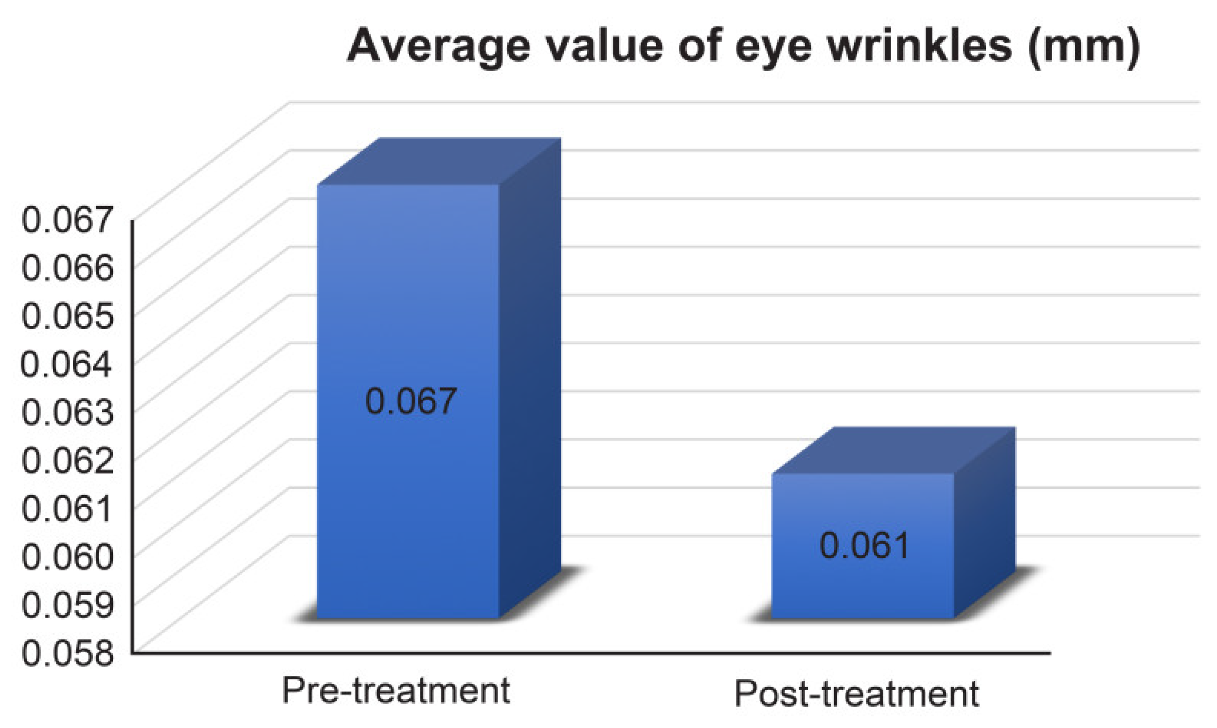
3.1. Wrinkles around the Eyes
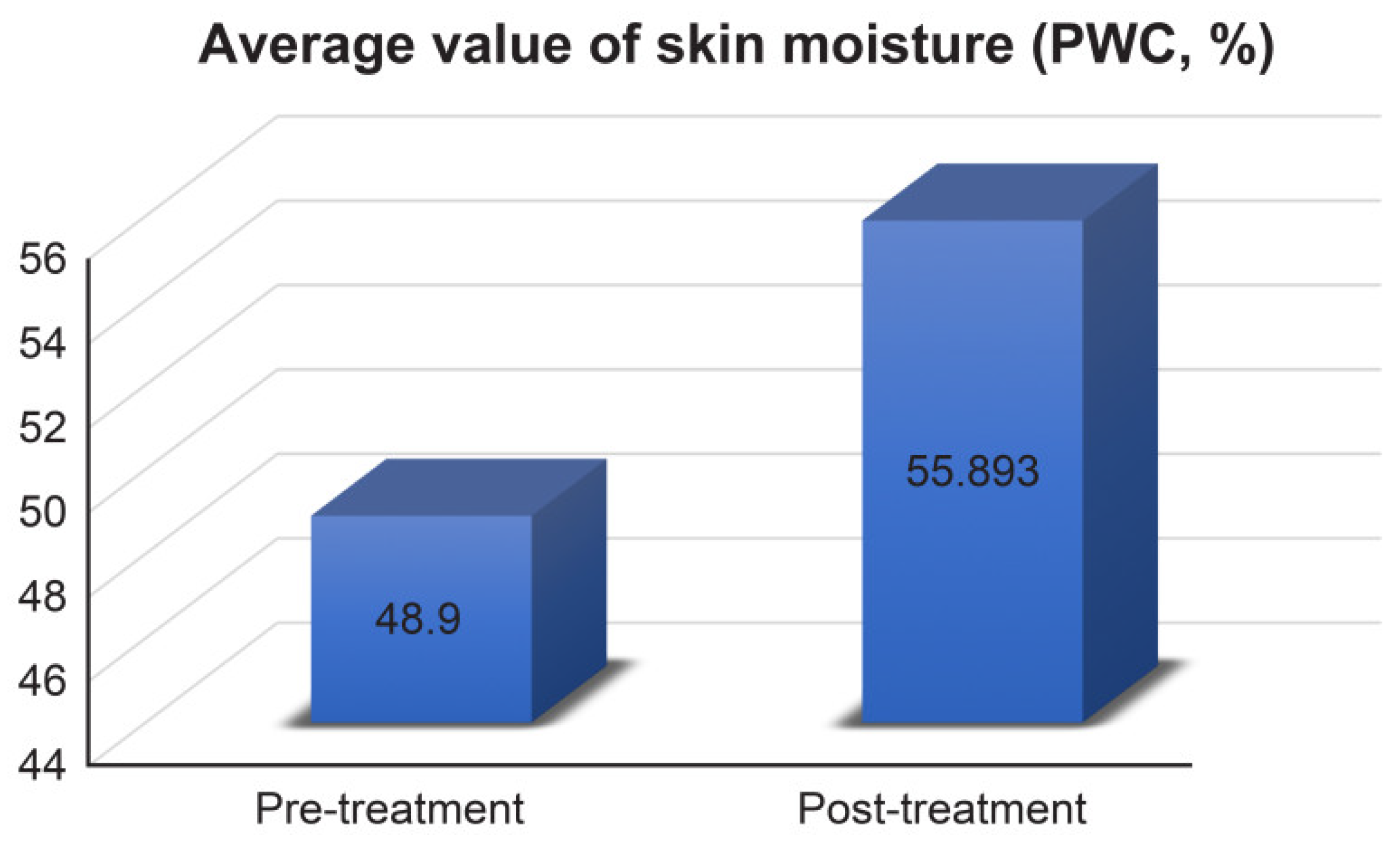
3.2. Skin Moisture
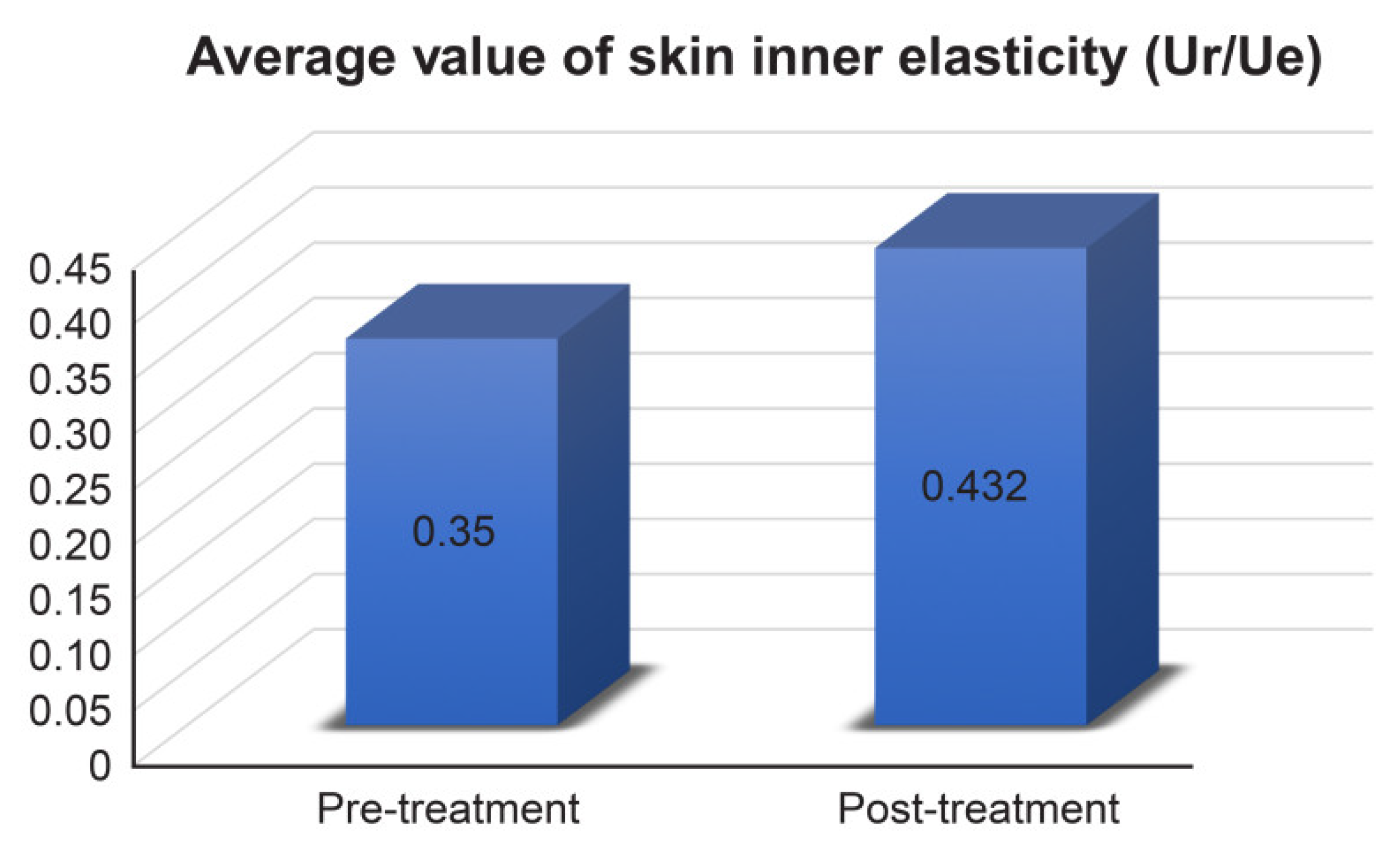
3.3. Skin Inner Elasticity
3.4. Skin Thickness
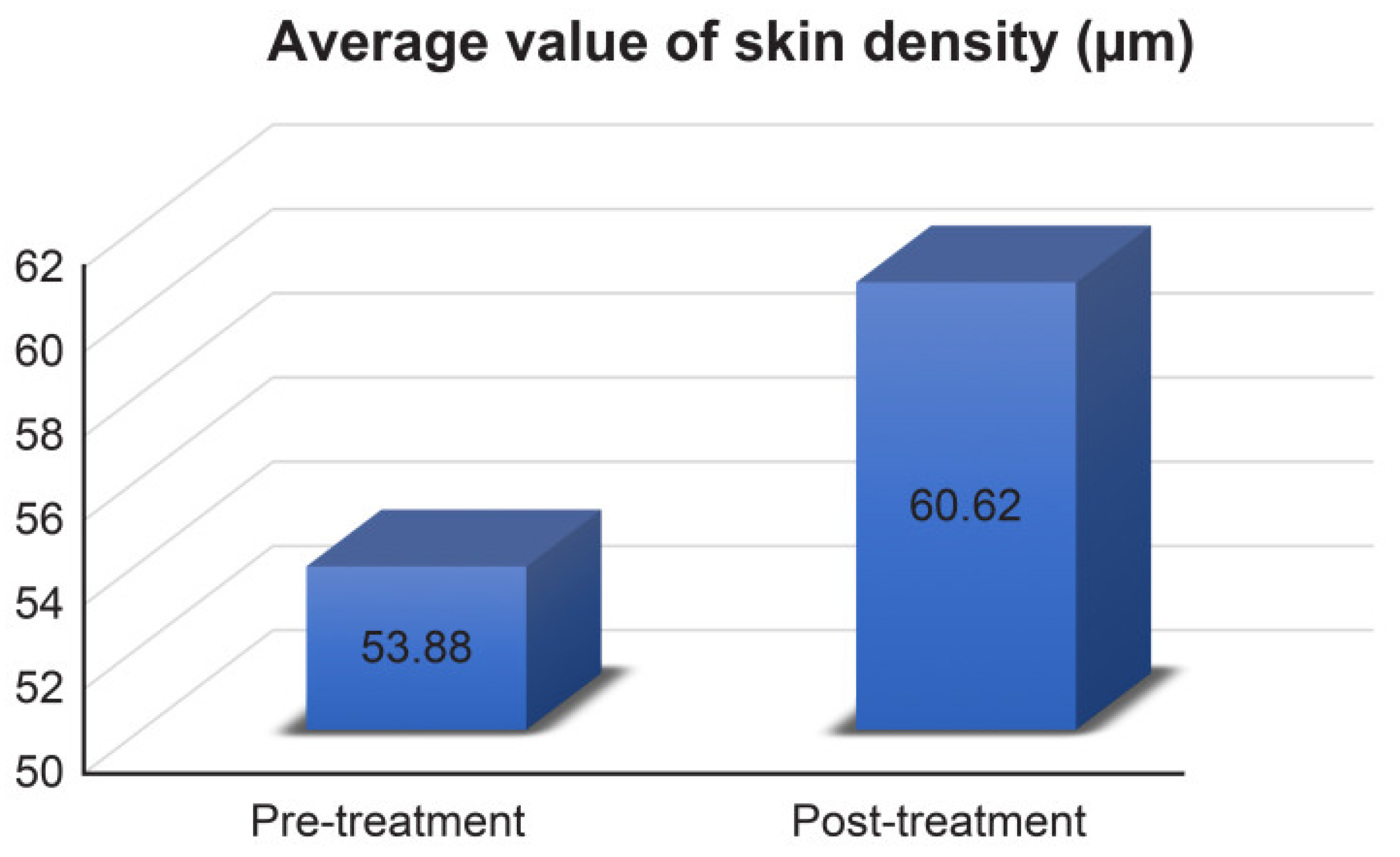
3.5. Skin Density
3.6. Safety Evaluation
3.7. Statistical Analysis
4. Discussion
5. Conclusions
Author Contributions
Funding
Institutional Review Board Statement
Informed Consent Statement
Data Availability Statement
Acknowledgments
Conflicts of Interest
References
- Zhang, S.; Duan, E. Fighting against skin aging: The way from bench to bedside. Cell Transplant. 2018, 27, 729–738. [Google Scholar] [CrossRef] [PubMed]
- Frantz, C.; Stewart, K.M.; Weaver, V.M. The extracellular matrix at a glance. J. Cell Sci. 2010, 123, 4195–4200. [Google Scholar] [CrossRef] [PubMed] [Green Version]
- Uitto, J.; Olsen, D.R.; Fazio, M.J. Extracellular matrix of the skin: 50 years of progress. J. Investig. Dermatol. 1989, 92, 61S–77S. [Google Scholar] [CrossRef] [PubMed] [Green Version]
- Kular, J.K.; Basu, S.; Sharma, R.I. The extracellular matrix: Structure, composition, age-related differences, tools for analysis and applications for tissue engineering. J. Tissue Eng. 2014, 5, 1–17. [Google Scholar] [CrossRef]
- Weihermann, A.C.; Lorencini, M.; Brohem, C.A.; de Carvalho, C.M. Elastin structure and its involvement in skin photoageing. Int. J. Cosmet. Sci. 2017, 39, 241–247. [Google Scholar] [CrossRef]
- Giro, M.G.; Oikarinen, A.I.; Oikarinen, H.; Sephel, G.; Uitto, J.; Davidson, J.M. Demonstration of elastin gene expression in human skin fibroblast cultures and reduced tropoelastin production by cells from a patient with atrophoderma. J. Clin. Investig. 1985, 75, 672–678. [Google Scholar] [CrossRef]
- Wen, Q.; Mithieux, S.M.; Weiss, A.S. Elastin biomaterials in dermal repair. Trends Biotechnol. 2020, 38, 280–291. [Google Scholar] [CrossRef]
- Baud, S.; Duca, L.; Bochicchio, B.; Brassart, B.; Belloy, N.; Pepe, A.; Dauchez, M.; Martiny, L.; Debelle, L. Elastin peptides in aging and pathological conditions. Biomol. Concepts 2013, 4, 65–76. [Google Scholar] [CrossRef]
- Truter, S.; Rosenbaum-Fiedler, J.; Sapadin, A.; Lebwohl, M. Calcification of elastic fibers in pseudoxanthoma elasticum. Mt. Sinai J. Med. 1996, 63, 210–215. [Google Scholar]
- Almine, J.F.; Wise, S.G.; Weiss, A.S. Elastin signaling in wound repair. Birth Defects Res. Part C 2012, 96, 248–257. [Google Scholar] [CrossRef]
- Rodríguez-Cabello, J.C.; de Torre, I.G.; Ibañez-Fonzeca, A.; Alonso, M. Bioactive scaffolds based on elastin-like materials for wound healing. Adv. Drug Deliv. Rev. 2018, 129, 118–133. [Google Scholar] [CrossRef] [PubMed] [Green Version]
- Daamen, W.F.; Veerkamp, J.H.; van Hest, J.C.; van Kuppevelt, T.H. Elastin as a biomaterial for tissue engineering. Biomaterials 2007, 28, 4378–4398. [Google Scholar] [CrossRef] [PubMed]
- Hashimoto, T.; Suzuki, Y.; Tanihara, M.; Kakimaru, Y.; Suzuki, K. Development of alginate wound dressings linked with hybrid peptides derived from laminin and elastin. Biomaterials 2004, 25, 1407–1414. [Google Scholar] [CrossRef] [PubMed]
- Hinek, A.; Rabinovitch, M.; Keeley, F.; Okamura-Oho, Y.; Callahan, J. The 67-kD elastin/laminin-binding protein is related to an enzymatically inactive, alternatively spliced form of beta-galactosidase. J. Clin. Investig. 1993, 91, 1198–1205. [Google Scholar] [CrossRef] [PubMed] [Green Version]
- Lodish, H.; Berk, A.; Zipursky, S.L. Collagen: The fibrous proteins of the matrix. In Molecular Cell Biology, 4th ed.; W. H. Freeman: New York, NY, USA, 2000; pp. 145–154. [Google Scholar]
- Shiratsuchi, E.; Nakaba, M.; Yamada, M. Elastin hydrolysate derived from fish enhances proliferation of human skin fibroblasts and elastin synthesis in human skin fibroblasts and improves the skin conditions. J. Sci. Food Agric. 2015, 96, 1672–1677. [Google Scholar] [CrossRef] [PubMed]
- Barati, M.; Jabbari, M.; Navekar, R.; Farahmand, F.; Zeinalian, R.; Salehi-Sahlabadi, A.; Abbaszadeh, N.; Mokari-Yamchi, A.; Davoodi, S.H. Collagen supplementation for skin health: A mechanistic systematic review. J. Cosmet. Dermatol. 2020, 19, 2820–2829. [Google Scholar] [CrossRef]
- Bianchi, F.M.; Angelinetta, C.; Rizzi, G.; Pratico, A.; Villa, R. Evaluation of the Efficacy of a Hydrolyzed Collagen Supplement for Improving Skin Moisturization, Smoothness, and Wrinkles. J. Clin. Aesthet. Dermatol. 2022, 15, 48–52. [Google Scholar]
- Chowdhury, A.; Nosoudi, N.; Karamched, S.; Parasaram, V.; Vyavahare, N. Polyphenol treatments increase elastin and collagen deposition by human dermal fibroblasts; Implications to improve skin health. J. Dermatol. Sci. 2021, 102, 94–100. [Google Scholar] [CrossRef]
- Bukhari, S.N.A.; Roswandi, N.L.; Waqas, M.; Habib, H.; Hussain, F.; Khan, S.; Sohail, M.; Ramli, N.A.; Thu, H.E.; Hussain, Z. Hyaluronic acid, a promising skin rejuvenating biomedicine: A review of recent updates and pre-clinical and clinical investigations on cosmetic and nutricosmetic effects. Int. J. Biol. Macromol. 2018, 120 Pt B, 1682–1695. [Google Scholar] [CrossRef]
- Rucker, R.B.; Kosonen, T.; Clegg, M.S.; Mitchell, A.E.; Rucker, B.R.; Uriu-Hare, J.Y.; Keen, C.L. Copper, Lysyl oxidase, and extracellular matrix protein cross-linking. Am. J. Clin. Nutr. 1998, 67 (Suppl. 5), 996S–1002S. [Google Scholar] [CrossRef] [Green Version]
- de Servi, B.; Orlandini, A.; Caviola, E.; Meloni, M. Amino acid and hyaluronic acid mixtures differentially regulate extracellular matrix genes in cultured human fibroblasts. J. Biol. Regul. Homeost. Agents 2018, 32, 517–527. [Google Scholar] [PubMed]
- Chung, K.W.; Song, S.H.; Kim, M.S. Synergistic effect of copper and amino acid mixtures on the production of extracellular matrix proteins in skin fibroblasts. Mol. Biol Rep. 2021, 48, 3277–3284. [Google Scholar] [CrossRef] [PubMed]
- Panwar, P.; Butler, G.S.; Jamroz, A.; Azizi, P.; Overall, C.M.; Brömme, D. Aging-associated modifications of collagen affect its degradation by matrix metalloproteinases. Matrix Biol. 2018, 65, 30–44. [Google Scholar] [CrossRef] [PubMed]
- Pitte, J.C.; Freis, O.; Vazqueq-Duchene, M.D.; Perie, G.; Pauly, G. Evaluation of elastin/collagen content in human dermis in-vivo by multiphoton tomography variation with depth and correlation with aging. Cosmetics 2014, 1, 11–221. [Google Scholar]
- Wang, H.; Shyr, T.; Fevola, M.J.; Cula, G.O.; Stamatas, G.N. Age-related morphological changes of the dermal matrix in human skin documented in vivo by multiphoton microscopy. J. Biomed. Opt. 2018, 23, 030501. [Google Scholar] [CrossRef] [Green Version]
- Shin, J.-W.; Kwon, S.-H.; Choi, J.-Y.; Na, J.-I.; Huh, C.-H.; Choi, H.-R.; Park, K.-C. Molecular Mechanisms of Dermal Aging and Antiaging Approaches. Int. J. Mol. Sci. 2019, 20, 2126. [Google Scholar] [CrossRef] [Green Version]
- Baumann, L.; Bernstein, E.F.; Weiss, A.S.; Bates, D.; Humphrey, S.; Silberberg, M.; Daniels, R. Clinical Relevance of Elastin in the Structure and Function of Skin. Aesthet. Surg. J. Open Forum. 2021, 14, 1–8. [Google Scholar] [CrossRef]
- McCabe, M.C.; Hill, R.C.; Calderone, K.; Cui, Y.; Yan, Y.; Quan, T.; Fisher, G.J.; Hansen, K.C. Alterations in extracellular matrix composition during aging and photoaging of the skin. Matrix Biol. Plus 2020, 8, 100041. [Google Scholar] [CrossRef]
- Haydonta, V.; Bernardb, B.A.; Fortunel, N.O. Age-related evolutions of the dermis: Clinical signs, fibroblast and extracellular matrix dynamics. Mech. Ageing Dev. 2019, 177, 150–156. [Google Scholar] [CrossRef]
- Cho, C.; Cho, E.; Kim, N.; Shin, J.; Woo, S.; Lee, E.; Hwang, J.; Ha, J. Age-related biophysical changes of the epidermal and dermal skin in Korean women. Skin Res. Technol. 2019, 25, 504–511. [Google Scholar] [CrossRef]
- Hill, K.E.; Davidson, J.M. Induction of increased collagen and elastin biosynthesis in copper-deficient pig aorta. Arteriosclerosis 1986, 6, 98–104. [Google Scholar] [CrossRef] [PubMed] [Green Version]
- Boyce, S.T.; Supp, A.P.; Wichkett, R.R.; Hoath, S.B.; Warden, G.D. Assessment with the Dermal Torque Meter of Skin Pliability After Treatment of Burns with Cultures Skin Substitutes. J. Bure Care Rehabil. 2000, 21, 55–63. [Google Scholar] [CrossRef] [PubMed]
- Murray, B.C.; Wickett, R.R. Correlations between Dermal Torque Meter®, Cutometer®, and Dermal Phase Meter® measurements of human skin. Skin Res. Technol. 1997, 3, 101–106. [Google Scholar] [CrossRef] [PubMed]


| Evaluation Items | Materials |
|---|---|
| Wrinkles Around the Eyes | Antera 3D CS |
| Skin Moisture | Moisture meter D Compact |
| Skin Inner Elasticity | Dermal Torque Meter |
| Skin Thickness and Density | Ultrascan UC22 |
| Inclusion Criteria | Exclusion Criteria |
|---|---|
|
|
| Evaluation Item | Mean | Rate of Change | p-Value | |
|---|---|---|---|---|
| Pre-Treatment | Post-Treatment | |||
| Wrinkles Around the Eyes | 0.067 ± 0.038 mm | 0.061 ± 0.033 mm | −8.955% | 0.042 |
| Skin Moisture | 48.9 ± 4.126% | 55.893 ± 3.371% | 14.301% | 0.043 |
| Skin Inner Elasticity | 0.35 ± 0.029 Ur/Ue | 0.432 ± 0.06 Ur/Ue | 23.429% | 0.042 |
| Skin Thickness | 1652.8 ± 302.06 μm | 1773.8 ± 293.58 μm | 7.321% | 0.043 |
| Skin Density | 53.88 ± 2.72 μm | 60.62 ± 4.201 μm | 12.509% | 0.042 |
Publisher’s Note: MDPI stays neutral with regard to jurisdictional claims in published maps and institutional affiliations. |
© 2022 by the authors. Licensee MDPI, Basel, Switzerland. This article is an open access article distributed under the terms and conditions of the Creative Commons Attribution (CC BY) license (https://creativecommons.org/licenses/by/4.0/).
Share and Cite
Kim, M.-S.; Chun, K.-E.; Lee, D.-K.; Song, S.-H. Evaluation of the Efficacy of an Elastin-Inducing Composition Containing Amino Acids, Copper, and Hyaluronic Acid: Results of an Open Single-Center Clinical Trial Study. Cosmetics 2022, 9, 51. https://doi.org/10.3390/cosmetics9030051
Kim M-S, Chun K-E, Lee D-K, Song S-H. Evaluation of the Efficacy of an Elastin-Inducing Composition Containing Amino Acids, Copper, and Hyaluronic Acid: Results of an Open Single-Center Clinical Trial Study. Cosmetics. 2022; 9(3):51. https://doi.org/10.3390/cosmetics9030051
Chicago/Turabian StyleKim, Man-Seok, Ko-Eun Chun, Dong-Keun Lee, and Seh-Hyon Song. 2022. "Evaluation of the Efficacy of an Elastin-Inducing Composition Containing Amino Acids, Copper, and Hyaluronic Acid: Results of an Open Single-Center Clinical Trial Study" Cosmetics 9, no. 3: 51. https://doi.org/10.3390/cosmetics9030051
APA StyleKim, M.-S., Chun, K.-E., Lee, D.-K., & Song, S.-H. (2022). Evaluation of the Efficacy of an Elastin-Inducing Composition Containing Amino Acids, Copper, and Hyaluronic Acid: Results of an Open Single-Center Clinical Trial Study. Cosmetics, 9(3), 51. https://doi.org/10.3390/cosmetics9030051