Article

Ecotoxicological Evaluation of Sunscreens on Marine Plankton

by
María Pilar González
1,
Alejandro Vilas
2 and
Ricardo Beiras
1,2,*
1
Departamento de Ecoloxía e Bioloxía, Universidade de Vigo, E-36310 Galicia, Spain
2
Grupo Ecotox (EcoCost) ECIMAT, Centro de investigación mariña, Universidade de Vigo, 36331 Galicia, Spain
*
Author to whom correspondence should be addressed.
Cosmetics 2022, 9(1), 20; https://doi.org/10.3390/cosmetics9010020
Submission received: 19 December 2021 / Revised: 17 January 2022 / Accepted: 28 January 2022 / Published: 1 February 2022
(This article belongs to the Special Issue Feature Papers in Cosmetics in 2022)

Abstract

:
In recent years, a large number of sunscreens have emerged to protect our skin. Most of them are made up of simple or compound aromatic structures, which can pose a threat to marine ecosystems. In order to understand their effects on the marine environment, different ecotoxicological bioassays were carried out using planktonic organisms from three phyla and two different trophic levels: larvae of the sea urchin Paracentrotus lividus, the copepod Acartia tonsa, and the microalga Tisochrysis lutea. The aim of these tests was to expose these organisms to leachates from eight sunscreen formulations. All of them showed a great variability in toxicity on the different plankton organisms. The highest toxicity level was found for cream number 4 when tested on sea urchin, exhibiting an EC50 = 122.4 mg/L. The toxicity of the UV filter 2-phenyl-5-benzimidazolesulfonic acid, exclusively present in that cream, was evaluated in sea urchin, where an EC10 = 699.6 mg/L was obtained under light exposure. According to our results, all tested creams become nontoxic to plankton upon 30,000-fold dilution in seawater; thus, only local effects are expected. This study highlights the need to understand the toxic effects generated by solar protection products, as well as their ingredients, on marine organisms.

1. Introduction

Sunscreens are defined as substances designed to protect our skin from the sun’s harmful rays. They reflect, absorb, and scatter ultraviolet A and B radiation [1]. Sunscreen formulations are composed of a wide variety of ingredients [2]. Their active components are organic and inorganic ultraviolet (UV) filters [3,4], each with different structures and solubilities. Most used UV filters such as octocrylene (OC), avobenzone, homosalate, and oxybenzone are organic molecules with conjugated aromatic structures whose function is mainly to absorb UV rays [3,5]. Organic UV filters are also added to plastics and other materials to provide them with resistance to light exposure [6]. In addition, sunscreens contain other components such as chemical preservatives, fragrances, and antioxidants substances [7,8]. These compounds have been detected in different seas at levels of ng/L [5,9,10,11]. UV filters reach the marine environment directly from the skin of bathers, which is the main source. In addition, there is an indirect route involving the effluents from wastewater treatment plants and atmospheric deposition [5,8].
The toxicity of sunscreens depends on their composition. Numerous effects on marine organisms have been observed; in fact, some UV filters pose a significant risk to marine invertebrates [6,12,13]. UV filters such as OC, titanium dioxide (TiO2), and ethylhexyl methoxycinnamate (EHMC) cause bleaching of some coral species by inducing the lytic cycle of viruses present in symbiont zooxanthellae [14,15,16], thus reducing their photosynthetic efficiency [17]. In addition, other effects have been seen in corals such as the induction of acute stress after exposure or abnormal fatty-acid metabolism [15,18]. Moreover, they can generate reactive oxygen species (ROS) and DNA damage in phytoplankton [19], mussels [17,20,21], copepods [22], and some fish [23]. In addition, they are known to cause endocrine disruption in thyroid regulation and neurotoxicity [7,24,25,26,27]. Furthermore, some UV filters and some chemical preservatives present in creams have estrogenic activity with positive regulation of the vitellogenin gene [27,28,29]. Other effects that have been studied are alterations in Paracentrotus lividus larval development [7,9,30,31], chromosomal aberrations [31], alterations in phytoplankton growth rate [9,20,30,32], and changes in communities favoring more resistant species [33].
In addition to these effects, some UV filters such as EHMC, OC, and TiO2 nanoparticles have been found to bioaccumulate after days of exposure in corals [15,18], in mussels [34,35,36], in phanerogams, and in fish [23,37].
It is important to highlight that all the components that make up sunscreens are present in the aquatic environment together; hence, they can interact with each other, which can modify their overall toxicity on biota [12,26]. In addition, other pollutants such as hydrocarbons or pesticides are present in the water and may be acting synergistically with these products [28]. Environmental factors may also influence the toxicity of these substances. The authors of [38] found that increased salinity, even with little variation, promoted a greater effect on marine organisms, as did temperature [39]. In addition, radiation in the water column generates new toxic byproducts and ROS via photooxidation of UV filters [5,8].
One of the organic UV filters used in sunscreens and cosmetics is 2-phenylbenzimidazole-5-sulfonic acid (PBSA). Although its fate in the environment is not well studied, it has been found to be present in wastewater at ng/L [40,41,42]. One of the effects on organisms is DNA damage due to the formation of ROS [43]. In addition, it has been found to change some biochemical parameters and enzymatic activities in the plasma of Oncorhynchus mykiss, such as increased cytochrome P450 activity [44] and increased lipid peroxidation in Danio rerio [45]. Its toxicity is thought to be due to compounds generated during its degradation by UV radiation [46].
The present study focused on evaluating and quantifying the toxicity on marine plankton of eight commercial sunscreens. To this end, we studied the effect on three marine plankton organisms: the microalga Tisochrysis lutea, larvae of the sea urchin Paracentrotus lividus, and the copepod Acartia tonsa. In addition, we tested PBSA individually because it was the only substance exclusively present in the most toxic sunscreen tested (cream 4). In this case, we also explored the effect that light exposure may have on the toxicity of this compound.

2. Materials and Methods

2.1. Samples

The eight sunscreens and their formulations (Table A1) were provided by the manufacturer, and PBSA (CAS number 27503-81-7) was obtained with 96% purity from Sigma-Aldrich (Darmstadt, Germany).

2.2. Exposure Media

All sunscreens and PBSA were tested for solubility in ultrapure water and in the nontoxic organic solvent DMSO (dimethyl sulfoxide). None of them were soluble; hence, creams were dosed according to standard methods for insoluble substances [47]. Briefly, a 10 g/L mixture was incubated with artificial seawater (ASW) of defined composition according to [48], in darkness, at 20 °C for 24 h on a rotary shaker at 1 rpm. After 24 h, the leachates were filtered through a glass fiber filter (GF/F Whatman™) and diluted in ASW. Dilutions tested were ×1/2, ×1/3, ×1/10, ×1/30, and ×1/100 for microalgae and ×1 (undiluted), ×1/3, ×1/10, and ×1/30 for zooplankton. In sea urchin, when toxicity was detected at ×1/30, higher dilutions were carried out (×1/100, ×1/300, and ×1/1000).

2.3. Test Organisms and Bioassays

The algal growth inhibition test was carried out according to the standard method from [49] adapted to T. lutea by [50]. The initial strain of T. lutea was obtained from the ECIMAT-Universidade de Vigo collection. Tests started with a 10,000 cells/mL inoculum in exponential growth phase. Cell numbers were recorded by using a Multisizer™ 3 Coulter Counter®, Beckman-Coulter. Eight replicates for the control and four for the test solutions were prepared. Test tubes were incubated in an isothermal room at 20 °C and light conditions for 72 h. After 3 days, growth was measured as
G R   d 1 = [ ln f i n a l   c e l l   n u m b e r ln i n i t i a l   c e l l   n u m b e r ] d .
Growth inhibition (I) was calculated as
I = G R c G R i G R c ,
where GRc and GRi are the growth rate in the control and growth rate in each test tube, respectively.
Responses were corrected by control and expressed as R = GRi/GRc.
Adult sea urchins (P. lividus) in ripe conditions were supplied by ECIMAT, and the sea urchin embryo test (SET) was carried out following standard methods [51]. Briefly, mature oocytes were fertilized in a 50 mL graduated cylinder with ASW, and fertilized eggs were transferred into glass vials (four replicates per treatment and eight controls) with airtight Teflon including 4 mL of exposure medium (final density of 40 per mL). Then, fertilized eggs were incubated at 20 °C in dark conditions for 2 days. After 48 h, the vials were fixed with three drops of 36% formaldehyde, observed in a Leica DMI 4000B inverted microscope, and the length (maximum linear dimension) of 35 individuals per vial was recorded using Leica LAS image analysis software (Leica microsystems, Wetzlar, Germany). The mean length increase for each treatment was expressed as the net response (R) in relation to the control response according to the following expression:
R = L t L e L c L e ,
where Lt is the mean length of a treatment, Le is the mean length of eggs, and Lc is the mean size of control.
In the case of the toxicity test with the PBSA filter, four additional replicates of each dilution and eight control vials were incubated in a room with a 16:8 h light/darkness photoperiod at 20 °C. According to [51], the pH of the PBSA leachate was adjusted with a drop of NaOH to meet the requirement of pH >7.
The acute lethal toxicity test with copepods followed standard methods [52] adapted by [53] to use nauplius larvae of Acartia tonsa. Mature copepods were obtained from a laboratory stock maintained by ECIMAT from 48 to 72 h before the start of the test. From the initial stock, adults were collected by a 300 µm mesh and incubated in laboratory conditions to produce nauplius <24 h following [53]. A total of 10 nauplii were transferred to 20 mL glass vials using a binocular stereoscope. Four vials for each dilution and eight vials for control were used. Copepod nauplius survival was recorded after 48 h and expressed as the percentage net response according to the expression R = (St · 100)/Sc, where St is the mean survival of a treatment, and Sc is the mean survival of the control.

2.4. Statistical Analysis

Statistical analyses were performed using IBM SPSS statistics (v.24) (provided from Universidade de Vigo, Vigo, Spain). Normal distribution of data and homoscedasticity was checked using the Shapiro–Wilk and Levene’s tests, respectively. Dilutions significantly different from the control (p < 0.05) were identified using Dunnett’s post hoc test or Dunnett’s T3, when the variances were not homogeneous, in order to find the lowest observed adverse effect concentration (LOEC) and the highest concentration with no observed adverse effects (NOEC). The dilutions that produced a 50% and 10% decrease in the endpoint (EC50 and EC10) and their 95% confidence intervals (CIs) were also calculated by fitting the data to a Probit dose–response model. Toxic units (TU) were calculated as TU = 1/EC50 [54].

3. Results

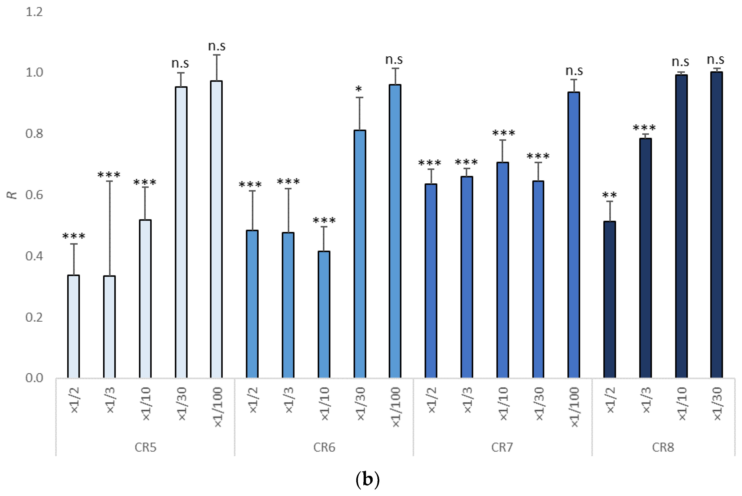
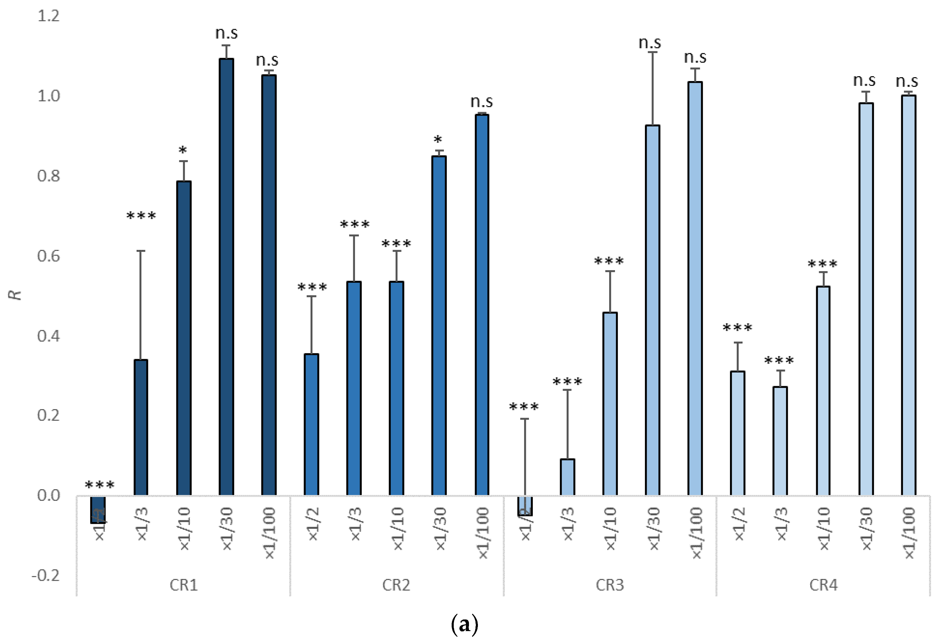
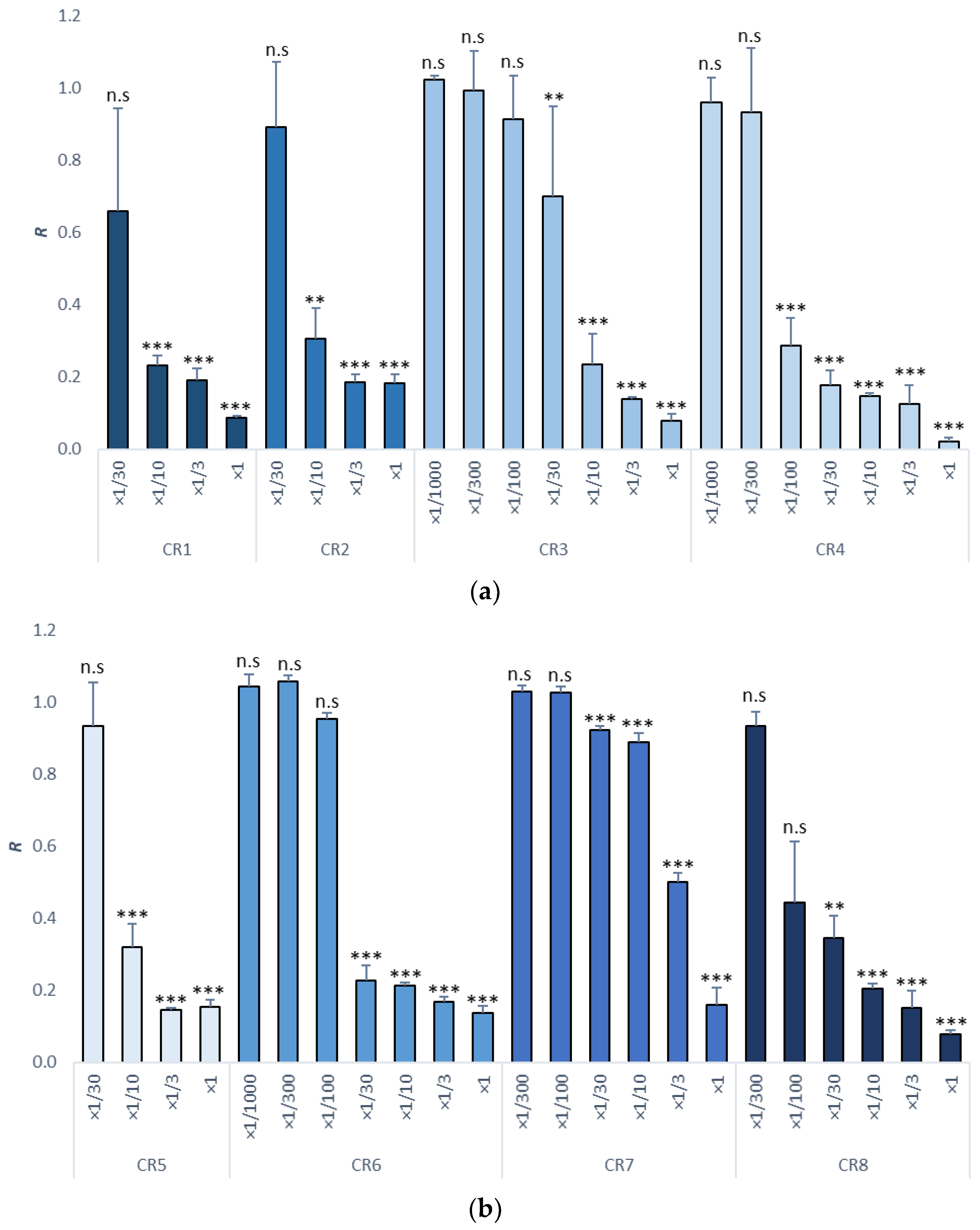
3.1. Growth Inhibition in Tisochrysis lutea

The toxicity of all sunscreens tested on T. lutea is shown in Table 1. As we can see, cream 3 was the most toxic for this species, (9.60 TU), whereas creams CR7 and CR8 were the least toxic to this microalga. Looking at the EC50 values, toxicity was ranked as CR3 > CR4 > CR5 > CR1 > CR6 > CR2 > CR8 > CR7.
The growth rate per day compared with control (R) is shown in Figure 1. For CR1 and CR3, the growth rate was negative in the undiluted leachate.

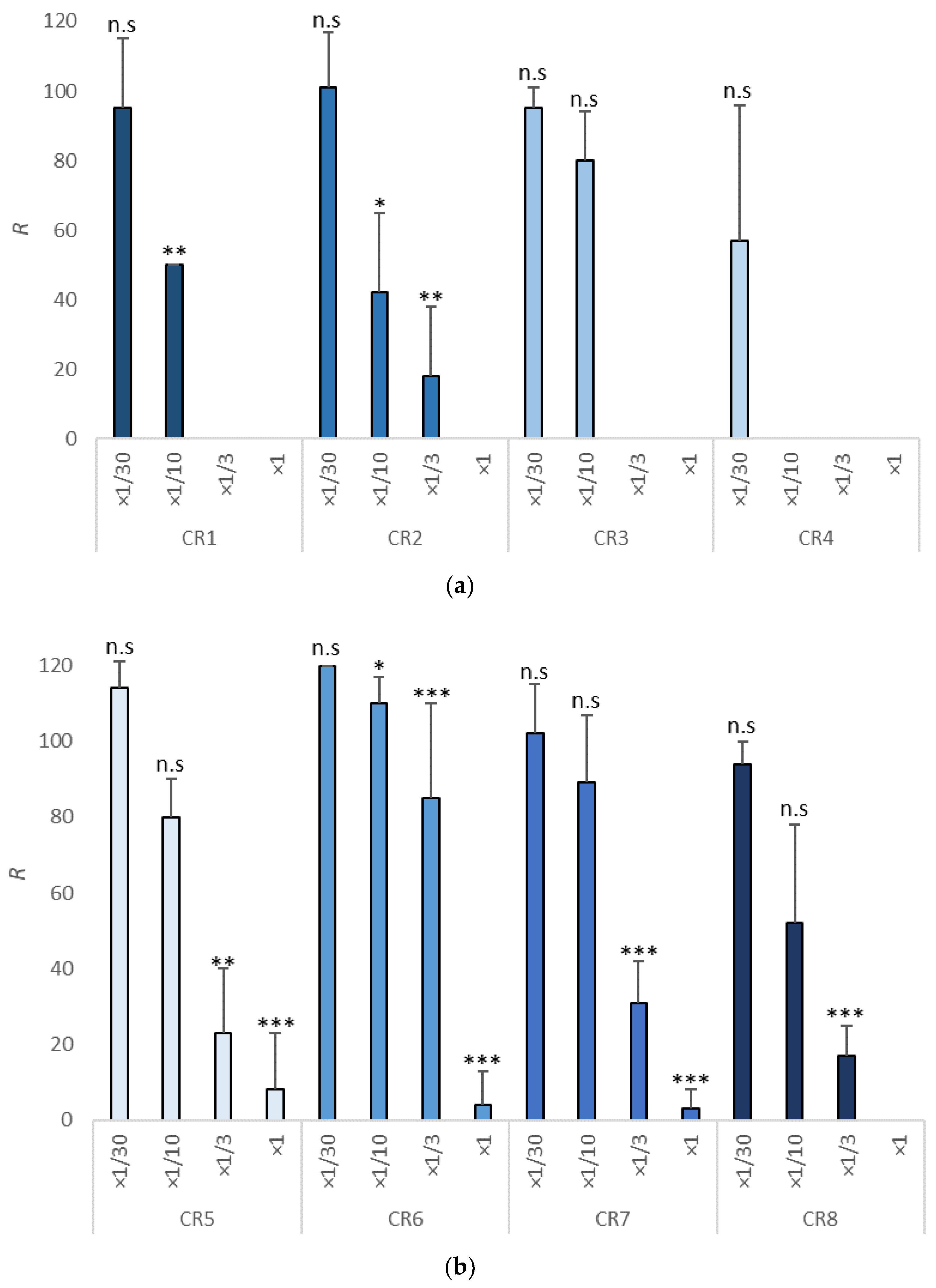
3.2. Sea Urchin Embryo Test (SET)

Results for the different dilutions of the sunscreen creams are shown in Table 2. According to the toxicity units and the EC50, cream CR4 stood out as the most toxic; its 10 g/L leachate needed to be diluted 300 times to lose toxicity. It was followed by creams CR8, CR6, and CR1, whereas creams CR7, CR5, CR3, and CR2 were classified as the least toxic.
The larval size increase compared with control data (R) is shown in Figure 2.

3.3. Survival Test in Copepods

The effects of all sunscreens on copepods are reflected in Table 3. As we saw previously in sea urchin, cream CR4 was once again the most toxic with an EC50 of 354.4 mg/L. Creams CR5, CR6, and CR7 were the least toxic to copepods.
The survival percentage compared to control data (R) is shown in Figure 3. Cream CR4 caused 100% mortality even after 10-fold dilution of the leachate. Cream CR6 was the least toxic, showing survival rates close to 100% after just threefold dilution of the leachate.

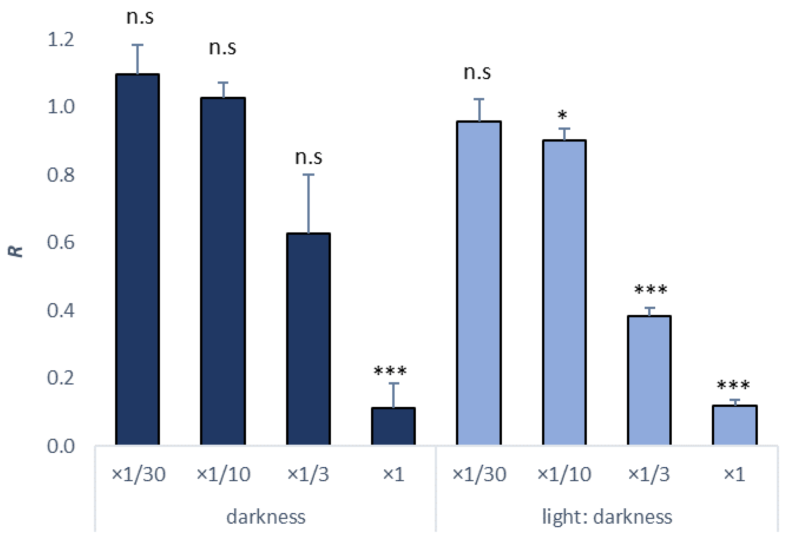
3.4. PBSA Sea Urchin Embryo Test (SET)

The SET was conducted with PBSA, the specific UV filter present only in the most toxic sunscreen for zooplankton, and the results are shown in Table 4. As we can see, light conditions affected the results, since the PBSA leachate had to be diluted just threefold to lose toxicity when tested in darkness, whereas it was necessary to dilute it 30-fold when incubations were conducted under light. However, in both cases, toxicity was moderate and could not explain the toxicity of this cream to the early life stages of sea urchins and copepods.
The larval size increase compared with control (R) is shown in Figure 4.

4. Discussion

According to our results, all creams were within the nontoxic category in all species tested according to the classification of chemical substances and mixtures for their labeling [55], since all EC50 values were above 100 mg/L. Creams CR4 and CR8 for Paracentrotus lividus were closest to the limit with EC50 = 122.4 mg/L and 191.6 mg/L, respectively. In the case of copepods, cream CR4 was again the closest to this limit with EC50 = 354.4 mg/L.
Some of the UV filters present in creams such as EHMC and OC have been previously studied in marine planktonic species [9,20,30]. For all sunscreens, higher amounts are needed to produce a 50% growth inhibition when they are part of a cream than independently [32]. The same has been observed in sea urchin [9,30] and in other marine organisms [12]. This may be due to the presence of humectants, hydrophobic excipients, and emulsifiers that have a high affinity for these filters and, therefore, can reduce their bioavailability. Thus, it is important to know the composition because the concentration of one single UV filter can differ between creams, as can be seen in Table A6 for undiluted leaching (10 g/L). In fact, from these values, it is possible to estimate at what concentration these filters present toxicity or not with the NOEC or LOEC values.
The sensitivity of plankton organisms to UV filters largely varies among different taxa [6,30]. In the case of TiO2, a relevant toxicity has been detected at a concentration of 1 mg/L in Tisochrysis lutea and in other species of microalgae [20]. However, for T. lutea, no toxicity was noted for this filter in creams CR1 and CR5 at 16 and 18 mg/L, respectively, although toxicity was noted in cream CR6 over 0.72 mg/L. For OC, toxicity has been detected at concentrations lower than 1 mg/L even at concentrations of 0.1 mg/L in marine plankton organisms [30]. Nonetheless, no toxicity was observed in creams CR2, CR4, and CR5 at 10, 33, and 27 mg/L, respectively. More toxicity of the EHMC filter has been detected in invertebrates than in microalgae [9].
The differences in the ranking of toxicity of the creams tested on the three plankton species point to the differential toxicity of some of the sunscreens’ components. In general, the toxicity on microalgae was lower with respect to the two species of zooplankton. This test is usually less sensitive, as has been seen with some UV filters [9,30]. The authors of [38] found that organic filters were less toxic than inorganic ones for Corophium orientale (crustacean), whereas the opposite was observed in Phaeodactylum tricornutum (phytoplankton). According to our results, this trend was not seen, since creams CR5 and CR6, which contain TiO2, were the least toxic toward the crustacean used. In addition, in Tisochrysis lutea, there were similar effects of creams with and without this type of filter.
Cream CR4 showed a remarkable toxicity on the two species of zooplankton tested, and this was the only formulation that included PBSA. Therefore, this UV filter was individually tested using the SET. Results (see Table 4) did not support the hypothesis that PBSA was the main cause of the toxicity of this formulation. However, the chemical speciation of this substance is highly influenced by pH. At high pH values, the nonionized form of the compound predominates, which is more toxic because it can readily pass through the lipid bilayer of the cell membrane [56]. For this reason, a bioassay in which the toxicity of PBSA is tested at different pH values is recommended before being able to rule out the contribution of this substance to the toxicity of cream CR4.
Lastly, in this UV filter studied separately, a significantly higher toxicity was observed when sea urchin larvae were incubated with light. This may be due to the generation of ROS [43] and the formation of transformation products resulting from the degradation of PBSA in the presence of UV radiation. The main degradation pathway has been found to generate four stable phototransformation products, which are indicated to be more dangerous to organisms than the parental compound [46]. Regarding the effects on biota, this UV filter is not well studied; the most severe concentration at which toxicity has been found is at 5 mg/L on zebrafish [45]. According to our results, a concentration of 1 g/L is necessary to cause observable effects on sea urchin larvae.

5. Conclusions

None of the tested sunscreens can be considered totally innocuous for the tested plankton species, since deleterious effects of undiluted 10 g/L leachates were observed in all cases on both phytoplankton and zooplankton. However, taking into account the high dilution factors of coastal environments, these effects are expected to be very limited on a geographical scale since a 300-fold dilution of the leachate produced at a 1:100 cream–seawater ratio (i.e., a 30,000-fold overall dilution) resulted in the absence of toxicity, even for the most toxic cream on the most sensitive test species (cream CR4 on sea urchin).
The toxicity of sunscreens on microalgae and copepods was lower than on sea urchin larvae, since a 2–28-fold higher concentration of sunscreen was needed to cause a level of effect of 50%. This highlights the need to use a battery of test species and not a single model when assessing the ecological risk of chemicals on the marine environment.
The toxicity of the UV filter PBSA on P. lividus larvae increased in the presence of light, likely due to formation of photo-oxidized metabolites upon exposure to UV radiation.
The results of this study suggest the importance of a better understanding of the toxicity of the components of sunscreens both alone and as part of mixtures, which is how they are most often found in the environment.

Author Contributions

Conceptualization, M.P.G., A.V. and R.B.; methodology, M.P.G., A.V. and R.B.; formal analysis, M.P.G. and A.V.; data curation, M.P.G. and A.V.; writing—original draft preparation, M.P.G.; writing—review and editing, M.P.G., A.V. and R.B.; supervision, R.B.; funding acquisition, R.B. All authors have read and agreed to the published version of the manuscript.

Funding

This research was supported by the RisBioPlas Project, financed by the Ministry of Science and Innovation—Spanish Agency of Research (AEI: 10.13039/501100011033), project reference PCI2020-112110), by the LABPLAS Project (Land-Based Solutions for Plastics in the Sea, Grant Agreement Nr.: 101003954, financed by the EU H2020 program), and by the Competitive Reference Group for Coastal Ecology (EZ1), funded by the call for grants for the consolidation and structuring of competitive research units of the Galician University System ED431C 2021/42.

Institutional Review Board Statement

Not applicable.

Informed Consent Statement

Not applicable.

Data Availability Statement

The data presented in this study are available on request from the corresponding author.

Acknowledgments

The authors thank Clara Mendoza Segura for her technical support during the tests and the ECIMAT staff for supplying the organisms.

Conflicts of Interest

The authors declare no conflict of interest.

Appendix A

Table A1. Complementary information for the eight sunscreens and their abbreviations.
ItemCharacteristics
CR1Colored fluid solution
CR2Body oil
CR3Fluid solution without color
CR4Gel cream
CR5High-protection fluid solution
CR6High-protection fluid solution
CR7Body spray
CR8Water fluid
Table A2. All toxicity parameters (NOEC, LOEC, EC10, EC50, and TU) for Tisochrysis lutea. The 95% confidence intervals are given in brackets. n.c., not calculable.
ItemDilutionsNOECLOECEC10EC50EC10 (mg/L)EC50 (mg/L)TU
CR1×1/100, ×1/30, ×1/10, ×1/3, ×1/2×1/30×1/101/12.311/4.96812.352016.134.96
(3.92–6.44)
CR2×1/100, ×1/30, ×1/10, ×1/3, ×1/2×1/100×1/301/62.711/4.09159.4624454.09
(2.66–5.74)
CR3×1/100, ×1/30, ×1/10, ×1/3, ×1/2×1/30×1/101/31.361/9.59318.881042.759.60
(7.25–13)
CR4×1/100, ×1/30, ×1/10, ×1/3, ×1/2×1/30×1/101/27.711/5.52360.881811.605.52
(4.34–6.98)
CR5×1/100, ×1/30, ×1/10, ×1/3, ×1/2×1/30×1/101/40.531/5.10246.731960.785.10
(3.16–7.65)
CR6×1/100, ×1/30, ×1/10, ×1/3, ×1/2×1/100×1/301/103.111/4.1696.982403.854.16
(1.84–7.13)
CR7×1/100, ×1/30, ×1/10, ×1/3, ×1/2×1/100×1/301/415.331/0.4924.0820,430.04<2
(0.01–1.67)
CR8×1/100, ×1/30, ×1/10, ×1/3, ×1/2×1/10×1/31/4.641/1.85215.335411.26<2
(1.10–2.29)
Table A3. All toxicity parameters (NOEC, LOEC, EC10, EC50, and TU) for Paracentrotus lividus. The 95% confidence intervals are given in brackets.
ItemDilutionsNOECLOECEC10EC50EC10 (mg/L)EC50 (mg/L)TU
CR1×1/30, ×1/10, ×1/3, ×1×1/30×1/101/258.921/20.1438.62496.5220.14
(11.87–51.23)
CR2×1/30, ×1/10, ×1/3, ×1×1/30×1/101/1001/10.14100986.1910.14
(6.20–20)
CR3×1/1000, ×1/300, ×1/100,
×1/30, ×1/10, ×1/3, ×1
×1/100×1/301/120.651/16.2382.90616.1416.23
(11.46–23)
CR4×1/1000, ×1/300, ×1/100,
×1/30, ×1/10, ×1/3, ×1
×1/300×1/1001/977.201/81.6710.23122.4481.67
(52.21–134.56)
CR5×1/30, ×1/10, ×1/3, ×1×1/30×1/101/67.171/9.7148.901030.939.70
(6.3–16.80)
CR6×1/1000, ×1/300, ×1/100,
×1/30, ×1/10, ×1/3, ×1
×1/100×1/301/203.861/21.8649.10457.4621.86
(13.85–35.10)
CR7×1/1000, ×1/300, ×1/100,
×1/30, ×1/10, ×1/3, ×1
×1/100×1/301/15.531/3.10643.923225.813.10
(2.67–3.56)
CR8×1/1000, ×1/300, ×1/100,
×1/30, ×1/10, ×1/3, ×1
×1/100×1/301/1097.461/52.209.11191.5752.20
(34.20–86.70)
Table A4. All toxicity parameters (NOEC, LOEC, EC10, EC50, and TU) for Acartia tonsa. The 95% confidence intervals are given in brackets. n.c., not calculable.
ItemDilutionsNOECLOECEC10EC50EC10 (mg/L)EC50 (mg/L)TU
CR1×1/30, ×1/10, ×1/3, ×1×1/30×1/101/25.71/11.17389.11895.2611.17
(1.5–42.78)
CR2×1/30, ×1/10, ×1/3, ×1×1/30×1/101/301/9.43333.331060.459.43
(2.11–48.02)
CR3×1/30, ×1/10, ×1/3, ×1×1/10×1/31/16.401/7.85609.761273.887.85
(1.10–21.23)
CR4×1/30, ×1/10, ×1/3, ×1n.cn.c1/52.101/28.22191.94354.3628.22
(n.c)
CR5×1/30, ×1/10, ×1/3, ×1×1/10×1/31/14.651/4.65682.602150.574.65
(1.21–16.42)
CR6×1/30, ×1/10, ×1/3, ×1×1/3×11/11.371/4.41879.512267.574.41
(1.31–13.14)
CR7×1/30, ×1/10, ×1/3, ×1×1/10×1/31/14.271/4.60700.792181.114.60
(1.36–13. 85)
CR8×1/30, ×1/10, ×1/3, ×1×1/10×1/31/27.311/8.47366.171180.648.46
(2.57–34.56)
Table A5. All toxicity parameters (NOEC, LOEC, EC10, EC50, and TU) for PBSA filter in Paracentrotus lividus. The 95% confidence intervals are given in brackets.
Incubation ConditionsDilutionsNOECLOECEC10EC50EC10 (mg/L)EC50 (mg/L)TU
Darkness×1/30, ×1/10, ×1/3, ×1×1/3×1/11/5.371/2.321861.504312.22.32
(1.99–2.70)
Light/darkness (16:8)×1/30, ×1/10, ×1/3, ×1×1/30×1/101/14.291/3.63699.592751.793.63
(2.91–4.50)

Appendix B

Table A6. Presence of UV filter in undiluted leachates according to their nominal composition (mg/L).
UV FilterCR1CR2CR3CR4CR5CR6CR7CR8
EHMC 499 999
OC 1000 1000800
TiO2479 53372
PBSA 200

References

  1. NCI Dictionary of Cancer Terms—National Cancer Institute. Available online: https://www.cancer.gov/publications/dictionaries/cancer-terms/def/sunscreen (accessed on 1 November 2020).
  2. Mildau, G. General Review of Official Methods of Analysis of Cosmetics. In Analysis of Cosmetic Products, 2nd ed.; Salvador, A., Chisvert, A., Eds.; Elsevier: Amsterdam, The Netherlands, 2018; pp. 67–83. [Google Scholar]
  3. Daly, S.; Ouyang, H.; Maitra, P. Chemistry of Sunscreens. In Principles and Practice of Photoprotection; Springer: Berlin/Heidelberg, Germany, 2016; pp. 159–178. [Google Scholar] [CrossRef]
  4. Bowman, D.M.; May, N.D.; Maynard, A.D. Nanomaterials in Cosmetics: Regulatory Aspects. In Analysis of Cosmetic Products, 2nd ed.; Salvador, A., Chisvert, A., Eds.; Elsevier: Amsterdam, The Netherlands, 2018; pp. 289–302. [Google Scholar]
  5. Giokas, D.L.; Salvador, A.; Chisvert, A. UV filters: From sunscreens to human body and the environment. TrAC Trends Anal. Chem. 2007, 26, 360–374. [Google Scholar] [CrossRef]
  6. Beiras, R. Environmental Risk Assessment of Pharmaceutical and Personal Care Products in Estuarine and Coastal Waters. In Pharmaceuticals in Marine and Coastal Environments; Elsevier: Amsterdam, The Netherlands, 2021; pp. 195–252. [Google Scholar]
  7. Corinaldesi, C.; Damiani, E.; Marcellini, F.; Falugi, C.; Tiano, L.; Brugè, F.; Danovaro, R. Sunscreen products impair the early developmental stages of the sea urchin Paracentrotus lividus. Sci. Rep. 2017, 7, 7815. [Google Scholar] [CrossRef] [PubMed] [Green Version]
  8. Sánchez-Quiles, D.; Blasco, J.; Tovar-Sánchez, A. Sunscreen Components Are a New Environmental Concern in Coastal Waters: An Overview. In Handbook of Environmental Chemistry; Springer: Cham, Switzerland, 2020; Volume 94, pp. 1–14. [Google Scholar]
  9. Paredes, E.; Perez, S.; Rodil, R.; Quintana, J.B.; Beiras, R. Ecotoxicological evaluation of four UV filters using marine organisms from different trophic levels Isochrysis galbana, Mytilus galloprovincialis, Paracentrotus lividus, and Siriella armata. Chemosphere 2014, 104, 44–50. [Google Scholar] [CrossRef] [PubMed]
  10. Bratkovics, S.; Wirth, E.; Sapozhnikova, Y.; Pennington, P.; Sanger, D. Baseline monitoring of organic sunscreen compounds along South Carolina’s coastal marine environment. Mar. Pollut. Bull. 2015, 101, 370–377. [Google Scholar] [CrossRef]
  11. Apel, C.; Joerss, H.; Ebinghaus, R. Environmental occurrence and hazard of organic UV stabilizers and UV filters in the sediment of European North and Baltic Seas. Chemosphere 2018, 212, 254–261. [Google Scholar] [CrossRef]
  12. Park, C.-B.; Jang, J.; Kim, S.; Kim, Y.J. Single- and mixture toxicity of three organic UV-filters, ethylhexyl methoxycinnamate, octocrylene, and avobenzone on Daphnia magna. Ecotoxicol. Environ. Saf. 2017, 137, 57–63. [Google Scholar] [CrossRef]
  13. Beiras, R.; Schönemann, A.M. Water quality criteria for selected pharmaceuticals and personal care products for the protection of marine ecosystems. Sci. Total Environ. 2021, 758, 143589. [Google Scholar] [CrossRef]
  14. Danovaro, R.; Bongiorni, L.; Corinaldesi, C.; Giovannelli, D.; Damiani, E.; Astolfi, P.; Greci, L.; Pusceddu, A. Sunscreens Cause Coral Bleaching by Promoting Viral Infections. Environ. Health Perspect. 2008, 116, 441–447. [Google Scholar] [CrossRef] [Green Version]
  15. Jovanović, B.; Guzmán, H.M. Effects of titanium dioxide (TiO2) nanoparticles on caribbean reef-building coral (Montastraea faveolata). Environ. Toxicol. Chem. 2014, 33, 1346–1353. [Google Scholar] [CrossRef]
  16. Tsui, M.M.; Leung, H.; Wai, T.-C.; Yamashita, N.; Taniyasu, S.; Liu, W.; Lam, K.S.P.; Murphy, M.B. Occurrence, distribution and ecological risk assessment of multiple classes of UV filters in surface waters from different countries. Water Res. 2014, 67, 55–65. [Google Scholar] [CrossRef]
  17. Lozano, C.; Givens, J.; Stien, D.; Matallana-Surget, S.; Lebaron, P. Bioaccumulation and Toxicological Effects of UV-Filters on Marine Species. In The Handbook of Environmental Chemistry; Springer Science and Business Media LLC.: Berlin/Heidelberg, Germany, 2020; Volume 94, pp. 85–130. [Google Scholar]
  18. Stien, D.; Clergeaud, F.; Rodrigues, A.M.S.; Lebaron, K.; Pillot, R.; Romans, P.; Fagervold, S.; Lebaron, P. Metabolomics Reveal That Octocrylene Accumulates in Pocillopora damicornis Tissues as Fatty Acid Conjugates and Triggers Coral Cell Mitochondrial Dysfunction. Anal. Chem. 2018, 91, 990–995. [Google Scholar] [CrossRef] [PubMed]
  19. Schiavo, S.; Oliviero, M.; Philippe, A.; Manzo, S. Nanoparticles Based Sunscreens Provoke Adverseeffects on Marine Microalgae Dunaliella Tertiolecta. Environ. Sci. Nano 2018, 5, 3011. [Google Scholar] [CrossRef]
  20. Miller, R.J.; Bennett, S.; Keller, A.A.; Pease, S.; Lenihan, H.S. TiO2 Nanoparticles Are Phototoxic to Marine Phytoplankton. PLoS ONE 2012, 7, e30321. [Google Scholar] [CrossRef] [PubMed] [Green Version]
  21. Barmo, C.; Ciacci, C.; Canonico, B.; Fabbri, R.; Cortese, K.; Balbi, T.; Marcomini, A.; Pojana, G.; Gallo, G.; Canesi, L. In Vivo Effects of N-TiO2 on Digestive Gland and Immune Function of the Marine Bivalve Mytilus Galloprovincialis. Aquat. Toxicol. 2013, 132–133, 9–18. [Google Scholar] [CrossRef]
  22. Chen, L.; Li, X.; Hong, H.; Shi, D. Multigenerational effects of 4-methylbenzylidene camphor (4-MBC) on the survival, development and reproduction of the marine copepod Tigriopus japonicus. Aquat. Toxicol. 2018, 194, 94–102. [Google Scholar] [CrossRef]
  23. Zhou, R.; Lu, G.; Yan, Z.; Bao, X.; Zhang, P.; Jiang, R. Bioaccumulation and biochemical effects of ethylhexyl methoxy cinnamate and its main transformation products in zebrafish. Aquat. Toxicol. 2019, 214, 105241. [Google Scholar] [CrossRef]
  24. Zucchi, S.; Oggier, D.M.; Fent, K. Global gene expression profile induced by the UV-filter 2-ethyl-hexyl-4-trimethoxycinnamate (EHMC) in zebrafish (Danio rerio). Environ. Pollut. 2011, 159, 3086–3096. [Google Scholar] [CrossRef]
  25. Guan, X.; Shi, W.; Zha, S.; Rong, J.; Su, W.; Liu, G. Neurotoxic impact of acute TiO2 nanoparticle exposure on a benthic marine bivalve mollusk, Tegillarca granosa. Aquat. Toxicol. 2018, 200, 241–246. [Google Scholar] [CrossRef]
  26. Guo, Y.; Chen, L.; Wu, J.; Hua, J.; Yang, L.; Wang, Q.; Zhang, W.; Lee, J.S.; Zhou, B. Parental Co-Exposure to Bisphenol A and Nano-TiO2 Causes Thyroid Endocrine Disruption and Developmental Neurotoxicity in Zebrafish Offspring. Sci. Total Environ. 2019, 650, 557–565. [Google Scholar] [CrossRef]
  27. Lee, I.; Lee, J.; Jung, D.; Kim, S.; Choi, K. Two-Generation Exposure to 2-Ethylhexyl 4-Methoxycinnamate (EHMC) in Japanese Medaka (Oryzias latipes) and Its Reproduction and Endocrine Related Effects. Chemosphere 2019, 228, 478–484. [Google Scholar] [CrossRef]
  28. Schlumpf, M.; Schmid, P.; Durrer, S.; Conscience, M.; Maerkel, K.; Henseler, M.; Gruetter, M.; Herzog, I.; Reolon, S.; Ceccatelli, R.; et al. Endocrine activity and developmental toxicity of cosmetic UV filters—An update. Toxicology 2004, 205, 113–122. [Google Scholar] [CrossRef]
  29. Christen, V.; Zucchi, S.; Fent, K. Effects of the UV-filter 2-ethyl-hexyl-4-trimethoxycinnamate (EHMC) on expression of genes involved in hormonal pathways in fathead minnows (Pimephales promelas) and link to vitellogenin induction and histology. Aquat. Toxicol. 2011, 102, 167–176. [Google Scholar] [CrossRef]
  30. Giraldo, A.; Montes, R.; Rodil, R.; Quintana, J.B.; Vidal-Liñán, L.; Beiras, R. Ecotoxicological Evaluation of the UV Filters Ethylhexyl Dimethyl p-Aminobenzoic Acid and Octocrylene Using Marine Organisms Isochrysis galbana, Mytilus galloprovincialis and Paracentrotus lividus. Arch. Environ. Contam. Toxicol. 2017, 72, 606–611. [Google Scholar] [CrossRef]
  31. Oliviero, M.; Schiavo, S.; Rametta, G.; Miglietta, M.L.; Manzo, S. Different sizes of ZnO diversely affected the cytogenesis of the sea urchin Paracentrotus lividus. Sci. Total Environ. 2017, 607-608, 176–183. [Google Scholar] [CrossRef]
  32. Thorel, E.; Clergeaud, F.; Jaugeon, L.; Rodrigues, A.M.S.; Lucas, J.; Stien, D.; Lebaron, P. Effect of 10 UV Filters on the Brine Shrimp Artemia salina and the Marine Microalga Tetraselmis sp. Toxics 2020, 8, 29. [Google Scholar] [CrossRef] [Green Version]
  33. Sendra, M.; Sánchez-Quiles, D.; Blasco, J.; Moreno-Garrido, I.; Lubián, L.M.; Pérez-García, S.; Tovar-Sánchez, A. Effects of TiO2 Nanoparticles and Sunscreens on Coastal Marine Microalgae: Ultraviolet Radiation Is Key Variable for Toxicity Assessment. Environ. Int. 2017, 98, 62–68. [Google Scholar] [CrossRef]
  34. Bachelot, M.; Li, Z.; Munaron, D.; Le Gall, P.; Casellas, C.; Fenet, H.; Gomez, E. Organic UV filter concentrations in marine mussels from French coastal regions. Sci. Total Environ. 2012, 420, 273–279. [Google Scholar] [CrossRef]
  35. Gomez, E.; Bachelot, M.; Boillot, C.; Munaron, D.; Chiron, S.; Casellas, C.; Fenet, H. Bioconcentration of two pharmaceuticals (benzodiazepines) and two personal care products (UV filters) in marine mussels (Mytilus galloprovincialis) under controlled laboratory conditions. Environ. Sci. Pollut. Res. 2012, 19, 2561–2569. [Google Scholar] [CrossRef]
  36. Vidal-Liñán, L.; Villaverde-De-Sáa, E.; Rodil, R.; Quintana, J.B.; Beiras, R. Bioaccumulation of UV filters in Mytilus galloprovincialis mussel. Chemosphere 2018, 190, 267–271. [Google Scholar] [CrossRef]
  37. Asztemborska, M.; Jakubiak, M.; Stęborowski, R.; Chajduk, E.; Bystrzejewska-Piotrowska, G. Titanium Dioxide Nanoparticle Circulation in an Aquatic Ecosystem. Water Air Soil Pollut. 2018, 229, 208. [Google Scholar] [CrossRef] [Green Version]
  38. Fastelli, P.; Renzi, M. Exposure of key marine species to sunscreens: Changing ecotoxicity as a possible indirect effect of global warming. Mar. Pollut. Bull. 2019, 149, 110517. [Google Scholar] [CrossRef]
  39. Prato, E.; Scardicchio, C.; Biandolino, F. Effects of temperature on the acute toxicity of cadmium to Corophium Insidiosum. Environ. Monit. Assess. 2007, 136, 161–166. [Google Scholar] [CrossRef]
  40. Rodil, R.; Benito Quintana, J.; López-Mahía, P.; Muniategui-Lorenzo, S.; Prada-Rodríguez, D. Multiclass Determination of Sunscreen Chemicals in Water Samples by Liquid Chromatography-Tandem Mass Spectrometry. Anal. Chem. 2008, 80, 1307–1315. [Google Scholar] [CrossRef]
  41. Orlikowska, A.; Fisch, K.; Schulz-Bull, D.E. Organic polar pollutants in surface waters of inland seas. Mar. Pollut. Bull. 2015, 101, 860–866. [Google Scholar] [CrossRef]
  42. Fisch, K.; Waniek, J.J.; Schulz-Bull, D.E. Occurrence of pharmaceuticals and UV-filters in riverine run-offs and waters of the German Baltic Sea. Mar. Pollut. Bull. 2017, 124, 388–399. [Google Scholar] [CrossRef]
  43. Bastien, N.; Millau, J.-F.; Rouabhia, M.; Davies, R.J.H.; Drouin, R. The Sunscreen Agent 2-Phenylbenzimidazole-5-Sulfonic Acid Photosensitizes the Formation of Oxidized Guanines in Cellulo after UV-A or UV-B Exposure. J. Investig. Dermatol. 2010, 130, 2463–2471. [Google Scholar] [CrossRef] [Green Version]
  44. Grabicova, K.; Fedorova, G.; Burkina, V.; Steinbach, C.; Schmidt-Posthaus, H.; Žlábek, V.; Kroupova, H.; Grabic, R.; Randak, T. Presence of UV filters in surface water and the effects of phenylbenzimidazole sulfonic acid on rainbow trout (Oncorhynchus mykiss) following a chronic toxicity test. Ecotoxicol. Environ. Saf. 2013, 96, 41–47. [Google Scholar] [CrossRef]
  45. Huang, X.; Li, Y.; Wang, T.; Liu, H.; Shi, J.; Zhang, X. Evaluation of the Oxidative Stress Status in Zebrafish (Danio rerio) Liver Induced by Three Typical Organic UV Filters (BP-4, PABA and PBSA). Int. J. Environ. Res. Public Health 2020, 17, 651. [Google Scholar] [CrossRef] [PubMed] [Green Version]
  46. Westphal, J.; Kümmerer, K.; Olsson, O. Experimental and in silico assessment of fate and effects of the UV filter 2-phenylbenzimidazole 5-sulfonic acid and its phototransformation products in aquatic solutions. Water Res. 2020, 171, 115393. [Google Scholar] [CrossRef]
  47. Beiras, R. Towards standard methods for the classification of aquatic toxicity for biologically active household chemicals (BAHC) present in plastics, pharmaceuticals, and cosmetic products. Environ. Monit. Assess. 2021, 193, 685. [Google Scholar] [CrossRef] [PubMed]
  48. Lorenzo, J.; Nieto, O.; Beiras, R. Effect of humic acids on speciation and toxicity of copper to Paracentrotus lividus larvae in seawater. Aquat. Toxicol. 2002, 58, 27–41. [Google Scholar] [CrossRef]
  49. ISO 10253; Marine Algal Growth Inhibition Test with Skeletonema sp. and Phaeodactylum Tricornutum-Water Quality. ISO: Geneva, Switzerland, 2016.
  50. Tato, T.; Beiras, R. The Use of the Marine Microalga Tisochrysis lutea (T-iso) in Standard Toxicity Tests; Comparative Sensitivity With Other Test Species. Front. Mar. Sci. 2019, 6, 488. [Google Scholar] [CrossRef] [Green Version]
  51. Saco-Álvarez, L.; Durán, I.; Lorenzo, J.I.; Beiras, R. Methodological basis for the optimization of a marine sea-urchin embryo test (SET) for the ecological assessment of coastal water quality. Ecotoxicol. Environ. Saf. 2010, 73, 491–499. [Google Scholar] [CrossRef] [PubMed]
  52. ISO 14669; Determination of Acute Lethal Toxicity to Marine Copepods (Copepoda, Crustacea)-Water Quality. ISO: Geneva, Switzerland, 1999.
  53. Beiras, R.; Tato, T.; López-Ibáñez, S. A 2-Tier standard method to test the toxicity of microplastics in marine water using Paracentrotus lividu s and Acartia clausi larvae. Environ. Toxicol. Chem. 2019, 38, 630–637. [Google Scholar] [CrossRef]
  54. Alonso-López, O.; López-Ibáñez, S.; Beiras, R. Assessment of Toxicity and Biodegradability of Poly(vinyl alcohol)-Based Materials in Marine Water. Polymer 2021, 13, 3742. [Google Scholar] [CrossRef]
  55. European Commission. Regulation (EC) No 1272/2008 of the European Parliament and of the Council of 16 December 2008 on Classification, Labelling and Packaging of Substances and Mixtures. Off. J. Eur. Union 2008, 353. Available online: https://eur-lex.europa.eu/eli/reg/2008/1272/oj (accessed on 17 January 2022).
  56. Beiras, R. Marine Pollution: Sources, Fate and Effects of Pollutants in Coastal Ecosystems; Elsevier: Amsterdam, The Netherlands, 2018. [Google Scholar]
Figure 1. Growth rate per day compared to control treatment (R) for Tisochrysis lutea: (a) for sunscreens 1 to 4; (b) for sunscreens 5 to 8. * p < 0.05, ** p < 0.01, *** p < 0.001; n.s., no significant difference with control.
Cosmetics 09 00020 g001aCosmetics 09 00020 g001b
Figure 2. Paracentrotus lividus larval size increase compared to control (R): (a) for sunscreens 1 to 4; (b) for sunscreens 5 to 8. ** p < 0.01, *** p < 0.001; n.s., no significant difference with control.
Cosmetics 09 00020 g002
Figure 3. Acartia tonsa survival percentage comparing to control treatment (R): (a) for sunscreens 1 to 4; (b) for sunscreens 5 to 8. * p < 0.05, ** p < 0.01, *** p < 0.001; n.s., no significant difference with control.
Cosmetics 09 00020 g003
Figure 4. Paracentrotus lividus larval size increase compared to control treatment (R) with PBSA filter under different incubation conditions. * p < 0.05, *** p < 0.001; n.s., no significant difference with control.
Cosmetics 09 00020 g004
Table 1. Toxicity parameters NOEC, LOEC, EC10, EC50, and TU from the T. lutea growth test for the eight sunscreens tested. See Table A2 for more information.
ItemNOECLOECEC10 (mg/L)EC50 (mg/L)TU
CR1×1/30×1/10812.3520164.96
CR2×1/100×1/30159.4624454.09
CR3×1/30×1/10318.881042.759.60
CR4×1/30×1/10360.881811.605.52
CR5×1/30×1/10246.731960.785.10
CR6×1/100×1/3096.982403.854.16
CR7×1/100×1/3024.0820,430.04<2
CR8×1/10 *×1/3 *215.335411.26<2
* Obtained by Dunnett’s T3.
Table 2. Toxicity parameters NOEC, LOEC, EC10, EC50, and TU from the P. lividus test for all sunscreens. See more details in Table A3.
ItemNOECLOECEC10 (mg/L)EC50 (mg/L)TU
CR1×1/30 *×1/10 *38.62496.5220.14
CR2×1/30 *×1/10 *100986.1910.14
CR3×1/100×1/3082.90616.1416.23
CR4×1/300×1/10010.23122.4481.67
CR5×1/30×1/10148.901030.939.70
CR6×1/100×1/3049.10457.4621.86
CR7×1/100×1/30643.923225.813.10
CR8×1/100 *×1/30*9.11191.5752.20
* Obtained by Dunnett´s T3
Table 3. Toxicity parameters NOEC, LOEC, EC10, EC50, and TU from the A. tonsa survival test for all sunscreens. See more details in Table A4. n.c., not calculable.
ItemNOECLOECEC10 (mg/L)EC50 (mg/L)TU
CR1×1/30×1/10389.11895.2611.17
CR2×1/30×1/10333.331060.459.43
CR3×1/10*×1/3 *609.761273.887.85
CR4n.c.n.c.191.94354.3628.22
CR5×1/10×1/3682.602150.574.65
CR6×1/3×1879.512267.574.41
CR7×1/10×1/3700.792181.114.60
CR8×1/10 *×1/3 *366.171180.648.46
* Obtained by Dunnett’s T3.
Table 4. Toxicity parameters NOEC, LOEC, EC10, EC50, and TU from the P. lividus length embryo test for PBSA filter. See more details in Table A5.
Incubation ConditionsNOECLOECEC10 (mg/L)EC50 (mg/L)TU
darkness×1/3×11861.504312.22.32
Light/darkness (16:8)×1/30×1/10699.592751.793.63
Publisher’s Note: MDPI stays neutral with regard to jurisdictional claims in published maps and institutional affiliations.

