Article

Broussonetia papyrifera Promotes Hair Growth Through the Regulation of β-Catenin and STAT6 Target Proteins: A Phototrichogram Analysis of Clinical Samples

1
Department of Pharmaceutical Science & Engineering, Seowon University, Cheongju 28674, Korea
2
Cosmeceutical R&D Center, Department of Cosmetics, Seowon University, Cheongju 28674, Korea
3
Department of Food Science & Engineering, Seowon University, Cheongju, Chungbuk 28674, Korea
*
Author to whom correspondence should be addressed.
Cosmetics 2020, 7(2), 40; https://doi.org/10.3390/cosmetics7020040
Submission received: 13 May 2020 / Revised: 25 May 2020 / Accepted: 28 May 2020 / Published: 1 June 2020

Abstract

:
Broussonetia papyrifera (B.papyrifera), belonging to the Moraceae family, is known to elicit anti-inflammatory, antioxidant, anti-tyrosinase, anticancer, antinociceptive, and antimicrobial effects. The present study has been designed to examine the effects of B. papyrifera extract on hair growth through in vitro and clinical samples. Real-time cell growth assay, T-cell factor/lymphoid enhancer-binding factor (TCF/LEF), activation of signal transducer and activator of transcription-6(STAT6) and STAT3 reporter gene function, and Western blotting was performed to examine whether B. papyrifera regulates the expression of target proteins implicated in the proliferation of human hair follicle dermal papilla (hHFDP) cells. In this human trial, using a phototrichogram, the effect of B. papyrifera on hair growth was examined by reconstitution analysis after shaving the hair of the clinical subject’s dorsal skin. B. papyrifera promoted growth equally in hHFDP cells, which is comparable to that of minoxidil and tofacitinib. Treatment with B. papyrifera extract enhanced the TCF/LEF-luciferase activity and increased the level of β-catenin protein. Moreover, B. papyrifera extract significantly suppressed interleukin-4 (IL4)-induced STAT6 phosphorylation. In clinical trial, using a phototrichogram, we assessed the hair density and total hair counts at 0, 6, and 12 weeks after the use of hair tonic containing B. papyrifera extract. After using the hair tonic for 12 weeks, the total hair count was significantly increased as compared with the subjects at the start date (n = 11). B. papyrifera promotes dermal papilla cells proliferation in vitro and clinically among human volunteers through the regulation of WNT-β-catenin and STAT6 pathways.

1. Introduction

Hair loss imposes a significant psychosocial impact on one’s quality of life. Diverse etiological factors can lead to progressive hair loss. These include, but are not limited to, genetic predisposition, use of certain medications (e.g., chemotherapy and radiotherapy), hormonal imbalances, stress, and exposure to environmental pollutants. With competitive and stressful living conditions and increasing exposure to environmental toxicants, there has been a steady rise in the incidence of hair loss in both male and female subjects, who gradually withdraw themselves from social interactions and suffer from psychological distresses [1]. Although a few therapeutic interventions are currently available for clinical use to prevent hair loss, there have been limitations of these therapies [1]. For alopecia patients, surgical methods and drugs therapies are usually used. For surgical treatment, the hair follicles are transplanted directly [2,3], whereas the 5-alpha reductase inhibitor (e.g., finasteride) or peripheral vasodialator (e.g., minoxidil) is used as hair growth promoting medications clinically [4]. However, these medications have potential side effects associated with their effectiveness [5]. Finasteride has been reported to cause erectile dysfunction, and to increase the chances of self-harm and depression. Minoxidil has been reported to have side effects which include rash and dermatitis-like reactions. Therefore, although currently available medications improve hair growth, they reduce quality of life [4,6,7]. Thus, there is a need for research focusing on understanding the mechanisms underlying hair loss, potential strategies to improve hair growth, and development of an effective therapy.
There are a bounty of natural products which have traditionally been used for hair growth in many societies for centuries, but the use of these products has not been scientifically validated. Thus, it would be important to search for clinically effective hair growth promoting medications from natural sources. In fact, a wide spectrum of research has focused on the development of bioactive natural compounds as effective hair tonics [8,9,10,11]. Until recently, substantial progress has been made to elucidate the intracellular signaling pathways involved in hair loss process, especially those linked with alteration in the cell cycle of dermal papillae cells. Multiple signal transduction molecules, including WNT-β-catenin, Janus-activated kinase (JAK), and signal transducer and activator of transcription (STAT), contribute to the anagen initiation of multipotent epithelial stem cells. Studies have shown that activation of these signal molecules promoted the cell cycle of hair follicles [12,13,14,15]. Thus, a rational approach is to find natural products that could modulate one or more of the cell signaling molecules, thereby promoting hair growth. In this study, we tested the effects of the extract of B. papyrifera (family, Moraceae) on the growth regulation of human hair follicle dermal papillae (hHFDP) cells, as well as the effect of a hair tonic containing B. papyrifera extract on hair growth in clinical subjects. B. papyrifera extract is traditionally used for herbal medicine. In previous study results, B. papyrifera has demonstrated anti-inflammatory [16,17], antioxidant [18], antityrosinase [19], anticancer [20], antinociceptive [21], and antimicrobial effects [22]. Our study demonstrated that B. papyrifera extract treatment regulated WNT/β-catenin and IL-4 / STAT6 signaling pathways in hHFDP cells and stimulated hair growth in clinical subjects.

2. Materials and Methods

2.1. Cell Culture, Chemicals and Antibodies

Human hair follicle dermal papilla (hHFDP) cells were obtained from Abm Inc. (Richmond, British Columbia, Canada). DMEM medium and fetal bovine serum (FBS) were procured from Invitrogen (Carlsbad, CA, USA). The hHFDP cells were cultured in DMEM medium containing 10% fetal bovine serum (FBS) and 100 U/ml penicillin-streptomycin. Cells were maintained in 5% CO2 incubator within a humidified atmosphere at 37 °C. B. papyrifera was extracted with 70% ethanol, and then lyophilized, and dissolved in dimethyl sulfoxide (DMSO). The CellTiter-Glo®Lumine Cell Viability Assay kit was purchased from Promega (Madison, WI, USA). The TCF/LEF Luciferase reporter gene stabled NIH3T3 cells were obtained from Enzo Life Sciences (Farmingdale, NY, USA). Stable STAT3 Luciferase-(LUCPorter™) reporter gene-expressing HEK293 cell lines were obtained from Novus Biologicals (Littleton, CO, USA). Stable STAT6 reporter (Luc) gene-expressing Ba/F3 cell lines were purchased from BPS bioscience (AcceGen, San Diego, CA, USA). Polyclonal antibodies against total β-catenin, phospho-specific β-catenin (Thr41/Ser45), STAT6, and phosphor-specific STAT6 (Tyr641) were purchased from Cell Signaling Technology (Beverly, MA, USA) and β-actin antibody, minoxidil, and tofactinb were purchased from Sigma-Aldrich (St. Louis, MO, USA). All other chemicals were molecular biology grade. For clinical study, a newly formulated scalp Tonic (BSKBP01: SJM19-2200) was prepared by adding 0.5% of B. papyrifera (leaf) extract to tonic scalp. The hair tonic containing the test material was given to the subjects in a random number in the same container.

2.2. Real-Time Cell Analyzer (RTCA) System

The xCELLigence System (ACEA Biosciences, San Diego, California, USA) uses an electronic cell sensor array technology that enables real-time monitoring of cell proliferation and cytotoxicity. A cell index (CI) value, determined by electrode impedance, provides an account of the cellular status including the number and viability. We performed real-time cell analysis following the procedure described by Y.E.Kim et al. [23]. In brief, cell culture medium was maintained at room temperature and an aliquot of 150 μl of media was added into each well of E-plate 8 of the xCELLigence system, and then to obtain a basal electrical impedance, placed in a cell culture incubator with E-plate 8 and the proper electrical contact to measure the background impedance for 24 hours. In order to determine the optimum cell concentration, the hHFDP cells (20,000 cells/well) were suspended in cell culture medium (50 μl) and added to each well (containing 150 μl medium) on E-plate 8. After 24 h, B. papyrifera (0–20 μg/ml) was suspended in 200 μl of cell culture medium and added to designate E-plate wells, and cells were monitored every 15 minutes for 72 hours. The electrical impedance CI was measured using the RTCA integrated software of the xCELLigence system.

2.3. CellTiter-Glo® Luminescent Cell Proliferation Assay

A CellTiter-Glo® Luminescent Cell Viability Assay Kit was used to evaluate cell proliferation and cytotoxicity (Promega, Madison, WI, USA). Briefly, hHFDP cells were seeded in 96-well plate (7000 cells/well) and after 24 h, cells were treated to B. papyrifera extract (0~20 μg/ml) in serum free medium for 72 h. CellTiter-Glo174 reagent was added in the same amount as the culture medium, and reacted at room temperature for 10 minutes. The amount of ATP was measured with a LuBi luminance meter (Micro Digital Ltd., Seoul, South Korea).

2.4. Measurement of Luciferase-Reporter Activity

The NIH3T3 cells stably transformed with TCF/LEF-luciferase constructor or HEK293 cells stable transformed with STAT3-luciferase constructs were seeded separately at 2 × 10⁴ cells in 96-well plates and maintained in DMEM media containing puromycin (3 mg/ml) and 5% FBS for 24 h. Then, the NIH3T3 (TCF/LEF) cells were cultured for 24 hours by treating WNT3a as a positive control or B. papyrifera (1 to 40 μg/ml) as a test group. The HEK293 (STAT3-luc) cells were treated with IL-6 (10 ng) and B. papyrifera (140 μg/ml) concentrations for 24 hours. The Ba/F3 cells stably transfected with STAT6-reporter (Luc) were seeded at 2 × 10⁴ cells in each well of a 96-well plate in DMEM containing 5% FBS for 24 h. Cells were treated with IL-4 (10 ng) and B. papyrifera (1–40 μg/ml) and incubated for 24 hours. To measure luciferase activity, 5x passive lysis buffer was added to each well and reacted for 10 minutes on an orbital shaker. The luciferase activity of TCF/LEF, STAT6, and STAT3 was measured using a LuBi microplate luminometer (Micro Digital Ltd.). All experiments were repeated three times and presented with the average and standard deviation.

2.5. Western Blotting

After the hHFDP cells were incubated in the presence or absence of B. papyrifera extract (0~40 μg/ml) for 48 h, cells were lysed by incubating on ice with a RIPA buffer (125 mM Tris pH 7.6, 750 mM NaCl, 5% NP-40, 5% sodium deoxycholate, and 0.5% SDS, protease inhibitor) for 2 h. The same amount of protein (20 μg) was separated from an 8% or 10% polyacrylamide gel and transferred to a polyvinylidene difluoride (PVDF) membrane (Thermo Scientific, Pittsburgh, PA, USA). The membrane was blocked by incubating with 5% (w/v) non-fat dried milk in Tris-buffered saline Tween-20 (TBST: 10mM Tris (pH 7.4) and 150 mM NaCl solution containing 0.05% Tween-20) for 1 h at room temperature, followed by hybridization using specific primary antibodies. The blot was washed with TBST, and then hybridized with horseradish peroxidase (HRP)-conjugated secondary antibody. The resulting image was observed using an enhanced chemiluminescence Western blotting detection system. (myECL imager, Thermo Scientific, Pittsburgh, PA, USA).

2.6. Clinical Subjects and Study Design

The subjects were healthy Korean males and females (n = 11), and the age ranged from 21 to 53 years. These volunteers were selected according to the basic and specific (BASP) classification [24,25]. Subjects included subjects diagnosed with M2, C2, and U1 ranges above the basic type and with specific types V1 and F1 ranges according to the BASP classification. The scalp tonic study was approved by the Regional Review Board (IRB) on 10 June 2019. The scalp tonic was tested twice in the morning and evening for 12 weeks. We reviewed the product usage schedule maintained by each participant and monitored each participant’s product usage and compliance by weighing the returned the test product, at the 6- and 12-week visits.

2.7. Hair Density and Total Hair Counts

A hair photographic device (SnT Lab, Seoul) was used to capture the head image of all participants with a fixed distance, angle, and lighting constant. Clinical images were collected at 6 and 12 weeks after treatment with the test product at baseline, and full comparative evaluations were performed by blind investigators, who evaluated the clinical images on a 7-point scale (−3, marked decrease; −2, intermediate decrease; −1, slightly decrease; 0, no change; +1, slightly increase; +2, intermediate increase; and +3, marked increase). To measure the number of hairs, the evaluation area was designated as 1 cm2, and after about 2 mm hairs were cut, red spots were marked on the hair loss area (vertex or forehead hairline). To determine the number of hairs, an optical trigram system (Folliscope ver 2.8, Lead M, Seoul, Korea) was used, and the total number of hairs was counted at 6 and 12 weeks after treatment with a hair tonic. The total number of hairs was counted within 1 cm2 area [26].

2.8. Statistical Analysis

Data measured from subjects are presented as the mean ± SD of the difference between the values observed at baseline and those obtained after using the scalp tonic. All statistical analyses were done through the SPSS® package program (SPSS Inc, Chicago, IL, U.S.A.). Statistical analysis of changes in hair density was performed using ANOVA (p < 0.05) and analysis of the total number of hairs was determined using a paired t-test. The p < 0.05 is an important change.

3. Results

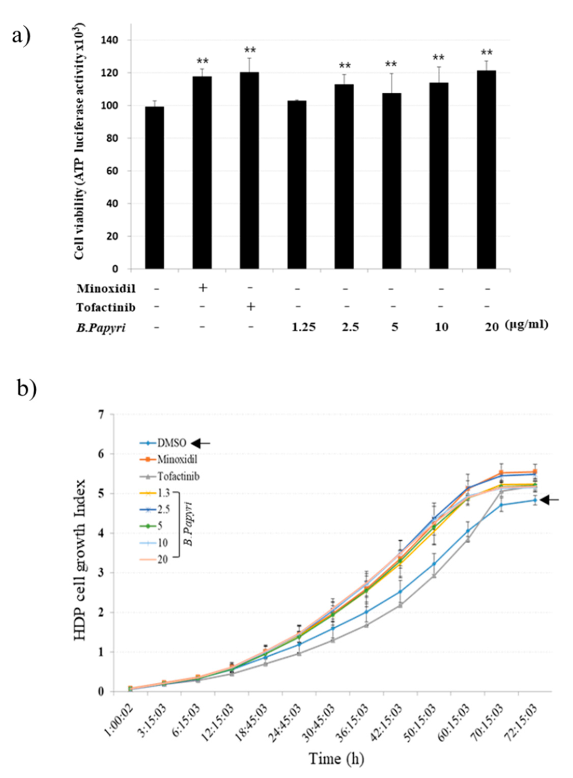
3.1. The Effects of B. papyrifera on the Growth of Hair Follicle Dermal Papilla Cells

In the present study, we examined the effects of ethanolic extract of B. papyrifera on the growth of hHFDP cells. Minoxidil and tofacitinib were used as positive controls. Treatment of cells with B. papyrifera extract enhanced the viability of the hHFDP cells in a dose-dependent manner (Figure 1a). The xCELLigence system-based analysis of real-time cell proliferation showed a time-dependent increase in the proliferation of hHFDP cells upon treatment with B. papyrifera extract (Figure 1b).

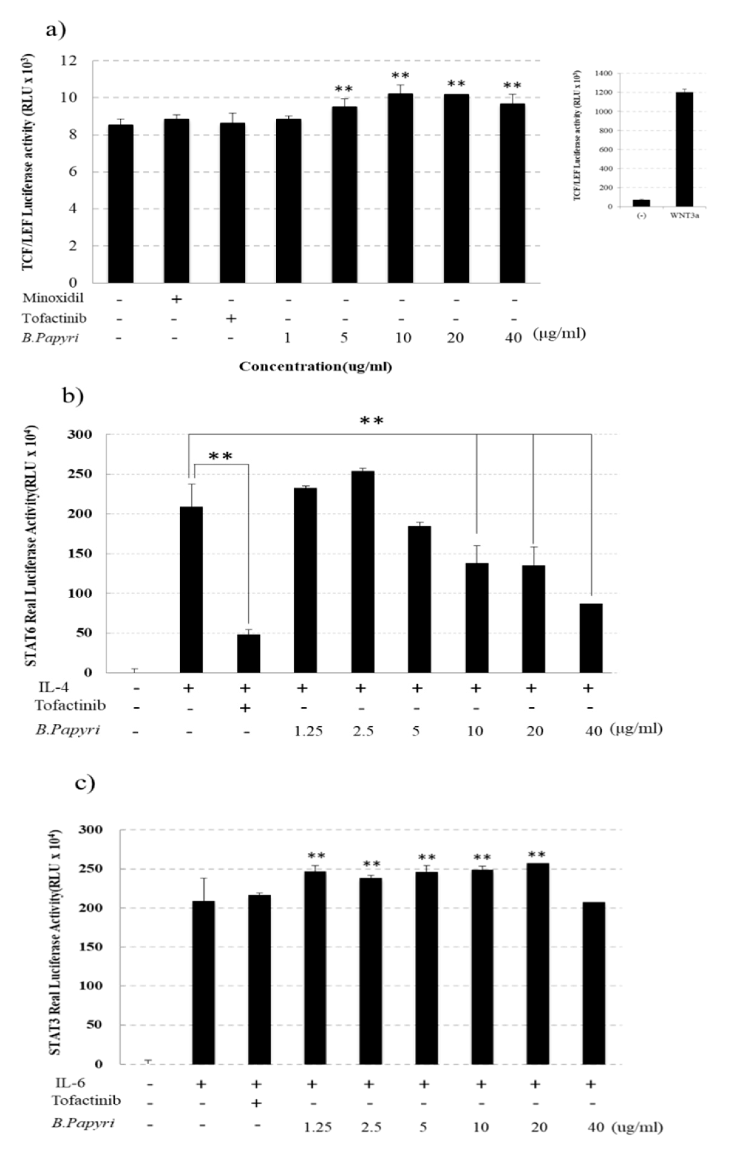
3.2. Effects of B. Papyrifera on the Reporter Gene Activity of Tcf/Lef, stat3 and stat6 in hHFDP Cell

To investigate the underlying mechanisms by which B. papyrifera promotes proliferation of hHFDP cells, we measured the effect of B. papyrifera on TCF/LEF and STAT6-luciferase reporter activity, which play an important role in regulating hair growth. As a result, we observed that incubation with B. papyrifera resulted in TCF/LEF luciferase activation as compared with that induced by minoxidil or tofacitinib (Figure 2a). To confirm the effect of B. papyrifera on STAT6 phosphorylation, which plays an important role in the regulation of hair growth, we observed STAT6 luciferase activity by treating the STAT6 reporter gene stabled BA/F3 cells with interleukin-4 and B. papyrifera. B. papyrifera decreased the STAT6 luciferase activity in a dose-dependent manner, which was comparable with that inhibited by minoxidil or tofacitinib (Figure 2b). The effect of B. papyrifera on STAT3-mediated transcriptional activity was also measured. As a result, the STAT3 transcriptional activity induced by IL-6 was further activated by B. papyrifera in a concentration-dependent manner (Figure 2c). This enhancement of IL-6-induced STAT3 transactivation was comparatively better than that activated by treatment with tofacitinib.

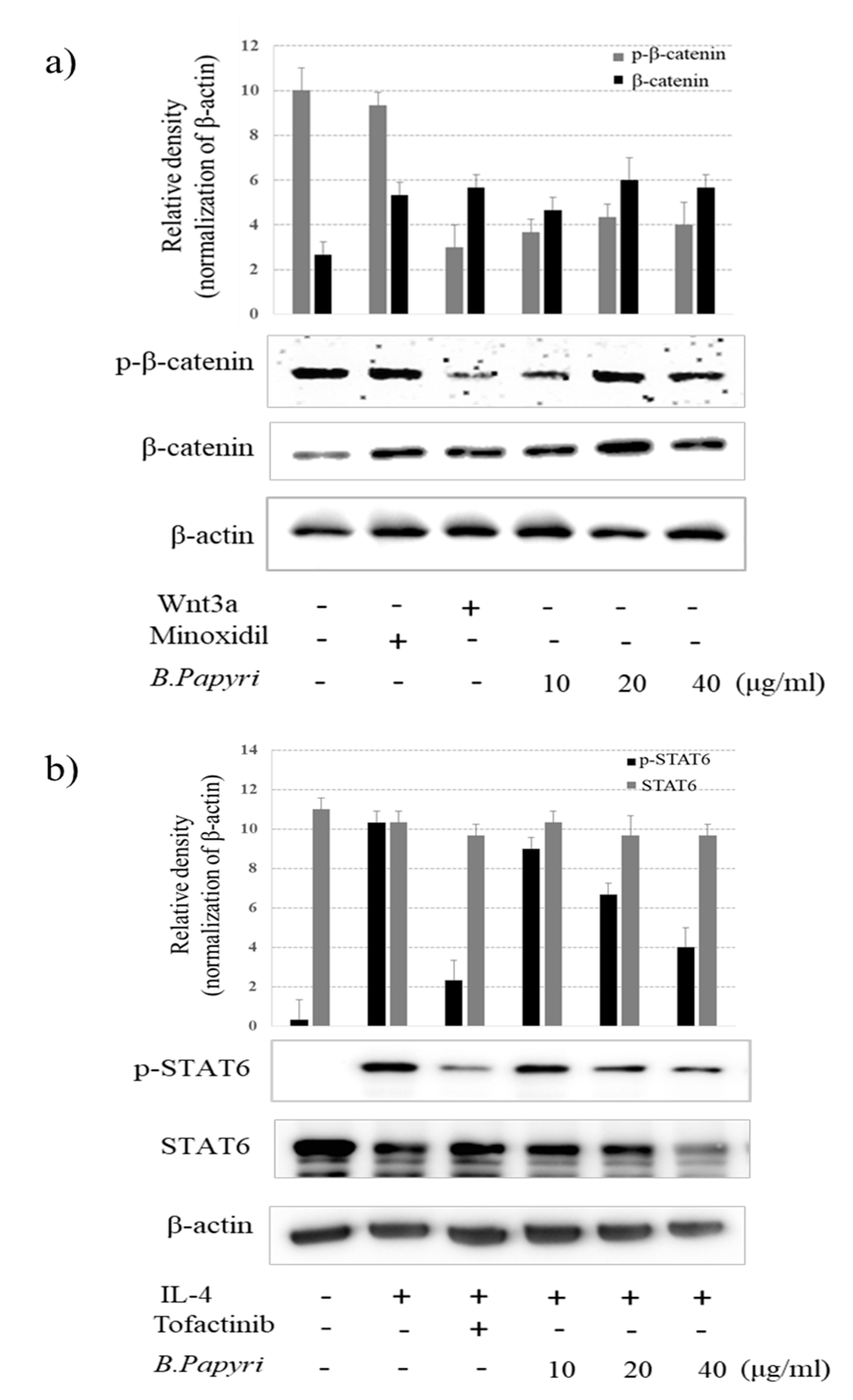
3.3. Effects of B. Papyrifera on Phosphorylation of β-Catenin and stat6 in hHFDP Cells.

Since the phosphorylation of β-catenin followed by its ubiquitination leads to the degradation of β-catenin, we examined the effect of B. papyrifera and the expression of phosphorylated β-catenin. As expected, incubation cells with B. papyrifera attenuated phosphorylation of β-catenin at Thr41 and Ser45 residues (Figure 3a) while increasing the level of β-catenin in a concentration-dependent manner. As WNT3a stabilizes β-catenin by inhibiting β-catenin phosphorylation in hHFDP cells, we compared the effect of B. papyrifera with that of Wnt3a, which also reduced β-catenin phosphorylation. Moreover, B. papyrifera diminished the phosphorylation of STAT6 in hHFDP cells stimulated with IL-4. The reference compound tofacitinib also attenuated IL-4-induced STAT6 phosphorylation at Tyr641 (Figure 3b).

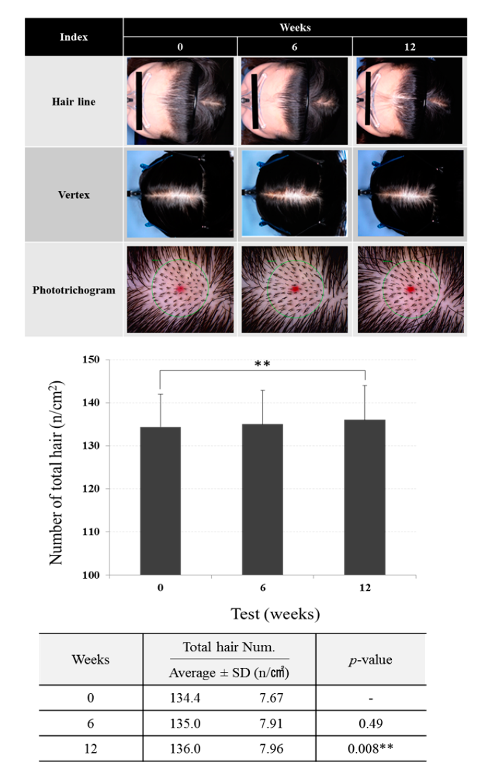
3.4 Effects of Scalp Tonic Enriched With B. papyrifera in Human Subjects

Background information of the Subjects:

A total of 15 subjects participated in the test group, however, four subjects were excluded in the middle of the clinical trial. Finally, the clinical trial was completed with 11 subjects. The test group was comprised of both male and female subjects (male/female = 2:9) with the mean age 43.4 ± 9.25 years. The demographic characteristics of the subjects are summarized in Table 1. There were no statistically significant differences between the 11 subjects who participated in the test group.

3.5. Effects of Scalp Tonic on Hair Density and Total Hair Counts

The analysis of the clinical image showed a significant increase in hair density in the test group as compared with the baseline values. The phototrigram analysis showed significant results (p < 0.05) in the total number of hairs in the test group at 12 weeks after treatment with the scalp tonic as compared with the baseline values. The total number of hairs after 12 weeks of treatment was calculated as 136.0 ± 7.96 number/cm2 (Figure 4). No significant side effects were observed or reported by volunteers.

4. Discussion

The human hair follicle cycle consists of the anagen phase, the catagen phase, and the telogen phase. The anagen phase is the period of growth and the cells in the hair bulb undergo rapid cell division creating new hair. Catagen, the second phase of hair cycle, is relatively short and lasts about 2–3 weeks. Finally, the third and final stage of hair cycle called the telogen phase starts and this phase begins with a resting period, when club hairs rest in the root and new hair begins to grow underneath [27]. In general, the change of hair is an aesthetic pursuit for appearance of beauty and is related to the change of positive and negative form of hair loss. [28]. The dermal papilla, a group of mesenchymal cells located at the base of hair follicles, plays an important role in regulating hair morphology, growth, and circulation. [29]. In the new anagen phase, the secretory factors of dermal papillary cells promote peripheral matrix cells and stimulate new stem cells to induce proliferation and differentiation. [30]. Abnormal cycling of hair follicles results in hair loss. Alopecia or hair loss results from many different etiological factors, such as heredity, aging, hormonal imbalance, stress, and exposure to environmental toxicants. However, irrespective of its etiology, the impact is quite common being social embarrassment and psychological distress. Progressive hair loss in some cases is indicative of other pathological conditions. Thus, alopecia is not only a cosmetic issue but also holds importance from a medical point of view. The currently available therapies lack sustained hair growth potential. For example, the use of minoxidil needs continuous application, and once therapy is discontinued, hair growth promotion is halted. The other commonly used therapy is finasteride which elicits various unwanted effects. Thus, there is a need to search and develop new hair growth promoting agents.
In this study, we examined the potential of developing a hair growth promoting agent by using the extract of B. papyrifera, a medicinal plant belonging to the Moraceae family that is widely distributed in East Asia and China [18]. Previous studies have reported that B. papyrifera possesses anti-inflammatory, antioxidant, anti-tyrosinase, anticancer, antinociceptive, and antimicrobial properties [16,17,18,21,22]. Our study demonstrated that the native plant B. papyrifera is involved in cell cycle regulation of hair follicle cells, which are regulated via the activation of WNT-β-catenin and STAT6 signaling pathways. It has been reported that the signals from dermal papilla maintained the follicular epithelium at the time of anagen progression. One of these dermal papillary signals, β-catenin protein, maintained the ability to induce hair growth across several rounds of dermal papillary cells [1,31,32]. Recent studies have demonstrated that the activation of Wnt/β-catenin signaling by diverse stimuli including a wide variety of natural products promoted hair growth through increased proliferation of dermal papilla cells [33,34,35]. Our finding that B. papyrifera extract decreased the phosphorylation of β-catenin and stabilized β-catenin, thereby resulting in increased TCF/LEF reporter activity, suggests a plausible mechanism of hair growth promotion by this product. Sano et al. reported that STAT3 was required for spontaneous anagen progression in the hair cycle, however exogenous stimuli-induced anagen progression could occur independent of STAT3 activation [36]. Thus, the finding that B. papyrifera increased the STAT3 transcriptional activity suggests that the preparation can enhance anagen phase progression resulting in hair growth promotion. Alternatively, hair growth is promoted by stimulating the activation or proliferation of hair follicle stem cells. Recent studies have reported that hair follicle stem cell function is inhibited by increased Janus-activated kinase (JAK)-STAT signaling in aged mice [37]. STAT5/6 signaling has been shown to control hair follicle stem cell arrest during pregnancy and lactation [38]. Sivan Harel et al. (2015) reported that topical treatment with tofactinib, a small molecule inhibitor of the JAK-STAT pathway, resulted in rapid reentry into anagen and subsequent hair growth [39]. The inhibition of IL-6-induced STAT6 transcriptional activity, as well as downregulation of STAT6 phosphorylation by both tofacitinib and B. papyrifera suggested an alternative mechanism of hair growth induction by the test product. However, the determination that B. papyrifera extract could suppress the JAK activation in dermal papilla cells is yet to be examined.
On the basis of the results of dermal papilla cells, a trial involving volunteers with androgenetic alopecia was designed to evaluate the effect of a formulated scalp tonic enriched with B. papyrifera extract. The improvement in hair growth among subjects suggests that the hair tonic formulation containing B. papyrifera extract (0.5%) are effective in improving hair loss in androgenic alopecia patients. The results demonstrate that B. papyrifera stimulates hair growth by activating WNT-β-catenin and STAT3 signals and inhibiting STAT6 signals in hair follicle cells. During scalp hair loss, the number of hair follicle stem cells usually remains unaltered, whereas that of the proliferating progenitor cells declines [40]. Thus, activating the dormant hair follicle stem cells can promote hair regrowth. The adipose-derived mesenchymal stem cells (AD-MSCs) and stromal vascular fraction cells (SVFs) can stimulate the scalp epidermal stem cells. Gentile et al. carried out a randomized, placebo-controlled, and evaluator-blinded clinical study to examine the effect of micrografting of adipose-derived mesenchymal stem cells on hair regrowth. They demonstrated that after 58 weeks of micrografting of adipose-derived mesenchymal stem cells, 17 male and 10 female patients with androgenic alopecia showed remarkable hair regrowth. However, six of these patients lost their hair in six months and were required to undergo retreatment with micrografting [41]. Moreover, a number of growth factors, such as vascular endothelial growth factor (VEGF), plate-derived growth factor (PDGF), and insulin-like growth factor-1 (IGF-1) play vital roles in hair growth cycle regulation. VEGF stimulates hair follicles by inducing angiogenesis and increasing blood supply to DPCs, while PDGF enhances the anagen stage [42]. Thus, it would be interesting to examine the role of B. papyrifera-enriched hair tonic in the induction of these growth factors to strengthen the mechanistic basis of hair growth promotion by this product. The results of this preliminary preclinical and clinical study suggest that B. papyrifera could be considered to be a potential candidate as a hair growth-promoting agent and further research is deemed necessary for the development of hair care cosmeceutical products.

Author Contributions

Conceptualization, B.Y.C. and G.N.; methodology, Y.H.L.; software, M.-K.K.; validation, S.-C.C. and B.Y.C.; formal analysis, G.N. and B.Y.C.; investigation, M.-K.K..; resources, B.Y.C. and G.N.; data curation, G.N. and B.Y.C.; writing—original draft preparation, Y.H.L and B.Y.C.; writing—review and editing, B.Y.C.; visualization, M.-K.K. and S.-C.C.; supervision, G.N. and B.Y.C.; project administration, B.Y.C. All authors have read and agreed to the published version of the manuscript.

Funding

This research was financially supported by the Ministry of Trade, Industry and Energy (MOTIE, P0006061) and the Korea Institute for Advancement of Technology (KIAT) through the Promoting Regional specialized Industry

Acknowledgments

We wish to thank BK bio Co., Ltd. and kolmar Korea co. Ltd. for helpful comments on the research and Joydeb Kumar Kundu for helpful comments on the manuscript.

Conflicts of Interest

The authors declare no potential conflicts of interest.

References

  1. Cotsarelis, G.; Millar, S.E. Towards a molecular understanding of hair loss and its treatment. Trends Mol. Med. 2001, 7, 293–301. [Google Scholar] [CrossRef]
  2. Miyashita, H.; Hakamata, Y.; Kobayashi, E.; Kobayashi, K. Characterization of hair follicles induced in implanted, cultured rat keratinocyte sheets. Exp. Dermatol. 2004, 13, 491–498. [Google Scholar] [CrossRef] [PubMed]
  3. Park, J.H. A novel concept for determining the direction of implanted hair in hairline correction surgery in East Asian women. Arch. Plast. Surg. 2018, 45, 292–294. [Google Scholar] [CrossRef] [PubMed] [Green Version]
  4. Suchonwanit, P.; Srisuwanwattana, P.; Chalermroj, N.; Khunkhet, S. A randomized, double-blind controlled study of the efficacy and safety of topical solution of 0.25% finasteride admixed with 3% minoxidil vs. 3% minoxidil solution in the treatment of male androgenetic alopecia. J. Eur. Acad. Dermatol. Venereol. 2018, 32, 2257–2263. [Google Scholar] [CrossRef] [PubMed]
  5. Jain, R.; De-Eknamkul, W. Potential targets in the discovery of new hair growth promoters for androgenic alopecia. Expert Opin. Ther. Targets 2014, 18, 787–806. [Google Scholar] [CrossRef] [PubMed]
  6. Lopedota, A.; Denora, N.; Laquintana, V.; Cutrignelli, A.; Lopalco, A.; Tricarico, D.; Maqoud, F.; Curci, A.; Mastrodonato, M.; la Forgia, F.; et al. Alginate-Based Hydrogel Containing Minoxidil/Hydroxypropyl-beta-Cyclodextrin Inclusion Complex for Topical Alopecia Treatment. J. Pharm. Sci. 2018, 107, 1046–1054. [Google Scholar] [CrossRef]
  7. O’Leary, M. By the way, doctor. Finasteride has been prescribed for my BPH. I think that the most frequent side effect is erectile dysfunction or loss of sexual desire. Would Levita or a similar drug (like Viagra or Cialis) overcome the side effects of finasteride? Harv. Health Lett. 2009, 34, 8. [Google Scholar]
  8. Ahshawat, M.S.; Saraf, S. Preparation and characterization of herbal creams for improvement of skin viscoelastic properties. Int. J. Cosmet. Sci. 2008, 30, 183–193. [Google Scholar] [CrossRef]
  9. Patel, S.; Sharma, V.; Chauhan, N.S.; Thakur, M.; Dixit, V.K. Hair Growth: Focus on Herbal Therapeutic Agent. Curr. Drug Discov. Technol. 2015, 12, 21–42. [Google Scholar] [CrossRef]
  10. Pervin, M.; Hasnat, M.A.; Lee, Y.M.; Kim, D.H.; Jo, J.E.; Lim, B.O. Antioxidant activity and acetylcholinesterase inhibition of grape skin anthocyanin (GSA). Molecules 2014, 19, 9403–9418. [Google Scholar] [CrossRef]
  11. Zaid, A.N.; Jaradat, N.A.; Eid, A.M.; Al Zabadi, H.; Alkaiyat, A.; Darwish, S.A. Ethnopharmacological survey of home remedies used for treatment of hair and scalp and their methods of preparation in the West Bank-Palestine. BMC Complement. Altern. Med. 2017, 17, 355. [Google Scholar] [CrossRef] [PubMed] [Green Version]
  12. Gentile, P.; Garcovich, S. Advances in Regenerative Stem Cell Therapy in Androgenic Alopecia and Hair Loss: Wnt pathway, Growth-Factor, and Mesenchymal Stem Cell Signaling Impact Analysis on Cell Growth and Hair Follicle Development. Cells 2019, 8, 466. [Google Scholar] [CrossRef] [PubMed] [Green Version]
  13. Premanand, A.; Reena Rajkumari, B. Androgen modulation of Wnt/beta-catenin signaling in androgenetic alopecia. Arch. Dermatol. Res. 2018, 310, 391–399. [Google Scholar] [CrossRef]
  14. Ramot, Y.; Zlotogorski, A. [Jak Inhibitors for the Treatment of Alopecia Areata]. Harefuah 2020, 159, 38–42. [Google Scholar] [PubMed]
  15. Wang, E.H.C.; Sallee, B.N.; Tejeda, C.I.; Christiano, A.M. JAK Inhibitors for Treatment of Alopecia Areata. J. Investig. Dermatol. 2018, 138, 1911–1916. [Google Scholar] [CrossRef] [Green Version]
  16. Ryu, H.W.; Park, M.H.; Kwon, O.K.; Kim, D.Y.; Hwang, J.Y.; Jo, Y.H.; Ahn, K.S.; Hwang, B.Y.; Oh, S.R. Anti-inflammatory flavonoids from root bark of Broussonetia papyrifera in LPS-stimulated RAW264.7 cells. Bioorg. Chem. 2019, 92, 103233. [Google Scholar] [CrossRef]
  17. Wu, W.T. Evaluation of anti-inflammatory effects of Broussonetia papyrifera stem bark. Indian J. Pharmacol. 2012, 44, 26–30. [Google Scholar] [CrossRef]
  18. Si, B.; Tao, H.; Zhang, X.; Guo, J.; Cui, K.; Tu, Y.; Diao, Q. Effect of Broussonetia papyrifera L. (paper mulberry) silage on dry matter intake, milk composition, antioxidant capacity and milk fatty acid profile in dairy cows. Asian-Australas J. Anim. Sci. 2018, 31, 1259–1266. [Google Scholar] [CrossRef] [Green Version]
  19. Ko, H.H.; Chang, W.L.; Lu, T.M. Antityrosinase and antioxidant effects of ent-kaurane diterpenes from leaves of Broussonetia papyrifera. J. Nat. Prod. 2008, 71, 1930–1933. [Google Scholar] [CrossRef]
  20. Park, S.; Fudhaili, A.; Oh, S.S.; Lee, K.W.; Madhi, H.; Kim, D.H.; Yoo, J.; Ryu, H.W.; Park, K.H.; Kim, K.D. Cytotoxic effects of kazinol A derived from Broussonetia papyrifera on human bladder cancer cells, T24 and T24R2. Phytomedicine 2016, 23, 1462–1468. [Google Scholar] [CrossRef]
  21. Lin, L.W.; Chen, H.Y.; Wu, C.R.; Liao, P.M.; Lin, Y.T.; Hsieh, M.T.; Ching, H. Comparison with various parts of Broussonetia papyrifera as to the antinociceptive and anti-inflammatory activities in rodents. Biosci. Biotechnol. Biochem. 2008, 72, 2377–2384. [Google Scholar] [CrossRef] [PubMed] [Green Version]
  22. Sohn, H.Y.; Kwon, C.S.; Son, K.H. Fungicidal effect of prenylated flavonol, papyriflavonol A, isolated from Broussonetia papyrifera (L.) vent. against Candida albicans. J. Microbiol. Biotechnol. 2010, 20, 1397–1402. [Google Scholar] [CrossRef] [PubMed] [Green Version]
  23. Kim, Y.E.; Choi, H.C.; Lee, I.C.; Yuk, D.Y.; Lee, H.; Choi, B.Y. 3-Deoxysappanchalcone Promotes Proliferation of Human Hair Follicle Dermal Papilla Cells and Hair Growth in C57BL/6 Mice by Modulating WNT/beta-Catenin and STAT Signaling. Biomol. Ther. 2016, 24, 572–580. [Google Scholar] [CrossRef] [PubMed] [Green Version]
  24. Kaneko, A.; Kaneko, T. A New Classification of Early Female Pattern Hair Loss. Int. J. Trichology 2018, 10, 61–67. [Google Scholar] [CrossRef] [PubMed]
  25. Lee, W.S.; Ro, B.I.; Hong, S.P.; Bak, H.; Sim, W.Y.; Kim, D.W.; Park, J.K.; Ihm, C.W.; Eun, H.C.; Kwon, O.S.; et al. A new classification of pattern hair loss that is universal for men and women: Basic and specific (BASP) classification. J. Am. Acad. Dermatol. 2007, 57, 37–46. [Google Scholar] [CrossRef]
  26. Baek, J.H.; Lee, S.Y.; Yoo, M.; Park, W.S.; Lee, S.J.; Boo, Y.C.; Koh, J.S. Effects of a new mild shampoo for preventing hair loss in Asian by a simple hand-held phototrichogram technique. Int J. Cosmet. Sci. 2011, 33, 491–496. [Google Scholar] [CrossRef]
  27. Alonso, L.; Fuchs, E. The hair cycle. J. Cell Sci. 2006, 119, 391–393. [Google Scholar] [CrossRef]
  28. Zgonc Skulj, A.; Poljsak, N.; Kocevar Glavac, N.; Kreft, S. Herbal preparations for the treatment of hair loss. Arch. Dermatol. Res. 2019. [Google Scholar] [CrossRef]
  29. Botchkarev, V.A.; Kishimoto, J. Molecular control of epithelial-mesenchymal interactions during hair follicle cycling. J. Investig. Dermatol. Symp. Proc. 2003, 8, 46–55. [Google Scholar] [CrossRef] [Green Version]
  30. Kang, B.M.; Shin, S.H.; Kwack, M.H.; Shin, H.; Oh, J.W.; Kim, J.; Moon, C.; Moon, C.; Kim, J.C.; Kim, M.K.; et al. Erythropoietin promotes hair shaft growth in cultured human hair follicles and modulates hair growth in mice. J. Dermatol. Sci. 2010, 59, 86–90. [Google Scholar] [CrossRef]
  31. Kishimoto, J.; Burgeson, R.E.; Morgan, B.A. Wnt signaling maintains the hair-inducing activity of the dermal papilla. Genes Dev. 2000, 14, 1181–1185. [Google Scholar] [PubMed]
  32. Miller, J.; Djabali, K.; Chen, T.; Liu, Y.; Ioffreda, M.; Lyle, S.; Christiano, A.M.; Holick, M.; Cotsarelis, G. Atrichia caused by mutations in the vitamin D receptor gene is a phenocopy of generalized atrichia caused by mutations in the hairless gene. J. Investig. Dermatol. 2001, 117, 612–617. [Google Scholar] [CrossRef] [Green Version]
  33. Cheon, H.I.; Bae, S.; Ahn, K.J. Flavonoid Silibinin Increases Hair-Inductive Property Via Akt and Wnt/beta-Catenin Signaling Activation in 3-Dimensional-Spheroid Cultured Human Dermal Papilla Cells. J. Microbiol. Biotechnol. 2019, 29, 321–329. [Google Scholar] [CrossRef]
  34. Lee, Y.R.; Bae, S.; Kim, J.Y.; Lee, J.; Cho, D.H.; Kim, H.S.; An, I.S.; An, S. Monoterpenoid Loliolide Regulates Hair Follicle Inductivity of Human Dermal Papilla Cells by Activating the Akt/beta-Catenin Signaling Pathway. J. Microbiol. Biotechnol. 2019, 29, 1830–1840. [Google Scholar] [CrossRef]
  35. Zhang, H.; Shi, Q.; Nan, W.; Wang, Y.; Wang, S.; Yang, F.; Li, G. Ginkgolide B and bilobalide promote the growth and increase beta-catenin expression in hair follicle dermal papilla cells of American minks. Biofactors 2019, 45, 950–958. [Google Scholar] [CrossRef]
  36. Sano, S.; Kira, M.; Takagi, S.; Yoshikawa, K.; Takeda, J.; Itami, S. Two distinct signaling pathways in hair cycle induction: Stat3-dependent and -independent pathways. Proc. Natl. Acad. Sci. USA 2000, 97, 13824–13829. [Google Scholar] [CrossRef] [Green Version]
  37. Doles, J.; Storer, M.; Cozzuto, L.; Roma, G.; Keyes, W.M. Age-associated inflammation inhibits epidermal stem cell function. Genes Dev. 2012, 26, 2144–2153. [Google Scholar] [CrossRef] [PubMed] [Green Version]
  38. Goldstein, J.; Fletcher, S.; Roth, E.; Wu, C.; Chun, A.; Horsley, V. Calcineurin/Nfatc1 signaling links skin stem cell quiescence to hormonal signaling during pregnancy and lactation. Genes Dev. 2014, 28, 983–994. [Google Scholar] [CrossRef] [PubMed] [Green Version]
  39. Harel, S.; Higgins, C.A.; Cerise, J.E.; Dai, Z.; Chen, J.C.; Clynes, R.; Christiano, A.M. Pharmacologic inhibition of JAK-STAT signaling promotes hair growth. Sci. Adv. 2015, 1, e1500973. [Google Scholar] [CrossRef] [Green Version]
  40. Garza, L.A.; Yang, C.C.; Zhao, T.; Blatt, H.B.; Lee, M.; He, H.; Stanton, D.C.; Carrasco, L.; Spiegel, J.H.; Tobias, J.W.; et al. Bald scalp in men with androgenetic alopecia retains hair follicle stem cells but lacks CD200-rich and CD34-positive hair follicle progenitor cells. J. Clin. Investig. 2011, 121, 613–622. [Google Scholar] [CrossRef] [Green Version]
  41. Gentile, P.; Scioli, M.G.; Cervelli, V.; Orlandi, A.; Garcovich, S. Autologous Micrografts from Scalp Tissue: Trichoscopic and Long-Term Clinical Evaluation in Male and Female Androgenetic Alopecia. Biomed. Res. Int. 2020, 2020, 7397162. [Google Scholar] [CrossRef] [PubMed] [Green Version]
  42. Gentile, P. Autologous Cellular Method Using Micrografts of Human Adipose Tissue Derived Follicle Stem Cells in Androgenic Alopecia. Int. J. Mol. Sci. 2019, 20, 3446. [Google Scholar] [CrossRef] [PubMed] [Green Version]
Figure 1. Effects of B. papyrifera on hHFDP cells. (a) CellTiter-Glo® luminescent cell growth assay shows that B. papyrifera promote proliferation of hHFDP cells; (b) Real-time xCELLigence system as described in the Methods. All experiments were performed in triplicate. The asterisk indicates a significant statistical significance (** p < 0.05).
Cosmetics 07 00040 g001
Figure 2. Luciferase activity of reporter gene by B. papyrifera. (a) For TCF/LEF reporter gene assay, cells were treated with B. papyrifera (1–40 µg/ml) for 24. The positive control group was treated with minoxidil (0.4 µM) or tofactinib (0.4 µM); (b) IL-4 induced STAT6; and (c) IL-6 induced STAT3 reporter gene assay as determined by luciferase activity. Tofactinib (0.4 µM), a JAKs inhibitor, was treated as a positive control. Each assay is representative for 3 experiments. The asterisk(s) indicate a significant statistical significance (** p < 0.05).
Cosmetics 07 00040 g002
Figure 3. Effect of B. papyrifera on the level or activity of proteins implicated in hair growth. (a) The level of p-β-catenin and β-catenin was detected by Western blotting using specific antibodies in hHF DP cells; (b) The level of p-STAT6 and STAT6 was detected by Western blotting using specific antibodies in IL-4-induced HDPCs; (c) The level of p-STAT3 and STAT3 was detected by Western blotting using specific antibodies in IL-6-induced HDPCs. β-Actin protein was used as an internal control. Immunoblot assay was performed as described in the Materials and Methods. Each blot is representative for 3 experiments.
Cosmetics 07 00040 g003
Figure 4. Images of change in hair pattern following the application of scalp tonic for 12 consecutive weeks. The photographic assessment of hair density showed a tendency of an increase in hair density in test groups as compared with the baseline data. Bottom panel bar graph represents the change of total hair counts measured by folliscope image at baseline and after (** p < 0.05).
Cosmetics 07 00040 g004
Table 1. Demographic characteristics of the subjects.
CharacteristicsTest Group (n = 11)
n (%)
SexMale2 (18)
Female9 (81)
AgeMean ± SD43.4 ± 9.25
Median44
Max, Min53, 21
20–291 (9)
30–391 (9)
40–496 (55)
≥503 (27)

