Review

Under Persistent Assault: Understanding the Factors that Deteriorate Human Skin and Clinical Efficacy of Topical Antioxidants in Treating Aging Skin

by
Patricia K. Farris
1 and
Yevgeniy Krol
2,*
1
School of Medicine, Tulane University, New Orleans, LA 70112, USA
2
Skinceuticals, New York, NY 10017, USA
*
Author to whom correspondence should be addressed.
Cosmetics 2015, 2(4), 355-367; https://doi.org/10.3390/cosmetics2040355
Submission received: 12 August 2015 / Revised: 29 October 2015 / Accepted: 29 October 2015 / Published: 5 November 2015
(This article belongs to the Special Issue The Antioxidant Potential of the Skin)

Abstract

:
Recent studies contend that the skin is subject to far more damage than just ultraviolet (UV) light, with infrared radiation and pollution now clearly demonstrated to degrade cutaneous tissue. While consumers continue to strive for new ways to augment the aesthetic appeal and improve the health of their skin, awareness regarding environmental insults and effective ways to protect the skin remains low. New advances in dermatologic science have exponentially increased the available information on the underlying mechanism of cutaneous damage and potential of topical antioxidants to treat aging skin. Combining antioxidants that can work through multiple pathways holds great potential for a cumulative and synergistic way to treat aging skin. Our goal is to provide a comprehensive review on environmental factors that damage human skin, discuss scientifically proven benefits of topical antioxidants, understand challenges of formulating and administering topical antioxidants, evaluate novel mechanisms of antioxidant activity, and suggest practical ways of integrating topical antioxidants with aesthetic procedures to complement clinical outcomes.

1. Introduction

Human skin is constantly exposed to environmental insults, the most prominent of which is ultraviolet radiation (UVR). Other factors like cigarette smoke, infrared radiation, visible light, diesel fuel exhaust, and ozone are believed to damage skin, even though UVR remains as the predominant stressor with a plethora of scientific evidence supporting its deleterious role [1,2,3,4,5]. As a consequence of the normal aging process, both intrinsic and extrinsic forces contribute to the progressive depletion of cutaneous antioxidants, leading to an imbalance of cellular redox in favor of a state of oxidative stress. This imbalance manifests phenotypically as diminishing skin health and loss of its aesthetic appeal. Even though oxidation is a basic chemical reaction vital for existence of almost every living species, organisms had to evolve a complex system of antioxidants to protect themselves from extensive oxidative damage at a cellular level. Regardless of the effectiveness of an organisms’ defense system, exposure to environmental insults and the cumulative effects of advancing age can render this defense capacity diminished and eventually obsolete in fighting constant oxidative assaults. Additionally, environmental assaults like UVR and various pollutants exhibit potential to cause cutaneous inflammation leading to an immune response involving the recruitment of activated neutrophils and macrophages with a subsequent release of oxidative bursts capable of degrading connective tissue and collagen fibers [6]. While the process of cutaneous aging has been divided into intrinsic (or chronologic) and extrinsic (or environmental) aging, it is extremely difficult to separate the two in human facial skin as both occur simultaneously and through similar mechanisms. The goal of this paper is to briefly review the scientific literature highlighting damaging effects of the environment on skin, discuss the potential of topical antioxidants in mitigating the damage, understand the challenges of properly formulating topical antioxidants, and suggest ways of integrating topical antioxidants with aesthetic treatments to augment clinical outcomes.

2. Ultraviolet Irradiation

The observations of the degenerative effect of solar radiation on skin were initially made in the late 19th century by Paul Gerson Unna, evaluating cutaneous anomalies of sailors, and William Auguste Dubreuilh, noting the increased prevalence of hyperkeratoses in vineyard workers [7,8]. Ultraviolet Radiation (UVR) exerts its damage on the skin by increasing the production of free radicals, specifically reactive oxygen species (ROS), resulting in photo-oxidative damage to cellular components in the epidermis and extracellular matrix [9]. One of the first studied molecular mechanism of UV-induced oxidative stress and its deleterious role in skin aging involves the absorption of photon energy by cutaneous chromophores leading to excitation of oxygen and subsequent production of ROS like superoxide radical (O2), singlet oxygen (O2•), and hydroxyl radicals (OH•) which can directly damage biomolecules vital for skin function [6]. Superoxide, for one, is known to oxidize and inactivate a number of dehydratases by causing a loss of Fe(II) from the active site. In addition to the loss of the dehydratases catalytic activity, the liberated Fe(II) from the (4Fe–4S) cluster participates in the reduction of hydrogen peroxide (H2O2) to hydroxyl radical (OH•), an extremely reactive radical with indiscriminating capacity to damage proximal biomolecules, in the classical Fenton reaction [10]. An alternative cascade of free radical production involves the reaction of photons or excited photosensitizers with molecular oxygen to form superoxide anion radicals, which subsequently lead to the generation of lipid radicals in the cellular membrane. This oxidative degradation of cell membrane lipids is referred to as lipid peroxidation, which propagates the formation of cascading free radicals further compounding the degradation of intracellular components [11]. Lipid peroxides and their associated byproducts, including 4-hydroxy-2-nonenal and malondialdehyde, disrupt cellular homeostasis by damaging vital proteins and via release of proinflammatory mediators [12]. Signaling pathways of UVR damage tend to converge on the upregulation of transcription factor Activator Protein-1 (AP-1) that can further increase the production of matrix metalloproteinase (MMP) that triggers collagen breakdown [13]. ROS signaling also blocks transforming growth factor beta (TGF-β) causing a reduction in new collagen formation [14]. This net loss of dermal collagen, due both to increased collage breakdown and reduced collagen synthesis, is fundamental to wrinkling and laxity seen in photoaged skin [15]. Additionally, ROS stimulate the production of elastin mRNA and are intricately involved in elastotic changes seen in photoaged skin [16]. Serving as the main target for environmental attack, the skin has evolved a comprehensive antioxidant defense system to combat extrinsic oxidative stress. Nevertheless, the cumulative exposure to UVR leads to the progressive depletion of cutaneous antioxidants, and an imbalance of cellular redox in favor of a state of oxidative stress. Oxidation of proteins and nucleic acids by UVR has also been implicated in advancement of photoaging [17]. The progressive accumulation of oxidative damage to lipids, proteins, and nucleic acid causes a negative feedback loop that results in an overall greater production of ROS, further exacerbating cellular redox homeostasis in favor of a pro-oxidative state [18]. The mechanism of action of UVR is generally tied to a specific wavelength, causing either direct damage to cellular components like collagen or by increasing the formation of ROS [17].

3. Infrared A Radiation

Infrared A Radiation (IRA) is another example of an insult that holds substantial capacity to damage and prematurely age human skin. Increasing scientific evidence supports the conclusion that wavelengths of the solar spectrum between 770 and 1400 nm (IRA) can cause actinic damage and accelerate skin aging [19]. While there are similarities between the ways in which UVR and IRA function at a mechanistic level, the most profound difference is in the cellular chromophores absorbing the wavelength. Furthermore, unlike UV rays, IRA can penetrate through all three layers of the skin [20]. While UVR shown an absorption spectrum in cellular membrane lipids as well as nucleic acids, IRA is believed to be absorbed by the copper atoms of complex IV of the mitochondrial respiratory chain [21,22]. This indicates that the initial reaction in IRA molecular signaling cascade occurs at a mitochondrial level. Krutmann et al. has shown that IRA causes changes in transcription of human fibroblasts in the genomic region responsible for homeostasis of extracellular matrix, apoptosis, and stress response [23]. Furthermore, studies have demonstrated that IRA propagates the formation of ROS, leading to upregulation of matrix metalloproteinase and subsequent breakdown of cutaneous collagen [24,25,26]. Of note in these findings is the absence of associated increase in tissue inhibitor of metalloproteinases (TIMP-1), suggesting that the upregulation of MMP-1 continues unabated and eventually leads to substantial collagen breakdown and formation of fine lines and wrinkles, the classic phenotype of photoaged skin. IRA is also known to reduce the expression of procollagen-1, potentially further diminishing the collagen content of the skin. Lastly, IRA has ability to potentiate angiogenesis, via an upregulation of vascular endothelial growth factor (VEGF), another important molecular hallmark of photoaged skin. Taken together these findings suggest that protection against IRA is paramount in maintaining vitality of human skin.

4. Pollution

Air pollution is gaining acceptance as an environmental stressor exhibiting ability to damage human skin, which is often the primary target of gaseous and liquid pollutants [5]. While pollution is a broad term, many studies have described the ability of various individual pollutants to damage cutaneous tissue. The pollutants believed to be most deleterious to skin are polycyclic aromatic hydrocarbons, volatile organic compounds, heavy metals, and ozone. The effects of multiple pollutants on the skin are believed to compound one another and can be seen almost instantaneously, as inflammation, or over an extended period, as advanced skin aging. One particular German study found that exposure to car exhaust particles and soot was significantly correlated with an increase in pigment deposits and wrinkles [27]. Likewise, Valacchi et al. has demonstrated that ozone exposure causes a cascade of stress signaling in the outer cutaneous layer, as well as increase in oxidative stress proteins in deeper layers of the skin [28]. Studies suggest that ozone functions as a strong oxidative agent causing matrix remodeling by inducing expression of MMP-9 [29]. Valacchi et al. have also demonstrated the compounding effect of ozone and UV in causing greater depletion in stratum corneum vitamin E content, producing additive oxidative stress in the skin [30]. While the underlying mechanism of action of pollution in skin is not clearly defined, there is clear evidence implicating the generation of ROS and subsequent state of heightened oxidative stress in all layers of the skin [31,32]. The harmful effects of pollution are being substantiated by an increasing number of researchers worldwide, and more controlled studies are required to unravel the complicated role it plays in accelerated skin aging.

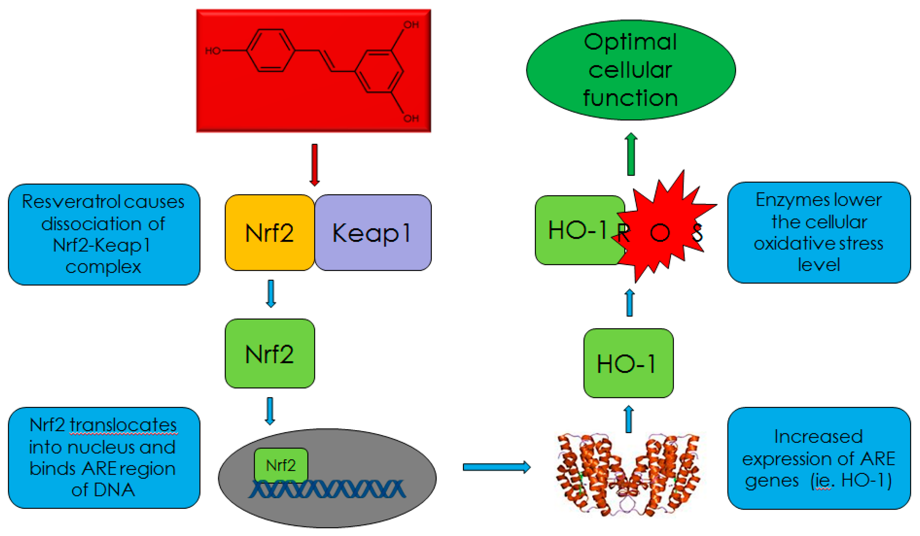
5. Topical Antioxidants as Treatment

Topically applied antioxidants are important agents to counteract the damaging effects of environmental insults and chronologic skin aging. Multiple studies have shown that topical application of antioxidants is an effective therapeutic approach to aid the skin’s defense against UV and other environmental insults [33,34,35,36,37]. In fact, the American Academy of Dermatology’s Guidelines of Care includes a recommendation for the application of topical antioxidants as a treatment for photodamage [38]. Fundamentally, antioxidants are believed to work directly by scavenging radical species, via a donation of an electron to neutralize the radical species. Specifically, l-ascorbic acid is hydrophilic and predominantly functions in the aqueous compartment of the cell [39].However, l-ascorbic acid function improves in the presence of alpha-tocopherol, which aggregates around the lipidic cellular membrane and potentiates the action of l-ascorbic acid four-fold [36]. Topical antioxidants can reduce the prevalence of ROS and subsequent inflammatory response, a vital component of their antiaging benefits [40]. Likewise, antioxidants prevent the oxidation and subsequent cross-linking of structural proteins like collagen and elastin, another commonality of aged skin. In addition to vitamins, botanical antioxidants have been shown to mitigate UV-induced damage. These phenolic and polyphenolic antioxidants have strong free radical scavenging capability and include green tea extract, genistein, silymarin, caffeic acid, ferulic acid, pycnogenol and fern extract [41].
One of the newer areas of interest in topical antioxidants is the “secondary” ability of antioxidants to not only directly terminate the propagation of radical species, but to increase the skin’s antioxidant capacity in order to prevent and correct environmentally and chronologically induced-oxidative damage of vital biomolecules. Instead of supplementing the skin with exogenous antioxidants, it is possible to strengthen the skin’s already prominent antioxidant network. The ability of our skin to synthesize antioxidants is tightly regulated by the Antioxidant Response Element (ARE) and transcription factor Nrf2 that control antioxidant gene expression. Enzymes such as superoxide dismutase, glutathione, catalase, and heme oxygenase-1 are under control of the Nrf2 pathway and produced de novo to protect various tissues against daily free radical attack [42,43]. Under standard conditions, this antioxidant defense system is activated by the production of ROS. However, stimulating this system independent of an increase in radical species prevalence can be an additional way to boost skin’s ability to fight oxidative damage. One molecule that is known to stimulate the Nrf2 pathway is Resveratrol, as has been shown in multiple studies in various tissues [44,45,46]. (Figure 1) in 2014, Farris et al. published a study that outlines the potential of a topical antioxidant formula containing 1.0% resveratrol, 0.5% baicalin, and 1.0% alpha-tocopherol to activate the Nrf2 pathway and improve skin’s ability to counteract extrinsic and intrinsic insults [47]. The benefits of using this antioxidant formula were also confirmed clinically as a statistically significant improvement in all skin quality parameters, including firmness and density.
Figure 1. Schematic representation of Resveratrol activating the Nrf2 pathway [47].
Cosmetics 02 00355 g001
Essentially, antioxidants slow the aging process by quenching the formation of ROS or by preventing the oxidation of vital biomolecules. However, the age-related depletion of cutaneous antioxidant molecules must be replenished in order to maintain significant defense against extrinsic and intrinsic insults. While it should come as no surprise that a diverse selection of topical antioxidants products are commercially available for this exact purpose, the prevalence of antioxidant products has not correlated with an effective way to differentiate the efficacy of these formulas in neutralizing ROS in vivo, boosting the skin’s antioxidant content, and ability to fight oxidative damage. For this reason, it is important to consider the underlying factors responsible for the efficacy of topical antioxidants.

6. Countermeasures by Antioxidants

When considering different formulations, containing antioxidants as primary active ingredients, factors like compatibility, stability, and penetration must be prioritized. One of the primary challenges in formulating antioxidants is to ensure the stability of the mixture while promoting the delivery of active ingredients into the skin. The ingredients must be protected from oxidation, in the delivery vehicle, in order to remain bioactive for topical application. The stability of antioxidants, like l-ascorbic acid, is greatly enhanced at an acidic pH and in an aqueous state where their individual pKa value is below that of the acid. Most importantly, antioxidants need to penetrate into the skin, and remain stable for a period of time required to attain the benefit. Penetration and utilization of antioxidants in biological systems is vital to achieve optimal efficacy. Antioxidants should be selected to exhibit synergistic action as opposed to neutralizing one another in a chosen vehicle. However, it is not enough to simply understand the factors required to formulate an efficacious antioxidant, controlled clinical studies must be conducted to validate the clinical effect. One example is the work of Pinnell et al., who published breakthrough research on the proper formulation parameters necessary for percutaneous absorption of topically applied vitamin C, an antioxidant widely utilized in dermatologic formulations [48]. His study determined that vitamin C must be formulated in its pure l-ascorbic acid form, at a pH below 3.5, in order to remove the ionic charge and promote delivery across the stratum corneum, and with a maximal concentration between 10% and 20% in order to reach optimal saturation in the skin [48]. As the most abundant antioxidant in skin, vitamin C or l-ascorbic acid works via a non-enzymatic manner to protect cells against various free radicals with capability to neutralize hydroxyl (OH), alkoxyl (OL), peroxyl (LOO) radicals, sulphur radicals, nitrogen–oxygen radicals, as well as reducing the radical state of other antioxidants [49,50,51]. An example being the ascorbic acid mediated reduction of tocopheroxyl radical back into alpha-tocopherol [52].Without the ability to penetrate the skin, a formulation containing vitamin C or any additional agent would be virtually ineffective.
The research published by Pinnell and colleagues, underscore the importance of designing a proper topical delivery system to achieve percutaneous absorption [48]. However, outside their compelling breakthrough, peer reviewed literature attesting to the ability of antioxidant products to penetrate live skin remains scarce. Furthering their initial discovery, Pinnell et al. were able to prove that a properly formulated mixture of antioxidants, containing vitamin C, E and ferulic acid exhibited a synergistic activity that increased the stability of the antioxidant cocktail and could significantly prevent UV-induced oxidative damage in skin [37,53]. They demonstrated that the combination of synergistic antioxidants provided 8-fold protection from the formation of sunburn cells and erythema. These studies supported the idea that vitamin C, E, and ferulic acid protected against solar-simulated radiation-triggered apoptosis as exemplified by their prevention of caspaces activation at eight times the minimal erythema dose (MED).
Antioxidants also display great potential to be combined with various in-office procedures to augment and complement the end benefit. However, when combining aggressive treatments with topical products, it is fundamentally important to limit the number of ingredients to avoid unnecessary irritation. Fractional lasers have been widely employed by aesthetic dermatologists for successful treatment of rhytids as well as the general skin aging phenotype [54,55]. While lasers remain one of the primary tools to treat cutaneous photoaging, the associated downtime often becomes a major limitation for patients considering this procedure. Combining the procedure with topical antioxidant application can have substantial benefits in reducing the subsequent cutaneous inflammation and aiding the wound healing process, thus exhibit potential of alleviating post-laser downtime. For example, l-ascorbic acid, a vital cofactor for prolyl and lysyl hydroxylase, is crucial for the synthesis and stability of collagen, which serves an important role in the wound healing process [56,57]. The thermal injury induced by fractional lasers is believed to substantially deplete cutaneous antioxidants well beyond standard conditions. For this reason, integrating this procedure with a topical antioxidant formula is an excellent method to complement the clinical efficacy while mitigating the unwanted side effects.
While the mechanism of action of topical antioxidant treatment in aiding wound healing is still not clearly understood, replenishing the skin’s antioxidant reservoir is vitally important in the proper repair of the laser-induced injury. Previously, a scientific poster presented at the 2014 American Academy of Dermatology annual conference demonstrated that the average duration of downtime resolved two days faster with the application of topical l-ascorbic acid, alpha-tocopherol, and ferulic acid serum immediately post-fractional ablative laser treatment [58]. This study also noted that the antioxidant serum was able to protect beta fibroblast growth factor (bFGF), and hypothesized this action to be important in the observed resolution of post-laser downtime. Overall, the antioxidant formula was well tolerated when used immediately post-fractional ablative laser and had the added benefit of inhibiting albicans infections following the laser treatment, as a consequence of the formulas’ low pH. This concept of integrating fractional lasers with topical antioxidants has been reproduced by Campos et al., who demonstrated that the use of the same topical l-ascorbic acid, alpha-tocopherol, and ferulic acid formula lead to an immediate benefit in the reducing post-operative pain, as well as a long-term benefits of enhanced clinical improvement in wrinkles and skin tone evenness after 90 days of post-laser application [59,60].
Integrated skincare is an evolving concept that combines in-office treatments with at-home product usage to achieve complementary clinical benefits and reduce unwanted side effects. Currently, treatment guidelines generally advise that the use of any products, exclusive of petrolatum ointment and sunscreen, should be avoided during the first week of recovery after an ablative procedure. However, incorporating a topical antioxidant formula may aid the wound healing process, minimize post-treatment downtime, and increase patient compliance with course of aesthetic treatments.

7. Conclusions

Scientific evidence is mounting that environmental factors beyond UVR can cause significant underlying damage to the skin. Thus, it comes as no surprise that a diverse selection of topical antioxidants products have been introduced over the last decade. Despite the prevalence of commercially available antioxidant formulas, a clear ability to differentiate their efficacy is not yet possible. For this reason, understanding the variation in topical antioxidant products, like the differences in delivery vehicle and synergistic action of active ingredients, is essential in determining their clinically relevant effect. While many topical formulations profess to provide similar benefits, they often lack controlled and comparative evidence supporting this claim. This presents a number of complexities in differentiating observed versus theoretical efficacy.
Even though a complete understanding of environmentally-induced skin damage remains elusive, it has been well accepted that continuous exposure to extrinsic assaults leads to an imbalance in favor of a state of oxidative stress causing a disruption of cellular homeostasis and deleterious biological effects phenotypically expressed as premature skin aging. In addition to escalating the production of free radicals, chronic exposure to UVR is known to substantially deplete the antioxidant content of the skin [61]. Scientists have also shown that other environmental pollutants, like polyaromatic hydrocarbon, and ozone, can deplete vital cutaneous antioxidants, weakening the skin defense system against oxidative stress [62,63]. Interestingly, Valacchi et al. showed that the combination of UV and ozone causes a significantly greater depletion of cutaneous antioxidants than UVR alone, leading to the conclusion that environmental factors in addition to UV can compound the subsequent cutaneous damage [30]. Free radicals produced by environmental insults can be neutralized by supplementing the skin with topical antioxidants provided that percutaneous absorption is achieved and antioxidant stability is maintained. However, published evidence on the clinical benefits of topical antioxidants is still limited. Instead, common tests to evaluate nutritional and pharmaceutical products, such as oxygen radical absorbance capacity (ORAC), hydroxyl radical antioxidant capacity (HORAC), hydrogen atom transfer (HAT), and electron transfer based in vitro assays are utilized to attest to the formulas’ benefit. Obviously, these testing methods are of limited clinical relevance and unreliable in correlating a products’ antioxidant capacity to its efficacy in fighting environmentally assaults. The limitation of these assays is that extrapolation of their in vitro efficacy to physiological benefits is highly inconsistent. In addition, these in vitro assays are unable to capture the formulations’ ability to penetrate the stratum corneum, a vital component necessary for an antioxidant to neutralize free radicals produced in live skin.
Antioxidants are known to work by neutralizing free radical species after exposure to environmental assault(s). Additionally, once cutaneous penetration is achieved, topically applied antioxidants allow the skin to build an internal reservoir defense that cannot be easily removed from the surface of the skin by mechanical force. The use of topical antioxidants to mitigate free radical propagation has been established in a few previous studies [50,51,52]. Antioxidants are also excellent candidates to be combined with in-office procedures in order to enhance overall patient satisfaction. This novel treatment modality is introducing a comprehensive way to augment clinical outcomes and changing the paradigm of post-procedure care. Likewise, further clinical data is needed to substantiate the benefits of the combination and uncover a better understanding of the underlying mechanism of action. Outside of the limited number of studies presented in this review, clinical trials involving antioxidant based therapies have been rather problematic. Questions like the insufficient ability of antioxidant derivatives to convert into their biologically active form in the skin, optimal treatment dosages, and proper duration of therapy for specific conditions are just some of the outstanding issues that should continue to be addressed to maximize the effect of antioxidants in humans.
Intuitively, not all products offer the same level of protection, but currently there is a shortage of published data confirming the clinical efficacy of topical antioxidant formulas. While this review offers increasing evidence on the damaging effects of environmental insults and provides further explanation on the role that proper formulation parameters play in overall efficacy, it can only serve as anecdotal evidence highlighting the importance of continued investment into clinical research on topical antioxidants.

Author Contributions

Yevgeniy Krol wrote the initial draft of the manuscript and performed the initial round of edits. Patricia Farris performed multiple rounds of review and made substantial edits to the manuscript.

Conflicts of Interest

Yevgeniy Krol is an employee of SkinCeuticals, New York, NY, USA. Patricia Farris declares no conflict of interest.

References

  1. Schroeder, P.; Schieke, S.M.; Morita, A. Premature Skin Aging by Infrared Radiation, Tobacco Smoke and Ozone. In Skin Aging; Springer Berlin Heidelberg: Berlin, Germany, 2006; pp. 45–53. [Google Scholar]
  2. Schroeder, P.; Lademann, J.; Darvin, M.E.; Stege, H.; Marks, C.; Bruhnke, S.; Krutmann, J. Infrared Radiation-Induced Matrix Metalloproteinase in Human Skin: Implications for Protection. J. Investig. Dermatol. 2008, 128, 2491–2497. [Google Scholar] [CrossRef] [PubMed]
  3. Kvam, E.; Tyrrell, R.M. Induction of oxidative DNA base damage in human skin cells by UV and near visible radiation. Carcinogenesis 1997, 18, 2379–2384. [Google Scholar] [CrossRef] [PubMed]
  4. Thiele, J.J.; Podda, M.; Packer, L. Tropospheric ozone: An emerging environmental stress to skin. Biol. Chem. 1997, 378, 1299–1305. [Google Scholar] [PubMed]
  5. Baudouin, C.; Charveron, M.; Tarroux, R.; Gall, Y. Environmental pollutants and skin cancer. Cell Biol. Toxicol. 2002, 18, 341–348. [Google Scholar] [CrossRef] [PubMed]
  6. Darr, D.; Fridovich, I. Free radicals in cutaneous biology. J. Investig. Dermatol. 1994, 102, 671–675. [Google Scholar] [CrossRef] [PubMed]
  7. Unna, P. Histopathologie der Hautkrankheiten; August Hirschwald: Berlin, Germany, 1984. (In German) [Google Scholar]
  8. Dubreuilh, W. Des hyperkeratoses circonscrites. Ann. Dermatol. Venereol. 1896, 7, 1158–1204. (In French) [Google Scholar]
  9. Scharffetter-Kochanek, K.; Wlaschek, M.; Brenneisen, P.; Schauen, M.; Blaudschun, R.; Wenk, J. UV induced reactive oxygen species in photocarcinogenesis and photoaging. Biol. Chem. 1997, 378, 1247–1257. [Google Scholar] [PubMed]
  10. Liochev, S.I.; Fridovich, I. The role of O2 in the production of HO: In vitro and in vivo. Free Radic. Biol. Med. 1994, 16, 29–33. [Google Scholar] [CrossRef]
  11. Dixit, R.; Mukhtar, H.; Bickers, D.R. Studies on the role of reactive oxygen species in mediating lipid peroxide formation in epidermal microsomes of rat skin. J. Investig. Dermatol. 1983, 81, 369–375. [Google Scholar] [CrossRef] [PubMed]
  12. Briganti, S.; Ricardo, M. Antioxidant activity, lipid peroxidation and skin disease. What’s new? JEADV 2003, 17, 663–669. [Google Scholar] [CrossRef] [PubMed]
  13. Rittie, L.; Fischer, G.J. UV-light-induced signal cascades and skin aging. Ageing Res. Rev. 2002, 1, 705–720. [Google Scholar] [CrossRef]
  14. Fisher, G.J.; Kang, S.; Varani, J.; Bata-Csorgo, Z.; Wan, Y.; Datta, S.; Voorhees, J.J. Mechaisms of photoaging and chronological skin aging. Arch. Dermatol. 2002, 138, 1462–1470. [Google Scholar] [CrossRef] [PubMed]
  15. Fisher, G.J.; Varani, J.; Voorhees, J.J. Looking older: Fibroblast collapse and therapeutic implications. Arch. Dermatol. 2008, 144, 666–672. [Google Scholar] [CrossRef] [PubMed]
  16. Traikovich, S.S. Use of Topical Ascorbic acid and its effect on photo damaged skin topography. Arch. Otorhinol. Head Neck Surg. 1999, 125, 1091–1098. [Google Scholar] [CrossRef]
  17. Fisher, G.J.; Wang, Z.; Datta, S.C.; Varani, J.; Kang, S.; Voorhees, J.J. Pathophysiology of premature skin aging induced by ultraviolet light. N. Engl. J. Med. 1997, 337, 1419–1429. [Google Scholar] [CrossRef] [PubMed]
  18. Breitenbach, M.; Bischof, J.; Streubel, M.K.; Trost, A.; Richer, K. Oxidative stress in aging human skin. Biomolecules 2015, 5, 545–589. [Google Scholar]
  19. Grether-Beck, S.; Marini, A.; Jaenicke, T.; Krutmann, J. Photoprotection of human skin beyond ultraviolet radiation. Photodermatol. Photoimmunol. Photomed. 2014, 30, 167–174. [Google Scholar] [CrossRef] [PubMed]
  20. Vierkotter, A.; Krutmann, J. Environmental influences on skin aging and ethnic-specific manifestations. Dermatoendocrinology 2012, 4, 227–231. [Google Scholar] [CrossRef] [PubMed]
  21. Grether-Beck, S.; Krutmann, J. Involvement of lipid rafts and caveolins in UVA signaling. Open Dermatol. J. 2009, 3, 153–159. [Google Scholar] [CrossRef]
  22. Karu, T.I. Mitochondrial signaling in mammalian cells activated by red and near-IR radiation. Photochem. Photobiol. 2008, 84, 1091–1099. [Google Scholar] [CrossRef] [PubMed]
  23. Calles, C.; Schneider, M.; Macaluso, F.; Benesova, T.; Krutmann, J.; Schroeder, P. Infrared a radiation influences the skin fibroblast transcriptome: Mechanisms and consequences. J. Investig. Dermatol. 2010, 130, 1524–1536. [Google Scholar] [CrossRef] [PubMed]
  24. Kim, H.H.; Lee, M.J.; Lee, S.R.; Kim, K.H.; Cho, K.H.; Eun, H.C.; Chung, J.H. Augmentation of UV-induced skin wrinkling by infrared irradiation in hairless mice: Implications for protection. Mech. Ageing Dev. 2005, 126, 1170–1177. [Google Scholar] [CrossRef] [PubMed]
  25. Kim, M.S.; Kim, Y.K.; Cho, K.H.; Chung, J.H. Regulation of type 1 procollagen and MMP-1 expression after single or repeated exposure to infrared radiation in human skin. Mech. Ageing Dev. 2006, 127, 875–882. [Google Scholar] [CrossRef] [PubMed]
  26. Chung, J.H.; Cho, S.; Shin, M.H.; Kim, Y.K.; Seo, J.-E.; Lee, Y.M.; Park, C.-H. Effects of Infrared Radiation and Heat on Human Skin Aging in Vivo. J. Investig. Dermatol. 2009, 14, 15–19. [Google Scholar]
  27. Vierkotter, A.; Schikowski, T.; Ranft, U.; Sugiri, D.; Matsui, M.; Kramer, U.; Krutmann, J. Airborne Particle Exposure and Extrinsic Skin Aging. J. Investig. Dermatol. 2010, 130, 2719–2726. [Google Scholar] [CrossRef] [PubMed]
  28. Valacchi, G.; van der Vliet, A.; Schock, B.C.; Okamoto, T.; Obermuller-Jevic, U.; Cross, C.E.; Packer, L. Ozone exposure activates oxidative stress response in murine skin. Toxicology 2002, 179, 163–170. [Google Scholar] [CrossRef]
  29. Valacchi, G.; Pagnin, E.; Okamoto, T.; Corbacho, A.M.; Olano, E.; Davis, P.A.; van der Vliet, A.; Packer, L.; Cross, C.E. Induction of stress proteins and MMP-9 by 0.8 ppm of ozone in murine skin. Biochem. Biophys. Res. Commun. 2003, 305, 741–746. [Google Scholar] [CrossRef]
  30. Valacchi, G.; Weber, S.U.; Luu, C.; Cross, C.E.; Packer, L. Ozone potentiates vitamin E depletion by ultraviolet radiation in murine stratum corneum. FEBS Lett. 2000, 466, 165–168. [Google Scholar] [CrossRef]
  31. Pryor, W.A. Mechanisms of radical formation from reactions of ozone with target molecules in the lung. Free Radic. Biol. Med. 1984, 17, 451–465. [Google Scholar] [CrossRef]
  32. Donaldson, K.; Tran, L.; Jimenez, L.A.; Duffin, R.; Newby, D.E.; Mills, N.; MacNee, W.; Stone, V. Combustion-derived nanoparticles: A review of their toxicology following inhalation exposure. Part Fibre Toxicol. 2005, 2. [Google Scholar] [CrossRef] [PubMed]
  33. Hamanaka, H.; Miyachi, Y.; Imamura, S. Photoprotective effect of topically applied SOD on sunburn reaction in comparison with sunscreen. J. Dermatol. 1990, 17, 595–598. [Google Scholar] [CrossRef] [PubMed]
  34. Mizushima, Y.; Hoshi, K.; Yamagawa, A.; Takana, K. Topical application of superoxide dismutase cream. Drugs Exp. Clin. Res. 1991, 17, 127–131. [Google Scholar] [PubMed]
  35. Lopez-Torres, M.; Thiele, J.J.; Shindo, Y.; Han, D.; Packer, L. Topical application of lpha-tocopherol modulates the antioxidant network and diminishes ultraviolet-induced oxidative damage in murine skin. Br. J. Dermatol. 1998, 138, 207–215. [Google Scholar] [CrossRef] [PubMed]
  36. Lin, J.Y.; Selim, M.A.; Shea, C.R.; Grichnik, J.M.; Omar, M.M.; Monteiro-Riviere, N.A.; Pinnell, S.R. UV photoprotection by combining topical antioxidants vitamin C and vitamin E. J. Am. Acad. Dermatol. 2003, 48, 866–874. [Google Scholar] [CrossRef] [PubMed]
  37. Murray, J.C.; Burch, J.A.; Streilein, R.D.; Iannacchione, M.A.; Hall, R.P.; Pinnell, S.R. A topical antioxidant solution containing vitamin C and E stabilized by ferulic acid provides protection for human skin against damage caused by ultraviolet irradiation. J. Am. Acad. Dermatol. 2008, 59, 418–425. [Google Scholar] [CrossRef] [PubMed]
  38. Drake, L.A.; Dinehart, S.M.; Farmer, E.R.; Goltz, R.W.; Graham, G.F.; Hordinsky, M.K.; Lewis, C.W.; Pariser, D.M.; Webster, S.B.; Whitaker, D.C.; et al. Guidelines of care for photoaging/photodamaged. J. Am. Acad. Dermatol. 1996, 35, 462–464. [Google Scholar] [PubMed]
  39. Davies, M.B.; Austin, J.A.; Patridge, D.A. Vitamin C: Its Chemistry and Biochemistry; Royal Society of Chemistry: London, UK, 1991. [Google Scholar]
  40. Oresajo, C.; Pillai, S.; Manco, M.; Yatskayer, M.; McDaniel, D. Antioxidants and the skin: Understanding formulation and efficacy. Dermatol. Ther. 2012, 25, 252–259. [Google Scholar] [CrossRef] [PubMed]
  41. Wang, S.; Balagula, Y.; Osterwalder, U. Photoprotection: A review of the current and future technologies. Dermtol. Ther. 2010, 23, 31–47. [Google Scholar] [CrossRef] [PubMed]
  42. Marrot, L.; Jones, C.; Perez, P.; Meunier, J.P. The significance of Nrf2 pathway in (photo)-oxidative stress response in melanocytes and keratinocytes of the human epidermis. Pigment Cell Melanoma Res. 2008, 21, 79–88. [Google Scholar] [CrossRef] [PubMed]
  43. Zhang, M.; An, C.; Gao, Y.; Leak, R.K.; Chen, J.; Zhang, F. Emerging Roles of Nrf2 and Phase II Antioxidant Enzymes in Neuroprotection. Prog. Neurobiol. 2013, 100, 30–47. [Google Scholar] [CrossRef] [PubMed]
  44. Ungvari, Z.; Baqi, Z.; Feher, A.; Recchia, F.A.; Sonntaq, W.E.; Pearson, K.; de Cabo, R.; Csiszar, A. Resveratrol confers endothelial protection via activation of the antioxidant transcription factor Nrf2. Am. J. Physiol. Heart Circ. Physiol. 2010, 299, H18–H24. [Google Scholar] [CrossRef] [PubMed]
  45. Kode, A.; Rajendrasozhan, S.; Caito, S.; Yang, S.R.; Megson, I.L.; Rahman, I. Resveratrol induces glutathione synthesis by activation of Nrf2 and protects against cigarette smoke-mediated oxidative stress in human lung epithelial cells. Am. J. Physiol. Lung Cell Mol. Physiol. 2008, 294, L478–L488. [Google Scholar] [CrossRef] [PubMed]
  46. Ren, J.; Fan, C.; Chen, N.; Huang, J.; Yang, Q. Resveratrol pretreatment attenuates cerebral ischemic injury by upregulating expression of transcription factor Nrf2 and HO-1 in rats. Neurochem. Res. 2011, 36, 2352–2362. [Google Scholar] [CrossRef] [PubMed]
  47. Farris, P.; Yatskayer, M.; Chen, N.; Krol, Y.; Oresajo, C. Evaluation of Efficacy and Tolerance of a Nightime Topical Antioxidant Containing Resveratrol, Baicalin, and Vitamin E for Treatment of Mild to Moderately Photodamaged Skin. J. Drugs Dermatol. 2014, 13, 1467–1472. [Google Scholar] [PubMed]
  48. Pinnell, S.R.; Yang, H.; Omar, M.; Monteiro-Riviere, N.; DeBuys, H.V.; Walker, L.C.; Wang, Y.; Levine, M. Topical l-ascorbic acid: Percutaneous absorption studies. Dermatol. Surg. 2001, 27, 137–142. [Google Scholar] [CrossRef] [PubMed]
  49. Shindo, Y.; Witt, E.; Han, D.; Epstein, W.; Packer, L. Enzymic and non-enzymic antioxidants in epidermis and dermis of human skin. J. Investig. Dermatol. 1994, 102, 122–124. [Google Scholar] [CrossRef] [PubMed]
  50. Frei, B.; England, L.; Ames, B.N. Ascorbate is an outstanding antioxidant in human blood plasma. Proc. Natl. Acad. Sci. USA 1989, 86, 6377–6381. [Google Scholar] [CrossRef] [PubMed]
  51. Hossain, M.A.; Asada, K. Monodehydroascorbate reductase from cucumber is a flavin adenine dinucleotide enzyme. J. Biol. Chem. 1985, 260, 12920–12926. [Google Scholar] [PubMed]
  52. Neuzil, J.; Thomas, S.R.; Stocker, R. Requirement for, promotion, or inhibition by alpha-tocopherol of radical-induced initiation of plasma lipoprotein lipid peroxidation. Free Radic. Biol. Med. 1997, 22, 57–71. [Google Scholar] [CrossRef]
  53. Lin, F.H.; Lin, J.Y.; Gupta, R.D.; Tournas, J.A.; Burch, J.A.; Selim, M.A.; Monteiro-Riviere, N.A.; Grichnik, J.M.; Zielinski, J.; Pinnell, S.R. Ferulic acid stabilizes a solution of vitamins C and E and doubles its photoprotection of skin. J. Investig. Dermatol. 2005, 125, 826–832. [Google Scholar] [CrossRef] [PubMed]
  54. Manstein, D.; Herron, G.S.; Sink, R.K.; Tanner, H.; Anderson, R.R. Fractional photothermolysis: A new concept for cutaneous remodeling using microscopic patterns of thermal injury. Lasers Surg. Med. 2004, 34, 426–438. [Google Scholar] [CrossRef] [PubMed]
  55. Waibel, J.; Beer, K.; Narurkar, V.; Alster, T. Preliminary observations on fractional ablative resurfacing devices: Clinical impressions. J. Drugs Dermatol. 2009, 8, 481–485. [Google Scholar] [PubMed]
  56. Pinnell, S.R.; Murad, S.; Darr, D. Induction of Collagen Synthesis by Ascorbic Acid. A Possible Mechanism. Arch. Dermatol. 1987, 123, 1684–1686. [Google Scholar] [CrossRef]
  57. Nusgens, B.V.; Humbert, P.; Rougier, A.; Richard, A.; Lapiere, C.M. Stimulation of collagen biosynthesis by topically applied vitamin C. Eur. J. Dermatol. 2002, 12, XXXII–XXXIV. [Google Scholar] [PubMed]
  58. Waibel, J.S.; Mi, Q.S.; Wulkan, A. Split Face Comparison of the Effect of 15% Vitamin C, 1% Vitamin E & Ferulic Acid Serum to Decrease Post-Operative Recovery in Fractional Ablative Laser Resurfacing for Photodamage. In Proceedings of AAD 2014, Denver, CO, USA, 21–25 March 2014.
  59. Campos, V.; Steiner, D.; Arbache, S.; Ferrara, F.; Santos, K.; Capucho, L. A Prospective Split-Face Double-Blind Randomized Placebo-Controlled Trial to Assess the Efficacy of Vitamin C and Ferulic Acid Serum Post-Fractional Ablative Laser for Skin Rejuvenation. In Proceedings of AAD 2014, Denver, CO, USA, 21–25 March 2014.
  60. Campos, V.; Steiner, D.; Arbache, S.; Ferrara, F.; Santos, K.; Capucho, L. A Prospective Split-Face Clinical Study to Evaluate the Long-Term Efficacy of Vitamin C and Ferulic Acid Serum Post-Fractional Ablative Laser for Skin Rejuvenation. In Proceedings of AAD 2015, San Francisco, CA, USA, 20–24 March 2015.
  61. Maeda, K.; Naganuma, M.; Fukuda, M. Effect of chronic exposure to ultraviolet-A including 2% ultraviolet-B on free radical reduction systems in hairless mice. Photochem. Photobiol. 1991, 54, 575–582. [Google Scholar] [CrossRef]
  62. Thiele, J.J.; Traber, M.G.; Tsang, K.G.; Cross, C.E.; Packer, L. In vivo exposure to ozone depletes vitamin C and E and induces lipid peroxidation in epidermal layers of murine skin. Free Radic. Biol. Med. 1997, 23, 85–91. [Google Scholar] [CrossRef]
  63. Shindo, Y.; Wit, E.; Han, D.; Packer, L. Dose-response effect of acute ultraviolet irradiation on antioxidants and molecular markers of oxidation in murine epidermis and dermis. J. Investig. Dermatol. 1994, 23, 470–475. [Google Scholar] [CrossRef]

