Article

Heat-Treated Lacticaseibacillus rhamnosus Skinbac™ SB06 Modulates Axillary Malodor-Associated Bacteria In Vitro and Demonstrates Antiperspirant and Deodorant Efficacy In Vivo


Probiotical Research Srl, 28100 Novara, Italy
*
Author to whom correspondence should be addressed.
Cosmetics 2026, 13(4), 178; https://doi.org/10.3390/cosmetics13040178
Submission received: 10 June 2026 / Revised: 7 July 2026 / Accepted: 8 July 2026 / Published: 10 July 2026
(This article belongs to the Section Cosmetic Technology)

Abstract

Background: The axillary microbiome is a major contributor to body malodor generation through bacterial metabolism of apocrine and eccrine secretions. Dysbiosis of this microbial community, particularly through overgrowth of odorigenic species such as Staphylococcus aureus, is associated with increased volatile compound production and local skin inflammation. Heat-treated postbiotics represent a promising class of cosmetic ingredients combining microbiological safety with retained bioactive properties. Objective: We aimed to evaluate the in vitro safety, molecular mechanisms, antipathogen and anti-inflammatory properties of heat-treated Lacticaseibacillus rhamnosus Skinbac™ SB06, and to assess the antiperspirant and deodorant efficacy of a deodorant spray formulation containing 1% SB06 in a controlled clinical study. Methods: In vitro studies assessed cytotoxicity (MTT/LDH assays), Aquaporin-3 (AQP3) expression, reactive oxygen species (ROS) production, antipathogen activity against Staphylococcus aureus (AlamarBlue assay), cytokine modulation (TNF-α, IL-6, IL-8, IL-23) in Normal Human Epidermal Keratinocytes (NHEK) and Peripheral Blood Mononuclear Cells (PBMCs), and axillary microbiome compatibility against Corynebacterium striatum, Staphylococcus epidermidis, and Staphylococcus hominis by viable plate count (CFU/mL). Clinically, a randomized split-body study (n = 20) evaluated antiperspirant effectiveness by gravimetric sweat collection and deodorant efficacy by expert olfactory panel (Likert 1–5) at 24 and 48 h. Results: In vitro testing confirmed the safety of SB06 (MTT and LDH, both non-significant vs. control). SB06 significantly increased AQP3 expression (+20%, p < 0.001) and significantly reduced ROS production (−48%, p < 0.05). Antipathogen testing showed significant reduction in S. aureus planktonic viability (−7%, p < 0.05). Microbiome compatibility testing on selected axillary-associated strains showed a differential compatibility profile, with the strongest inhibitory effect observed for C. striatum (13% residual viability at T24h, corresponding to 87% inhibition), near-complete preservation of S. epidermidis (92% residual viability at T48h), and a mild reduction in S. hominis (−15% at T48h). Cytokine modulation showed significant IL-8 and IL-23 reduction in NHEK (both p ≤ 0.05) and immunostimulatory activity in PBMCs. Clinically, SB06 reduced sweat production vs. placebo by −21.8% at T24 (p = 0.0009) and −10.0% at T48 (p = 0.0495), with significantly lower odor intensity at both timepoints (median score 3 vs. 4, p < 0.0001). Conclusions: Heat-treated L. rhamnosus SB06 showed a multimodal in vitro profile including antipathogen, anti-inflammatory, antioxidant, and AQP3-upregulating activities, and was associated with statistically significant antiperspirant and deodorant effects in a randomized controlled split-body study. These findings are consistent with SB06 being a functional postbiotic ingredient with potential for deodorant and antiperspirant applications, pending confirmation in larger controlled studies.

1. Introduction

The axillary region constitutes a unique anatomical microenvironment characterized by warmth, moisture, occlusion, and high densities of both eccrine and apocrine sweat glands, creating optimal conditions for a specialized resident microbial community [1,2]. Unlike other skin sites, axillary skin is continuously exposed to apocrine secretions containing odorless precursor molecules—including steroids, fatty acid conjugates, and sulfanyl alkanols—that are transformed by resident bacteria into the volatile compounds responsible for characteristic axillary malodor, including 3-methyl-2-hexenoic acid, 3-hydroxy-3-methylhexanoic acid, and 3-methyl-3-sulfanylhexan-1-ol [3,4,5].
The axillary microbiome is dominated by Corynebacterium, Staphylococcus, and Cutibacterium, whose relative abundances determine odor intensity and quality [2,6]. Corynebacterium species are considered the primary producers of thioalcohol-derived malodor through C-S lyase activity, while coagulase-negative staphylococci are associated with milder odor profiles [5,7]. Staphylococcus aureus, although typically a minor component, exerts a disproportionate odorigenic and inflammatory influence when its relative abundance increases: it converts amino acid conjugates and fatty acid precursors into potent volatile malodorous compounds, forms resilient biofilms, and promotes local barrier disruption and inflammatory responses [7,8,9]. In addition, epidermal tight junctions contribute to cutaneous barrier homeostasis and are relevant to the maintenance of skin integrity in sites exposed to repeated chemical and mechanical stress, such as the axilla [10]. Current antiperspirant and deodorant strategies rely predominantly on aluminum-based salts for sweat reduction and antimicrobial agents for odor control [11]. These approaches, while effective, can induce axillary microbiome dysbiosis by reducing protective species and enabling recolonization by more odorigenic populations [12,13]. Growing consumer demand for microbiome-compatible, naturally derived ingredients has directed cosmetic research toward biological alternatives targeting malodor at its microbial source without collateral disruption of the resident microbiota [14].
Postbiotics—defined as preparations of non-viable microorganisms and/or their components that confer a health benefit on the host [15]—represent an emerging class of cosmetic active ingredients with significant potential in this context. Heat inactivation of probiotic strains generates structurally intact bacterial cell corpses retaining bioactive cell wall components, metabolites, lipoteichoic acids, and peptidoglycans that mediate immunomodulatory, antimicrobial, antioxidant, and barrier-enhancing effects [16,17]. Thermal inactivation further confers practical formulation advantages including ambient stability, extended shelf life, and regulatory simplicity compared to live microorganism delivery [17,18].
Lacticaseibacillus rhamnosus (formerly Lactobacillus rhamnosus) is among the most extensively studied Lactobacillus species for both gastrointestinal and cutaneous applications [19,20]. Its production of organic acids contributes to local acidification inhibitory to pathogens, while its surface components facilitate competitive exclusion at epithelial adhesion sites [21]. In cutaneous applications, L. rhamnosus strains have demonstrated anti-inflammatory properties through pro-inflammatory cytokine downregulation, barrier-supporting tight junction upregulation, and oxidative stress reduction in keratinocytes [20,22], making heat-treated L. rhamnosus a candidate of mechanistic interest for deodorant formulations requiring simultaneous antipathogen efficacy, local anti-inflammation, and skin conditioning.
Aquaporin-3 (AQP3), the predominant water channel in the epidermis, mediates water and glycerol transport across keratinocyte membranes, supporting stratum corneum hydration, barrier repair, and cellular proliferation [23,24]. Its expression is suppressed by inflammatory cytokines and oxidative stress [25]—conditions relevant to axillary skin subjected to microbiome dysbiosis or repeated conventional deodorant use. AQP3 upregulation by postbiotic ingredients may therefore contribute to improved skin conditioning in the underarm region, particularly in users experiencing dryness or sensitivity.
This study aimed to provide a comprehensive in vitro and in vivo characterization of heat-treated L. rhamnosus Skinbac™ SB06 for deodorant formulation development. In vitro objectives were to: (I) establish safety by cytotoxicity assays; (II) quantify AQP3 modulation; (III) assess antioxidant capacity by ROS quantification; (IV) evaluate antipathogen activity against S. aureus planktonic cells and biofilm; and (V) characterize cytokine modulation in NHEK and PBMCs. The in vivo objective was to evaluate the antiperspirant effectiveness and deodorant efficacy of a 1% SB06 deodorant spray formulation compared to a matched placebo vehicle in a randomized split-body controlled clinical study.

2. Materials and Methods

2.1. Cell Culture

Normal Human Epidermal Keratinocytes (NHEK-Ad, Cat. No. 00192627; Lonza, Basel, Switzerland) were cultured in Keratinocyte Growth Medium (Cat. No. 00192060; Lonza) at 37 °C/5% CO2. Cells were seeded at 5 × 10⁴ cells/well in 24-well plates and allowed to adhere overnight before treatment. Cryopreserved peripheral blood mononuclear cells were obtained from Lonza (Basel, Switzerland). Cells were thawed according to the manufacturer’s protocol, washed twice with pre-warmed complete RPMI-1640 medium, and resuspended in complete medium supplemented with 10% heat-inactivated fetal bovine serum (FBS; Thermo Fisher Scientific, Waltham, MA, USA) and 2 mM L-glutamine. Cells were seeded at 1 × 10⁶ cells/mL and used as a model of systemic immune response.

2.2. Preparation of Heat-Treated Probiotic Strain

Lacticaseibacillus rhamnosus Skinbac™ SB06 was thermally inactivated at temperatures exceeding 75 °C for 30–90 min and spray-dried. Enumeration was performed by flow cytometry using Thiazole Orange/Propidium Iodide staining (TO/PI, Cat. No. 349483; BD Biosciences, Franklin Lakes, NJ, USA) to determine total fluorescent units (TFU), standardized to 109 TFU/g. The resulting powder was stored at 4 °C until use. All in vitro experiments were conducted at 107 TFU/mL.

2.3. In Vitro Testing

2.3.1. Safety Assessment

Cytotoxicity was evaluated using MTT tetrazolium reduction and Lactate Dehydrogenase (LDH) release assays. NHEK cells were treated with 107 TFU/mL SB06 for 24 h at 37 °C. LDH release was quantified from supernatants per manufacturer instructions (CytoTox 96®, Promega, Madison, WI, USA, Cat. No. G7891). For MTT, cells were incubated with 0.5 mg/mL MTT (Sigma-Aldrich, St. Louis, MO, USA, Cat. No. M-5655) for 4 h; formazan was dissolved in DMSO (Sigma-Aldrich, Cat. No. D5879) and absorbance read at 570 nm on a VarioskanLUX microplate reader (Thermo Fisher Scientific, Waltham, MA, USA). All experiments were performed in triplicate.

2.3.2. AQP3 Expression

NHEK cells were treated with 107 TFU/mL SB06 for 24 h at 37 °C. Following incubation, protein extraction was performed and AQP3 protein levels were quantified by ELISA (Human AQP3 ELISA Kit, Cat. No. HUFI00733; Assay Genie, Dublin, Ireland). Results are expressed as percentage relative to untreated control cells. All experiments were performed in triplicate.

2.3.3. Antioxidant Activity—ROS Quantification

NHEK cells were stimulated with 107 TFU/mL SB06 and incubated for 24 h at 37 °C. Supernatants were collected and ROS levels quantified by cytochrome C reduction colorimetric assay (Sigma-Aldrich, Cat. No. C3131; absorbance 550 nm). Results are expressed as percentage reduction relative to untreated controls. All experiments were performed in triplicate.

2.3.4. Antipathogen Activity Against Staphylococcus aureus

Staphylococcus aureus (SA) was plated at OD600 = 0.05 in 48-well plates and immediately co-incubated with SB06 at 107 TFU/mL. After 72 h, planktonic cells were separated from biofilm by medium aspiration and metabolic activity of both fractions assessed by AlamarBlue assay (resazurin sodium salt, 0.015%; Sigma-Aldrich). Fluorescence was recorded (excitation 560 nm/emission 590 nm) and expressed as Relative Fluorescence Units (RFU) relative to S. aureus monoculture controls. All experiments were performed in triplicate.

2.3.5. Axillary Microbiome Compatibility

The microbiome compatibility profile of SB06 was assessed against selected axillary-resident microbial strains of interest: Staphylococcus epidermidis ATCC 14990, Staphylococcus hominis ATCC 27844, and Corynebacterium striatum ATCC 6940 (ABICH S.r.l., Verbania, Italy; Report REL/1082/2025). Each strain was prepared from frozen stock and inoculated at OD600 ≈ 0.2 in Tryptic Soy Broth (TSB) medium, then immediately co-incubated with SB06 at 107 TFU/mL at 37 °C. Phosphate-buffered saline (PBS) served as the vehicle control. Microbial growth was monitored by absorbance at 600 nm and confirmed by viable plate counts (CFU/mL) at T0, T2h, T4h, T6h, T24h, and T48h. All conditions were tested in triplicate. Results are reported at the timepoints of primary interest: T4h and T48h for S. epidermidis, T24h for C. striatum, and T48h for S. hominis.

2.3.6. Cytokine Modulation in NHEK and PBMCs

Immunomodulatory properties were assessed in NHEK (skin model) and PBMCs (systemic immune model). For the NHEK model, UVC irradiation (30 J/cm2) was applied concurrently with SB06 stimulation to mimic an inflammatory skin stress condition, consistent with the methodology previously employed for this strain series [26]; cells were co-stimulated with 107 TFU/mL SB06 and UVC for 24 h. PBMCs were stimulated with 107 TFU/mL SB06 alone for 24 h. At the end of incubation, supernatants were collected and concentrations of TNF-α, IL-6, IL-8, and IL-23 were quantified by ELISA (R&D Systems, Minneapolis, MN, USA) according to the manufacturer’s instructions. Results are expressed as fold increase relative to untreated control cells (basal level = 1). Statistical comparisons were performed using Student’s t-test. All experiments were performed in triplicate.

2.4. Clinical Study

2.4.1. Study Design and Population

A randomized split-body open-label controlled study was conducted by ABICH S.r.l. (Clinical and Cosmetological Trials Center, Vimodrone, Italy; Report REL/3452/2025) on 20 healthy volunteers of both sexes, aged 18–65 years, presenting intense axillary sweating (minimum 100 mg of sweat collected from each axilla in 20 min under controlled conditions at screening, prior to randomization) (Table 1). Eligible participants were healthy adults considered suitable for participation by the investigators. Exclusion criteria included pregnancy or breastfeeding, local or systemic medication potentially affecting skin response, signs of irritation at the test site, active skin disease that could interfere with study outcomes, participation in other concurrent clinical studies, and any condition judged by the investigators to make the subject unsuitable for participation.
All participants underwent a pre-treatment washout period of at least 17 days during which the use of any antiperspirant product was prohibited. The study was conducted in accordance with the principles of Good Clinical Practice (GCP) and the Declaration of Helsinki. Written informed consent was obtained from all participants prior to enrolment. Data were processed in compliance with EU Regulation 679/2016 (GDPR).
Volunteers were randomized so that half applied the test product (Deo Vapo Base + SB06, COSM25_001SA) to the left axilla and the placebo (Deo Vapo Base, COSM25_001S) to the right axilla; the remaining half were assigned in the opposite manner. The INCI composition of the active formulation was: Aqua, Maltodextrin, PEG-40 Hydrogenated Castor Oil, PPG-26-Buteth-26, Lactobacillus, Glycerin, Sodium Benzoate, Potassium Sorbate, Citric Acid (SB06 included at 1% concentration). The placebo vehicle contained: Aqua, PEG-40 Hydrogenated Castor Oil, PPG-26-Buteth-26, Glycerin, Sodium Benzoate, Potassium Sorbate, Citric Acid. Both formulations were aqueous spray solutions; the active formulation appeared as a turbid aqueous solution owing to the postbiotic active ingredient.

2.4.2. Product Application

Both products were applied once daily for 4 consecutive days at a dose of 2 mg/cm2. For each sniff test session, volunteers washed both axillae in the laboratory using a fragrance-free, non-antibacterial neutral soap provided by ABICH prior to product application. Products were applied by trained ABICH technicians under standardized conditions.

2.4.3. Antiperspirant Effectiveness Assessment

Antiperspirant effectiveness was assessed gravimetrically at 24 h (T24) and 48 h (T48) after the last daily treatment. Subjects were placed in a controlled hot room (temperature 40 °C, relative humidity 30–40%) for a 40-min warm-up period to thermally induce perspiration, followed by a 20-min collection period during which pre-weighed absorbent underarm pads (One-Time Underarm Sweat Pad, 113 mm × 123 mm) were placed under both axillae. Pad weights were measured before and after the collection period using an analytical balance (LA214i) to determine sweat mass in milligrams. The percentage reduction in sweat production on the treated side versus the placebo side was calculated as the primary endpoint.

2.4.4. Deodorant Efficacy Assessment—Olfactory Sensory Analysis (Sniff Test)

Deodorant efficacy was assessed by olfactory sensory analysis (sniff test) performed by a panel of three trained expert evaluators from ABICH S.r.l. working in a well-ventilated, odor-free environment. Evaluators independently scored the intensity of axillary sweat odor immediately after product application (T0), and at 24 (T24) and 48 (T48) hours after application, using a Likert scale from 1 to 5 (1 = no sweat odor; 2 = light; 3 = moderate; 4 = intense; 5 = very intense). Median scores across the three evaluators were used as the summary statistic per volunteer and timepoint. Olfactory assessments at T0 served as the post-wash baseline, with both axillae starting at a score of 1. Because the study was conducted under an open-label design, evaluators were not blinded to treatment allocation.

2.5. Statistical Analysis

In vitro data are presented as mean ± standard deviation (SD). Normality was assessed using the Kolmogorov–Smirnov test. Comparisons between treatment groups and controls were performed using paired or independent t-tests as appropriate. Statistical significance was set at p < 0.05. No correction for multiple comparisons was applied in the exploratory in vitro analyses; results should be interpreted as hypothesis-generating. All in vitro experiments were performed in biological triplicate, consistent with standard practice for exploratory cell-based assays; findings should be interpreted as preliminary and require independent replication. In vitro analyses were performed using GraphPad Prism 9.0 (GraphPad Software, San Diego, CA, USA).
For the clinical study, antiperspirant effectiveness was analyzed by paired t-test comparing mean sweat mass (in mg) between product and placebo axillae at T24 and T48. Deodorant efficacy (olfactory scores) within each group over time was analyzed by the Friedman test (non-parametric repeated measures), with between-side comparisons at T24 and T48 performed using a paired statistical test. Statistical analyses for the clinical study were performed by ABICH S.r.l. using certified laboratory software according to FDA guidelines for antiperspirant effectiveness testing. p-values and significance levels are reported as follows: ns (p > 0.05), * (p ≤ 0.05), ** (p ≤ 0.01), *** (p ≤ 0.001), **** (p ≤ 0.0001).

3. Results

3.1. In Vitro Safety Assessment

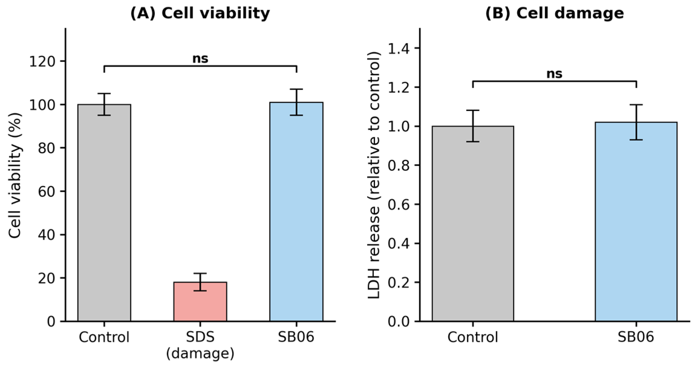
The safety profile of SB06 was evaluated in NHEK cells (Figure 1). The MTT assay demonstrated maintained metabolic activity (100% relative to untreated control), and the LDH assay showed no significant increase in membrane permeability following 24-h treatment with 107 TFU/mL SB06. Both assays confirm the absence of cytotoxic effects at the tested concentration, establishing the safety foundation required for topical formulation development.

3.2. AQP3 Expression

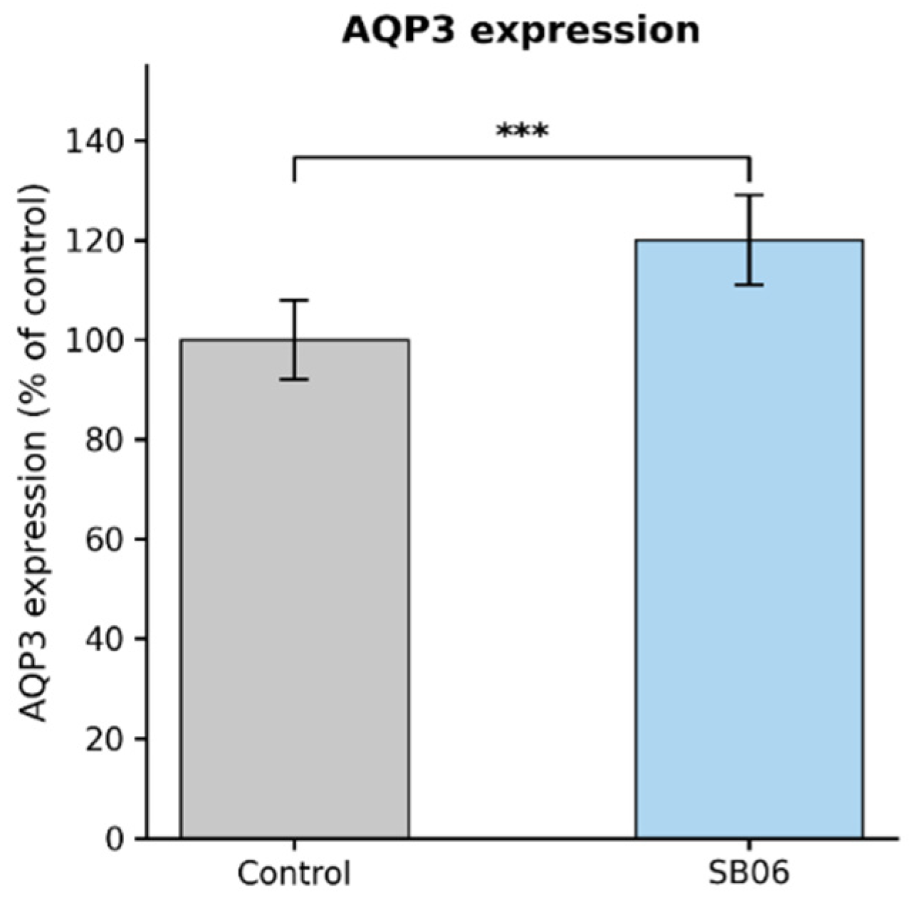
SB06 treatment induced a statistically significant increase in AQP3 protein expression in NHEK cells compared to untreated controls (+20% relative to control, p < 0.001; Figure 2). This upregulation was achieved without associated cytotoxic effects, indicating a specific biological response.

3.3. Antioxidant Activity—ROS Quantification

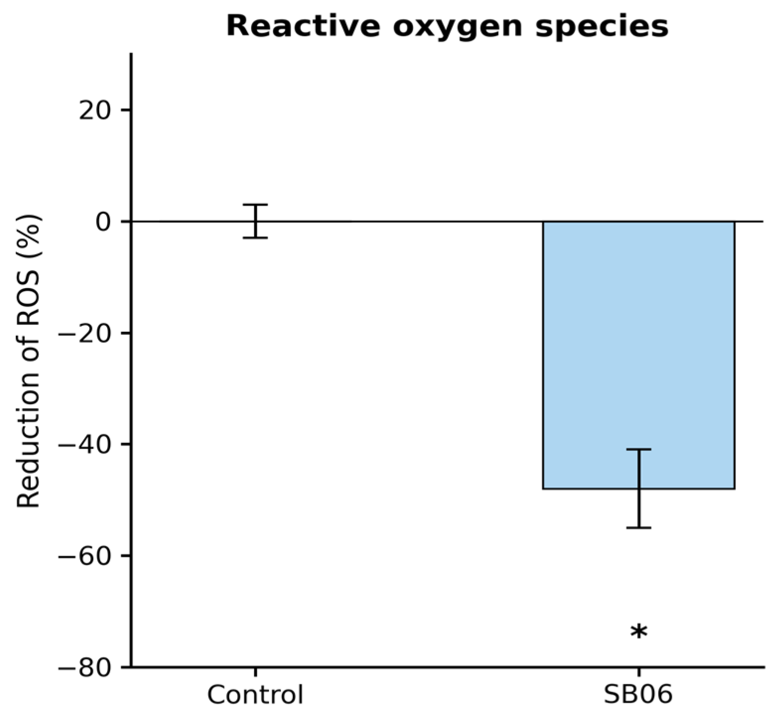
SB06 induced a statistically significant reduction in ROS production in NHEK cells (−48%, p < 0.05; Figure 3). This magnitude of antioxidant activity is relevant for attenuating oxidative-stress-driven skin damage associated with axillary dysbiosis and inflammatory episodes.

3.4. Antipathogen Activity Against Staphylococcus aureus

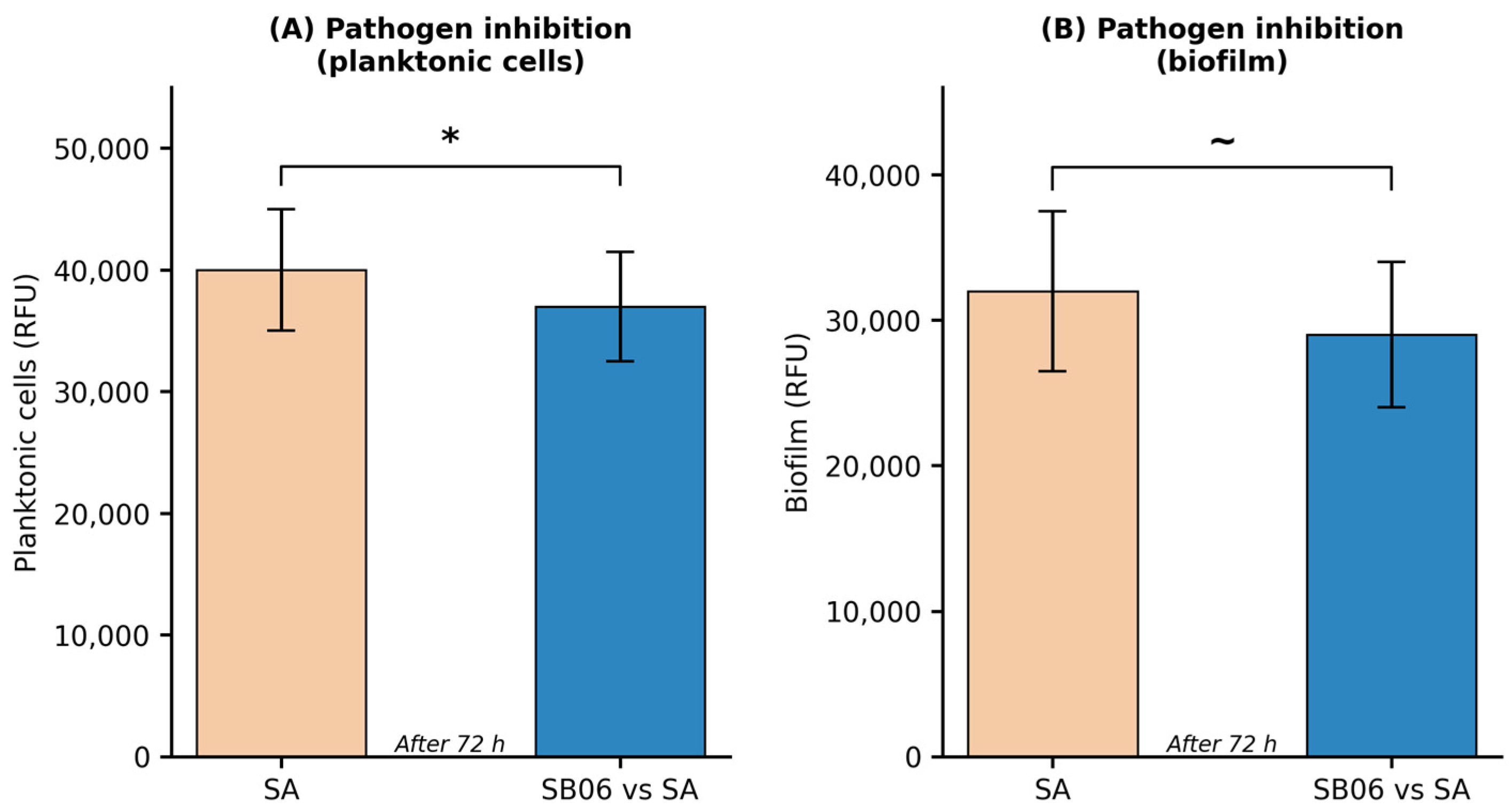
Co-incubation with SB06 for 72 h produced a statistically significant reduction in S. aureus planktonic cell metabolic activity compared to the monoculture control (−7%, p < 0.05; Figure 4). A directional reduction in biofilm metabolic activity was also observed (−8%), which did not reach statistical significance (p ≈ 0.1) and should not be interpreted as evidence of biological activity in the absence of formal significance. These results, while modest in magnitude, suggest a directional inhibitory effect of SB06 against the tested S. aureus model and warrant confirmation in adequately powered, strain-specific studies.

3.5. Axillary Microbiome Compatibility

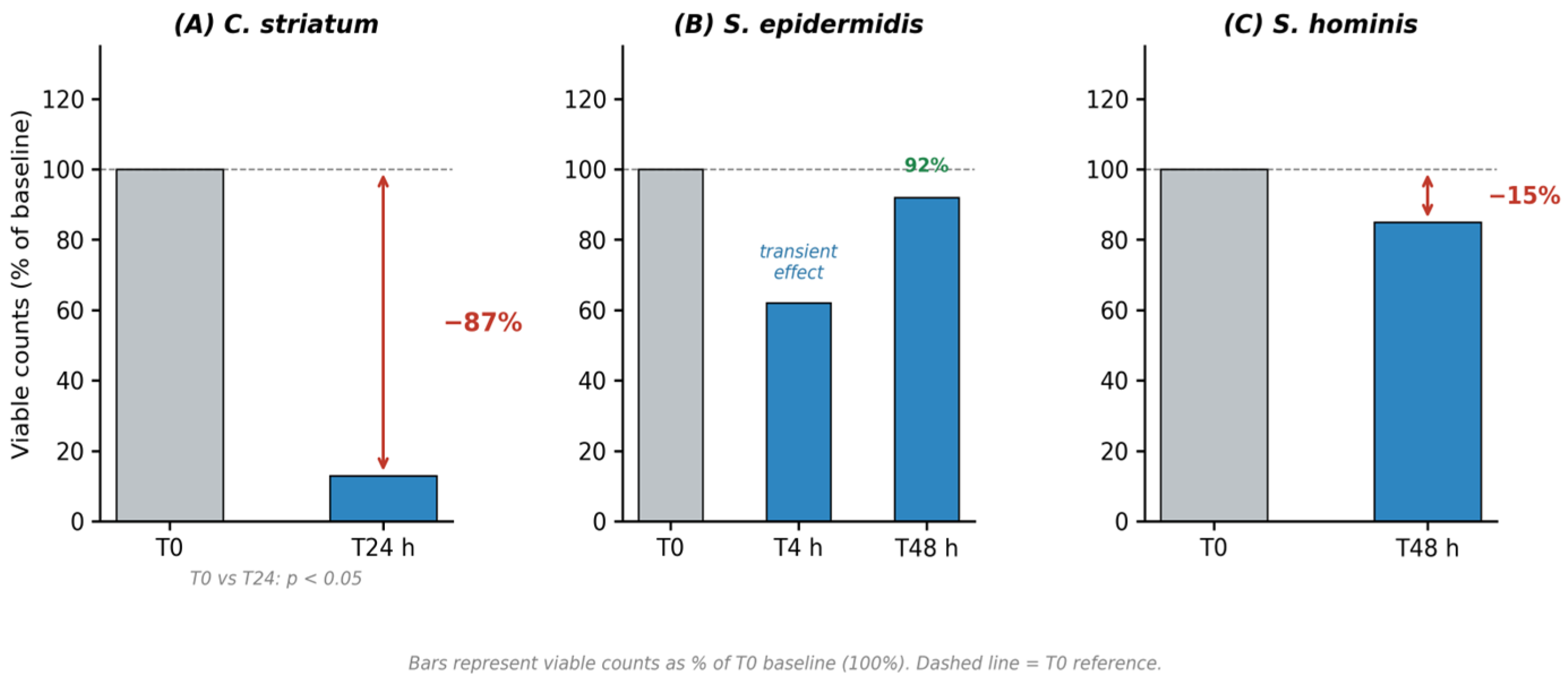
The microbiome compatibility profile of SB06 against selected axillary-resident species is shown in Figure 5. Within this selected panel, SB06 showed a differential effect across species, with the strongest inhibitory activity observed for C. striatum.
C. striatum showed a marked reduction in viable counts at T24h: SB06-treated cultures retained only 13% residual viability relative to the PBS vehicle control at the same timepoint, corresponding to an 87% inhibition of viable counts. This represents the strongest inhibitory effect observed across the tested species and is consistent with mechanistic relevance to the deodorant application context, given the established role of C. striatum as the primary thioalcohol-producing taxon in the axillary microbiome [5,7].
S. epidermidis exhibited a transient reduction at T4h (62% residual viability relative to control; −38%) with near-complete recovery at T48h (92% residual viability relative to control), demonstrating that the effect on this beneficial commensal is transient and does not persist over the full 48-h observation period.
S. hominis showed a consistent reduction in viable counts at T48h (85% residual viability relative to control; −15%), indicating a sustained, albeit moderate, inhibitory effect on this thioalcohol-contributing species over the 48-h observation period. A single type strain per species was tested; broader isolate panels are required before a general axillary-microbiome-compatibility claim can be supported.

3.6. Cytokine Modulation in NHEK and PBMCs

The cytokine modulation profile of SB06 was characterized across two cellular models (Table 2). In NHEK cells, SB06 induced a significant reduction in IL-8 (0.2×, p < 0.01) and IL-23 (0.2×, p < 0.05), while TNF-α was unaffected and IL-6 showed a non-significant increase (1.9×, p ≈ 0.1). In PBMCs, SB06 induced a marked upregulation of TNF-α (13.3×, p < 0.001) and IL-6 (7×, p < 0.001) alongside a significant reduction in IL-8 (0.94×, p < 0.01). IL-23 in PBMCs was not significantly altered.

3.7. Clinical Efficacy

3.7.1. Antiperspirant Effectiveness

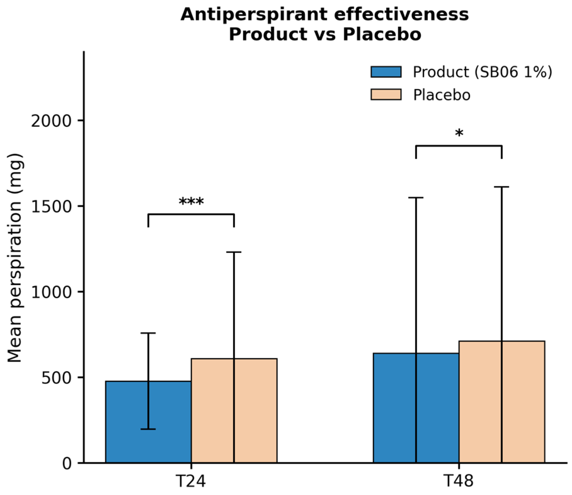
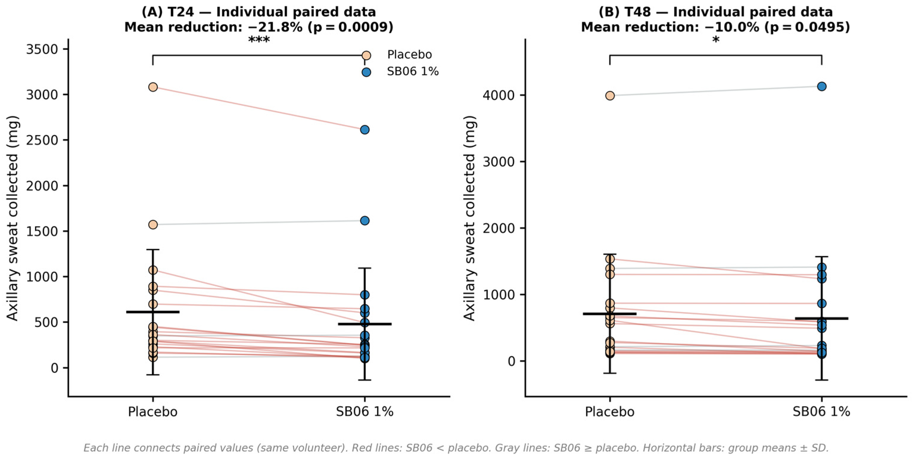
Gravimetric sweat collection data from 20 volunteers are summarized in Table 3 and Figure 6. At T24, the SB06-treated axilla produced a mean of 477 mg of sweat compared to 609 mg in the placebo-treated contralateral axilla, corresponding to a statistically significant reduction of −21.8% (p = 0.0009, ***). At T48, mean sweat production was 639 mg (SB06) versus 710 mg (placebo), representing a significant reduction of −10.0% (p = 0.0495, *). Individual-level data confirmed that the majority of volunteers responded to treatment in the direction of reduced sweat production at both timepoints. Effect size analysis indicated a large effect at T24 (Cohen’s d = 0.85; 95% CI for mean difference: −206 to −60 mg) and a medium effect at T48 (Cohen’s d = 0.56; 95% CI: −130 to −12 mg), supporting the practical magnitude of the observed antiperspirant effect at both timepoints, within the limitations of this exploratory study. Individual-level data are presented in Figure 10. At T24, 17 of 20 volunteers (85%) showed lower sweat production on the SB06-treated side compared to the placebo-treated contralateral axilla; at T48, this was observed in 16 of 20 volunteers (80%), confirming a majority-responder pattern at both timepoints.

3.7.2. Deodorant Efficacy—Olfactory Sensory Analysis
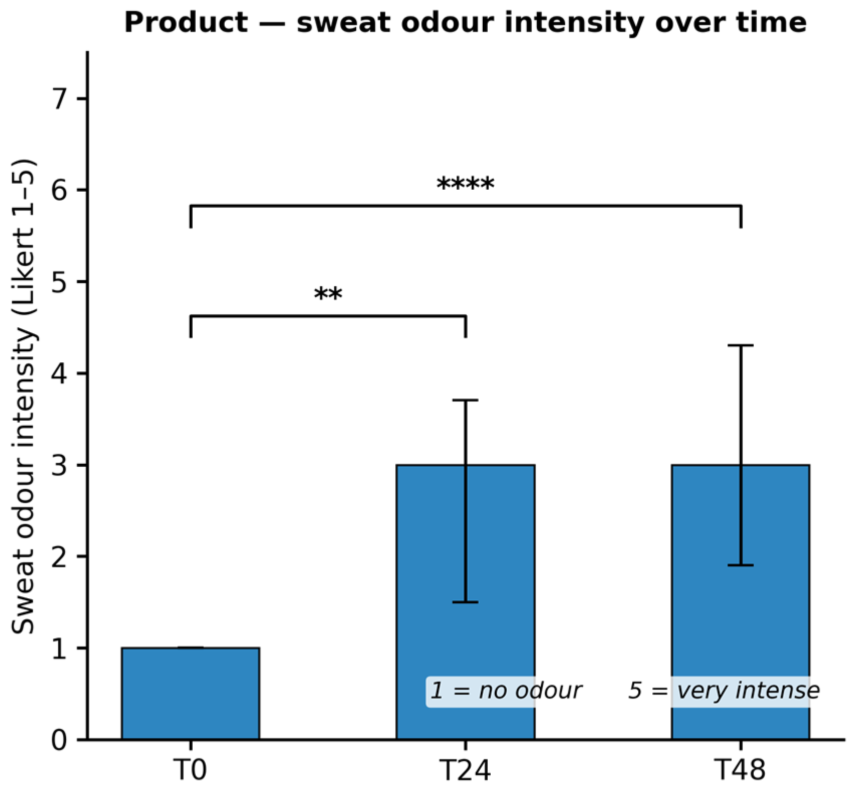
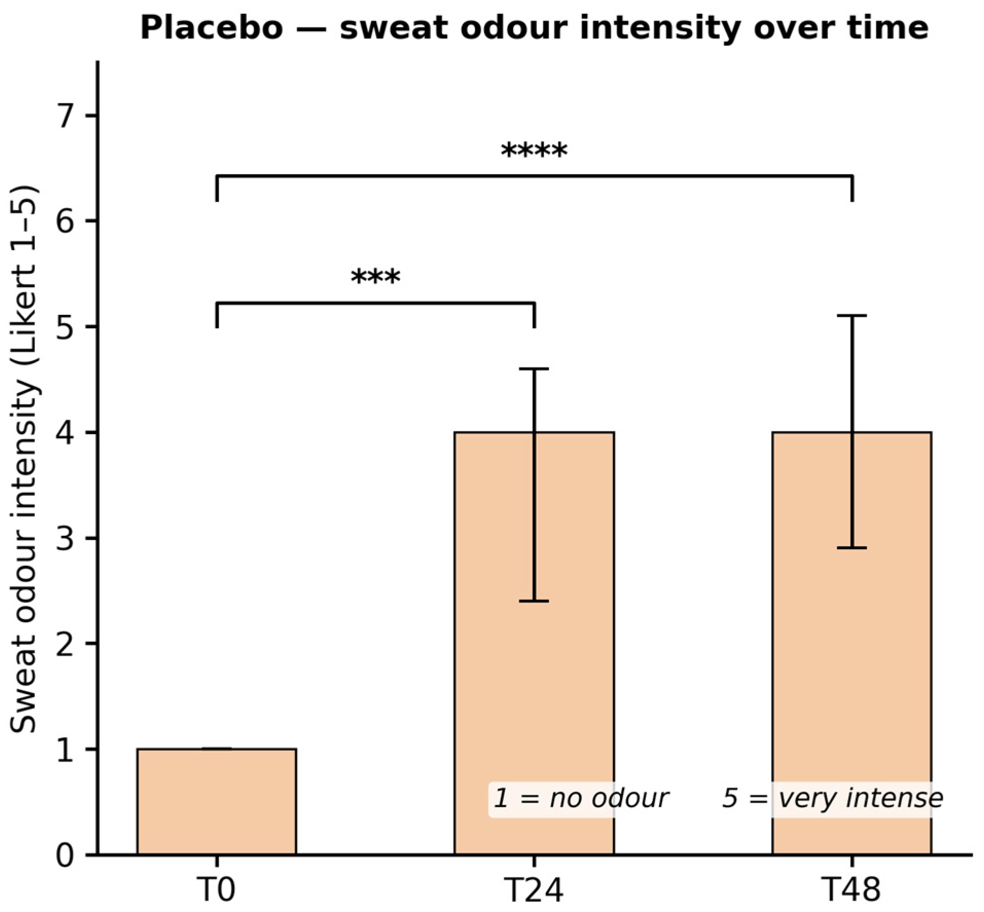
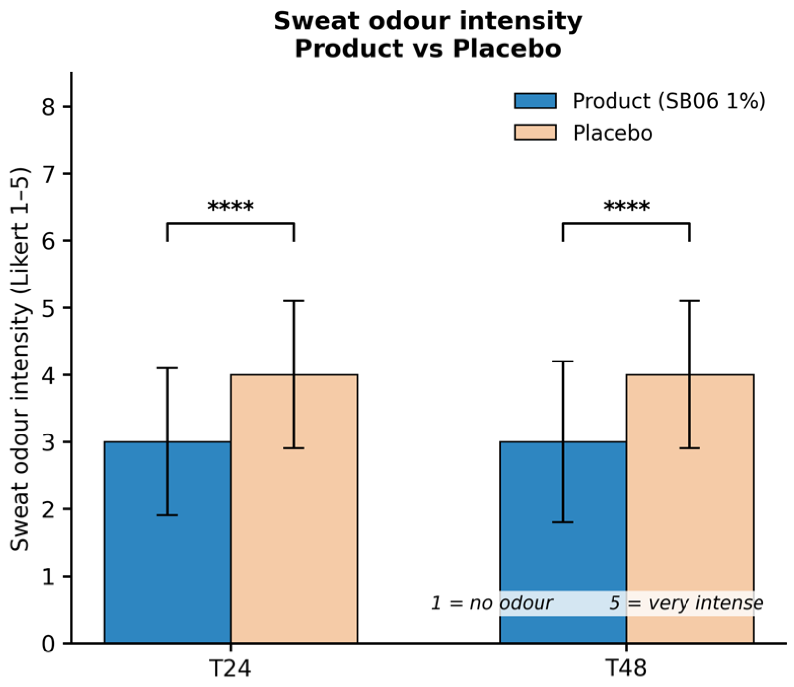
Olfactory sensory evaluation results are summarized in Table 4 and Figure 7, Figure 8 and Figure 9. At T0 (immediately after washing and product application), both axillae were rated at a median score of 1 (no sweat odor), confirming an equivalent and odor-free baseline for both conditions. At T24, the median odor intensity score in the SB06-treated axilla was 3 (moderate odor) compared to 4 (intense odor) on the placebo side. At T48, the same pattern persisted: SB06 3 versus placebo 4. Both T24 and T48 comparisons between product and placebo reached high statistical significance (p < 0.0001, **** for both timepoints).
Within-group analysis (Friedman test) confirmed that odor intensity increased significantly over time for both conditions: for the SB06 product, T0 vs. T24 (p = 0.0024, **) and T0 vs. T48 (p < 0.0001, ****); for the placebo, T0 vs. T24 (p = 0.0002, ***) and T0 vs. T48 (p < 0.0001, ****). Importantly, the rate and magnitude of odor increase were attenuated in the SB06-treated axilla versus the placebo at both measurement points, confirming a genuine deodorant effect of the postbiotic formulation.
Figure 10. Individual paired sweat production data (mg) at T24 (A) and T48 (B). Each line connects placebo and SB06 values from the same volunteer; red lines indicate volunteers in whom sweat production was lower on the SB06-treated side (T24: 17/20; T48: 16/20), gray lines indicate volunteers in whom sweat production on the SB06-treated side was equal to or higher than on the placebo-treated side (T24: 3/20; T48: 4/20). Horizontal bars represent group means ± SD. Statistical significance by paired t-test: *** p ≤ 0.001 (T24); * p ≤ 0.05 (T48).
Cosmetics 13 00178 g010

4. Discussion

This study provides a comprehensive characterization of heat-treated L. rhamnosus SB06 as a postbiotic ingredient for deodorant applications, integrating mechanistic in vitro evidence with controlled clinical data that collectively support its potential value as a functional active ingredient with in vitro microbiome-compatible properties. The combination of a split-body randomized design with matched placebo control and two orthogonal clinical endpoints—gravimetric antiperspirant assessment and expert olfactory evaluation—substantially strengthens the clinical interpretation relative to uncontrolled single-arm studies.
The confirmed safety of SB06 in NHEK cells established through complementary MTT and LDH assays represents an essential prerequisite for topical deodorant formulation. MTT reflects mitochondrial metabolic integrity while LDH quantifies membrane permeability, together excluding primary and secondary cytotoxicity at the tested concentration [27,28].
The statistically significant AQP3 upregulation (+20%, p < 0.001) contributes a skin conditioning dimension to the SB06 profile that is of relevance for axillary applications. AQP3 mediates water and glycerol transport across keratinocyte membranes, contributing to stratum corneum hydration, barrier repair, and cellular proliferation [23,24,29]. Its suppression by inflammatory cytokines and oxidative stress [25] is directly relevant to axillary skin subjected to repeated conventional deodorant application or microbiome dysbiosis.
AQP3 upregulation by SB06 suggests a potential contribution to skin hydration and barrier-supporting processes in the axillary region, independently of any mechanistic link to the observed antiperspirant effect. However, the present study was not designed to directly evaluate skin barrier function, and no direct relationship can be inferred between AQP3 modulation and the antiperspirant effect observed in the clinical study.
The statistically significant ROS reduction achieved by SB06 (−48%, p < 0.05) is notable compared to data on related heat-treated strains: whereas L. plantarum SB01 and B. animalis spp. lactis SB05 showed a non-significant directional ROS reduction (−33%, p = 0.06) at comparable concentrations [30], SB06 achieves formal statistical significance, suggesting a more potent or mechanistically distinct antioxidant activity for this specific strain. Postbiotic-derived antioxidant activity may be mediated by radical scavenging from cell wall components including exopolysaccharides and peptidoglycan fragments, and by indirect activation of endogenous antioxidant enzymes [31,32]. In the axillary context, ROS reduction may attenuate inflammation-driven malodor amplification and limit oxidative degradation of skin lipids serving as volatile compound precursors [33]. The observed activity of SB06 against the tested S. aureus model is mechanistically relevant to the deodorant application context, although confirmation across additional strains would strengthen generalizability. The statistically significant reduction in planktonic cell viability (p ≤ 0.05) and consistent directional biofilm trend (p ≈ 0.1) indicate competitive or antagonistic pressure on this odorigenic species, possibly mediated by organic acid production, competitive adhesion site exclusion, or bacteriocin-like substances released during heat inactivation [16,34]. While a 7% reduction in planktonic viability may appear modest in absolute terms, its biological relevance should be interpreted in the context of the axillary microenvironment rather than against conventional antimicrobial benchmarks. In the axillary ecosystem, S. aureus typically represents a minority taxon whose odorigenic and inflammatory influence is disproportionate to its relative abundance [7,8]. Partial competitive suppression of S. aureus—combined with the concurrent anti-inflammatory activity observed in NHEK cells—may be sufficient to attenuate its contribution to malodor and local irritation without requiring bactericidal eradication. Furthermore, the postbiotic mechanism of action is inherently distinct from that of chemical antimicrobials: rather than direct killing, it likely involves competitive exclusion and microenvironmental modulation, for which even modest reductions in viable counts may translate to meaningful ecological effects [16,34].
The broader microbiome selectivity profile of SB06, including selective activity against Corynebacterium striatum, is discussed in the following paragraph.
The microbiome compatibility data obtained on the selected axillary-associated strains are consistent with a differential activity profile that may support microbiome-compatible deodorant development. The marked reduction in C. striatum viable counts at T24h (13% residual viability relative to the PBS control at the same timepoint, corresponding to an 87% inhibition) is relevant given that C. striatum is recognized as the primary thioalcohol-producing taxon in the axillary microbiome, responsible for the conversion of odorless cysteine conjugates into the volatile sulfanylalkanols characteristic of intense underarm malodor [5,7,33]. S. hominis, which contributes to thioalcohol release through distinct enzymatic pathways [7], also showed a consistent inhibitory trend at T48h (−15% residual viability relative to control). By contrast, S. epidermidis—a beneficial commensal involved in skin barrier homeostasis, antimicrobial peptide production, and competitive exclusion of opportunistic pathogens [9,10,35]—showed only a transient reduction at T4h (62% residual viability) with recovery to 92% of control viability at T48h, confirming that SB06 does not persistently suppress this key protective species. This differential profile—characterized by stronger inhibition of C. striatum together with limited and partly transient effects on selected commensal species—is consistent with a microbiome-compatible mode of action in vitro and may differentiate postbiotic-based formulations from conventional broad-spectrum antimicrobials. It should be noted, however, that no direct microbiological assessment of the axillary microbiome was performed during the clinical study, and whether similar microbial modulation occurs under in vivo conditions remains to be established [12,13].
The cytokine modulation profile of SB06 reveals a context-dependent immunomodulatory pattern coherent for a deodorant ingredient. In NHEK cells, the significant reductions in IL-8 and IL-23 support a local anti-inflammatory effect: IL-8 is a potent neutrophil-attracting chemokine overproduced during axillary inflammatory episodes contributing to erythema and irritation [36], and its suppression suggests improved axillary skin comfort during product use. The concurrent IL-23 downregulation supports attenuation of the Th17-axis, a pathway implicated in chronic skin inflammatory conditions [37]. In PBMCs, upregulation of TNF-α and IL-6 reflects a pattern of innate immune activation, functionally distinct from tissue-pathological inflammation and consistent with the capacity for enhanced antimicrobial surveillance demonstrated by the antipathogen data [38,39]. The simultaneous IL-8 reduction in PBMCs supports a targeted rather than dysregulated immune response [36]. It should be explicitly noted that the pronounced TNF-α and IL-6 induction observed in PBMCs (13.3× and 7×, respectively) may represent a cell-type-specific inflammatory activation pattern in this in vitro model, distinct from the anti-inflammatory profile observed in keratinocytes. However, two considerations limit the clinical relevance of this interpretation for a leave-on topical product. First, systemic exposure of circulating mononuclear cells to a topically applied deodorant spray is negligible under normal use conditions, making the PBMC model a worst-case surrogate rather than a physiologically representative one. Second, the local keratinocyte compartment—the tissue directly exposed during axillary application—showed a clearly anti-inflammatory profile (IL-8↓, IL-23↓), which is the endpoint most directly relevant to the safety and tolerability of the formulation. No adverse events or signs of local inflammation were observed in any of the 20 clinical volunteers, further supporting the absence of pro-inflammatory effects under actual use conditions. Given the exploratory nature of the in vitro analyses and the absence of multiple testing correction, these findings should be interpreted as mechanistic hypothesis-generating observations rather than definitive conclusions.
The clinical results are consistent with the in vitro mechanistic profile and provide controlled pilot support for its translational relevance. The −21.8% reduction in sweat production at T24 (p = 0.0009) and −10.0% at T48 (p = 0.0495) represent statistically significant and methodologically rigorous antiperspirant effects, assessed according to FDA guidelines using pre-weighed absorbent pads under standardized heat stress conditions in a split-body randomized design. The attenuation of the antiperspirant effect from T24 (−21.8%) to T48 (−10.0%) is consistent with the expected duration of action of a postbiotic formulation applied once daily. Unlike aluminum-based antiperspirants, which form physical plugs within eccrine ducts that persist for 24–72 h, postbiotic-based formulations likely act through biological mechanisms—including competitive modulation of axillary bacteria and local immunomodulation—whose effects are inherently transient and dependent on sustained application. The observed waning at T48 therefore reflects the natural dissipation of a single-dose biological effect rather than loss of intrinsic activity and would be expected to stabilize with continued daily use over longer application periods. This interpretation is consistent with the study design, which assessed efficacy after only 4 days of application—a period likely insufficient to establish the microbiome-modulating effects that may underlie longer-term deodorant efficacy. Future studies employing longer application periods and microbiome profiling will be required to characterize the full duration-of-action profile of SB06-based formulations.
The deodorant efficacy results were consistent across both assessment timepoints: the SB06-treated axilla maintained a significantly lower odor intensity score (median 3, “moderate”) compared to the placebo-treated contralateral axilla (median 4, “intense”) at both T24 and T48, with a between-group difference reaching p < 0.0001 at both timepoints. These findings support a deodorant effect of the postbiotic formulation relative to the base vehicle over 48 h following the final application. It is noteworthy that neither the product nor the placebo fully prevented odor development over 48 h—both arms showed significant within-group increases from T0 (Friedman test)—which reflects the physiological inevitability of progressive odor development in high-sweating individuals over this period. However, the SB06 formulation consistently attenuated this progression relative to the vehicle-only control, suggesting a cosmetically relevant effect under the present test conditions.
Taken together, the in vitro and in vivo data suggest that heat-treated L. rhamnosus SB06 possesses biological activities that may be relevant for deodorant applications, including antipathogen activity against the tested S. aureus model, modulation of inflammatory markers in keratinocytes, antioxidant activity, AQP3 upregulation, and a reduction in sweat production and odor intensity under placebo-controlled conditions. While these findings are consistent with a multifactorial mechanism of action, the relative contribution of each pathway to the observed clinical effects remains to be established.
This multimodal profile distinguishes SB06 from conventional deodorant actives and aligns with the growing demand for non-aluminum alternatives with potential microbiome-compatible properties [14].

Study Limitations

Several limitations should be acknowledged. The in vitro models, while well-validated for skin research, do not fully recapitulate the complexity of the axillary microenvironment including multispecies microbial interactions, eccrine and apocrine secretion chemistry, and the physical–chemical milieu of the underarm. Antipathogen activity against S. aureus was assessed using the AlamarBlue metabolic assay, which is methodologically distinct from the OD600/CFU viability approach used in Section 3.5; direct cross-assay comparison is therefore not appropriate. The modest magnitude of S. aureus planktonic inhibition and the non-significant biofilm trend require confirmation in mixed-species biofilm systems representative of the axillary environment. The immunostimulatory PBMC signals (TNF-α, IL-6) were not accompanied by functional antimicrobial killing readouts, and their translational relevance to topical application—where systemic PBMC exposure is negligible—should be interpreted in context.
Regarding the clinical study, a specific limitation of the deodorant efficacy assessment is the absence of evaluator blinding: olfactory assessors were aware of which axilla received the active product, introducing potential assessment bias that cannot be excluded. This represents a meaningful limitation of the olfactory endpoint and should be considered when interpreting the odor intensity scores. Future studies should implement blinded olfactory assessment protocols, for example through coded sample presentation or independent panel evaluation, to minimize this source of bias. The sample size of 20 volunteers, while adequate for exploratory evaluation according to FDA antiperspirant testing guidelines, limits statistical power for detecting small between-subgroup differences and precludes definitive conclusions regarding responder characteristics. The biological basis of the observed antiperspirant effect remains unclear. Although SB06 significantly increased AQP3 expression in keratinocytes, AQP3 is a water channel expressed in epidermal keratinocytes and is not known to directly regulate eccrine sweat gland secretion. The study was not designed to investigate the mechanistic basis of the antiperspirant effect, and no causal link can be established between AQP3 modulation and reduced perspiration. The antiperspirant effect observed clinically may reflect distinct biological mechanisms that remain to be elucidated. Dedicated mechanistic studies will be required to clarify this observation. The clinical study population was not fully characterized in terms of sex distribution and mean age, as these variables were not systematically recorded in the study protocol; future studies should prospectively collect complete demographic data to allow subgroup analyses. Additional baseline variables including BMI, smoking status, physical activity level, and dermatological history were not systematically collected in the study protocol and therefore cannot be assessed as potential confounding factors for the sweating and body odor endpoints. Future studies should prospectively record these variables to enable covariate-adjusted analyses. The inclusion of volunteers requiring a minimum baseline sweat threshold (100 mg/20 min collected from each axilla under controlled conditions at screening) ensures methodological rigor for the antiperspirant endpoint but may reduce generalizability to the broader population of light sweaters. A major limitation of the present work is the absence of direct microbiological assessment of the axillary microbiome during the clinical study. Although the in vitro experiments demonstrated selective activity against representative axillary-associated species, it cannot be assumed that similar microbial shifts occurred in vivo under real-use conditions. Consequently, the proposed microbiome-mediated mechanism underlying the observed deodorant efficacy should be considered biologically plausible but not directly demonstrated. Future studies incorporating longitudinal microbiome profiling (e.g., 16S rRNA sequencing or metagenomic analysis) before and after treatment will be required to establish whether the in vitro selectivity profile translates to measurable microbiome modulation in vivo and whether clinical effects are associated with changes in microbial composition and function. Finally, the study duration of 4 application days, while consistent with standard antiperspirant efficacy testing protocols, does not permit assessment of effects associated with longer-term daily use.

5. Conclusions

This study suggests that heat-treated L. rhamnosus Skinbac™ SB06 possesses a multimodal activity profile that is mechanistically coherent and clinically supported for deodorant applications. In vitro, SB06 showed: (I) confirmed safety in NHEK; (II) significant AQP3 upregulation (+20%, p < 0.001); (III) statistically significant antioxidant activity (−48% ROS, p < 0.05), with a stronger signal than that previously observed for related postbiotic strains under comparable experimental conditions; (IV) antipathogen activity against S. aureus planktonic cells (p < 0.05) with a consistent directional biofilm effect; (V) a dual immunomodulatory profile combining local keratinocyte anti-inflammatory activity (IL-8↓, IL-23↓ in NHEK) with immune activation capacity in PBMCs; and (VI) a preferential inhibitory effect against C. striatum (13% residual viability at T24h, corresponding to 87% inhibition), the principal axillary malodor taxon, together with near-complete preservation of the beneficial commensal S. epidermidis (92% residual viability at T48h). Clinically, a 1% SB06 deodorant spray formulation was associated with statistically significant antiperspirant effectiveness versus a matched placebo in a split-body randomized study: −21.8% sweat reduction at T24 (p = 0.0009) and −10.0% at T48 (p = 0.0495). Deodorant efficacy was confirmed by olfactory sensory analysis, with the SB06-treated axilla showing significantly lower sweat odor intensity than placebo at both T24 and T48 (median score 3 vs. 4, both p < 0.0001).
These findings, obtained across in vitro assays and selected axillary-associated strains together with a controlled clinical study, are consistent with heat-treated L. rhamnosus SB06 being a functional postbiotic ingredient with potential for deodorant and antiperspirant formulations with in vitro microbiome-compatible properties that target malodor-associated microbial activity while also supporting skin conditioning. Larger, double-blind controlled studies incorporating axillary microbiome profiling and extended application periods are warranted to fully characterize the mechanistic and clinical potential of SB06-based deodorant formulations.

Author Contributions

Conceptualization, M.P. and A.A.; methodology, A.V. and G.D.; investigation, G.D. and A.V.; data curation, G.D. and A.V.; writing, original draft preparation, G.D.; writing, review and editing, G.D. and A.A.; supervision, M.P. and A.A.; project administration, M.P. and A.A. All authors have read and agreed to the published version of the manuscript.

Funding

This research was supported by Probiotical S.p.A. (Novara, Italy).

Institutional Review Board Statement

Ethical review and approval were not required for this study pursuant to EU Regulation (EC) No 1223/2009 on cosmetic products and applicable national regulations governing non-invasive cosmetic product testing in adult healthy volunteers. The study was conducted in accordance with the Declaration of Helsinki and Good Clinical Practice (GCP) principles. Data were processed in compliance with EU Regulation 679/2016 (GDPR). The study was conducted by ABICH S.r.l. (Vimodrone, Italy), an accredited cosmetic contract research organisation.

Informed Consent Statement

Informed consent was obtained from all subjects involved in the study.

Data Availability Statement

The original contributions presented in this study are included in the article. Further inquiries can be directed to the corresponding author.

Acknowledgments

The in vivo study was conducted by ABICH S.r.l. (Clinical and Cosmetological Trials Center, Verbania and Vimodrone, Italy), whose contribution to clinical data acquisition is gratefully acknowledged. The authors would like to thank Paolo Saronni for his technical support and valuable contributions throughout the project. During the preparation of this work the authors used Claude (Anthropic, San Francisco, CA, USA) for language editing and manuscript revision assistance. The authors reviewed and edited all AI-assisted content and take full responsibility for the final manuscript.

Conflicts of Interest

G.D., A.V., A.A. and M.P. are employees of Probiotical Research Srl. The authors declare that, despite these financial and commercial relationships, the research was conducted with complete scientific autonomy and independence. The funders had no role in study design; collection, analyses, or interpretation of data; writing; or the decision to publish.

References

  1. Haykal, D.; Cartier, H.; Dréno, B. Dermatological Health in the Light of Skin Microbiome Evolution. J. Cosmet. Dermatol. 2024, 23, 3836–3846. [Google Scholar] [CrossRef] [PubMed]
  2. Callewaert, C.; Ravard Helffer, K.; Lebaron, P. Skin Microbiome and its Interplay with the Environment. Am. J. Clin. Dermatol. 2020, 21, 4–11. [Google Scholar] [CrossRef] [PubMed] [PubMed Central]
  3. Medrano, A.C.; Cantu, A.; Aviles-Rosa, E.O.; Hall, N.J.; Maughan, M.N.; Gadberry, J.D.; Prada-Tiedemann, P.A. Chemical Characterization of Human Body Odor Headspace Components. Separations 2024, 11, 85. [Google Scholar] [CrossRef]
  4. James, A.G.; Austin, C.J.; Cox, D.S.; Taylor, D.; Calvert, R. Microbiological and biochemical origins of human axillary odour. FEMS Microbiol. Ecol. 2013, 83, 527–540. [Google Scholar] [CrossRef] [PubMed]
  5. Troccaz, M.; Gaïa, N.; Beccucci, S.; Schrenzel, J.; Cayeux, I.; Starkenmann, C.; Lazarevic, V. Mapping axillary microbiota responsible for body odours using a culture-independent approach. Microbiome 2015, 3, 3. [Google Scholar] [CrossRef] [PubMed] [PubMed Central]
  6. Urban, J.; Fergus, D.J.; Savage, A.M.; Ehlers, M.; Menninger, H.L.; Dunn, R.R.; Horvath, J.E. The effect of habitual and experimental antiperspirant and deodorant product use on the armpit microbiome. PeerJ 2016, 4, e1605. [Google Scholar] [CrossRef] [PubMed] [PubMed Central]
  7. Bawdon, D.; Cox, D.S.; Ashford, D.; James, A.G.; Thomas, G.H. Identification of axillary Staphylococcus sp. involved in the production of the malodorous thioalcohol 3-methyl-3-sufanylhexan-1-ol. FEMS Microbiol. Lett. 2015, 362, fnv111. [Google Scholar] [CrossRef] [PubMed]
  8. Herman, R.; Kinniment-Williams, B.; Rudden, M.; James, A.G.; Wilkinson, A.J.; Murphy, B.; Thomas, G.H. Identification of a staphylococcal dipeptidase involved in the production of human body odor. J. Biol. Chem. 2024, 300, 107928. [Google Scholar] [CrossRef] [PubMed]
  9. Gallo, R.L.; Hooper, L.V. Epithelial antimicrobial defence of the skin and intestine. Nat. Rev. Immunol. 2012, 12, 503–516. [Google Scholar] [CrossRef] [PubMed] [PubMed Central]
  10. Brandner, J.M. Tight junctions and tight junction proteins in mammalian epidermis. Eur. J. Pharm. Biopharm. 2009, 72, 289–294. [Google Scholar] [CrossRef] [PubMed]
  11. Callewaert, C.; Hutapea, P.; Van de Wiele, T.; Boon, N. Deodorants and antiperspirants affect the axillary bacterial community. Arch. Dermatol. Res. 2014, 306, 701–710. [Google Scholar] [CrossRef] [PubMed]
  12. Fredrich, E.; Barzantny, H.; Brune, I.; Tauch, A. Daily battle against body odor: Towards the activity of the axillary microbiota. Trends Microbiol. 2013, 21, 305–312. [Google Scholar] [CrossRef] [PubMed]
  13. Nakatsuji, T.; Gallo, R.L. The role of the skin microbiome in atopic dermatitis. Ann. Allergy Asthma Immunol. 2019, 122, 263–269, Erratum in Ann. Allergy Asthma Immunol. 2019, 123, 529. [Google Scholar] [CrossRef] [PubMed] [PubMed Central]
  14. Han, J.H.; Kim, H.S. Skin Deep: The Potential of Microbiome Cosmetics. J. Microbiol. 2024, 62, 181–199. [Google Scholar] [CrossRef] [PubMed]
  15. Salminen, S.; Collado, M.C.; Endo, A.; Hill, C.; Lebeer, S.; Quigley, E.M.M.; Sanders, M.E.; Shamir, R.; Swann, J.R.; Szajewska, H.; et al. The International Scientific Association of Probiotics and Prebiotics (ISAPP) consensus statement on the definition and scope of postbiotics. Nat. Rev. Gastroenterol. Hepatol. 2021, 18, 649–667, Erratum in Nat. Rev. Gastroenterol. Hepatol. 2021, 18, 671. Erratum in Nat. Rev. Gastroenterol. Hepatol. 2022, 19, 551. [Google Scholar] [CrossRef] [PubMed] [PubMed Central]
  16. Piqué, N.; Berlanga, M.; Miñana-Galbis, D. Health Benefits of Heat-Killed (Tyndallized) Probiotics: An Overview. Int. J. Mol. Sci. 2019, 20, 2534. [Google Scholar] [CrossRef] [PubMed] [PubMed Central]
  17. Mehta, J.P.; Ayakar, S.; Singhal, R.S. The potential of paraprobiotics and postbiotics to modulate the immune system: A Review. Microbiol. Res. 2023, 275, 127449. [Google Scholar] [CrossRef] [PubMed]
  18. Aguilar-Toalá, J.E.; Garcia-Varela, R.; Garcia, H.S.; Mata-Haro, V.; González-Córdova, A.F.; Vallejo-Cordoba, B.; Hernández-Mendoza, A. Postbiotics: An evolving term within the functional foods field. Trends Food Sci. Technol. 2018, 75, 105–114. [Google Scholar] [CrossRef]
  19. Capurso, L. Thirty Years of Lactobacillus rhamnosus GG: A Review. J. Clin. Gastroenterol. 2019, 53, S1–S41. [Google Scholar] [CrossRef] [PubMed]
  20. Ratanapokasatit, Y.; Laisuan, W.; Rattananukrom, T.; Petchlorlian, A.; Thaipisuttikul, I.; Sompornrattanaphan, M. How Microbiomes Affect Skin Aging: The Updated Evidence and Current Perspectives. Life 2022, 12, 936. [Google Scholar] [CrossRef] [PubMed] [PubMed Central]
  21. Lopes, E.G.; Moreira, D.A.; Gullón, P.; Gullón, B.; Cardelle-Cobas, A.; Tavaria, F.K. Topical application of probiotics in skin: Adhesion, antimicrobial and antibiofilm in vitro assays. J. Appl. Microbiol. 2017, 122, 450–461. [Google Scholar] [CrossRef] [PubMed]
  22. Habeebuddin, M.; Karnati, R.K.; Shiroorkar, P.N.; Nagaraja, S.; Asdaq, S.M.B.; Khalid Anwer, M.; Fattepur, S. Topical Probiotics: More Than a Skin Deep. Pharmaceutics 2022, 14, 557. [Google Scholar] [CrossRef] [PubMed] [PubMed Central]
  23. Bollag, W.B.; Aitkens, L.; White, J.; Hyndman, K.A. Aquaporin-3 in the epidermis: More than skin deep. Am. J. Physiol. Cell Physiol. 2020, 318, C1144–C1153. [Google Scholar] [CrossRef] [PubMed] [PubMed Central]
  24. Qin, H.; Zheng, X.; Zhong, X.; Shetty, A.K.; Elias, P.M.; Bollag, W.B. Aquaporin-3 in keratinocytes and skin: Its role and interaction with phospholipase D2. Arch. Biochem. Biophys. 2011, 508, 138–143. [Google Scholar] [CrossRef] [PubMed] [PubMed Central]
  25. Karimi, N.; Ahmadi, V. Aquaporin Channels in Skin Physiology and Aging Pathophysiology: Investigating Their Role in Skin Function and the Hallmarks of Aging. Biology 2024, 13, 862. [Google Scholar] [CrossRef] [PubMed]
  26. Mondadori, G.; Amoruso, A.; Visciglia, A.; Deusebio, G.; Pinto, D.; Pane, M.; Rinaldi, F. Heat-Treated Probiotics’ Role in Counteraction of Skin UVs-Induced Damage In Vitro. Cosmetics 2025, 12, 121. [Google Scholar] [CrossRef]
  27. Mosmann, T. Rapid colorimetric assay for cellular growth and survival: Application to proliferation and cytotoxicity assays. J. Immunol. Methods 1983, 65, 55–63. [Google Scholar] [CrossRef] [PubMed]
  28. Decker, T.; Lohmann-Matthes, M.L. A quick and simple method for the quantitation of lactate dehydrogenase release in measurements of cellular cytotoxicity and tumor necrosis factor (TNF) activity. J. Immunol. Methods 1988, 115, 61–69. [Google Scholar] [CrossRef] [PubMed]
  29. Schey, K.L.; Wang, Z.; Wenke, J.L.; Qi, Y. Aquaporins in the eye: Expression, function, and roles in ocular disease. Biochim. Biophys. Acta 2014, 1840, 1513–1523. [Google Scholar] [CrossRef] [PubMed] [PubMed Central]
  30. Deusebio, G.; Visciglia, A.; Amoruso, A.; Pane, M. Heat-Treated Strains of Lactiplantibacillus Plantarum Skinbac™ SB01 and Bifidobacterium animalis spp. Lactis Skinbac™ SB05 Visibly Fight Aging Signs Both In Vitro and In Vivo. Cosmetics 2026, 13, 76. [Google Scholar] [CrossRef]
  31. Papaccio, F.; D’Arino, A.; Caputo, S.; Bellei, B. Focus on the Contribution of Oxidative Stress in Skin Aging. Antioxidants 2022, 11, 1121. [Google Scholar] [CrossRef] [PubMed] [PubMed Central]
  32. Pourzand, C.; Albieri-Borges, A.; Raczek, N.N. Shedding a New Light on Skin Aging, Iron- and Redox-Homeostasis and Emerging Natural Antioxidants. Antioxidants 2022, 11, 471. [Google Scholar] [CrossRef] [PubMed] [PubMed Central]
  33. Natsch, A.; Emter, R. The specific biochemistry of human axilla odour formation viewed in an evolutionary context. Philos. Trans. R. Soc. Lond. B Biol. Sci. 2020, 375, 20190269. [Google Scholar] [CrossRef] [PubMed] [PubMed Central]
  34. Żółkiewicz, J.; Marzec, A.; Ruszczyński, M.; Feleszko, W. Postbiotics-A Step Beyond Pre- and Probiotics. Nutrients 2020, 12, 2189. [Google Scholar] [CrossRef] [PubMed] [PubMed Central]
  35. Nakatsuji, T.; Chen, T.H.; Narala, S.; Chun, K.A.; Two, A.M.; Yun, T.; Shafiq, F.; Kotol, P.F.; Bouslimani, A.; Melnik, A.V.; et al. Antimicrobials from human skin commensal bacteria protect against Staphylococcus aureus and are deficient in atopic dermatitis. Sci. Transl. Med. 2017, 9, eaah4680. [Google Scholar] [CrossRef] [PubMed] [PubMed Central]
  36. Xu, J.; Lupu, F.; Esmon, C.T. Inflammation, innate immunity and blood coagulation. Hamostaseologie 2010, 30, 5–6, 8–9. [Google Scholar] [CrossRef] [PubMed]
  37. Lunjani, N.; Hlela, C.; O’Mahony, L. Microbiome and skin biology. Curr. Opin. Allergy Clin. Immunol. 2019, 19, 328–333. [Google Scholar] [CrossRef] [PubMed]
  38. Woo, Y.R.; Kim, H.S. Interaction between the microbiota and the skin barrier in aging skin: A comprehensive review. Front. Physiol. 2024, 15, 1322205. [Google Scholar] [CrossRef] [PubMed] [PubMed Central]
  39. Chilicka, K.; Dzieńdziora-Urbińska, I.; Szyguła, R.; Asanova, B.; Nowicka, D. Microbiome and Probiotics in Acne Vulgaris—A Narrative Review. Life 2022, 12, 422. [Google Scholar] [CrossRef] [PubMed] [PubMed Central]
Figure 1. Safety assessment of SB06 in NHEK cells. (A) Cell viability by MTT assay; (B) Cytotoxicity by LDH release assay after 24-h treatment (107 TFU/mL). Data represent mean ± SD of three independent experiments. ns = not significant vs. untreated control.
Cosmetics 13 00178 g001
Figure 2. AQP3 protein expression in NHEK cells after 24-h treatment with SB06 (107 TFU/mL), measured by ELISA. Data represent mean ± SD of three independent experiments. *** p < 0.001 vs. untreated control.
Cosmetics 13 00178 g002
Figure 3. ROS production in NHEK cells after 24-h treatment with SB06 (107 TFU/mL), quantified by cytochrome C reduction assay. Results expressed as percentage reduction vs. untreated control. Data represent mean ± SD of three independent experiments. * p < 0.05 vs. untreated control.
Cosmetics 13 00178 g003
Figure 4. Activity of SB06 against the tested S. aureus model after 72-h co-incubation. (A) Planktonic cell metabolic activity (RFU). (B) Biofilm metabolic activity (RFU). Data represent mean ± SD of three independent experiments. * p < 0.05 vs. S. aureus monoculture control (statistically significant); ~ p = 0.1 vs. S. aureus monoculture control (not significant, directional trend only).
Cosmetics 13 00178 g004
Figure 5. Viable counts expressed as percentage of T0 baseline (T0 = 100%) are shown for SB06-treated cultures relative to the corresponding PBS control at the same timepoint for (A) C. striatum at T0 and T24h, (B) S. epidermidis at T0, T4h, and T48h, and (C) S. hominis at T0 and T48h. Values represent means from triplicate experiments conducted under standardized conditions. Replicate-level variability data were not reported in the primary laboratory report (ABICH S.r.l., Report REL/1082/2025); error bars are therefore omitted. Annotated percentages indicate residual viability or inhibition relative to the PBS control at the same timepoint.
Cosmetics 13 00178 g005
Figure 6. Mean axillary perspiration (mg) in the SB06-treated and placebo-treated axillae at T24 and T48 after the last daily treatment. Statistical significance: *** p ≤ 0.001; * p ≤ 0.05 (paired t-test, SB06 vs. placebo).
Cosmetics 13 00178 g006
Figure 7. Variation in sweat odor intensity (Likert 1–5) in the SB06-treated axilla over time (T0, T24, T48). Within-group Friedman test: T0 vs. T24 p = 0.0024 (**); T0 vs. T48 p < 0.0001 (****).
Cosmetics 13 00178 g007
Figure 8. Variation in sweat odor intensity in the placebo-treated axilla over time (T0, T24, T48). Within-group Friedman test: T0 vs. T24 p = 0.0002 (***); T0 vs. T48 p < 0.0001 (****).
Cosmetics 13 00178 g008
Figure 9. Direct comparison of sweat odor intensity between SB06 and placebo at T24 and T48. Paired comparison: both p < 0.0001 (****).
Cosmetics 13 00178 g009
Table 1. Demographic and study design characteristics of the clinical study population. Mean age ± SD and sex distribution were not reported in the final clinical report provided by ABICH S.r.l. (Report REL/3452/2025).
CharacteristicValue
Study designRandomized within-subject (split-body), controlled, open-label
Sample size (n)20
SexMale and female
Age range (years)18–65
Inclusion criterion—sweating≥100 mg axillary sweat/20 min under controlled conditions
Washout period≥17 days (no antiperspirant use)
Study duration~33 days per volunteer
Adverse events/dropoutsNone/0
Table 2. Cytokine modulation profile of SB06 in NHEK and PBMC models. Results expressed as fold increase ± SD relative to untreated basal level (=1).
CytokineCell ModelBasal LevelSB06p-Value
TNF-αPBMCs1 ± 0.0513.3 ± 0.02p < 0.001
NHEK1 ± 0.011.1 ± 0.1ns
IL-6PBMCs1 ± 0.187 ± 0.2p < 0.001
NHEK1 ± 0.041.9 ± 0.01p ≈ 0.1
IL-8PBMCs1 ± 0.060.94 ± 0.01p < 0.01
NHEK1 ± 0.010.2 ± 0.1p < 0.01
IL-23PBMCs1 ± 0.021 ± 0.04ns
NHEK1 ± 0.000.2 ± 0.03p < 0.05
ns = not significant.
Table 3. Mean axillary sweat collected (mg) and percentage reduction for SB06 versus placebo at T24 and T48.
TimepointSB06 (mg)Placebo (mg)% Reduction vs. Placebo95% CI (mg)Cohen’s dp-Value
T24477609−21.8%[−206, −60]0.850.0009 (***)
T48639710−10.0%[−130, −12]0.560.0495 (*)
Table 4. Median sweat odor intensity scores (Likert 1–5) for SB06 and placebo at T0, T24, and T48.
TimepointSB06 (Median)Placebo (Median)SB06 vs. Placebo p-Value
T01 (no odor)1 (no odor)—(baseline)
T243 (moderate odor)4 (intense odor)<0.0001 (****)
T483 (moderate odor)4 (intense odor)<0.0001 (****)
**** p < 0.0001.
Disclaimer/Publisher’s Note: The statements, opinions and data contained in all publications are solely those of the individual author(s) and contributor(s) and not of MDPI and/or the editor(s). MDPI and/or the editor(s) disclaim responsibility for any injury to people or property resulting from any ideas, methods, instructions or products referred to in the content.