Article

Rejuvenating Complex of Hyaluronic Acid, Amino Acids and Vitamins Promotes Cutaneous Microcirculation in Human Skin

by
Gabriel Siquier-Dameto
1,2,
Pere Boadas-Vaello
1 and
Enrique Verdú
1,*
1
Research Group of Clinical Anatomy, Embryology and Neuroscience (NEOMA), Department of Medical Sciences, University of Girona, E-17003 Girona, Spain
2
Dameto Clinics International, 07310 Campanet, Spain
*
Author to whom correspondence should be addressed.
Cosmetics 2026, 13(4), 177; https://doi.org/10.3390/cosmetics13040177
Submission received: 1 June 2026 / Revised: 30 June 2026 / Accepted: 8 July 2026 / Published: 10 July 2026
(This article belongs to the Section Cosmetic Dermatology)

Abstract

Background: Cutaneous microcirculation is crucial for skin appearance and rejuvenation, influencing color, luminosity and texture. Aging reduces microcirculation and alters skin color parameters. This study investigated the long-term effects of intradermal CellBooster® Lift (CBL), a mechanically stabilized hyaluronic acid complex enriched with amino acids and vitamins, on skin microcirculation, color, and aesthetics in healthy adults. This study was registered at clinicaltrial.gov (NCT06000839). Methods: 36 women and 4 men aged 35–55 received three intradermal injections of 3 mL CBL across the face at two-week intervals. Cutaneous microcirculation was assessed using a PeriFlux 5000® laser Doppler system at 7 and 84 days post-treatment. Skin color parameters, including lightness (L*), redness (a*), yellow-blue component (b*), individual typology angle (ITA°), individual whitening angle (IWA°), color homogeneity (H76), saturation, and luminance-saturation ratio, were analyzed using Visia® CR 2D imaging. Aesthetic improvement was evaluated by both participants and medical practitioners using the Global Aesthetic Improvement Scale (GAIS). Results: At 84 days post-treatment, CBL significantly enhanced cutaneous microcirculation and induced favorable changes in all measured skin color parameters: L*, ITA°, IWA°, and luminance-saturation ratio increased, while a*, b*, H76, and saturation decreased in women, but no changes were seen in men. No significant differences were observed between female age groups. On the GAIS, approximately 70–73% of participants reported improvement or significant improvement, while medical practitioners rated improvement in up to 93% of cases. Conclusions: Intradermal CBL administration improves cutaneous microcirculation and skin color characteristics, contributing to a more youthful appearance in adults aged 35–55, with consistent effects across age groups and high subjective and clinical aesthetic ratings.

1. Introduction

There are different treatments that have been used to improve cutaneous microcirculation, such as the use of ozone (ozone therapy) [1], negative pressure wound therapy (NPWT) [2], compressive skin treatment with a moisturizer containing ceramide [3], vibrotherapy [4], cutaneous application of cold atmospheric plasma [5], transcutaneous application of gaseous carbon dioxide [6], intradermal injection of L-arginine combined with local skin heating [7], topical administration of capsaicin [8], cutaneous application of platelet-rich plasma [9], cutaneous microneedle application [10], and laser therapy [11].
The vasodilatory capacity of the cutaneous microcirculation decreases with age, while its vasoconstrictive response increases. Likewise, there is a loss in the number and density of cutaneous blood vessels with age. The increase in skin pallor with age is attributable to the reduction in skin microcirculation [12]. The density of the cutaneous capillary network decreases with age, as the cutaneous microcirculation becomes thicker and more twisted in aging skin, with an increase in total vascular length, while the epidermis becomes thinner with aging, which makes the transparency of the skin increase and facilitates the observation of the subpapillary vascular plexus; hence the redness of the skin (a*) increases with age [13,14]. In this context, various parameters of skin color change with age. There is a reduction in skin luminosity (L*) and an increase in skin redness (a*), without changes in the yellow component of the skin (b*), all of which makes aged skin appear darker than younger skin [15]. In addition, the perception of age is influenced, among other parameters, by the uniformity of skin tone, which decreases with age [16].
In addition to various physical and some topical chemical treatments (e.g., cold atmospheric plasma, platelet-rich plasma) to improve skin microcirculation, and thus the skin’s appearance and rejuvenation potential, oral consumption of green tea polyphenols increases dermal blood flow, skin hydration by limiting trans-epidermal water loss, and reduces skin redness (a*) [17]. The oral use of a dietary supplement based on grape seed extract (polyphenols), melon concentrate providing SOD activity, zinc, and vitamin C—in other words, a supplement based on antioxidant compounds—reduces the red-pink color and shine of the skin, but increases skin luminosity, giving it a more radiant, luminous, even, smooth, and firm appearance, while also increasing skin hydration levels [18]. Other natural products including escin, which comes from horse chestnut seeds (Aesculus hippocastanum), promote cutaneous blood flow and the generation of new cutaneous vessels when applied topically [19]; topical application of menthol increases cutaneous blood flow in a dose-dependent manner under thermoneutral conditions; therefore, menthol acts as a vasodilator when applied topically to the skin [20]; consuming capsules made from plant concentrates and vitamin complexes, rich in polyphenols, promotes cutaneous blood flow, skin thickness, and reduces trans-epidermal water loss [21]; and taking the mixture of four polyphenolic botanical extracts (Rosmarinus officinalis, Olea europaea, Lippia citriodora, Sophora japonica) in capsule form helps reduce trans-epidermal water loss, improves skin elasticity, reduces skin pigmentation (ITA°), and gives the skin a more radiant appearance, in subjects exposed to a highly polluted environment [22]. Preclinical studies demonstrated that ginsenoside Rb1 from ginseng (Panax ginseng), astragaloside IV from astragalus (Astragalus membranaceus), asiaticoside from Centella asiatica, and lupeol from Lupinus albus are natural products that promote increased skin microcirculation [23]. On the other hand, the topical application of creams containing N, N-dimethylglycine sodium salt (DMG-Na) [24,25] and antiseptic agents (octenidine dihydrochloride, hexamethylenbiguanide) [26] also promotes cutaneous microcirculation and skin blood flow.
All this scientific evidence suggests the existence of different approaches that allow for the implementation of cutaneous microcirculation, resulting in skin with a more youthful or rejuvenated appearance, improved color, luminosity, and overall appearance. Therefore, it is important to continue implementing cosmetic approaches that promote microcirculation and these skin rejuvenation changes. In this context, the intradermal application of a hyaluronic acid complex supplemented with vitamins and amino acids in healthy adult subjects with signs of aging showed that it improved viscoelasticity, the degree of hydration, skin density, and cutaneous microcirculation, giving a brightening and whitening appearance of the skin color, but without causing changes in skin wrinkles, 42 days post-treatment [27]. In the present study, the results at 84 days post-treatment are shown, obtained in these same healthy adult subjects recruited, when applying this same cosmetic product, but focusing on changes in microcirculation and on changes associated with skin color characteristics. Since this cosmetic compound is indicated for women, an analysis of these parameters was carried out by separating the recruited women into four age groups (35–40 years, 41–45 years, 46–50 years, and 51–55 years) and trying to find out if there are differences dependent on the age of the women. Therefore, the objective of the present study is to assess the long-term effects on cutaneous microcirculation, skin color and appearance in healthy volunteer subjects after intradermal application of the cosmetic product. The study aimed to determine whether these effects translate into measurable aesthetic improvements across age groups and both genders. The principal conclusions are that intradermal CBL administration enhances microcirculation, improves skin color parameters, and produces measurable aesthetic and rejuvenating effects.

2. Materials and Methods

This study was conducted at the Eurofins Dermscan Pharmascan international center (69100 Villeurbanne, Lyon, France), under the direction and supervision of Dr. Gabriel Siquier-Dameto, and its design was carried out by the NEOMA research group (University of Girona, 17003 Girona, Spain). Eurofins Dermscan Pharmascan is an international clinical trial center for cosmetic and pharmaceutical products that offers methods for evaluating the safety and efficacy of dermocosmetic products.
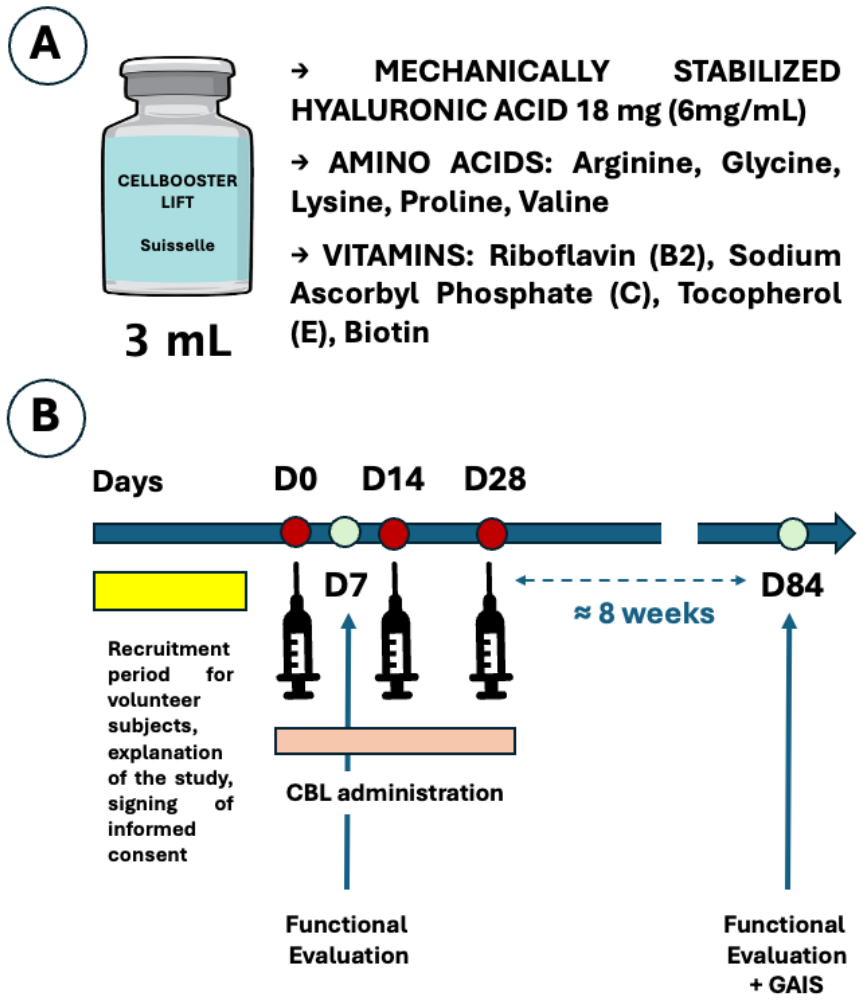
In the present study, 40 healthy adult subjects aged 35 to 55 years were recruited, 36 women and 4 men, following the inclusion and exclusion criteria described above [27]. Every two weeks, subjects received a 3 mL injection of CELLBOOSTER® Lift (CBL; Suisselle, 1400 Yverdon-les-Bains, Switzerland) in the entire face (excluding the forehead) using the micropapule technique. This treatment was repeated three times (D0, D14, D28). The product contains hyaluronic acid mechanically stabilized by CHAC technology (6 mg/mL) supplemented with amino acids (arginine, glycine, lysine, proline, valine) and vitamins (riboflavin, biotin, sodium ascorbyl phosphate, tocopherol) in a phosphate-buffered solution (pH 6.8–7.6) [27]. At 7 and 84 days post-administration of CBL, cutaneous microcirculation was assessed using a PeriFlux 5000® laser Doppler system equipped with thermostatic probes no. 457, which allows simultaneous measurements of blood perfusion and skin temperature (Perimed AB, Stockholm, Sweden) [27,28,29]. Using the Visia® CR 2D camera (Canfield Scientific, Inc.; Parsippany–Troy Hills, NJ 07054, USA), skin photographs were obtained and processed with appropriate software to analyze the following skin color parameters: (i) degree of skin pigmentation from the individual typological angle (ITA°), the individual whitening angle (IWA°), and the H76 parameter or degree of skin color homogeneity; (ii) skin luminosity from a luminescence map; (iii) degree of saturation in the skin color; (iv) and the relationship between lightness and degree of saturation [27,30,31,32] (Figure 1).
The aesthetic improvement after CBL injection was assessed using the Global Aesthetic Improvement Scale (GAIS), which is a subjective rating of the improvement in treatment results at a given time, compared to pretreatment, based on the following score: (1) exceptional improvement, (2) great improvement, (3) improvement, (4) unchanged, and (5) worse [27,33,34].
This research was conducted in accordance with the principles of the Declaration of Helsinki (1964) and complied with ISO 14155:2020 [35] and Regulation (EU) 2017/745 of 5 April 2017. The study protocol was approved by the ethics committee “Comité de protection des personnes Illes-de-France I” (Hôpital Hôtel Dieu, 1, place du Parvis Notre Dame, 75004 Paris, France; approval code: 22.02507.000118; approval date: 12 September 2022). Recruited subjects were informed both verbally and in writing about the clinical research prior to undergoing any study-specific procedures. All participants provided written informed consent prior to inclusion in the study. The informed consent form was provided during the screening visit, and all participant questions were addressed at that time. Furthermore, treatment with this product demonstrated a favorable safety profile, as no severe adverse reactions were observed during the 42-day follow-up period [27].
Data for this study were collected 7 and 84 days after the first CBL injection. Day 84 represents the subjects’ last visit after treatment with CBL, which is approximately 8 weeks after the last CBL injection. Data are shown as mean ± standard error of the mean (SEM). The Shapiro–Wilk test was used to verify the normality of the groups, and the Student’s t test was used to compare the parameters. Finally, when comparing different age groups of women and when the group of men was analyzed, the Kruskal–Wallis and Mann–Whitney U statistical tests were used. A significance level of p < 0.05 was applied. The SPSS 25.0 program for Windows was used in this statistical analysis.
It should be noted that this study was registered on clinicaltrial.gov (NCT06000839; 15 August 2023).

3. Results

3.1. Cutaneous Microcirculation Evaluated with Laser-Doppler Technique

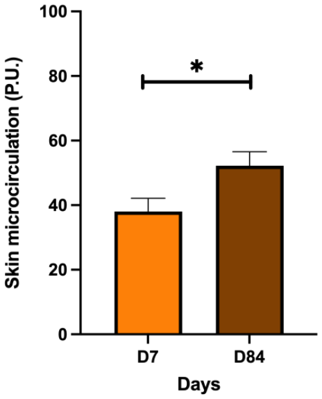
At 84 days (D84) after the start of CBL treatment, healthy women volunteers showed a significant increase in cutaneous microcirculation, assessed by laser-Doppler, compared to day 7 (D7) (p < 0.05) (Figure 2), suggesting that CBL treatment increases blood flow through the skin.
In the small group of men, this parameter at D7 was 76.19 ± 19.86 and at D84 was 77.05 ± 14.92, with no significant differences observed (p > 0.05) between the two days.
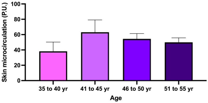
On the other hand, when the women’s group is analyzed for cutaneous microcirculation 84 days after the start of CBL treatment by age groups, a low microcirculation value is observed in the 35–40 age group, which increases in the 41–45 age group, and then gradually decreases in the following older age groups, with no significant differences (p > 0.05) observed between the different age groups (Figure 3).
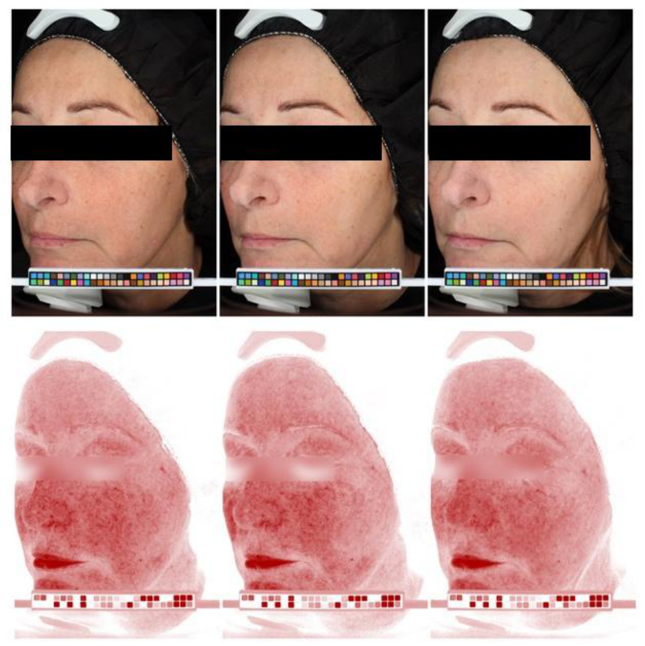
Figure 4 shows photographs of a subject before treatment (D7, left), 42 days (D42, center) and 84 days (D84, right) after treatment. The upper images demonstrate that, following treatment, the skin -which initially exhibited a reddish appearance- became lighter and less red. The images below illustrate hemoglobin distribution in the skin. Following treatment, the distribution appeared more homogeneous, suggesting improved cutaneous microcirculation.

3.2. Skin Color Parameters

Table 1 shows the results obtained from the different parameters evaluated regarding skin color in the baseline situation (D7) and after treatment with CBL (D84) in women.
In women, the CBL treatment produces significant changes in all skin color parameters, specifically producing an increase in lightness (L*), pigmentation level (ITA°), whiteness ratio (IWA°), and Luminance-Saturation ratio of the skin, while this treatment causes a decrease in redness (a*), yellow component (b*), and saturation, but no significant changes in color homogeneity (H76).
The analysis performed on the small group of men recruited for all these skin color parameters on days 7 and 84 is shown in Table 2.
Although there are temporary changes in these parameters in the small group of men, no significant differences were observed between days 7 and 84 for all these skin color parameters.
On the other hand, when analyzing the results of the different skin color parameters at 84 days (D84) of CBL treatment in women by different age groups, no significant differences (p > 0.05; ns) were observed for any of the parameters between the different age groups of women recruited in the study. In other words, in our study, we found no significant differences in skin color parameters related to the age of the women (Table 3).
Although in our study, the application of CBL does not induce significant changes in skin color parameters among the different age groups of women, a trend of age-related changes should be noted. Parameters such as lightness (L*), ITA°, IWA°, and L-S ratio tend to decrease slightly with age, while redness (a*), yellow component ratio (b*), H76, and saturation (S) tend to increase slightly with age.
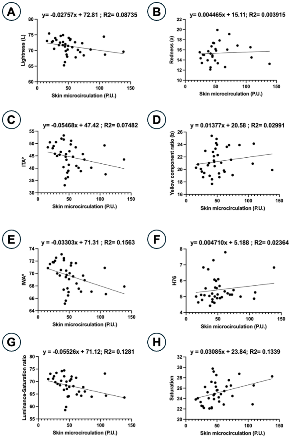
When examining the relationship between skin microcirculation and various skin color parameters at 84 days (D84), it was observed that some color parameters increased with skin microcirculation, while others decreased (Figure 5). Thus, lightness (L), ITA°, IWA°, and the luminance-saturation ratio decreased as skin microcirculation increased (Figure 5A, 5C, 5E, and 5G, respectively), while skin redness (a*), the yellow component of the skin (b*), H76, and saturation tended to increase with increasing skin microcirculation (Figure 5B, 5D, 5F, and 5H, respectively).
In the relationship between skin microcirculation and lightness (L*), the p-value was 0.0869. This p-value was 0.7518 in the relationship between skin microcirculation and redness (a*). For the relationship between skin microcirculation and degree of pigmentation (ITA°), the p-value was 0.1065, while the p-value was 0.3132 in the relationship between skin microcirculation and the yellow component ratio (b*). For the relationship between skin microcirculation and skin whiteness ratio (IWA°), a p-value of 0.0170 was observed, while in the relationship between skin microcirculation and skin color homogeneity (H76), the p-value was 0.3706. In the relationship between cutaneous microcirculation and luminance-saturation ratio, the p-value was 0.0321, and finally, in the relationship between cutaneous microcirculation and the saturation parameter, the p-value was 0.0282.

3.3. Global Aesthetic Improvement Scale (GAIS) Assessed by the Subject and by Medical Practitioner

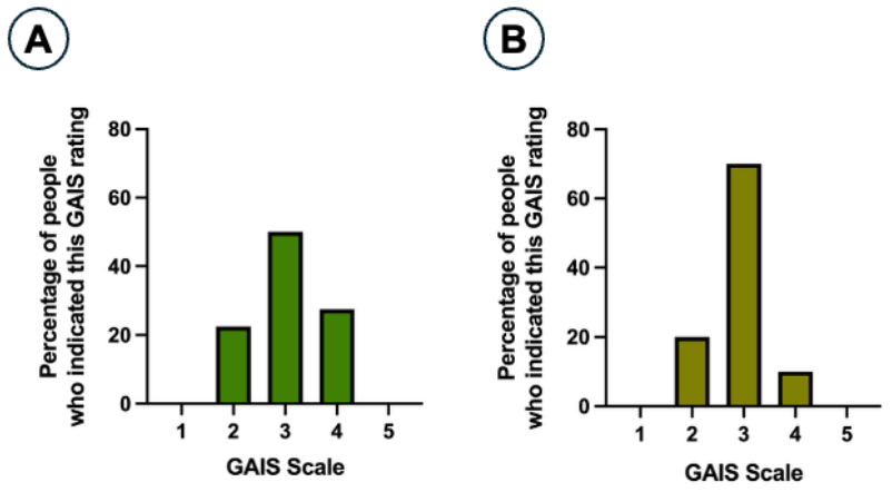
The results of the aesthetic improvement with the CBL treatment evaluated by the GAIS are shown in Figure 6. At 84 days after the start of CBL treatment, 22.5% of treated subjects reported great improvement, 50% reported improvement, and 27.5% reported no change. No subjects reported worse or exceptional improvement (Figure 6A). Regarding the Medical Practitioner assessment, 20% reported great improvement, 70% reported improvement, and 10% reported no change. None of these assessments reported worse or exceptional improvement (Figure 6B). In general, Medical Practitioners rated the aesthetic changes after CBL treatment better than the treated subjects.
At 84 days (D84), in men, the responses on the GAIS were 75% unchanged and 25% greatly improved among subjects treated with CBL, while among Medical Practitioners the response was 75% improved and 25% unchanged. And in relation to the response of women (Subjects) on the GAIS, by age groups, it was as shown in Table 4.
All women aged 35 to 40 yr reported improvement or significant improvement after CBL treatment. 67.67% of women aged 41 to 45 yr reported improvement or significant improvement with the treatment. This pattern of improvement was reported by 81.81% of women aged 46 to 50 yr and 73.13% of women aged 51 to 55 yr (Table 4).
At 84 days (D84), the Medical Practitioners’ GAIS response for women aged 35 to 40 yr was improved in 75% of women and greatly improved in 25% of women in this age group. For women aged 41 to 45 yr, it was improved in 66.67% of women, greatly improved in 16.17% of women, and the same percentage of women showed no change after CBL treatment. The Medical Practitioners’ response for women aged 46 to 50 yr was improved in 63.64% of women in this age group, greatly improved in 27.27% of women, and no change in 9.09% of women. Finally, for women aged 51 to 55 yr, it was improved in 73.33% of women, greatly improved in 20% of women, and no change in 6.67% of women in this age group.
Overall, in older women (51–55 years old), the perception of improvement or much improvement was 73.13% in these women, which for Medical Practitioners increased to 93.33% of women of this age, while in young women (35–40 years old), the perception of improvement or much improvement was 100%, and the same percentage of perception of the Medical Practitioner.

4. Discussion

Intradermal treatment with CellBooster®-Lift (CBL) in healthy Caucasian women volunteers aged 35 to 55 years significantly increased cutaneous microcirculation, accompanied by significant changes in various skin color parameters 84 days after the start of treatment, including an increase in lightness (L*; L* ≈ 71), degree of skin pigmentation (ITA°; ITA° ≈ 44), individual whitening angle (IWA°; IWA° ≈ 69), and luminescence-saturation ratio (≈67). The treatment also caused a significant decrease in skin redness (a*; a* ≈ 16), the yellow component of the skin (b*; b* ≈ 21), skin color homogeneity (H76; H76 ≈ 5.5), and color saturation of the skin (≈25.5). In the small group of men in whom this product was tested, no significant changes in these parameters were observed between days 7 and 84. Therefore, in this strictly preliminary study with men, the changes detected in the larger study with women were not observed.
On the Global Aesthetic Improvement Assessment Scale (GAIS), participants indicated that the treatment received resulted in an improvement or a significant improvement in 72.5% of cases, while only 27.5% of subjects indicated that they perceived no change with the treatment. It should be noted that no participant indicated that the treatment resulted in a worsening of their skin.
The analysis performed on the majority group of women treated with CBL reveals that with age there is a tendency for skin microcirculation to increase between 35 and 40 years and 41–45 years slightly, then decrease slightly until 51–55 years, although no significant differences were observed between the different age groups for this parameter. Regarding skin color parameters, no significant differences were found between the different age groups of the female volunteers. On the Global Aesthetic Improvement Scale (GAIS), 100% of women aged 35–40 reported that CBL treatment had improved or greatly improved their skin’s appearance. In women aged 41–45, this percentage was approximately 77%, with 33% reporting no change. In women aged 46–50, the percentage of improvement or great improvement was approximately 82%, and 18% indicated no change. Finally, in women aged 51–55, the percentage of improvement or great improvement was 73%, with 27% reporting no change. None of the women surveyed reported that CBL treatment had worsened their skin appearance.
Overall, the results of this study conducted on healthy women volunteers suggest that at 84 days (D84), CBL treatment increases cutaneous microcirculation, accompanied by significant changes in skin color, and about 70% of participants reported an improvement or significant improvement in their appearance, while only 30% indicated no change. None of the participants reported any worsening of their skin.
The CBL cosmetic product contains mostly hyaluronic acid (HA), and there is scientific evidence that HA promotes the healing of skin wounds by increasing the density of skin vascularization thanks to its angiogenic role [36,37], which is mediated by its binding to CD44 receptors of endothelial cells [38,39]. The binding of hyaluronic acid to the CD44 receptor of the endothelial cell promotes its proliferation [40]. This proliferation requires a sufficient supply of nutrients, such as essential amino acids, some of which are already provided by the CBL cosmetic product and are absorbed by the endothelial cells [41]. Furthermore, vitamin C, or ascorbic acid, participates in the physiology of endothelial cells, including stimulating endothelial proliferation and maintaining endothelial nitric oxide secretion, which helps modulate blood flow [42]. However, vitamin E is an anti-angiogenic factor [43], but it is a vitamin that reduces skin redness by reducing the activation of mast cells and the release of the contents of their granules [44]. Both vitamins are also part of the composition of the cosmetic compound CBL. Topical application of vitamin C prevents photoaging and promotes collagen synthesis by dermal fibroblasts [45]. Topical application of vitamins C and E has anti-aging and skin-brightening effects [46]. Vitamin C inhibits melanogenesis, reducing skin hyperpigmentation, and is therefore considered a skin-lightening treatment [47]. Vitamin E and vitamin E derivatives also reduce melanogenesis and therefore may be effective ingredients in cosmetics for skin whitening or improving skin pigmentation [48].
In the present study, the effect of changes in cutaneous microcirculation on various skin color parameters was analyzed in a Caucasian women population, aged between 35 and 55 years. Under these conditions, the results show that with increased cutaneous microcirculation (P.U.), lightness (L*), ITA°, IWA°, and luminance-saturation ratio decrease, while redness (a*), the yellow component (b*), H76, and skin color saturation increase (see Figure 5). An increase in cutaneous microcirculation associated with increased skin redness can be explained by greater blood flow to the skin and the generation of erythema or redness. The cutaneous application of vasodilating substances promotes an increase in cutaneous blood flow and skin reddening [20,49,50], but angiogenesis that occurs during skin wound repair also increases blood flow to the skin, causing it to appear redder [51]. The L* parameter for skin color ranges from 0 (darkest color) to 100 (lightest color). Increased cutaneous microcirculation is associated with a decrease in this parameter, meaning it approaches zero, which implies a slight darkening of the skin color [52]. There is evidence to suggest that variations in the value of the L* parameter are related to changes in the amount of hemoglobin in the skin, and in general, when hemoglobin levels in the skin increase, the value of the L* parameter decreases [53]; therefore, greater cutaneous microcirculation, which provides more blood flow and more hemoglobin in the skin, leads to a decrease in parameter L*. Since the parameter L* influences the ITA° value [54,55], a decrease in L also causes a decrease in ITA°, which is what is observed in the present study, and this is associated with an increase in cutaneous microcirculation. The yellow component (b*) of skin color also influences the ITA° value [54,55], and since it is in the denominator of the ITA° calculation formula, an increase in the b parameter also leads to a decrease in ITA°. The yellow component of the skin, or rather the yellow-blue component of the skin (parameter b*), refers to the color axis between yellow and blue, with yellow being warmer (mediated by melanin, carotenoids, or bilirubin) and blue being cooler (due to deoxygenated hemoglobin). A shift in value towards positive or increasing values is a shift towards yellow, while a shift towards negative values or a decrease in value is a shift towards blue [56]. With increased cutaneous microcirculation, a very slight increase in the b* value is observed, which means a slight shift towards the yellow color, which is interpreted as slightly more tanned skin [57], but also slightly more aged skin [58]. Scientific evidence suggests that activated melanocytes not only increase melanin production, a factor that enhances skin tanning, but also secrete factors that induce cutaneous angiogenesis [59]. Therefore, increased cutaneous microcirculation can be accompanied by increased skin tanning, resulting in a more yellow skin tone, and consequently, an increase in the b* parameter value. On the other hand, the individual whitening angle (IWA°) decreases with increased cutaneous microcirculation. It is generally expected that with increased cutaneous microcirculation, the skin will appear pinker and less whitish [60]. Since the IWA° is the skin whitening angle, it is reasonable to expect this parameter to decrease with increased cutaneous microcirculation. Regarding homogeneity, or the homogeneous tone of skin color (parameter H76), it increases with increased cutaneous microcirculation. Normally, an increase in cutaneous microcirculation leads to greater homogeneity of skin color, meaning that the skin tone is uniform. Increased cutaneous microcirculation implies a more uniform distribution of hemoglobin throughout the vascular network of the skin, which at the level of skin color translates into an increase in the homogeneity of skin tone [60]. Color saturation is the intensity and/or purity of a color. A color with high saturation is intense and vibrant, while a color with low saturation is dull or grayish [61]. In the CIELAB system, color saturation is represented by Chroma (C*), which depends on the chromatic dimensions of the red/green (a*) and yellow/blue (b*) axes. A high Chroma implies high saturation, resulting in a bright, vivid, and intense color, while a low Chroma is a color with low saturation, a pale color close to gray [56]. Since the chromatic dimensions of the red/green (a*) and yellow/blue (b*) axes increase with cutaneous microcirculation, the saturation of skin color (Chroma) also increases with cutaneous microcirculation. Skin tanning includes the tone of the skin color and/or the degree of saturation of the skin color [62]. And as already mentioned, a deeper tan implies greater angiogenesis mediated by activated melanocytes, and a more yellow skin tone, resulting in higher Chroma (saturation). Similarly, increased skin reddening due to cutaneous microcirculation also contributes to increased Chroma on the red/green axis. Finally, if the saturation of skin color increases with microcirculation, the luminance/saturation ratio will decrease, which is what is observed in the present study. It should be noted that all these changes in skin color parameters in the study subjects in relation to cutaneous microcirculation were observed 84 days after treatment with CBL.
Previous studies have described changes in cutaneous microcirculation with aging, such as a decrease in blood vessel density, disorganization of the vascular network, and increased blood vessel stiffness [12,13,15]. These age-related changes in cutaneous microcirculation are accompanied by age-related changes in skin pigmentation, including decreased luminescence (L*) and increased skin redness (a*) in elderly subjects [15]. In the present study, where the recruited female population was divided into four age ranges, the L* parameter decreased slightly between women aged 35–40 and 41–45 (from approximately 74 to approximately 71), before remaining constant at 71 in the 46–50 and 51–55 age groups. Despite this trend, no significant differences were observed between women in the different age groups. Regarding the a* parameter, a slight increase in value was observed, from 14 to 16 between the 35–40 and 41–45 age groups, then remaining at 16 in the 46–50 and 51–55 age groups, with no age-related differences observed. In the young Caucasian population (between 18 and 30 years old), the value of the parameter L* ranges between 55 and 65, and the value of the parameter a* between 10 and 15. With age, the value of L* decreases and the value of a* increases [63]. In the present study, 84 days after CBL administration, the L* and a* values were observed in the entire recruited population to be approximately 71 and 16, respectively. When analyzed in the female population of different age groups, these values ranged from 71 to 74 and 14–16, respectively. Cutaneous treatment with CBL yielded L* and a* values that were slightly higher or very similar to those observed in young subjects (18–30 years of age), suggesting that CBL treatment may have a skin rejuvenation effect, that is, it could give a more youthful appearance. Previous studies have also described skin rejuvenation with cutaneous injection of hyaluronic acid, where changes in pigmentation and/or skin color are described [64,65,66,67,68]. This skin-rejuvenating effect has led most people in the study (≈73%) to indicate on the GAIS that CBL treatment has resulted in an improvement or a great improvement in the appearance of their skin, and only 27% indicated that the treatment produced no change. Previous studies have also reported improvements on the GAIS after cutaneous treatment with hyaluronic acid, with a 30% to 100% improvement on the GAIS between 160 and 180 days [69,70,71].
It should be noted that other age-related skin changes include thinning of the epidermis, loss of elasticity, dryness, wrinkles, age spots, and a reduced layer of subcutaneous fat [72,73,74,75]. Furthermore, the main changes in skin color with age include the appearance of age spots or sunspots (hyperpigmentation due to accumulated UV damage), uneven pigmentation, and areas of paleness or discoloration due to a decrease in melanocytes [76,77,78,79]. Cosmetic treatments have attempted to improve these age-related skin changes, trying to give a rejuvenated appearance. Specifically, hyaluronic acid-based treatments have been shown to produce more hydrated and radiant skin [80], and they improve skin elasticity, firmness, texture, and hydration, providing a healthier appearance and reducing the number and depth of wrinkles [81,82,83]. Treatment with hyaluronic acid improves skin lightness (L*), reduces skin redness (a*) and increases skin yellowness (b*), over time up to 70 days (D70), which improves skin’s radiance and tone evenness [84]. The hyaluronic acid treatment, supplemented with amino acids and vitamins, promotes skin hydration, increased skin density with greater viscoelasticity, and a brighter, more even complexion, resulting in a more youthful appearance [27]. The novelty of this study lies in the fact that CBL treatment promotes long-term cutaneous microcirculation (D84), which influences skin color parameters such as IWA° and the luminance-saturation ratio, thus contributing to a more youthful appearance. Furthermore, while several studies analyze skin color parameters following hyaluronic acid treatments [67,68,80,83,84], very few examine cutaneous microcirculation with these cosmetic treatments [27], and the influence of microcirculation on skin color after hyaluronic acid treatment, as analyzed in the present study.
One of the limitations of the present study is that intradermal injection can generate a mechanical skin healing effect that in turn stimulates angiogenesis and microcirculation; that is, the needles have an effect of micro-lesions on the skin that promote angiogenesis/microcirculation, and this may mask the effects of the injected product. Injecting a placebo product in parallel with CBL treatment would have allowed the mechanical effect of the intradermal injection by the needle to be unmasked. The microneedling technique on the skin is known to promote the release of growth factors and angiogenesis [85], and specifically in mice, it has been shown that microneedle application induces an increase in TNF-alpha and VEGF synthesis [86], two potent chemical mediators that induce angiogenesis in the skin [87,88]. In a study conducted on rats using the IBAM technique, which involves striking a cylindrical punch placed on the animal’s abdominal skin with a hammer to create a mechanical skin injury, a skin hardening process was observed because of the skin healing process. When the skin subjected to IBAM mechanical injury was treated with an intradermal injection of hyaluronic acid, a decrease in the stiffness of the superficial tissues was observed in response to the hyaluronic acid injection during the first few weeks. These results suggest that hyaluronic acid modulates the stiffness of soft tissues such as skin, affecting the skin healing process caused by mechanical injury [89].
Another limitation of the study is the very small number (n = 4) of adult men recruited and treated with CBL. Therefore, the results from this group of men should be considered preliminary, with the intention of repeating the study with a larger number of adult men. It should be noted that men represent a small proportion of the total patient population in aesthetic medicine. Thus, in 2018, only 10% of minimally invasive, non-surgical facial treatments with hyaluronic acid or botulinum toxin were performed on men, and this percentage has increased to almost 20% by 2024 [90]. Despite these statistical results, it has been proven that subcutaneous injection with hyaluronic acid in men aged 23 to 60 years increases the masculinization of their faces, which these patients rate very favorably on the GAIS [91]. The use of hyaluronic acid-based fillers has proven effective in men for the subtle correction of signs of aging, such as wrinkle reduction, volumizing, and facial contouring [92].
Overall, the findings demonstrate that intradermal administration of CellBooster®-Lift (CBL) enhances cutaneous microcirculation, modulates skin color parameters, and produces measurable aesthetic improvements in adult women aged 35–55 years. These results align with previous studies reporting skin rejuvenation effects of hyaluronic acid and associated compounds, likely mediated through stimulation of endothelial cell proliferation, enhanced angiogenesis, and modulation of melanogenesis. These observations support the working hypothesis that improving microcirculation can positively influence skin color and perceived appearance. Future studies could explore longer-term outcomes, optimal dosing regimens and efficacy in more diverse populations to confirm and extend these findings, as well as further elucidate the underlying biological mechanisms.

Author Contributions

All authors listed above have contributed sufficiently to be included as authors. Conceptualization, methodology, and validation, G.S.-D. and E.V.; formal analysis and investigation, G.S.-D., P.B.-V. and E.V.; resources, G.S.-D. and E.V.; data curation, G.S.-D., P.B.-V. and E.V.; writing—original draft preparation, E.V.; writing—review and editing, G.S.-D., P.B.-V. and E.V.; visualization and supervision, G.S.-D., P.B.-V. and E.V.; project administration, G.S.-D. and E.V.; funding acquisition, G.S.-D., P.B.-V. and E.V. All authors have read and agreed to the published version of the manuscript.

Funding

This research was funded by Suisselle SA, Rue Galilée 6, 1400 Yverdon-les-Bains, Switzerland (grant number 2022-A01427-36 and grant number 045/22).

Institutional Review Board Statement

The study was conducted in accordance with the Declaration of Helsinki and according to EN ISO standard 14155:2020 and EU Regulation 2017/745 of 5 April 2017. The research documents of this study (CIP #21E4451) were reviewed and approved by an independent ethics committee to which Eurofins Dermscan Pharmascan assigns all of its projects, on 9 December 2022. The ethics committee that evaluated the project was “Comité de protection des personnes Ile-de-France” (address: Hôpital Hôtel Dieu—1, place du Parvis Notre dame 75004 PARIS France). In addition, Eurofins Dermscan Pharmascan processed personal data of the subjects on behalf of Suisselle SA in accordance with the rules on the protection of personal data and, in particular, Regulation (EU) 2016/679 of the European Parliament and of the Council of 27 April 2016 on the protection of natural persons with regard to the processing of personal data and on the free circulation of said data.

Informed Consent Statement

Informed consent was obtained from all subjects involved in the study. Written informed consent has been obtained from the patients to publish this paper.

Data Availability Statement

All data generated or analyzed during this study are included in this published article.

Acknowledgments

The authors would like to thank the technical staff of Eurofins Dermscan Pharmascan for their contribution to the technical execution of this study, which was based on a research project submitted by the authors and subsequently accepted by Suisselle. The authors also thank nurses Audrey Cruz Menarini and Yassine Kbaier, as well as physician Pressian Paraskevov, for their assistance to Gabriel Siquier-Dameto during the treatment administration phases. Finally, the authors would like to express their sincere gratitude to all study participants. Without their participation, this study would not have been possible.

Conflicts of Interest

The authors declare no conflicts of interest.

Abbreviations

The following abbreviations are used in this manuscript:
a*Redness of the skin
b*Yellow-blue component of the skin
C*Chroma
CBLCellBooster-Lift
CD44Cell-surface glycoprotein 44
CIELABInternational Commission on Illumination
CHACCohesive Polydensified Matrix Technology
DMG-NaN,N-dimethylglycine sodium salt
D7Day 7 post-treatment
D70Day 70 post-treatment
D84Day 84 post-treatment
GAISGlobal Aesthetic Improvement Scale
HAHyaluronic acid
H76Skin color homogeneity
IBAMImpact-Based Analysis Method
ITA°Individual Typology Angle
IWA°Individual whitening angle
L*Skin luminosity, lightness, and/or luminescence
NPWTNegative pressure wound therapy
TNF-alphaTumor necrosis factor alpha
SEMStandard error of the mean
SODSuperoxide dismutase
SPSSStatistical Package for the Social Sciences
UVUltraviolet
VEGFVascular Endothelial Growth Factor

References

  1. Liu, L.; Zeng, L.; Gao, L.; Zeng, J.; Lu, J. Ozone therapy for skin diseases: Cellular and molecular mechanisms. Int. Wound J. 2023, 20, 2376–2385. [Google Scholar] [PubMed]
  2. Sogorski, A.; Becker, A.; Dadras, M.; Wallner, C.; Wagner, J.M.; Glinski, M.V.; Lehnhardt, M.; Behr, B. Superior Enhancement of Cutaneous Microcirculation Due to “Cyclic” Application of a Negative Pressure Wound Therapy Device in Humans—Local and Remote Effects. Front. Surg. 2022, 9, 822122. [Google Scholar] [CrossRef]
  3. Kirsner, R.S.; Andriessen, A.; Hanft, J.R.; Hu, S.; Marston, W.A.; Ruotsi, L.C.; Yosipovitch, G. Improvement of Chronic Venous Insufficiency Related Leg Xerosis and Dermatitis with Ceramide-Containing Cleansers and Moisturizers: An Expert-Based Consensus. J. Drugs Dermatol. 2024, 23, 61–66. [Google Scholar] [CrossRef] [PubMed]
  4. Piotrowska, A.; Czerwińska-Ledwig, O.; Stefańska, M.; Pałka, T.; Maciejczyk, M.; Bujas, P.; Bawelski, M.; Ridan, T.; Żychowska, M.; Sadowska-Krępa, E.; et al. Changes in Skin Microcirculation Resulting from Vibration Therapy in Women with Cellulite. Int. J. Environ. Res. Public Health 2022, 19, 3385. [Google Scholar] [CrossRef] [PubMed]
  5. Kisch, T.; Helmke, A.; Schleusser, S.; Song, J.; Liodaki, E.; Stang, F.H.; Mailaender, P.; Kraemer, R. Improvement of cutaneous microcirculation by cold atmospheric plasma (CAP): Results of a controlled, prospective cohort study. Microvasc. Res. 2016, 104, 55–62. [Google Scholar] [CrossRef]
  6. Finzgar, M.; Melik, Z.; Cankar, K. Effect of transcutaneous application of gaseous carbon dioxide on cutaneous microcirculation. Clin. Hemorheol. Microcirc. 2015, 60, 423–435. [Google Scholar] [CrossRef]
  7. Kim, K.; Hurr, C.; Patik, J.C.; Matthew Brothers, R. Attenuated cutaneous microvascular function in healthy young African Americans: Role of intradermal l-arginine supplementation. Microvasc. Res. 2018, 118, 1–6. [Google Scholar] [CrossRef]
  8. Schäfer, R.C.; Sohn, A.; Kersten, A.; Amr, A.; Held, M.; Wenger, A. Quantification of Dermal Microcirculatory Changes after Topical Administration of Capsaicin: A Randomized Placebo-Controlled Study in 46 Subjects. J. Investig. Surg. 2022, 35, 1673–1678. [Google Scholar] [CrossRef]
  9. Lippi, L.; Ferrillo, M.; Losco, L.; Folli, A.; Marcasciano, M.; Curci, C.; Moalli, S.; Ammendolia, A.; de Sire, A.; Invernizzi, M. Aesthetic Rehabilitation Medicine: Enhancing Wellbeing beyond Functional Recovery. Medicina 2024, 60, 603. [Google Scholar] [CrossRef] [PubMed]
  10. McCrudden, M.T.; McAlister, E.; Courtenay, A.J.; González-Vázquez, P.; Singh, T.R.; Donnelly, R.F. Microneedle applications in improving skin appearance. Exp. Dermatol. 2015, 24, 561–566. [Google Scholar] [CrossRef] [PubMed]
  11. Ihsan, F.R. Low-level laser therapy accelerates collateral circulation and enhances microcirculation. Photomed. Laser Surg. 2005, 23, 289–294. [Google Scholar] [CrossRef] [PubMed]
  12. Bentov, I.; Reed, M.J. The effect of aging on the cutaneous microvasculature. Microvasc. Res. 2015, 100, 25–31. [Google Scholar] [CrossRef] [PubMed]
  13. Li, L.; Mac-Mary, S.; Sainthillier, J.M.; Nouveau, S.; de Lacharriere, O.; Humbert, P. Age-related changes of the cutaneous microcirculation in vivo. Gerontology 2006, 52, 142–153. [Google Scholar] [CrossRef] [PubMed]
  14. Humbert, P.; Sainthillier, J.M.; Mac-Mary, S.; Petitjean, A.; Creidi, P.; Aubin, F. Capillaroscopy and videocapillaroscopy assessment of skin microcirculation: Dermatologic and cosmetic approaches. J. Cosmet. Dermatol. 2005, 4, 153–162. [Google Scholar] [CrossRef] [PubMed]
  15. Kelly, R.I.; Pearse, R.; Bull, R.H.; Leveque, J.L.; de Rigal, J.; Mortimer, P.S. The effects of aging on the cutaneous microvasculature. J. Am. Acad. Dermatol. 1995, 33, 749–756. [Google Scholar] [CrossRef] [PubMed]
  16. Nkengne, A.; Bertin, C.; Stamatas, G.N.; Giron, A.; Rossi, A.; Issachar, N.; Fertil, B. Influence of facial skin attributes on the perceived age of Caucasian women. J. Eur. Acad. Dermatol. Venereol. 2008, 22, 982–991. [Google Scholar] [CrossRef] [PubMed]
  17. Heinrich, U.; Moore, C.E.; De Spirt, S.; Tronnier, H.; Stahl, W. Green tea polyphenols provide photoprotection, increase microcirculation, and modulate skin properties of women. J. Nutr. 2011, 141, 1202–1208. [Google Scholar] [CrossRef] [PubMed]
  18. Dumoulin, M.; Gaudout, D.; Lemaire, B. Clinical effects of an oral supplement rich in antioxidants on skin radiance in women. Clin. Cosmet. Investig. Dermatol. 2016, 9, 315–324. [Google Scholar] [CrossRef] [PubMed]
  19. Kim, J.; Joo, J.H.; Rim, H.; Kim, S.H.; Shin, J.Y.; Jun, S.H.; Kang, N.G. Microcirculation-Promoting Effect of Escin on Cutaneous Tissue via Gsk3β Down-Regulation. Curr. Issues Mol. Biol. 2025, 47, 840. [Google Scholar] [CrossRef] [PubMed]
  20. Craighead, D.H.; Alexander, L.M. Topical menthol increases cutaneous blood flow. Microvasc. Res. 2016, 107, 39–45. [Google Scholar] [CrossRef] [PubMed]
  21. De Spirt, S.; Sies, H.; Tronnier, H.; Heinrich, U. An encapsulated fruit and vegetable juice concentrate increases skin microcirculation in healthy women. Skin. Pharmacol. Physiol. 2012, 25, 2–8. [Google Scholar] [PubMed]
  22. Nobile, V.; Cestone, E.; Ghirlanda, S.; Poggi, A.; Navarro, P.; García, A.; Jones, J.; Caturla, N. Skin and Scalp Health Benefits of a Specific Botanical Extract Blend: Results from a Double-Blind Placebo-Controlled Study in Urban Outdoor Workers. Cosmetics 2024, 11, 139. [Google Scholar] [CrossRef]
  23. Liu, E.; Gao, H.; Zhao, Y.; Pang, Y.; Yao, Y.; Yang, Z.; Zhang, X.; Wang, Y.; Yang, S.; Ma, X.; et al. The potential application of natural products in cutaneous wound healing: A review of preclinical evidence. Front. Pharmacol. 2022, 13, 900439. [Google Scholar] [CrossRef] [PubMed]
  24. Grada, A.; Issa, N. Boosting Skin Microcirculation for Androgenetic Alopecia: Does It Work? J. Investig. Dermatol. 2025, 145, 1839–1841. [Google Scholar] [CrossRef] [PubMed]
  25. Béke, G.; Lendvai, A.; Hollósi, E.; Braun, N.; Theek, C.; Kállai, J.; Lányi, Á.; Becker, M.; Völker, J.M.; Schulze Zur Wiesche, E.; et al. Topically Applied N,N-Dimethylglycine Sodium Salt Enhances Human Skin Blood Flow by Inducing Endothelial Nitric Oxide Release. J. Investig. Dermatol. 2024, 144, 2823–2827. [Google Scholar] [CrossRef] [PubMed]
  26. Langer, S.; Sedigh Salakdeh, M.; Goertz, O.; Steinau, H.U.; Steinstraesser, L.; Homann, H.H. The impact of topical antiseptics on skin microcirculation. Eur. J. Med. Res. 2004, 9, 449–454. [Google Scholar] [PubMed]
  27. Siquier-Dameto, G.; Boadas-Vaello, P.; Verdú, E. Intradermal Treatment with a Hyaluronic Acid Complex Supplemented with Amino Acids and Antioxidant Vitamins Improves Cutaneous Hydration and Viscoelasticity in Healthy Subjects. Antioxidants 2024, 13, 770. [Google Scholar] [CrossRef] [PubMed]
  28. Mosdósi, B.; Bölcskei, K.; Helyes, Z. Impairment of microcirculation and vascular responsiveness in adolescents with primary Raynaud phenomenon. Pediatr. Rheumatol. 2018, 16, 20. [Google Scholar] [CrossRef]
  29. Choi, C.M.; Bennett, R.G. Laser Dopplers to determine cutaneous blood flow. Dermatol. Surg. 2003, 29, 272–280. [Google Scholar] [CrossRef] [PubMed]
  30. Uhoda, E.; Piérard-Franchimont, C.; Petit, L.; Piérard, G. Skin weathering and ashiness in black Africans. Eur. J. Dermatol. 2003, 13, 574–578. [Google Scholar] [PubMed]
  31. Hourblin, V.; Nouveau, S.; Roy, N.; de Lacharrière, O. Skin complexion and pigmentary disorders in facial skin of 1204 women in 4 Indian cities. Indian J. Dermatol. Venereol. Leprol. 2014, 80, 395–401. [Google Scholar] [CrossRef] [PubMed]
  32. Campiche, R.; Trevisan, S.; Séroul, P.; Rawlings, A.V.; Adnet, C.; Imfeld, D.; Voegeli, R. Appearance of aging signs in different pigmented facial skin by a novel imaging system. J. Cosmet. Dermatol. 2019, 18, 614–627. [Google Scholar] [PubMed]
  33. Savoia, A.; Vannini, F.; Baldi, A. Radiofrequency waves with filling and peeling substances: An innovative minimally invasive technique for facial rejuvenation. Dermatol. Ther. 2011, 1, 2–10. [Google Scholar] [CrossRef]
  34. Savoia, A.; Accardo, C.; Vannini, F.; Di Pasquale, B.; Baldi, A. Outcomes in thread lift for facial rejuvenation: A study performed with happy lift™ revitalizing. Dermatol. Ther. 2014, 4, 103–114. [Google Scholar] [CrossRef]
  35. EN ISO 14155:2020; Clinical Investigation of Medical Devices for Human Subjects—Good Clinical Practice. International Organization for Standardization: Geneva, Switzerland, 2020.
  36. King, S.R.; Hickerson, W.L.; Proctor, K.G. Beneficial actions of exogenous hyaluronic acid on wound healing. Surgery 1991, 109, 76–84. [Google Scholar] [PubMed]
  37. Gao, F.; Liu, Y.; He, Y.; Yang, C.; Wang, Y.; Shi, X.; Wei, G. Hyaluronan oligosaccharides promote excisional wound healing through enhanced angiogenesis. Matrix Biol. 2010, 29, 107–116. [Google Scholar] [CrossRef] [PubMed]
  38. Slevin, M.; Krupinski, J.; Gaffney, J.; Matou, S.; West, D.; Delisser, H.; Savani, R.C.; Kumar, S. Hyaluronan-mediated angiogenesis in vascular disease: Uncovering RHAMM and CD44 receptor signaling pathways. Matrix Biol. 2007, 26, 58–68. [Google Scholar] [CrossRef] [PubMed]
  39. Wang, Y.Z.; Cao, M.L.; Liu, Y.W.; He, Y.Q.; Yang, C.X.; Gao, F. CD44 mediates oligosaccharides of hyaluronan-induced proliferation, tube formation and signal transduction in endothelial cells. Exp. Biol. Med. 2011, 236, 84–90. [Google Scholar] [CrossRef]
  40. Slevin, M.; Krupinski, J.; Kumar, S.; Gaffney, J. Angiogenic oligosaccharides of hyaluronan induce protein tyrosine kinase activity in endothelial cells and activate a cytoplasmic signal transduction pathway resulting in proliferation. Lab. Investig. 1998, 78, 987–1003. [Google Scholar] [PubMed]
  41. Hayashi, K.; Jutabha, P.; Kamai, T.; Endou, H.; Anzai, N. LAT1 is a central transporter of essential amino acids in human umbilical vein endothelial cells. J. Pharmacol. Sci. 2014, 124, 511–513. [Google Scholar] [CrossRef] [PubMed]
  42. May, J.M.; Harrison, F.E. Role of vitamin C in the function of the vascular endothelium. Antioxid. Redox Signal. 2013, 19, 2068–2083. [Google Scholar] [CrossRef] [PubMed]
  43. Nakagawa, K.; Shibata, A.; Yamashita, S.; Tsuzuki, T.; Kariya, J.; Oikawa, S.; Miyazawa, T. In vivo angiogenesis is suppressed by unsaturated vitamin E, tocotrienol. J. Nutr. 2007, 137, 1938–1943. [Google Scholar] [CrossRef] [PubMed]
  44. Shaik-Dasthagirisaheb, Y.B.; Varvara, G.; Murmura, G.; Saggini, A.; Caraffa, A.; Antinolfi, P.; Tete, S.; Tripodi, D.; Conti, F.; Cianchetti, E.; et al. Role of vitamins D, E and C in immunity and inflammation. J. Biol. Regul. Homeost. Agents 2013, 27, 291–295. [Google Scholar] [PubMed]
  45. Al-Niaimi, F.; Chiang, N.Y.Z. Topical Vitamin C and the Skin: Mechanisms of Action and Clinical Applications. J. Clin. Aesthetic Dermatol. 2017, 10, 14–17. [Google Scholar] [CrossRef]
  46. Rattanawiwatpong, P.; Wanitphakdeedecha, R.; Bumrungpert, A.; Maiprasert, M. Anti-aging and brightening effects of a topical treatment containing vitamin C, vitamin E, and raspberry leaf cell culture extract: A split-face, randomized controlled trial. J. Cosmet. Dermatol. 2020, 19, 671–676. [Google Scholar] [CrossRef] [PubMed]
  47. Boo, Y.C. Ascorbic Acid (Vitamin C) as a Cosmeceutical to Increase Dermal Collagen for Skin Antiaging Purposes: Emerging Combination Therapies. Antioxidants 2022, 11, 1663. [Google Scholar] [CrossRef] [PubMed]
  48. Kamei, Y.; Otsuka, Y.; Abe, K. Comparison of the inhibitory effects of vitamin E analogues on melanogenesis in mouse B16 melanoma cells. Cytotechnology 2009, 59, 183–190. [Google Scholar] [CrossRef] [PubMed]
  49. Jacobi, U.; Kaiser, M.; Koscielny, J.; Schütz, R.; Meinke, M.; Sterry, W.; Lademann, J. Comparison of blood flow to the cutaneous temperature and redness after topical application of benzyl nicotinate. J. Biomed. Opt. 2006, 11, 014025. [Google Scholar] [CrossRef] [PubMed]
  50. Elawa, S.; Mirdell, R.; Farnebo, S.; Tesselaar, E. Skin blood flow response to topically applied methyl nicotinate: Possible mechanisms. Skin. Res. Technol. 2020, 26, 343–348. [Google Scholar] [PubMed]
  51. Johnson, K.E.; Wilgus, T.A. Vascular Endothelial Growth Factor and Angiogenesis in the Regulation of Cutaneous Wound Repair. Adv. Wound Care 2014, 3, 647–661. [Google Scholar] [CrossRef]
  52. Jung, I.; Jo, A.R.; Kwon, Y.J.; Kwon, S.; An, I.S. Facial L*a*b* values and preferred base makeup products among native Korean women: A clinical study. Biomed. Dermatol. 2017, 1, 2. [Google Scholar] [CrossRef]
  53. Takiwaki, H.; Miyaoka, Y.; Kohno, H.; Arase, S. Graphic analysis of the relationship between skin colour change and variations in the amounts of melanin and haemoglobin. Skin. Res. Technol. 2002, 8, 78–83. [Google Scholar] [CrossRef] [PubMed]
  54. Del Bino, S.; Bernerd, F. Variations in skin colour and the biological consequences of ultraviolet radiation exposure. Br. J. Dermatol. 2013, 169, 33–40. [Google Scholar] [CrossRef] [PubMed]
  55. Chardon, A.; Cretois, I.; Hourseau, C. Skin colour typology and suntanning pathways. Int. J. Cosmet. Sci. 1991, 13, 191–208. [Google Scholar] [CrossRef] [PubMed]
  56. Ly, B.C.K.; Dyer, E.B.; Feig, J.L.; Chien, A.L.; Del Bino, S. Research Techniques Made Simple: Cutaneous Colorimetry: A Reliable Technique for Objective Skin Color Measurement. J. Investig. Dermatol. 2020, 140, 3–12. [Google Scholar] [CrossRef] [PubMed]
  57. Seitz, J.C.; Whitmore, C.G. Measurement of erythema and tanning responses in human skin using a tri-stimulus colorimeter. Dermatologica 1988, 177, 70–75. [Google Scholar] [CrossRef] [PubMed]
  58. Ogura, Y.; Kuwahara, T.; Akiyama, M.; Tajima, S.; Hattori, K.; Okamoto, K.; Okawa, S.; Yamada, Y.; Tagami, H.; Takahashi, M.; et al. Dermal carbonyl modification is related to the yellowish color change of photo-aged Japanese facial skin. J. Dermatol. Sci. 2011, 64, 45–52. [Google Scholar] [CrossRef] [PubMed]
  59. Adini, I.; Ghosh, K.; Adini, A.; Chi, Z.L.; Yoshimura, T.; Benny, O.; Connor, K.M.; Rogers, M.S.; Bazinet, L.; Birsner, A.E.; et al. Melanocyte-secreted fibromodulin promotes an angiogenic microenvironment. J. Clin. Investig. 2014, 124, 425–436. [Google Scholar] [PubMed]
  60. Stephen, I.D.; Coetzee, V.; Law Smith, M.; Perrett, D.I. Skin blood perfusion and oxygenation colour affect perceived human health. PLoS ONE 2009, 4, e5083. [Google Scholar] [CrossRef] [PubMed]
  61. Schiller, F.; Valsecchi, M.; Gegenfurtner, K.R. An evaluation of different measures of color saturation. Vis. Res. 2018, 151, 117–134. [Google Scholar] [CrossRef] [PubMed]
  62. Prichard, I.; Kneebone, A.; Hutchinson, A.D.; Wilson, C. The relationship between skin tone dissatisfaction and sun tanning behaviour. Aust. J. Psychol. 2014, 66, 168–174. [Google Scholar] [CrossRef]
  63. Kikuchi, K.; Masuda, Y.; Yamashita, T.; Kawai, E.; Hirao, T. Image analysis of skin color heterogeneity focusing on skin chromophores and the age-related changes in facial skin. Skin. Res. Technol. 2015, 21, 175–183. [Google Scholar] [PubMed]
  64. Sparavigna, A.; Tenconi, B.; De Ponti, I. Antiaging, photoprotective, and brightening activity in biorevitalization: A new solution for aging skin. Clin. Cosmet. Investig. Dermatol. 2015, 8, 57–65. [Google Scholar] [CrossRef] [PubMed]
  65. Cui, Y.; Wang, F.; Voorhees, J.J.; Fisher, G.J. Rejuvenation of Aged Human Skin by Injection of Cross-linked Hyaluronic Acid. Plast. Reconstr. Surg. 2021, 147, 43–49. [Google Scholar]
  66. Adel, N.; Stankovic, N. Dual-plane Product Technique for Earlobe Rejuvenation: Apriline Hyaluronic Acid Filler and Apriline Skin Booster Injection. Plast. Reconstr. Surg. Glob. Open 2025, 13, e7294. [Google Scholar] [CrossRef] [PubMed]
  67. Oku, M.; Oku, K. The Efficacy and Safety of Combining Cross-Linked Hyaluronic Acid Filler VYC-12L and Energy-Based Devices for Facial Skin Quality Improvement in Asians. J. Cosmet. Dermatol. 2025, 24, e70485. [Google Scholar] [CrossRef] [PubMed]
  68. Seo, S.B.; Park, H.; Jo, J.Y.; Ryu, H.J. Skin rejuvenation effect of the combined PDLLA and non-cross-linked hyaluronic acid: A preliminary study. J. Cosmet. Dermatol. 2024, 23, 794–802. [Google Scholar] [PubMed]
  69. Cavallini, M.; Papagni, M.; Ryder, T.J.; Patalano, M. Skin Quality Improvement With VYC-12, a New Injectable Hyaluronic Acid: Objective Results Using Digital Analysis. Dermatol. Surg. 2019, 45, 1598–1604. [Google Scholar] [CrossRef] [PubMed]
  70. Barone, M.; Salzillo, R.; De Bernardis, R.; Carruthers, J.; Persichetti, P. Three Key Submuscular Points for Nonsurgical Rejuvenation of the Midface in Caucasian Patients: A Methodological Approach Using Injectable Hyaluronic Acid Fillers. Aesthetic Plast. Surg. 2024, 48, 3154–3162. [Google Scholar] [CrossRef] [PubMed]
  71. Scarano, A.; Qorri, E.; Sbarbati, A.; Desiderio, V.; Amuso, D. Mesotherapy with HA and Choline Against Facial Skin Aging: An Open-Label Uncontrolled, Monocentric Study. J. Clin. Med. 2025, 14, 2303. [Google Scholar] [CrossRef] [PubMed]
  72. Franco, A.C.; Aveleira, C.; Cavadas, C. Skin senescence: Mechanisms and impact on whole-body aging. Trends Mol. Med. 2022, 28, 97–109. [Google Scholar] [CrossRef] [PubMed]
  73. Wyles, S.P.; Carruthers, J.D.; Dashti, P.; Yu, G.; Yap, J.Q.; Gingery, A.; Tchkonia, T.; Kirkland, J. Cellular Senescence in Human Skin Aging: Leveraging Senotherapeutics. Gerontology 2024, 70, 7–14. [Google Scholar] [PubMed]
  74. Thau, H.; Gerjol, B.P.; Hahn, K.; von Gudenberg, R.W.; Knoedler, L.; Stallcup, K.; Emmert, M.Y.; Buhl, T.; Wyles, S.P.; Tchkonia, T.; et al. Senescence as a molecular target in skin aging and disease. Ageing Res. Rev. 2025, 105, 102686. [Google Scholar] [CrossRef] [PubMed]
  75. Chin, T.; Lee, X.E.; Ng, P.Y.; Lee, Y.; Dreesen, O. The role of cellular senescence in skin aging and age-related skin pathologies. Front. Physiol. 2023, 14, 1297637. [Google Scholar] [CrossRef] [PubMed]
  76. Dorf, N.; Maciejczyk, M. Skin senescence-from basic research to clinical practice. Front. Med. 2024, 11, 1484345. [Google Scholar] [CrossRef]
  77. Armenta, A.M.; Henkel, E.D.; Ahmed, A.M. Pigmentation Disorders in the Elderly. Drugs Aging 2019, 36, 235–245. [Google Scholar] [CrossRef] [PubMed]
  78. Ambagaspitiya, S.S.; Appuhamillage, G.A.; Wimalawansa, S.J. Impact of Vitamin D on Skin Aging, and Age-Related Dermatological Conditions. Front. Biosci. 2025, 30, 25463. [Google Scholar] [CrossRef]
  79. Ortonne, J.P. Pigmentary changes of the ageing skin. Br. J. Dermatol. 1990, 122, 21–28. [Google Scholar] [CrossRef]
  80. Zhou, R.; Yu, M. The Effect of Local Hyaluronic Acid Injection on Skin Aging: A Systematic Review and Meta-Analysis. J. Cosmet. Dermatol. 2025, 24, e16760. [Google Scholar] [CrossRef] [PubMed]
  81. Dziabas, D.C.; Kasai, M.; Chicone, G. Use of Hyaluronic Acid Hybrid Complex to Treat Wrinkles in the Upper Third of the Face: A Case Report. Cureus 2024, 16, e73451. [Google Scholar] [CrossRef] [PubMed]
  82. Chylińska, N.; Maciejczyk, M. Hyaluronic Acid and Skin: Its Role in Aging and Wound-Healing Processes. Gels 2025, 11, 281. [Google Scholar] [CrossRef] [PubMed]
  83. Bravo, B.; Correia, P.; Gonçalves Junior, J.E.; Sant’Anna, B.; Kerob, D. Benefits of topical hyaluronic acid for skin quality and signs of skin aging: From literature review to clinical evidence. Dermatol. Ther. 2022, 35, e15903. [Google Scholar] [CrossRef] [PubMed]
  84. Hsin, S.; Lourenço, K.; Porcello, A.; Chemali, M.; Marques, C.; Raffoul, W.; Cerrano, M.; Applegate, L.A.; Laurent, A.E. Clinical Safety and Efficacy of Hyaluronic Acid-Niacinamide-Tranexamic Acid Injectable Hydrogel for Multifactorial Facial Skin Quality Enhancement with Dark Skin Lightening. Gels 2025, 11, 495. [Google Scholar] [CrossRef] [PubMed]
  85. Fertig, R.M.; Gamret, A.C.; Cervantes, J.; Tosti, A. Microneedling for the treatment of hair loss? J. Eur. Acad. Dermatol. Venereol. 2018, 32, 564–569. [Google Scholar] [PubMed]
  86. Jin, G.R.; Hwang, S.B.; Park, H.J.; Lee, B.H.; Boisvert, W.A. Microinjury-Induced Tumor Necrosis Factor-α Surge Stimulates Hair Regeneration in Mice. Skin. Pharmacol. Physiol. 2023, 36, 27–37. [Google Scholar] [CrossRef] [PubMed]
  87. Micali, G.; Lacarrubba, F.; Musumeci, M.L.; Massimino, D.; Nasca, M.R. Cutaneous vascular patterns in psoriasis. Int. J. Dermatol. 2010, 49, 249–256. [Google Scholar] [CrossRef] [PubMed]
  88. Detmar, M. The role of VEGF and thrombospondins in skin angiogenesis. J. Dermatol. Sci. 2000, 24, 78–84. [Google Scholar] [CrossRef]
  89. Bouffandeau, A.; Poisbleau, D.; Ouidja, M.O.I.; Karasinski, P.; Papy, D.; Flouzat-Lachaniette, C.H.; Bosc, R.; Hersant, B.; Meningaud, J.P.; Haiat, G. Multimodal assessment of the skin biomechanical properties during wound healing and hyaluronic acid response. J. Mech. Behav. Biomed. Mater. 2026, 180, 107451. [Google Scholar] [CrossRef] [PubMed]
  90. American Society of Plastic Surgeons. National Plastic Surgery Statistics. Available online: https://www.plasticsurgery.org/news/plastic-surgery-statistics (accessed on 26 June 2026).
  91. Mastroluca, E.; Patalano, M.; Bertossi, D. Minimally invasive aesthetic treatment of male patients: The importance of consultation and the lower third of the face. J. Cosmet. Dermatol. 2021, 20, 2086–2092. [Google Scholar] [CrossRef] [PubMed]
  92. Monheit, G.D.; Prather, C.L. Hyaluronic acid fillers for the male patient. Dermatol. Ther. 2007, 20, 394–406. [Google Scholar] [CrossRef] [PubMed]
Figure 1. Schematic representation of the cosmetic product used in this study and of the experimental design of this study. (A) Each vial of CellBooster Lift (CBL) from Suisselle contains 3 mL of CBL, the composition of which is indicated on the right of the vial. (B) Study design scheme. After a period of recruitment of volunteers, CBL was injected on days 0, 14 and 28, with functional assessments performed on days 7 and 84, as well as GAIS assessment on day 84.
Cosmetics 13 00177 g001
Figure 2. Results of the skin microcirculation of the women volunteers in the baseline situation (D7) and after treatment with CBL (D84) using the laser–Doppler technique. Values are mean ± SEM. Thirty-six subjects (n = 36) were analyzed. * p < 0.05 with respect to baseline (D7).
Cosmetics 13 00177 g002
Figure 3. Results of the skin microcirculation in the women recruited from the different age groups, 84 days after the CBL injection. The values are mean ± standard error of the mean (SEM). The number of women recruited from each age group was 4 women in the 35–40 age group, 6 in the 41–45 age group, 11 women in the 46–50 age group and 15 women in the 51–55 age group.
Cosmetics 13 00177 g003
Figure 4. Representative images obtained at 7 days (D7, left), 42 days (D42, center), and 84 days (D84, right) following treatment. The upper images were acquired using the Visia® CR 2D camera system, while the lower images were obtained using a laser Doppler imaging system to visualize microcirculation through hemoglobin distribution.
Cosmetics 13 00177 g004
Figure 5. Results of the relationship between skin microcirculation and various skin color parameters in women recruited at 84 days (D84) after CBL treatment. (A) relationship between skin microcirculation and Lightness (L*); (B) relationship between skin microcirculation and redness (a*); (C) relationship between skin microcirculation and degree of pigmentation (ITA°); (D) relationship between skin microcirculation and yellow component ratio (b*); (E) relationship between skin microcirculation and skin whiteness ratio (IWA°); (F) relationship between skin microcirculation and skin color homogeneity (H76); (G) relationship between skin microcirculation and luminance-saturation ratio; (H) relationship between skin microcirculation and saturation. Each relationship includes the mathematical formula of the line with its slope and R-squared (R2). Thirty-six (n = 36) women were analyzed.
Cosmetics 13 00177 g005
Figure 6. Results of the global aesthetic improvement scale (GAIS) at 84 days (D84). (A) Results from subjects treated with CBL; (B) Medical Practitioner Results. Forty subjects (n = 40) were analyzed.
Cosmetics 13 00177 g006
Table 1. Results of the various parameters of the skin color in women recruited (n = 36) on days 7 (D7) and 84 (D84).
ParameterD7D84
Lightness (L*)68.83 ± 0.4171.37 ± 0.41 p < 0.0001
Redness (a*)17.11 ± 0.3615.33 ± 0.33 p < 0.0005
Yellow component ratio (b*)23.09 ± 0.3721.30 ± 0.34 p < 0.0006
ITA°38.80 ± 0.9444.57 ± 0.86 p < 0.0001
IWA°67.08 ± 0.3969.58 ± 0.36p < 0.0001
H765.63 ± 0.105.43 ± 0.13p = 0.2295 (ns)
Saturation27.34 ± 0.3625.15 ± 0.36p < 0.0001
Luminance-Saturation ratio64.74 ± 1.1368.23 ± 0.66p < 0.01
Table 2. Results of the various parameters of the skin color in men recruited (n = 4) on days 7 (D7) and 84 (D84).
ParameterD7D84
Lightness (L*)62.41 ± 1.6166.13 ± 1.01p = 0.1143
Redness (a*)21.98 ± 0.9419.72 ± 0.82p = 0.2000
Yellow component ratio (b*)23.80 ± 2.0921.85 ± 2.09p = 0.4857
ITA°27.49 ± 4.5636.40 ± 4.14p = 0.3429
IWA°62.23 ± 1.8165.72 ± 1.36p = 0.2000
H766.91 ± 0.526.08 ± 0.32p = 0.2000
Saturation31.18 ± 1.5728.33 ± 1.18p = 0.2000
Luminance-Saturation ratio54.96 ± 2.5260.54 ± 1.79p = 0.1143
Table 3. Results of the different parameters of skin color among the women recruited from the different age groups, at 84 days (D84) after treatment with CBL.
Age Groups of Recruited Women
Parameter35–40 yr41–45 yr46–50 yr51–55 yr
Lightness (L*)73.77 ± 0.7570.97 ± 1.0170.66 ± 0.8271.40 ± 0.58p > 0.05 (ns)
Redness (a*)13.56 ± 0.7915.55 ± 1.0615.83 ± 0.6315.35 ± 0.42p > 0.05 (ns)
Yellow (b*)20.36 ± 0.5520.25 ± 0.9622.07 ± 0.7421.41 ± 0.44p > 0.05 (ns)
ITA°49.08 ± 1.8345.17 ± 2.1342.48 ± 1.7244.51 ± 1.25p > 0.05 (ns)
IWA°71.52 ± 0.7169.91 ± 0.9868.75 ± 0.7069.54 ± 0.49p > 0.05 (ns)
H764.83 ± 0.176.06 ± 0.525.35 ± 0.135.41 ± 0.19p > 0.05 (ns)
Saturation (S)23.99 ± 0.9324.95 ± 1.1925.61 ± 0.7025.19 ± 0.50p > 0.05 (ns)
L-S ratio71.56 ± 1.4068.03 ± 1.8467.14 ± 1.3168.23 ± 0.94p > 0.05 (ns)
NOTE: The number of women in the 35–40 age group was 4, in the 41–45 age group there were 6 women, in the 46–50 age group there were 11 women, and in the 51–55 age group there were 15 women.
Table 4. GAIS results in women recruited from different age groups, at 84 days (D84) after CBL treatment.
GAIS35–40 yr41–45 yr46–50 yr51–55 yr
Exceptional
improved (1)
0%0%0%0%
Great
improved (2)
25%16.67%36.36%13.13%
Improved (3)75%50%45.45%60%
No change
(unchanged) (4)
0%33.33%18.19%26.67%
Worse (5)0%0%0%0%
Disclaimer/Publisher’s Note: The statements, opinions and data contained in all publications are solely those of the individual author(s) and contributor(s) and not of MDPI and/or the editor(s). MDPI and/or the editor(s) disclaim responsibility for any injury to people or property resulting from any ideas, methods, instructions or products referred to in the content.