Article

Cannabidiol-Loaded Hyaluronic Acid-Based Nanogel for Inflammatory Acne: In Vitro and Open-Label, Non-Randomized Clinical Evaluation of Efficacy and Tolerability


by
Peerawas Kopongpanich
1,2,
Kittima Lekmanee
2,
Kittipong Sanookpan
2,
Vipaporn Panapisal
3,
Chavee Laomeephol
4,5,
Sornkanok Vimolmangkang
6,7,
Visarut Buranasudja
8 and
Jittima Amie Luckanagul
3,5,7,*
1
MSc. Program in Research for Enterprise, Faculty of Pharmaceutical Sciences, Chulalongkorn University, Bangkok 10330, Thailand
2
Nabsolute Co., Ltd., Bangkok 10700, Thailand
3
Department of Pharmaceutics and Industrial Pharmacy, Faculty of Pharmaceutical Sciences, Chulalongkorn University, Bangkok 10330, Thailand
4
Department of Biochemistry and Microbiology, Faculty of Pharmaceutical Sciences, Chulalongkorn University, Bangkok 10330, Thailand
5
Center of Excellence in Biomaterial Engineering in Medical and Health, Chulalongkorn University, Bangkok 10330, Thailand
6
Department of Pharmacognosy and Pharmaceutical Botany, Faculty of Pharmaceutical Sciences, Chulalongkorn University, Bangkok 10330, Thailand
7
Center of Excellence in Plant-Produced Pharmaceuticals, Chulalongkorn University, Bangkok 10330, Thailand
8
Department of Pharmacology and Physiology, Faculty of Pharmaceutical Sciences, Chulalongkorn University, Bangkok 10330, Thailand
*
Author to whom correspondence should be addressed.
Cosmetics 2026, 13(4), 165; https://doi.org/10.3390/cosmetics13040165
Submission received: 25 May 2026 / Revised: 25 June 2026 / Accepted: 25 June 2026 / Published: 28 June 2026

Abstract

Acne is a common inflammatory skin condition that significantly impacts quality of life. Standard treatments often cause skin irritation or contribute to antibiotic resistance. Cannabidiol (CBD) has demonstrated anti-inflammatory and sebum-regulating properties; however, its application is limited by poor solubility and stability. This study investigated the physicochemical properties of a CBD-loaded hyaluronic acid–graft-poly(N-isopropylacrylamide) nanogel (Hy-CBD). The biological activities of Hy-CBD, including its anti-inflammatory and antioxidant effects, were also evaluated. In addition, an exploratory clinical study was conducted to assess the safety and efficacy of the formulation in 22 Asian participants with inflammatory acne. In this open-label, non-randomized study, participants applied the gel twice daily for seven days. Assessments of skin tolerance, lesion size, redness, and pigmentation were performed at baseline, Day 2, and Day 7 using clinical examination and imaging analysis. The Hy-CBD gel was clinically tolerated, with no evidence of comedogenic or acnegenic potential. By Day 7, inflammatory lesion size was reduced by 46%, with significant improvements in redness and post-inflammatory pigmentation. All participants reported a subjective reduction in acne severity and expressed satisfaction with the treatment outcomes. These findings suggest that the Hy-CBD gel is a safe and promising delivery system for acne management. Nevertheless, larger randomized controlled studies are required to validate these preliminary findings.

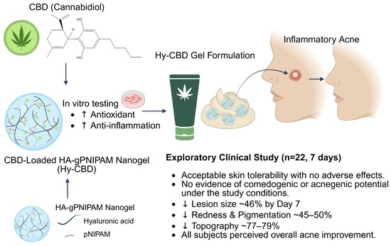
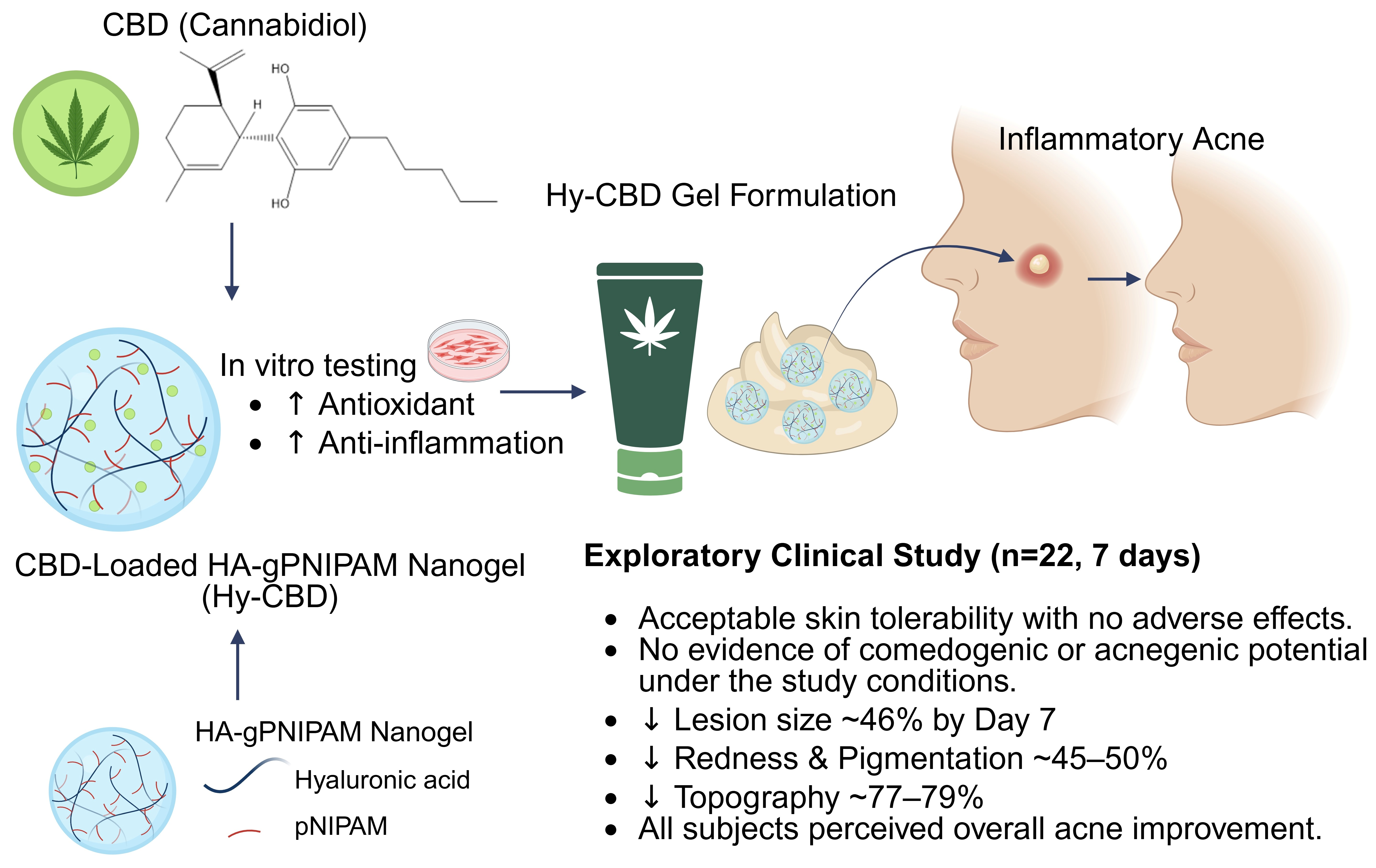
Graphical Abstract

1. Introduction

Acne vulgaris is a common chronic inflammatory skin disorder affecting the pilosebaceous unit. It is caused by several factors, including excess sebum production, follicular hyperkeratinization, hormonal dysregulation, bacterial colonization, and inflammation [1]. Approximately 85% of individuals aged 12–24 years are affected by acne, which can lead to significant psychosocial impacts such as anxiety, depression, and reduced self-esteem [2,3,4].
Conventional management relies on topical therapies (e.g., benzoyl peroxide, antibiotics, retinoids, azelaic acid, and dapsone) and systemic agents (e.g., isotretinoin, oral antibiotics, hormonal therapy) [5]. To target multiple pathogenic pathways and mitigate antimicrobial resistance, current guidelines recommend a multimodal approach tailored to disease severity, patient tolerance, and preference [6]. However, standard treatments are frequently limited by adverse effects; topical agents commonly cause localized skin irritation, sensitivity, and dryness, whereas systemic therapies can induce systemic toxicities, or accelerate global antimicrobial resistance [7,8,9]. As a result, alternative microbiome-targeted interventions, such as probiotics, postbiotics, bacteriophages, and gut–skin axis modulators, are being actively explored [10,11]. In parallel, natural compounds are gaining attention as complementary options due to their broad biological activities and favorable safety profiles [12], with several phytochemicals demonstrating antimicrobial, anti-inflammatory, sebum-regulating, and wound-healing properties [13,14,15,16,17,18].
Cannabidiol (CBD) is a major non-psychoactive phytochemical derived from Cannabis sativa (hemp) that exhibits potent antioxidant, anti-inflammatory, antimicrobial, and sebum-regulating properties [19]. CBD exerts these effects largely by interacting with cannabinoid receptors (CB1 and CB2) expressed on skin cells, including keratinocytes, fibroblasts, melanocytes, and sebocytes [20]. Mechanistically, CBD-rich extracts inhibit Cutibacterium acnes proliferation, suppress core inflammatory pathways, such as NF-κB and MAPK, and promote collagen synthesis [21,22,23]. Clinical data support these multi-targeted actions, demonstrating that topical CBD formulations effectively reduce sebum production and erythema with a highly favorable safety profile [21].
The regulatory landscape of CBD in cosmetic products remains fragmented and rapidly evolving, directly impacting the commercialization and global applicability of CBD-based formulations. In the European Union and the United Kingdom, CBD is permitted in cosmetics provided that Δ9-tetrahydrocannabinol (THC) content falls within strict, predefined limits; however, a unified EU legislation is still lacking, and products making therapeutic claims risk classification as medicines [24]. In the United States, hemp-derived CBD has been federally legal since 2018, contingent on a THC threshold below 0.3%, though individual state regulations vary and are not harmonized with international standards [25]. Regulatory frameworks across East Asia are considerably more restrictive: China strictly prohibits all Cannabis sativa-derived ingredients in cosmetics, Japan permits only THC-free preparations, and South Korea tolerates only exceptionally low residual levels with no clear pathway for mainstream consumer cosmetic commercialization [24,25].
In contrast, Thailand presents a unique regulatory environment highly relevant to this work. Having become the first Southeast Asian nation to legalize medical cannabis in 2019 [26], Thailand has since shifted toward a strictly monitored, prescription-based medical model for cannabis biomass while maintaining a distinct, permissible framework for hemp-derived cosmetics [27]. Domestic manufacturers are permitted to utilize CBD and hemp extracts under the condition that THC concentrations do not exceed 0.2% by weight [26,27]. However, these formulations are classified as high-risk cosmetics that mandate formal registration with the Thai Food and Drug Administration (FDA), and the commercial importation of finished cosmetics containing these ingredients remains prohibited.
Despite its promising biological activities, the clinical use of CBD is constrained by an unfavorable physicochemical profile [28]. It is highly lipophilic with poor aqueous solubility (approximately 0.1 µg/mL), which limits its skin permeability and results in a low baseline bioavailability of around 6% [29,30]. Furthermore, CBD is highly unstable, degrading readily when exposed to light, elevated temperature, and atmospheric oxygen [31]. Light exposure markedly accelerates active compound loss during storage [32,33], while thermal stress promotes degradation, particularly in oil-based vehicles under accelerated storage conditions [34]. It is likewise highly vulnerable to oxidative degradation [35]. Because these combined properties complicate the development of stable topical vehicles, advanced formulation strategies are essential to protect the molecule. To address these limitations, nanocarrier systems have emerged as a promising approach to simultaneously enhance the physicochemical stability, aqueous solubility, and cutaneous bioavailability of CBD [36].
Nanogels are nanoscale, crosslinked polymer networks capable of retaining high amounts of water while maintaining structural and colloidal stability. Their amphiphilic nature allows for the simultaneous encapsulation of both hydrophilic and hydrophobic compounds, facilitating controlled release, enhanced skin penetration, and prolonged cutaneous hydration [37,38,39]. By reducing particle size and drastically increasing the surface-area-to-volume ratio, these nanotechnology-based platforms significantly improve the apparent solubility and bioavailability of poorly water-soluble molecules [40,41,42]. While other nanocarriers like liposomes, solid lipid nanoparticles, and nanoemulsions are available, nanogels offer superior colloidal stability, tunable swelling behavior, and higher hydration capacities [43]. Furthermore, their key physicochemical attributes, such as matrix porosity, surface charge, and stimuli-responsiveness, can be tailored through custom polymer selection and synthesis methods [42,44,45,46].
Hyaluronic acid-graft poly(N-isopropylacrylamide) (HA-pNIPAM) is a thermo-responsive amphiphilic copolymer that has successfully enhanced the aqueous solubility and bioactivity of lipophilic phytochemicals, such as curcumin and asiatic acid [47,48]. Mechanistically, above its lower critical solution temperature (about 32 °C), the pNIPAM segments undergo a conformational collapse, self-assembling into hydrophobic core domains that solubilize and shield lipophilic molecules. Concurrently, the hydrophilic hyaluronic acid outer shell remains hydrated, conferring biocompatibility, hydration, and cellular uptake [47,49,50].
In a preliminary study applying this platform to CBD encapsulation, HA-pNIPAM nanogels significantly enhanced the aqueous solubility of CBD and markedly improved its stability during room-temperature storage [51]. Beyond these initial efficacy findings, previous in vitro and in vivo evaluations have confirmed the systemic safety of HA-pNIPAM carriers [47,48,51,52,53,54], and their safety profile for human topical application has been established by the patent holder [55]. Consequently, these dual-functional nanogels present a highly promising approach to overcome the intrinsic physicochemical limitations of CBD.
Building on these foundational encapsulation and stability findings, the present study evaluates the biological efficacy and clinical potential of the CBD-loaded HA-pNIPAM nanogel platform. Specifically, we investigated the in vitro biological properties of the CBD-nanogel formulation, including its anti-inflammatory and cellular activities. This was coupled with a preliminary clinical trial in human subjects with active inflammatory acne to assess short-term cutaneous tolerability, localized clinical responses, and the practical feasibility of this advanced nanogel vehicle for topical acne management.

2. Methods

2.1. Materials

Hyaluronic acid–graft-poly(N-isopropylacrylamide) (HA-pNIPAM) was obtained from Nabsolute Co., Ltd., Bangkok, Thailand. The ethanolic Cannabis sativa extract containing 3% w/w of CBD was sourced from Leapdelab Co., Ltd., Samut Prakan, Thailand. The final Hy-CBD gel formulation (containing 3% w/w Hy-CBD corresponding to 0.0003% w/w CBD with 0.0075% w/w of HA-pNIPAM) was developed according to a patent-pending process (Thailand Patent No. 2401008454) by the Faculty of Pharmaceutical Sciences, Chulalongkorn University, using Caprylic/Capric Triglyceride and Cetyl Phosphate as emollients, Ammonium Acryloyldimethyltaurate/VP Copolymer as the gelling agent, and pentylene glycol as the preservative system.

2.2. In Vitro Biological Activity Testing

2.2.1. Anti-Inflammatory Activity

RAW 264.7 macrophages were used as an inflammatory cell model, with lipopolysaccharide (LPS) serving as the inflammatory inducer and nitric oxide (NO) as the inflammatory biomarker. Cells were seeded at a density of 40,000 cells/well in a 24-well plate and incubated for 24 h prior to treatment with LPS (1 µg/mL), LPS combined with CBD (2.5 µg/mL), or LPS combined with CBD (2.5 µg/mL) loaded in 0.003–0.005% w/v HA-pNIPAM nanogels for 24 h. NO production was determined using the Griess assay by mixing the culture supernatant with Griess reagent and measuring absorbance at 540 nm using a CLARIOstar Plus microplate reader (CLARIOstar, BMG Labtech, Ortenberg, Germany).

2.2.2. Anti-Oxidation Activity

The antioxidant activities of CBD-loaded HA-pNIPAM nanogels were determined using a DCFH-DA-based assay. Briefly, HaCaT keratinocyte cells were seeded into a black 96-well microplate with a clear bottom at a density of 20,000 cells/well and cultured for 24 h prior to treatment. Cells were then treated with CBD (10 µg/mL) and 0.001% w/v HA-pNIPAM nanogels for 24 h. Following treatment, the cells were gently rinsed twice with PBS and stained in the dark with DCFH-DA solution (5 μM in serum-free MEM). After 30 min of staining, the cells were washed with PBS, and serum-free medium was added to each well. The cells were subsequently treated with 500 μM H2O2 in serum-free medium at 37 °C for 1 h. Finally, the cells were washed twice with PBS. The intracellular fluorescence signal was recorded using a CLARIOstar microplate reader (BMG Labtech, Ortenberg, Germany) (excitation wavelength: 485 nm; emission wavelength: 530 nm).

2.3. Clinical Study Design and Sample

The clinical evaluations were conducted by Dermscan Asia Co., Ltd. (Bangkok, Thailand). The study was approved by the Specialty Independent Research Ethics Committee—SIREC (certificate number DA22A520 (5_2023), approved on 12 January 2023), and was carried out in accordance with the Declaration of Helsinki. Prior to participation, all subjects were informed about the study procedures and provided written informed consent. The study was retrospectively registered with the Thai Clinical Trials Registry (TCTR20250507005) on 7 May 2025.
Participants were selected based on the following inclusion criteria: healthy Asian males or females aged 18–45 years; Fitzpatrick skin type I to IV; presenting with inflammatory facial acne; and willing to comply with study procedures.
Exclusion criteria included: pregnancy, breastfeeding, or plans to become pregnant; active cutaneous conditions in the study area (e.g., eczema); allergies to cosmetic products; use of retinoids within the past six months; use of other acne medication within the past month; changes in hormonal treatment (including contraceptives) within the previous three months; and use of topical or systemic treatments in the preceding weeks that may interfere with cutaneous response. Other exclusions were recent dermatologic procedures or surgery under general anesthesia in the past two months, high sun or UV exposure in the past month, or current participation in another clinical study.

2.4. Interventions

A total of 22 participants were enrolled and treated with Hy-CBD gel. Participants applied the formulation to affected acne areas twice daily (morning and evening) for seven consecutive days. To ensure complete coverage of varying lesion distributions, the gel was applied topically over active lesions rather than in a fixed mass per area; consequently, the absolute quantity applied was not standardized across subjects. For clinical evaluation, two inflammatory lesions of similar type and baseline type, size, and severity were selected per participant. These target lesions were longitudinally assessed by a single trained dermatologist at every visit (Day 0, Day 2, and Day 7).

2.5. Outcomes

2.5.1. Cutaneous Tolerance

A dermatologist assessed facial skin tolerance based on clinical signs, including erythema, edema, dryness, desquamation, and roughness. On Day 0, participants were asked about common skin sensations such as tightness, stinging, itching, and burning. Final acceptability was determined by comparing these indicators across visits.

2.5.2. Comedogenic Potential

To assess comedogenicity, lesion counts on the face were compared at Day 2 and Day 7 relative to baseline (Day 0). The dermatologist counted blackheads, microcysts, papules, and pustules (excluding the nasal pyramid, vermillion border, chin crease, and scalp rim). Changes in lesion numbers (Day 2−Day 0 and Day 7−Day 0) were analyzed descriptively.

2.5.3. Effect on Inflammatory Acne

Two similar inflammatory lesions were selected for each subject at baseline. On Day 0, 2, and 7, the dermatologist evaluated lesion size (measured with a vernier caliper), color, residual pigmentation, desquamation, and topography/depth.

2.5.4. Imaging Using the Visia® System

The VISIA® imaging system (Canfield Scientific, Parsippany, NJ, USA) was used to capture high-resolution facial images under white, UV, and polarized light. Image repositioning was standardized using an overlay display for accurate comparisons across timepoints.

2.5.5. Subjective Evaluation

On Day 2 and 7, participants completed a questionnaire to evaluate product characteristics, perceived efficacy, and willingness to continue use.

2.6. Calculation Formulas

Data are presented as raw values and descriptive statistics, including mean, median, standard error of the means (SEM), and 95% confidence intervals (CI 95%). The absolute variations (∆) and percentage variations (∆%) from baseline were calculated as follows:
  =   T Z t i T Z t 0
% = T Z t i T Z t 0 T Z t 0 × 100
where T Z refers to the measured value in the zone treated with the tested product, t 0 represents the baseline (before product use), and t i represents the time point after product application.

2.7. Statistical Analysis

Background and demographic data were summarized using descriptive statistics. Data normality was assessed using the Shapiro–Wilk test (α = 0.01). For non-normally distributed data, Friedman’s test was used, followed by the Wilcoxon signed-rank test for post hoc analysis. For normally distributed data, repeated measures one-way ANOVA and t-tests were applied. For cohort analysis, t-tests and one-way ANOVA followed by Tukey’s post hoc test were used for normal data, while the Mann–Whitney U or Kruskal–Wallis tests were used for non-normal data. Analyses were performed using Microsoft Excel for data handling, SPSS (version 21.0) for statistical testing, and GraphPad Prism 9 for graphing. A p-value < 0.05 was considered statistically significant.

3. Results

3.1. Biological Activities of CBD-Loaded HA-pNIPAM Nanogel

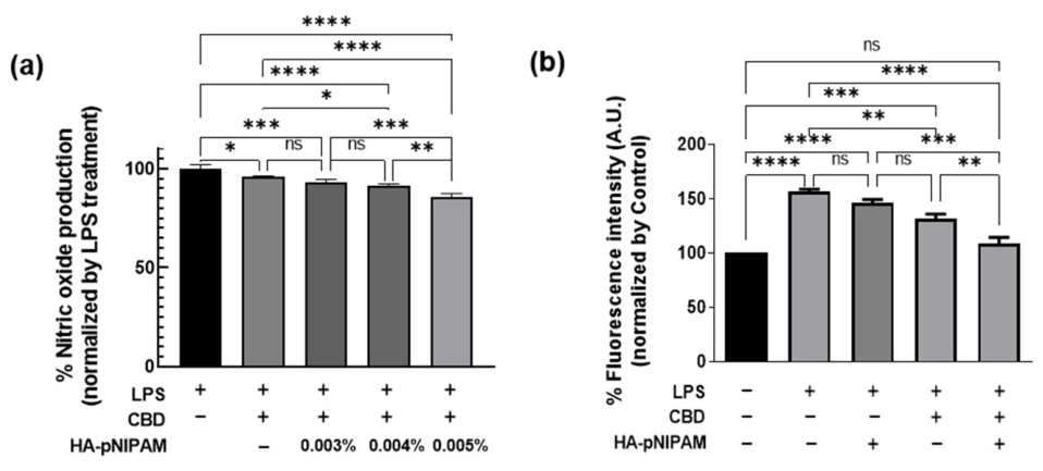
Figure 1a illustrates that CBD-loaded 0.003–0.005% w/v HA-pNIPAM nanogels reduced nitric oxide production to 6.72%, 8.88%, and 14.14%, respectively, in a concentration-dependent manner. In contrast, free CBD alone produced a slight reduction in nitric oxide levels of approximately 4%. These results suggest that encapsulation of CBD within the HA-pNIPAM nanogel enhanced its anti-inflammatory activity compared with free CBD. Results from the DCFH-DA assay in Figure 1b demonstrated that CBD in combination with HA-pNIPAM enhanced antioxidant activity by approximately 48%, exceeding the effect observed with free CBD alone. These findings suggest that incorporation of CBD into the HA-pNIPAM nanogel system can enhance the antioxidant activity of the active compound.

3.2. Exploratory Clinical Results

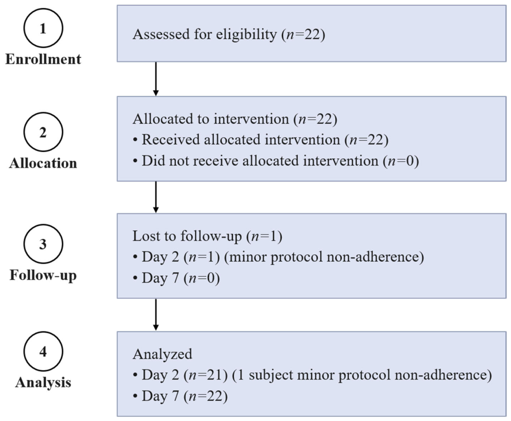
A total of 22 participants (18 females and 4 males), aged between 19 and 44 years (mean age: 26 ± 2 years), were enrolled in this study (Figure 2). Participant demographics are summarized in Table 1. All participants were of Asian descent with Fitzpatrick skin phototypes III to IV. The distribution of skin types was as follows: 59.1% greasy, 27.3% combination, and 13.6% dry skin. One participant missed the Day 2 follow-up visit, which was recorded as a minor protocol deviation.
Cutaneous tolerance was assessed by a dermatologist through clinical examination. On Days 2 and 7 of product use, 95% of participants showed no clinical signs, while 5% showed no relevant signs. One participant reported very mild itching on Day 2, which was considered unrelated to the product. Overall, the findings from this short-term study suggest that the Hy-CBD gel demonstrated acceptable clinical tolerability.
Table 2 blackhead counts did not change, and fewer than one-third of participants showed variations in microcysts, papules, or pustules. By Day 7, no significant differences were observed in blackhead, microcyst, or papule counts. Changes in pustule counts were limited to fewer than one-third of participants. These observations suggest that the formulation did not cause retentional or inflammatory lesions, and the formulation did not demonstrate evidence of comedogenicity or acnegenicity under the study conditions (Table 2).
Clinical efficacy was evaluated by measuring the size and characteristics of inflammatory acne lesions using a vernier caliper and clinical scoring scales. Two representative inflammatory lesions were selected per participant. The use of Hy-CBD gel for 2 and 7 days resulted in a visible and statistically significant reduction in lesion size (Table 3). For the first lesion, the average diameter decreased by 27% on Day 2 and by 46% on Day 7 (p < 0.001). For the second lesion, reductions of 23% and 42% were observed on the same days (p < 0.001). These findings provide preliminary evidence of a potential benefit of the product on inflammatory acne lesion size within a short treatment period.
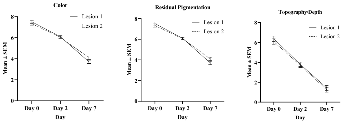
Clinical evaluation of lesion characteristics, including color, residual pigmentation, desquamation, and topography/depth, showed significant improvements in most parameters (Table 4 and Figure 3). Color scores decreased significantly by Day 7, with reductions of 50% for Lesion 1 and 45% for Lesion 2 (both p < 0.001, Wilcoxon signed-rank test). Residual pigmentation followed a similar pattern, with reductions of 50% and 45%, respectively.
As presented in Table 4, the numerical scores reported for lesion color and residual pigmentation are identical across the evaluated time points. Because both parameters were assessed visually on the same target lesions by a single dermatologist using a subjective ordinal scoring scale, the assigned visual grades overlapped across the observation period (Day 0, Day 2, and Day 7). We acknowledge that subjective ordinal scoring has limited sensitivity to fully discriminate between these two closely related visual attributes. Because erythema was the predominant visible feature of the active inflammatory lesions during this short-term study, both parameters were graded concordantly. The raw data were provided as a non-editable file in Supplementary Figure S1. These outcomes are therefore retained for transparency but should be interpreted as reflecting the same underlying visual change rather than as independent measures. Consequently, future studies should incorporate objective instrumental measurements, such as colorimetry or narrow-band spectrophotometry, to independently resolve subtle changes in erythema and melanin content.
Desquamation and scaling showed minimal variation and the changes were not statistically significant, likely due to the low prevalence of these features among participants. In contrast, topography and depth improved significantly, with reductions of 77% for Lesion 1 and 79% for Lesion 2 by Day 7 (both p < 0.001). These results support the clinical efficacy of the Hy-CBD gel in improving the visual and structural features of inflammatory acne lesions over a short treatment period.
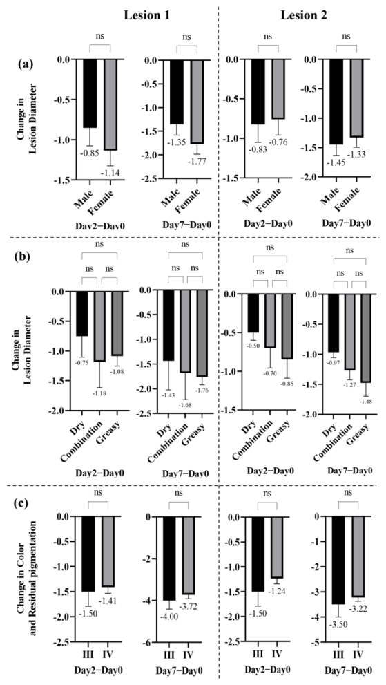
To investigate how sex, skin type, and skin phototype affected the clinical efficacy of the Hy-CBD acne gel, participants were grouped based on these characteristics. First, the impact of sex on the reduction in inflammatory lesion size was assessed. As shown in Figure 4a, no statistically significant difference was observed between male and female participants, suggesting that sex did not affect the outcomes. Subsequently, skin type (dry, combination, or greasy) was examined. While participants with greasy skin showed a trend toward a greater lesion size reduction (Figure 4b), this did not reach statistical significance. Finally, the effect of skin phototypes on lesion color and residual pigmentation was analyzed. A comparison of phototypes III and IV showed no statistically significant differences (Figure 4c).
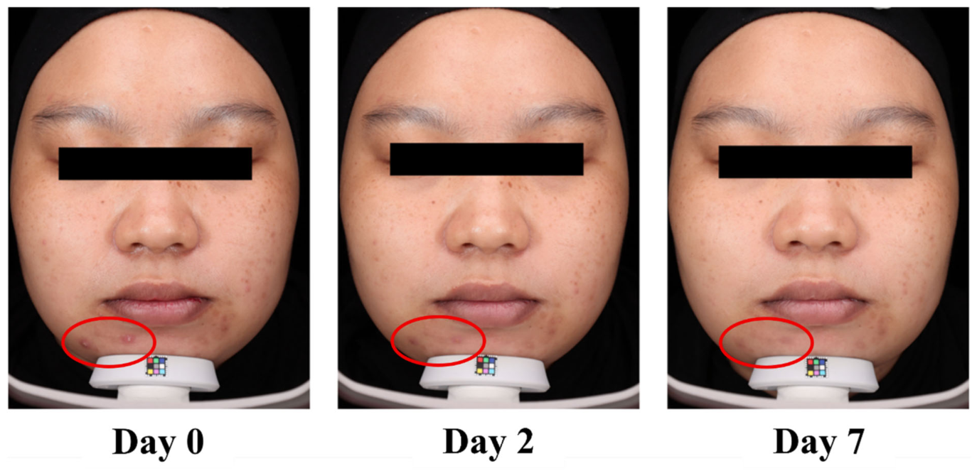
Figure 5 shows VISIA® images of inflammatory acne progression in a representative participant from baseline (Day 0) through Day 2 and Day 7. At baseline, the lesion showed marked redness, swelling, and elevation. After 2 days of treatment, noticeable improvements included reduced size, redness, and swelling. By Day 7, the lesion had nearly flattened with minimal redness and no swelling. These results show the gel’s effectiveness in improving inflammatory acne within a short treatment period.
In the subjective evaluation, all participants (100%) rated the Hy-CBD gel positively for its appearance, texture, and the absence of skin staining. A total of 91% reported a non-sticky finish and quick absorption. All participants (100%) reported reductions in both the number and severity of acne lesions, starting on Day 2 and continuing through Day 7. Reduced redness (erythema) was reported in 95% of participants by Day 2 and in 100% by Day 7.

4. Discussion

Topical treatments are essential for managing mild-to-moderate acne, while severe cases often require a combination of systemic and topical therapies. However, conventional topical agents can cause side effects such as dryness, redness, peeling, and itching, which often lead to patient discomfort and reduced compliance [56]. A primary goal in topical acne treatment is to achieve an optimal balance between therapeutic efficacy and patient tolerability. Therefore, recent research has focused on developing vehicle formulations that can improve drug delivery while minimizing local irritation [57].
Physicochemical characterization confirmed that Hy-CBD exhibits superior properties compared to free CBD, consistent with findings by May et al. [51]. An encapsulation efficiency of 87.57 ± 3.72% was achieved at 0.25% w/w HA-pNIPAM, yielding a loading capacity of 1117.42 ± 47.73% and a drug content of 100.54 ± 0.58% (Supplementary Figure S2). Nanoparticle tracking analysis and transmission electron microscopy revealed discrete spherical particles with a mean size of 258.9 ± 104.2 nm. Temperature-dependent shifts in size and polydispersity aligned with the characteristic pNIPAM thermal transition, which drove significantly higher CBD release at 37 °C than at 25 °C.
Incorporation of phytochemicals into the HA-pNIPAM nanogel significantly enhanced entrapment efficiency and formulation stability during storage, consistent with previous reports [48,54]. In addition, the Hy-CBD formulation retained the intrinsic biological activities of CBD, including anti-inflammatory and antioxidant effects, while further enhancing these biological responses. This improvement may be associated with enhanced cellular uptake facilitated by the nanogel delivery system, as previously observed in studies investigating the antiproliferative activity of curcumin-loaded HA-pNIPAM nanogels [47].
Preliminary screening also confirmed that encapsulation preserved the antimicrobial efficacy of CBD against Staphylococcus aureus, a clinically relevant opportunistic skin pathogen [58] (Supplementary Figure S3). Although Cutibacterium acnes is the primary organism implicated in acne pathogenesis, it was not evaluated in this study due to limited availability in our current testing facility. Based on these favorable in vitro results, only the optimized Hy-CBD formulation was advanced to the clinical phase. Free CBD and empty HA-pNIPAM polymer arms were excluded from the human evaluation because the individual baseline properties, safety profiles, and therapeutic limitations of these separate components have already been well-established in previous literature [24,55]. Consequently, these laboratory findings provided the necessary mechanistic rationale to transition the formulation into a preliminary human trial.
This preliminary clinical study indicated favorable short-term skin tolerability for the Hy-CBD nanogel, with no clinical incidents observed by the dermatologist on Day 2 or Day 7. One participant reported mild symptoms after two days of use; however, the physician did not consider these to be related to the product. Because this evaluation was limited to a 7-day period, long-term cutaneous safety and cumulative toxicity cannot be determined from these data alone. Nevertheless, these short-term findings help minimize initial concerns regarding localized irritation often associated with topical nanocarriers.
In terms of clinical response, significant reductions in lesion size, erythema, residual pigmentation, and acne depth were observed within the 2-to-7-day window (p < 0.001). Subjective self-evaluations supported these results, with 95% of participants reporting a noticeable decrease in localized inflammation.
To put these preliminary outcomes into context, our results can be compared with BTX 1503, one of the most clinically advanced topical CBD formulations evaluated to date [59,60]. In a large-scale, 12-week Phase II trial, BTX 1503 demonstrated acceptable safety but missed its primary efficacy endpoint for inflammatory lesions due to a high vehicle response. The key clinical and structural differences between BTX 1503 and our Hy-CBD formulation are summarized in Table 5.
The differences in outcomes shown in Table 5 may be explained by distinct delivery characteristics. Conventional vehicles often struggle with CBD’s high lipophilicity and poor aqueous solubility, leading to variable skin penetration. In contrast, the HA-pNIPAM nanogel system was designed to bypass these limitations through enhanced solubilization and sustained-release properties, which likely contributed to improved cutaneous delivery.
Nevertheless, our findings must be interpreted with strict caution. Because of the open-label, non-randomized design and the lack of a vehicle-control arm, these 7-day results represent a preliminary proof-of-concept. Further randomized, double-blind, vehicle-controlled trials over an extended duration and in larger cohorts are mandatory to validate these observations and establish true clinical efficacy.
Subgroup analyses provide preliminary evidence that the acute response to the Hy-CBD gel remained consistent across diverse patient characteristics, including sex, skin type, and Fitzpatrick phototype. Although hormonal factors substantially contribute to acne pathogenesis [61], comparable reductions in lesion size were observed in both male and female participants, suggesting a broad mechanism of action. The trend toward greater lesion-size reduction in participants with oily or combination skin is noteworthy and aligns with the documented sebostatic properties of CBD [62]. Additionally, participants with phototype III showed a slight tendency toward greater improvements in lesion color and pigmentation compared to phototype IV.
Because these subgroup trends did not reach statistical significance, they must not be over-interpreted; the small sample size and short 7-day duration prevent any definitive conclusions regarding long-term demographic or phenotypic advantages. However, they indicate that the formulation’s potential benefits, such as modulating post-inflammatory pigmentation, warrant deeper investigation. Future, adequately powered trials with extended observation periods and objective colorimetric analyses are necessary to confirm these specific clinical responses.
The initial clinical improvements observed in this study likely reflect the underlying biological activity of CBD at the cellular level. Several anti-inflammatory mechanisms of CBD have been proposed, though the exact pathways are not yet fully understood. The endocannabinoid system (ECS), involving G protein-coupled receptors such as CB1 and CB2, is the primary system through which phytocannabinoids work [63]. In the skin, CB1 receptors are mostly found in keratinocytes of the stratum spinosum and stratum granulosum, while CB2 receptors are found in the stratum basale. Both CB1 and CB2 receptors are found in epidermal nerve fibers and in immune cells such as lymphocytes, natural killer cells, mast cells, and macrophages [64]. While CBD has low affinity for CB1 and CB2 receptors, it can modulate their activity at low concentrations [65,66]. CBD also acts as a CB2 inverse agonist, potentially reducing immune cell migration and lowering inflammatory responses.
Beyond the classical ECS, CBD interacts with other molecular targets, including GPR55, PPAR-γ, and TRPVs [67]. It has been shown to inhibit the adenylyl cyclase–cAMP pathway associated with TRPV1 signaling [68]. By modulating calcium influx through TRPV channels, CBD can suppress the release of pro-inflammatory cytokines [69]. As a PPAR-γ agonist, CBD inhibits NF-κB-mediated transcription, which downregulates inflammatory mediators such as TNF-α, IL-1β, IL-6, and cyclooxygenases [70]. Furthermore, CBD acts as a GPR55 antagonist, promoting IL-10 release and exerting an anti-inflammatory effect [71,72].
Sebum secretion is a key factor in acne severity [73]. Combined with abnormal desquamation, a process known as hyperkeratinization leads to microcomedones, which can develop into visible acne lesions [74]. Comedogenesis refers to the formation of comedones due to follicular blockage, while acnegenesis involves inflammatory changes (folliculitis) [75]. In-use clinical studies are considered the gold standard for evaluating these effects due to their high sensitivity [76]. In our evaluation, no significant increase in either retentional or inflammatory lesions was observed. These results suggest that the formulation is unlikely to be comedogenic or acnegenic. Since acne-prone skin is particularly reactive, testing comedogenic potential of the final formulation provides valuable insights for clinicians and patients [77].
Although the final CBD concentration (0.0003% w/w) was low on a mass basis, it corresponded to approximately 9.5 µM, which falls within the concentration range previously reported to exert biological activity on sebocytes and keratinocytes in vitro [78,79]. While clinical evidence at this concentration remains unavailable, a study by Oláh et al. using a full-thickness human skin organ culture (hSOC) model demonstrated that exposure to CBD at 10 µM over 6 days was able to reduce lipogenesis [23]. The effects observed in the present study may therefore be associated with enhanced delivery by the HA-pNIPAM nanogel system despite the low CBD concentration. Nevertheless, due to the absence of vehicle and free-CBD control groups and the lack of cutaneous pharmacokinetic data, the observed clinical improvements cannot be attributed solely to CBD and may also reflect contributions from the nanogel vehicle or the natural resolution of lesions. Accordingly, these findings should be considered preliminary and hypothesis-generating.
The expanding application of nanotechnology in topical therapeutics offers new opportunities to optimize the delivery and clinical performance of lipophilic compounds like CBD. However, because nanocarriers can alter skin penetration dynamics, cellular interactions, and local immune responses, rigorous safety assessments remain mandatory [29]. In this study, the Hy-CBD nanogel showed encouraging short-term tolerability, with no clinically significant irritation or acnegenic exacerbations observed over the 7-day evaluation period. When considered alongside the observed reductions in inflammatory lesion characteristics, these initial findings suggest that HA-based nanogels may serve as a promising, adaptable platform for topical cannabinoid therapy. Nevertheless, larger, randomized clinical trials with extended observation windows are essential to validate these findings and fully characterize the long-term safety and therapeutic efficacy of this delivery system in acne and other inflammatory dermatoses.

Limitation

Several limitations must be considered when interpreting the findings. First, the absence of a vehicle, placebo, or untreated control groups limits the ability to establish a definitive causal relationship between the formulation and the observed clinical improvements, as spontaneous lesion resolution or baseline skincare practices may have contributed. Future studies should employ randomized, vehicle-controlled, double-blinded designs. Second, the small sample size restricted statistical power and limits the generalizability of these findings to broader acne populations. Larger and more diverse cohorts are therefore needed to strengthen the external validity of these observations.
Third, while the 7-day treatment period is standard for evaluating acute cosmetic tolerability, it is entirely insufficient to determine sustained efficacy or long-term safety, particularly regarding cumulative exposure to the nanogel vehicle itself. Extended treatment and follow-up periods are mandatory in future trials. Finally, Hy-CBD nanogel was evaluated without a direct head-to-head comparison against free CBD or the empty HA-pNIPAM nanogel, preventing an exact quantification of how much the carrier system itself contributed to the clinical response. Future investigations should incorporate free CBD and blank nanogel comparator arms alongside standard treatments to better define the therapeutic advantages of this delivery platform. Despite these limitations, this study provides valuable preliminary evidence supporting the feasibility and acute tolerability of the formulation, establishing a clear foundation for future randomized controlled trials.

5. Conclusions

This study demonstrated the successful development of a CBD-loaded HA-pNIPAM nanogel formulation with enhanced biological properties compared with free CBD. Encapsulation within the HA-pNIPAM nanogel enhanced the biological efficacy of CBD, supporting its potential for further therapeutic application. In the preliminary clinical study, the Hy-CBD formulation exhibited acceptable cutaneous tolerability and was associated with a reduction in inflammatory acne lesions over a short treatment period. As conventional acne therapies are often associated with undesirable side effects, phytochemical-based alternatives with favorable safety profiles, such as CBD, may represent a valuable therapeutic option either as a first-line treatment or as an adjunct to standard therapies. Importantly, this study evaluated CBD in its final formulation, providing clinically relevant insight into the efficacy and safety profile under practical conditions of use. The Hy-CBD gel demonstrated no observable comedogenic or acnegenic effects under the study conditions, with participants reporting satisfactory treatment outcomes. Collectively, these findings support the potential of the Hy-CBD gel as a safe and well-tolerated topical formulation for the management of inflammatory acne. Furthermore, the HA-pNIPAM nanogel system may represent a promising delivery platform for other phytochemical-based topical formulations. Nevertheless, larger-scale studies with longer follow-up periods are required to further confirm these preliminary findings and establish long-term clinical efficacy and safety of the formulation.

Supplementary Materials

The following supporting information can be downloaded at: https://www.mdpi.com/article/10.3390/cosmetics13040165/s1, Figure S1: Raw clinical evaluation scores for inflammatory acne lesions 1 and 2; Figure S2: Physicochemical properties of the CBD-loaded HA-pNIPAM nanogel; Figure S3: Antimicrobial activity of free CBD-loaded HA-pNIPAM nanogel against Staphylococcus aureus by disk diffusion assay.

Author Contributions

P.K. prepared the Hy-CBD gel, interpretation, visualization, writing—original draft, and writing—review & editing. K.L. contributed to the project administration, data collection, and writing—review & editing. K.S. contributed to investigation, formal analysis, and resources. V.P. contributed to writing—original draft, interpretation, and writing—review & editing. C.L. contributed to writing—review & editing. S.V. contributed resources. V.B. and J.A.L. contributed to methodology, study conception and design, funding acquisition, and supervision. All authors have read and agreed to the published version of the manuscript.

Funding

This study was funded by The Innovation Institute for Industry (N72B640104) with support from the NSRF via the Program Management Unit for Human Resources & Institutional Development, Research and Innovation (PMU-B) [grant number B13F660137].

Institutional Review Board Statement

This study was conducted following protocols approved by the Specialized Independent Research Ethics Committee—SIREC (certificate number DA22A520 (5_2023), dated 12 January 2023). All procedures complied with the ethical principles of the Declaration of Helsinki. This study was registered with the Thai Clinical Trials Registry (TCTR), Number TCTR20250507005.

Informed Consent Statement

All participants provided written informed consent prior to participation.

Data Availability Statement

The corresponding authors will provide the datasets used and/or analyzed during the present work upon reasonable request.

Acknowledgments

The authors thank Leapdelab Co., Ltd. for providing the CBD raw material. We are also grateful to Nabsolute Co., Ltd. and its staff for supplying the HA-pNIPAM and for their support throughout the study. We acknowledge the Pharmaceutical Research Instrument Center, Faculty of Pharmaceutical Sciences, Chulalongkorn University, for providing access to research facilities. Finally, PK acknowledges the Second Century Fund (C2F), Chulalongkorn University, for financial support during his master’s degree studies.

Conflicts of Interest

JL was a co-founder and chief technology officer (CTO) of Nabsolute Co., Ltd., while VB was the chief compliance officer (CCO). In addition, PK, KL, and KS were employed by Nabsolute Co., Ltd., and SV is the chief executive officer (CEO) of Leapdelab. These affiliations are disclosed to ensure transparency regarding potential conflicts of interest. The other authors declare no financial or personal relationships that influenced the results or interpretation of this study.

References

  1. Tuchayi, S.M.; Makrantonaki, E.; Ganceviciene, R.; Dessinioti, C.; Feldman, S.R.; Zouboulis, C.C. Acne vulgaris. Nat. Rev. Dis. Primers 2015, 1, 15029. [Google Scholar] [CrossRef] [PubMed]
  2. Bhate, K.; Williams, H.C. Epidemiology of acne vulgaris. Br. J. Dermatol. 2013, 168, 474–485. [Google Scholar] [CrossRef] [PubMed]
  3. Tan, J.K.L.; Bhate, K. A global perspective on the epidemiology of acne. Br. J. Dermatol. 2015, 172, 3–12. [Google Scholar] [CrossRef] [PubMed]
  4. Samuels, D.V.; Rosenthal, R.; Lin, R.; Chaudhari, S.; Natsuaki, M.N. Acne vulgaris and risk of depression and anxiety: A meta-analytic review. J. Am. Acad. Dermatol. 2020, 83, 532–541. [Google Scholar] [CrossRef] [PubMed]
  5. Oge, L.K.; Broussard, A.; Marshall, M.D. Acne Vulgaris: Diagnosis and Treatment. Am. Fam. Physician 2019, 100, 475–484. [Google Scholar] [PubMed]
  6. Habeshian, K.A.; Cohen, B.A. Current Issues in the Treatment of Acne Vulgaris. Pediatrics 2020, 145, S225–S230. [Google Scholar] [CrossRef] [PubMed]
  7. Tripathi, S.V.; Gustafson, C.J.; Huang, K.E.; Feldman, S.R. Side effects of common acne treatments. Expert. Opin. Drug Saf. 2013, 12, 39–51. [Google Scholar] [CrossRef] [PubMed]
  8. Coondoo, A.; Phiske, M.; Verma, S.; Lahiri, K. Side-effects of topical steroids: A long overdue revisit. India. Dermatol. Online J. 2014, 5, 416–425. [Google Scholar] [CrossRef] [PubMed] [PubMed Central]
  9. Manson, S.C.; Brown, R.E.; Cerulli, A.; Vidaurre, C.F. The cumulative burden of oral corticosteroid side effects and the economic implications of steroid use. Respir. Med. 2009, 103, 975–994. [Google Scholar] [CrossRef] [PubMed]
  10. Burckhardt-Bravo, V.; Funes-Ferrada, R.; Valenzuela, F. Update on novel acne treatments: A narrative review focused on microbiome modulation and non-pharmacological approaches. An. Bras. Dermatol. 2026, 101, 501249. [Google Scholar] [CrossRef] [PubMed]
  11. Łukańko, K.; Lipska, P.; Sobczak, J.; Lorek, J.; Duda-Madej, A. Rethinking Acne Vulgaris: The Gut–Skin Axis as a Central Mechanism and Therapeutic Target. Appl. Sci. 2026, 16, 4527. [Google Scholar] [CrossRef]
  12. Fisk, W.A.; Lev-Tov, H.A.; Sivamani, R.K. Botanical and Phytochemical Therapy of Acne: A Systematic Review. Phytother. Res. 2014, 28, 1137–1152. [Google Scholar] [CrossRef] [PubMed]
  13. Chen, T.; Zhu, Z.; Du, Q.; Wang, Z.; Wu, W.; Xue, Y.; Wang, Y.; Wu, Y.; Zeng, Q.; Jiang, C.; et al. A Skin Lipidomics Study Reveals the Therapeutic Effects of Tanshinones in a Rat Model of Acne. Front. Pharmacol. 2021, 12, 675659. [Google Scholar] [CrossRef] [PubMed]
  14. Gad, H.A.; Roberts, A.; Hamzi, S.H.; Gad, H.A.; Touiss, I.; Altyar, A.E.; Kensara, O.A.; Ashour, M.L. Jojoba Oil: An Updated Comprehensive Review on Chemistry, Pharmaceutical Uses, and Toxicity. Polymers 2021, 13, 1711. [Google Scholar] [CrossRef] [PubMed]
  15. Luo, J.; He, W.; Li, X.; Ji, X.; Liu, J. Anti-acne vulgaris effects of chlorogenic acid by anti-inflammatory activity and lipogenesis inhibition. Exp. Dermatol. 2021, 30, 865–871. [Google Scholar] [CrossRef] [PubMed]
  16. Ogunbiyi, A.; Enechukwu, N.A. African black soap: Physiochemical, phytochemical properties, and uses. Dermatol. Ther. 2021, 34, e14870. [Google Scholar] [CrossRef] [PubMed]
  17. Wei, M.P.; Qiu, J.D.; Li, L.; Xie, Y.F.; Yu, H.; Guo, Y.H.; Yao, W. Saponin fraction from Sapindus mukorossi Gaertn as a novel cosmetic additive: Extraction, biological evaluation, analysis of anti-acne mechanism and toxicity prediction. J. Ethnopharmacol. 2021, 268, 113552. [Google Scholar] [CrossRef] [PubMed]
  18. Duda-Madej, A.; Viscardi, S.; Grabarczyk, M.; Topola, E.; Kozłowska, J.; Mączka, W.; Wińska, K. Is Camphor the Future in Supporting Therapy for Skin Infections? Pharmaceuticals 2024, 17, 715. [Google Scholar] [CrossRef] [PubMed]
  19. Baswan, S.M.; Klosner, A.E.; Glynn, K.; Rajgopal, A.; Malik, K.; Yim, S.; Stern, N. Therapeutic Potential of Cannabidiol (CBD) for Skin Health and Disorders. Clin. Cosmet. Investig. Dermatol. 2020, 13, 927–942. [Google Scholar] [CrossRef] [PubMed]
  20. Martinelli, G.; Magnavacca, A.; Fumagalli, M.; Dell’Agli, M.; Piazza, S.; Sangiovanni, E. Cannabis sativa and Skin Health: Dissecting the Role of Phytocannabinoids. Planta Med. 2021, 88, 492–506. [Google Scholar] [CrossRef] [PubMed]
  21. Ali, A.; Akhtar, N. The safety and efficacy of 3% Cannabis seeds extract cream for reduction of human cheek skin sebum and erythema content. Pak. J. Pharm. Sci. 2015, 28, 1389–1395. [Google Scholar] [PubMed]
  22. Oláh, A.; Markovics, A.; Szabó-Papp, J.; Szabó, P.T.; Stott, C.; Zouboulis, C.C.; Bíró, T. Differential effectiveness of selected non-psychotropic phytocannabinoids on human sebocyte functions implicates their introduction in dry/seborrhoeic skin and acne treatment. Exp. Dermatol. 2016, 25, 701–707. [Google Scholar] [CrossRef] [PubMed]
  23. Oláh, A.; Tóth, B.I.; Borbíró, I.; Sugawara, K.; Szöllõsi, A.G.; Czifra, G.; Pál, B.; Ambrus, L.; Kloepper, J.; Camera, E.; et al. Cannabidiol exerts sebostatic and antiinflammatory effects on human sebocytes. J. Clin. Investig. 2014, 124, 3713–3724. [Google Scholar] [CrossRef] [PubMed]
  24. Rusu, A.; Farcaș, A.-M.; Oancea, O.-L.; Tanase, C. Cannabidiol in Skin Health: A Comprehensive Review of Topical Applications in Dermatology and Cosmetic Science. Biomolecules 2025, 15, 1219. [Google Scholar] [CrossRef] [PubMed]
  25. Fafaliou, M.; Papadopoulos, A.; Pavlou, P.; Varvaresou, A. Cannabidiol’s Antioxidant Properties in Skin Care Products and Legislative Regulations. Plants 2025, 14, 3521. [Google Scholar] [CrossRef] [PubMed]
  26. Assanangkornchai, S.; Thaikla, K.; Talek, M.; Saingam, D. Medical cannabis use in Thailand after its legalization: A respondent-driven sample survey. PeerJ 2022, 10, e12809. [Google Scholar] [CrossRef] [PubMed] [PubMed Central]
  27. Erlen Joni, E.K.; Abdullah Sanek, S.; Rahayu, I. From Green Rush to Regulation: Assessing the Impact of Medical Cannabis Legalisation in Thailand. Int. J. Res. Innov. Soc. Sci. 2025, 4, 9728–9738. [Google Scholar] [CrossRef]
  28. Millar, S.A.; Maguire, R.F.; Yates, A.S.; O’Sullivan, S.E. Towards Better Delivery of Cannabidiol (CBD). Pharmaceuticals 2020, 13, 219. [Google Scholar] [CrossRef] [PubMed]
  29. Assadpour, E.; Rezaei, A.; Das, S.S.; Krishna Rao, B.V.; Singh, S.K.; Kharazmi, M.S.; Jha, N.K.; Jha, S.K.; Prieto, M.A.; Jafari, S.M. Cannabidiol-Loaded Nanocarriers and Their Therapeutic Applications. Pharmaceuticals 2023, 16, 487. [Google Scholar] [CrossRef] [PubMed] [PubMed Central]
  30. Koch, N.; Jennotte, O.; Gasparrini, Y.; Vandenbroucke, F.; Lechanteur, A.; Evrard, B. Cannabidiol aqueous solubility enhancement: Comparison of three amorphous formulations strategies using different type of polymers. Int. J. Pharm. 2020, 589, 119812. [Google Scholar] [CrossRef] [PubMed]
  31. Lapteva, M.; Faro Barros, J.; Kalia, Y.N. Cutaneous Delivery and Biodistribution of Cannabidiol in Human Skin after Topical Application of Colloidal Formulations. Pharmaceutics 2024, 16, 202. [Google Scholar] [CrossRef] [PubMed]
  32. Mazzetti, C.; Ferri, E.; Pozzi, M.; Labra, M. Quantification of the content of cannabidiol in commercially available e-liquids and studies on their thermal and photo-stability. Sci. Rep. 2020, 10, 3697. [Google Scholar] [CrossRef] [PubMed]
  33. Trofin, I.G.; Dabija, G.; Vaireanu, D.I.; Filipescu, L. The influence of long-term storage conditions on the stability of cannabinoids derived from cannabis resin. Rev. Chim. 2012, 63, 422–427. [Google Scholar]
  34. Kosović, E.; Sýkora, D.; Kuchař, M. Stability Study of Cannabidiol in the Form of Solid Powder and Sunflower Oil Solution. Pharmaceutics 2021, 13, 412. [Google Scholar] [CrossRef] [PubMed]
  35. Fraguas-Sánchez, A.I.; Fernández-Carballido, A.; Martin-Sabroso, C.; Torres-Suárez, A.I. Stability characteristics of cannabidiol for the design of pharmacological, biochemical and pharmaceutical studies. J. Chromatogr. B 2020, 1150, 122188. [Google Scholar] [CrossRef] [PubMed]
  36. Ramalho, Í.M.d.M.; Pereira, D.T.; Galvão, G.B.L.; Freire, D.T.; Amaral-Machado, L.; Alencar, É.d.N. Current trends on cannabidiol delivery systems: Where are we and where are we going? Expert Opin. Drug Deliv. 2021, 18, 1577–1587. [Google Scholar] [CrossRef] [PubMed]
  37. Sharma, M.; Ojha, B.; Khan, A.; Gupta, M. Nanogel and its Utilization in Cosmeceuticals. Skin 2015, 2016, 2017. [Google Scholar] [CrossRef]
  38. Oh, J.K.; Drumright, R.; Siegwart, D.J.; Matyjaszewski, K. The development of microgels/nanogels for drug delivery applications. Prog. Polym. Sci. 2008, 33, 448–477. [Google Scholar] [CrossRef]
  39. Kaewruethai, T.; Laomeephol, C.; Pan, Y.; Luckanagul, J.A. Multifunctional Polymeric Nanogels for Biomedical Applications. Gels 2021, 7, 228. [Google Scholar] [CrossRef] [PubMed]
  40. Losada-Barreiro, S.; Celik, S.; Sezgin-Bayindir, Z.; Bravo-Fernández, S.; Bravo-Díaz, C. Carrier Systems for Advanced Drug Delivery: Improving Drug Solubility/Bioavailability and Administration Routes. Pharmaceutics 2024, 16, 852. [Google Scholar] [CrossRef] [PubMed]
  41. Kumar, R. Nanotechnology based approaches to enhance aqueous solubility and bioavailability of griseofulvin: A literature survey. J. Drug Deliv. Sci. Technol. 2019, 53, 101221. [Google Scholar] [CrossRef]
  42. Khan, K.U.; Minhas, M.U.; Badshah, S.F.; Suhail, M.; Ahmad, A.; Ijaz, S. Overview of nanoparticulate strategies for solubility enhancement of poorly soluble drugs. Life Sci. 2022, 291, 120301. [Google Scholar] [CrossRef] [PubMed]
  43. Ferozekhan, S.; Umashankar, M.S.; Narayanasamy, D. A Comprehensive Review of Nanogel-Based Drug Delivery Systems. Cureus 2024, 16, e68633. [Google Scholar] [CrossRef] [PubMed] [PubMed Central]
  44. Soni, K.S.; Desale, S.S.; Bronich, T.K. Nanogels: An overview of properties, biomedical applications and obstacles to clinical translation. J. Control. Release 2016, 240, 109–126. [Google Scholar] [CrossRef] [PubMed] [PubMed Central]
  45. Brianna Anwar, A.; Teow, S.-Y.; Wu, Y.S. Nanogel-based drug delivery system as a treatment modality for diverse diseases: Are we there yet? J. Drug Deliv. Sci. Technol. 2024, 91, 105224. [Google Scholar] [CrossRef]
  46. Sabir, F.; Asad, M.I.; Qindeel, M.; Afzal, I.; Dar, M.J.; Shah, K.U.; Zeb, A.; Khan, G.M.; Ahmed, N.; Din, F.-U. Polymeric Nanogels as Versatile Nanoplatforms for Biomedical Applications. J. Nanomater. 2019, 2019, 1526186. [Google Scholar] [CrossRef]
  47. Luckanagul, J.A.; Ratnatilaka Na Bhuket, P.; Muangnoi, C.; Rojsitthisak, P.; Wang, Q.; Rojsitthisak, P. Self-Assembled Thermoresponsive Nanogel from Grafted Hyaluronic Acid as a Biocompatible Delivery Platform for Curcumin with Enhanced Drug Loading and Biological Activities. Polymers 2021, 13, 194. [Google Scholar] [CrossRef] [PubMed] [PubMed Central]
  48. Win, Y.Y.; Charoenkanburkang, P.; Limprasutr, V.; Rodsiri, R.; Pan, Y.; Buranasudja, V.; Luckanagul, J.A. In Vivo Biocompatible Self-Assembled Nanogel Based on Hyaluronic Acid for Aqueous Solubility and Stability Enhancement of Asiatic Acid. Polymers 2021, 13, 4071. [Google Scholar] [CrossRef] [PubMed] [PubMed Central]
  49. Charoenkanburkang, P.; Kaewruethai, T.; Rojsitthisak, P.; Luckanagul, J. The study of physical properties and formulation of self-assembling nanogel from grafted hydrophilic polymer backbone as stabilization system for curcumin. Thai J. Pharm. Sci. (TJPS) 2021, 45, 50–53. [Google Scholar] [CrossRef]
  50. Umar, A.K.; Laomeephol, C.; Pannarai, N.; Lueantor, C.; Thamnium, S.; Kaewruethai, T. Mechanistic insights into the self-assembly and cellular uptake of thermoresponsive pNIPAM-grafted hyaluronic acid nanogels. J. Pharm. Investig. 2025. [Google Scholar] [CrossRef]
  51. Kyaw, M.T.T. Developemnt of Hyaluronic Acid Grafted with Poly N-Isopropyl Acrylamide Nanogel Incorporating Hemp Extract Containing CBD. Master’s Thesis, Chulalongkorn University, Bangkok, Thailand, 2024. [Google Scholar]
  52. Kotcharat, N.; Charoenkanburkang, P.; Luckanagul, J.A. Grafted hyaluronic acid nanogel for the incorporation of poly (I: C) as an immunostimulatory adjuvant. SRP 2020, 11, 247–253. [Google Scholar]
  53. Eakpunyakul, N.; Kopongpanich, P.; Bhalang, K.; Luckanagul, J.A. EFFICACY OF HYALURONIC NANOGEL SPRAY FOR TREATING DRY MOUTH: A RANDOMIZED CROSSOVER STUDY. J. Evid.-Based Dent. Pract. 2025, 25, 102142. [Google Scholar] [CrossRef] [PubMed]
  54. Boonpetcharat, N.M.; Thu Kyaw, M.T.; Boonkanokwong, V.; Luckanagul, J.A. Development of a Curcumin-Loaded Hyaluronic Acid Nanogel Formulation Using Wet Granulation Method for Enhanced Dissolution and Stability. Gels 2025, 11, 585. [Google Scholar] [CrossRef] [PubMed]
  55. Luckanagul Jittima, A.; Buranasudja, V. A Method for Preparing Hyaluronic Acid Grafted Poly(N-Isopropylacrylamide) (HA-G-PNI-PAM). US patent US 2024/0336748 A1. 2024, 14 July 2021. [Google Scholar]
  56. Zhao, J.; Wang, Y.; Jiang, L.; Mu, Y.-Z. The application of skin care product in acne treatment. Dermatol. Ther. 2020, 33, e14287. [Google Scholar] [CrossRef] [PubMed]
  57. Otlewska, A.; Baran, W.; Batycka-Baran, A. Adverse events related to topical drug treatments for acne vulgaris. Expert. Opin. Drug Saf. 2020, 19, 513–521. [Google Scholar] [CrossRef] [PubMed]
  58. Popov, L.; Kovalski, J.; Grandi, G.; Bagnoli, F.; Amieva, M.R. Three-Dimensional Human Skin Models to Understand Staphylococcus aureus Skin Colonization and Infection. Front. Immunol. 2014, 5, 2014. [Google Scholar] [CrossRef] [PubMed]
  59. ASX/Media Release: BTX 1503 Acne Phase 2 Study Results Presentation [Internet]. Botanix Pharmaceuticals. 2019. Available online: https://www.asx.com.au/asxpdf/20191023/pdf/449s36d2bz6vfr.pdf (accessed on 17 April 2023).
  60. A Randomized, Double-Blind, Vehicle-Controlled Study to Evaluate the Safety and Efficacy of BTX 1503 in Patients with Moderate to Severe Acne Vulgaris. 2018. Available online: https://clinicaltrials.gov/study/NCT03573518 (accessed on 13 June 2026).
  61. Zhang, R.; Zhou, L.; Lv, M.; Yue, N.; Fei, W.; Wang, L.; Liu, Z.; Zhang, J. The Relevant of Sex Hormone Levels and Acne Grades in Patients with Acne Vulgaris: A Cross-Sectional Study in Beijing. Clin. Cosmet. Investig. Dermatol. 2022, 15, 2211–2219. [Google Scholar] [CrossRef] [PubMed] [PubMed Central]
  62. Ferreira, I.; Lopes, C.M.; Amaral, M.H. Treatment advances for acne vulgaris: The scientific role of cannabinoids. Cosmetics 2024, 11, 22. [Google Scholar] [CrossRef]
  63. Sheriff, T.; Lin, M.J.; Dubin, D.; Khorasani, H. The potential role of cannabinoids in dermatology. J. Dermatol. Treat. 2020, 31, 839–845. [Google Scholar] [CrossRef] [PubMed]
  64. Ständer, S.; Schmelz, M.; Metze, D.; Luger, T.; Rukwied, R. Distribution of cannabinoid receptor 1 (CB1) and 2 (CB2) on sensory nerve fibers and adnexal structures in human skin. J. Dermatol. Sci. 2005, 38, 177–188. [Google Scholar] [CrossRef] [PubMed]
  65. Nikan, M.; Nabavi, S.M.; Manayi, A. Ligands for cannabinoid receptors, promising anticancer agents. Life Sci. 2016, 146, 124–130. [Google Scholar] [CrossRef] [PubMed]
  66. Pertwee, R.G. The diverse CB1 and CB2 receptor pharmacology of three plant cannabinoids: Δ9-tetrahydrocannabinol, cannabidiol and Δ9-tetrahydrocannabivarin. Br. J. Pharmacol. 2008, 153, 199–215. [Google Scholar] [CrossRef] [PubMed]
  67. Alsherbiny, M.A.; Bhuyan, D.J.; Low, M.N.; Chang, D.; Li, C.G. Synergistic Interactions of Cannabidiol with Chemotherapeutic Drugs in MCF7 Cells: Mode of Interaction and Proteomics Analysis of Mechanisms. Int. J. Mol. Sci. 2021, 22, 10103. [Google Scholar] [CrossRef] [PubMed] [PubMed Central]
  68. Anand, U.; Jones, B.; Korchev, Y.; Bloom, S.R.; Pacchetti, B.; Anand, P.; Sodergren, M.H. CBD Effects on TRPV1 Signaling Pathways in Cultured DRG Neurons. J. Pain Res. 2020, 13, 2269–2278. [Google Scholar] [CrossRef] [PubMed] [PubMed Central]
  69. Iannotti, F.A.; Hill, C.L.; Leo, A.; Alhusaini, A.; Soubrane, C.; Mazzarella, E.; Russo, E.; Whalley, B.J.; Di Marzo, V.; Stephens, G.J.; et al. Nonpsychotropic plant cannabinoids, cannabidivarin (CBDV) and cannabidiol (CBD), activate and desensitize transient receptor potential vanilloid 1 (TRPV1) channels in vitro: Potential for the treatment of neuronal hyperexcitability. ACS Chem. Neurosci. 2014, 5, 1131–1141. [Google Scholar] [CrossRef] [PubMed]
  70. Atalay, S.; Jarocka-Karpowicz, I.; Skrzydlewska, E. Antioxidative and Anti-Inflammatory Properties of Cannabidiol. Antioxidants 2019, 9, 21. [Google Scholar] [CrossRef] [PubMed] [PubMed Central]
  71. Sunda, F.; Arowolo, A. A molecular basis for the anti-inflammatory and anti-fibrosis properties of cannabidiol. FASEB J. 2020, 34, 14083–14092. [Google Scholar] [CrossRef] [PubMed]
  72. Lanuti, M.; Talamonti, E.; Maccarrone, M.; Chiurchiù, V. Activation of GPR55 Receptors Exacerbates oxLDL-Induced Lipid Accumulation and Inflammatory Responses, while Reducing Cholesterol Efflux from Human Macrophages. PLoS ONE 2015, 10, e0126839. [Google Scholar] [CrossRef] [PubMed]
  73. Esler, W.P.; Tesz, G.J.; Hellerstein, M.K.; Beysen, C.; Sivamani, R.; Turner, S.M.; Watkins, S.M.; Amor, P.A.; Carvajal-Gonzalez, S.; Geoly, F.J.; et al. Human sebum requires de novo lipogenesis, which is increased in acne vulgaris and suppressed by acetyl-CoA carboxylase inhibition. Sci. Transl. Med. 2019, 11, eaau8465. [Google Scholar] [CrossRef] [PubMed]
  74. Thiboutot, D.M. The role of follicular hyperkeratinization in acne. J. Dermatol. Treat. 2000, 11, 5–8. [Google Scholar] [CrossRef]
  75. American Academy of Dermatology invitational symposium on comedogenicity. American Academy of Dermatology invitational symposium on comedogenicity. J. Am. Acad. Dermatol. 1989, 20, 272–277. [CrossRef] [PubMed]
  76. Weiss, C.; Caswell, M. Non-comedogenic and non-acnegenic claim substantiation. J. Cosmet. Sci. 2017, 68, 253–256. [Google Scholar] [PubMed]
  77. Mills, O.H.; Kligman, A.M. External Factors Aggravating Acne. Dermatol. Clin. 1983, 1, 365–370. [Google Scholar] [CrossRef]
  78. Lee, J.H.; Yoon, J.Y.; Kim, D.H.; Kwon, Y.G.; Kim, G.H.; Park, B.J.; Suh, D.H. Potential of cannabidiol as acne and acne scar treatment: Novel insights into molecular pathways of pathophysiological factors. Arch. Dermatol. Res. 2024, 316, 428. [Google Scholar] [CrossRef] [PubMed] [PubMed Central]
  79. Jiang, Z.; Jin, S.; Fan, X.; Cao, K.; Liu, Y.; Wang, X.; Ma, Y.; Xiang, L. Cannabidiol Inhibits Inflammation Induced by Cutibacterium acnes-Derived Extracellular Vesicles via Activation of CB2 Receptor in Keratinocytes. J. Inflamm. Res. 2022, 15, 4573–4583. [Google Scholar] [CrossRef] [PubMed] [PubMed Central]
Figure 1. (a) Effect of CBD-loaded HA-pNIPAM against LPS-induced inflammation in RAW 264.7 cells. (b) Effect of CBD-loaded HA-pNIPAM against H2O2-induced ROS generation in HaCaT keratinocyte cells. Data presented are mean ± SEM values of the three replications. ns indicates no significant difference; * p < 0.05, ** p < 0.005, *** p < 0.001, and **** p < 0.0001 indicate statistically significant differences between groups.
Cosmetics 13 00165 g001
Figure 2. Flowchart of participant progression through the study using the Transparent Reporting of Evaluations with Nonrandomized Designs (TREND).
Cosmetics 13 00165 g002
Figure 3. Mean clinical scores (±SEM) for color, residual pigmentation, and topography/depth of inflammatory acne lesions at Day 0, Day 2 and Day 7. Color and Residual Pigmentation were recorded separately by a single dermatologist but were identical at all time points. These two reflected the same erythema-driven visual change and therefore should not be interpreted as independent outcomes.
Cosmetics 13 00165 g003
Figure 4. Subgroup analyses of the clinical efficacy of Hy-CBD gel at Day 2 and Day 7 compared to baseline. Effects of (a) sex and (b) skin type on the reduction in lesion diameter, and (c) skin phototype on changes in lesion color and residual pigmentation. Data presented are mean ± SEM values of the three replications. ns indicates no significant differences between groups.
Cosmetics 13 00165 g004
Figure 5. Representative VISIA® photographic images showing lesion improvement at Day 0, Day 2, and Day 7 following treatment with the Hy-CBD gel.
Cosmetics 13 00165 g005
Table 1. Demographic characteristics of study participants.
VariableSubjects (n = 22)
Age, mean ± SD26 ± 2
Sex
Male, n (%)4 (18.2)
Female, n (%)18 (81.8)
Skin type
Dry, n (%)3 (13.6)
Combination, n (%)6 (27.3)
Greasy, n (%)13 (59.1)
Skin phototypes (Fitzpatrick skin type)
Type III, n (%)4 (18.2)
Type IV, n (%)18 (81.8)
Table 2. Changes in retentional and inflammatory lesions counts at Day 2 and Day 7 compared to baseline (Day 0).
Lesion TypeD0–D2 (Mean ± SEM)D0–D7 (Mean ± SEM)
Blackheads0.0 ± 0.00.0 ± 0.0
Microcysts−0.1 ± 0.20.2 ± 0.8
Papules0.1 ± 0.1−1.0 ± 0.8
Pustules−0.05 ± 0.10.0 ± 0.2
Table 3. Changes in inflammatory lesion diameter compared to baseline (Day 0).
Lesion NumberKineticsChange in
Lesion Diameter
(Mean ± SEM)
Reduction (%)Subjects Showing Reduction (%)Statistical Significance
Lesion 1Day 2−Day 0−1.08 ± 0.16 *−27%100%p < 0.001 (paired t-test)
Day 7−Day 0−1.70 ± 0.18 *−46%100%
Lesion 2Day 2−Day 0−0.77 ± 0.16 **−23%86%p < 0.001 (Wilcoxon test)
Day 7−Day 0−1.35 ± 0.14 **−42%100%
* Significant difference from the initial measurement (p < 0.001) using a paired t-test. ** Significant difference from the initial measurement (p < 0.001) using the Wilcoxon signed-rank test.
Table 4. Clinical scores for color, residual pigmentation, desquamation/scaling and topography/depth at Day 0, Day 2, and Day 7.
Clinical
Score
Inflammatory
Lesion
DayVariations in
Clinical Scores
(Mean ± SEM)
% of Cutaneous Variation
% (Di-D0)
Statistical Significance
ColorLesion 107.52 ± 0.15 p < 0.001 (Wilcoxon test)
26.10 ± 0.11 *−19%
73.71 ± 0.16 *−50%
Lesion 207.33 ± 0.15 p < 0.001 (Wilcoxon test)
26.05 ± 0.11 *−17%
74.10 ± 0.16 *−45%
Residual PigmentationLesion 107.52 ± 0.15 p < 0.001 (Wilcoxon test)
26.10 ± 0.11 *−19%
73.71 ± 0.16 *−50%
Lesion 207.33 ± 0.15 p < 0.001 (Wilcoxon test)
26.05 ± 0.11 *−17%
74.10 ± 0.16 *−45%
Desquamation/
Scaling
Lesion 100.43 ± 0.36 NA
20.05 ± 0.08NA
70.00 ± 0.03NA
Lesion 200.38 ± 0.36 NA
20.24 ± 0.08NA
70.05 ± 0.03NA
Topography/
Depth
Lesion 106.38 ± 0.28 p < 0.001 (Wilcoxon test)
23.81 ± 0.22 *−40%
71.43 ± 0.25 *−77%
Lesion 206.10 ± 0.28 p < 0.001 (Wilcoxon test)
23.71 ± 0.22 *−39%
71.24 ± 0.25 *−79%
* Significant difference from baseline (p < 0.001) using the Wilcoxon signed-rank test. NA: Not Applicable; fewer than one-third of the panel presented a variation. Color and Residual Pigmentation were recorded separately by a single dermatologist but were identical at all time points. These two reflected the same erythema-driven visual change and therefore should not be interpreted as independent outcomes.
Table 5. Comparison of Clinical and Formulation Profiles between BTX 1503 and Hy-CBD.
Feature/OutcomeBTX 1503 Formulation [59,60]Hy-CBD Nanogel
(Present Study)
Vehicle or delivery systemSolution (Conventional vehicle)Thermo-responsive HA-pNIPAM Nanogel
Study Design & ScalePhase II, Randomized, Double-blind, Vehicle-controlled (n = 368; 276 active, 92 vehicle)Preliminary, Open-label, Non-randomized (n = 22)
Evaluation Period12 weeks (Long-term safety/efficacy)7 days (Acute tolerability/initial response)
Primary Clinical FindingsAcceptable tolerability; missed primary endpoint for inflammatory lesions due to high vehicle effect.Favorable acute tolerability; rapid reduction in lesion size and erythema (p < 0.001).
Delivery System CharacteristicsBound by conventional CBD lipophilicity and variable skin penetration.Enhanced aqueous solubility and thermo-responsive sustained-release properties.
Disclaimer/Publisher’s Note: The statements, opinions and data contained in all publications are solely those of the individual author(s) and contributor(s) and not of MDPI and/or the editor(s). MDPI and/or the editor(s) disclaim responsibility for any injury to people or property resulting from any ideas, methods, instructions or products referred to in the content.