Article

Transtemporal Endoscopic Subcutaneous Face-and-Neck Lift with SMAS Plication

by
Alexey E. Avdeev
,
Valentin I. Sharobaro
,
Anastasiya S. Borisenko
and
Anna S. Bairamova
*
Department of Plastic Surgery, Sechenov University, Bolshaya Pirogovskaya St. 6, Building 1, 119435 Moscow, Russia
*
Author to whom correspondence should be addressed.
Cosmetics 2026, 13(2), 81; https://doi.org/10.3390/cosmetics13020081
Submission received: 14 February 2026 / Revised: 8 March 2026 / Accepted: 19 March 2026 / Published: 24 March 2026
(This article belongs to the Section Cosmetic Technology)

Abstract

Introduction: Facial aging is characterized by progressive soft-tissue descent, affecting all anatomical layers—from bone structures to the skin envelope. Early manifestations include downward displacement of the midface soft tissues, deepening of the nasolacrimal and nasolabial folds, and the appearance of soft-tissue “puckering” in the lower third of the face. At this stage, patients typically seek aesthetic correction to restore youthful facial contours with minimal or no visible signs of surgical intervention. Methods: This study is an observational analysis of a prospectively maintained surgical database including 201 female patients who underwent TESL between 2006 and 2024. Patient demographic data, surgical technique specifics, and postoperative outcomes were collected. A total of 612 procedures were performed. The cohort was stratified into two age groups: 30–35 years (n = 72) and 36–45 years (n = 129). Results: No cases of facial nerve injury or neurological complications were observed. Complications included 13 cases of localized cicatricial alopecia (6.47%) and four postoperative hematomas (1.99%). Eleven patients (5.47%) required minor secondary revision to address preauricular skin pleating. The technique demonstrated consistent and favorable outcomes in restoring soft-tissue volume and positioning, eliminating early lower-face “puckering,” and improving the cervicomental and mandibular contours. Conclusions: For patients under 45 years of age presenting with early signs of facial soft-tissue ptosis, endoscopic subcutaneous midface elevation with vertical SMAS plication is a safe, effective, and minimally invasive approach to rejuvenating the mid and lower face.

1. Introduction

Age-related facial changes represent a gradual and dynamic process involving the descent and redistribution of soft tissues under the influence of gravity. The wide variety of facelift techniques proposed by different generations of surgeons reflects the lack of a one-size-fits-all approach. Given the anatomical and morphological variability of the face—affected by factors such as ethnicity, age, and genetics—many authors have emphasized the need to combine different surgical techniques to achieve optimal outcomes. They also stress the importance of tailoring the surgical approach to the individual characteristics of each patient [1].
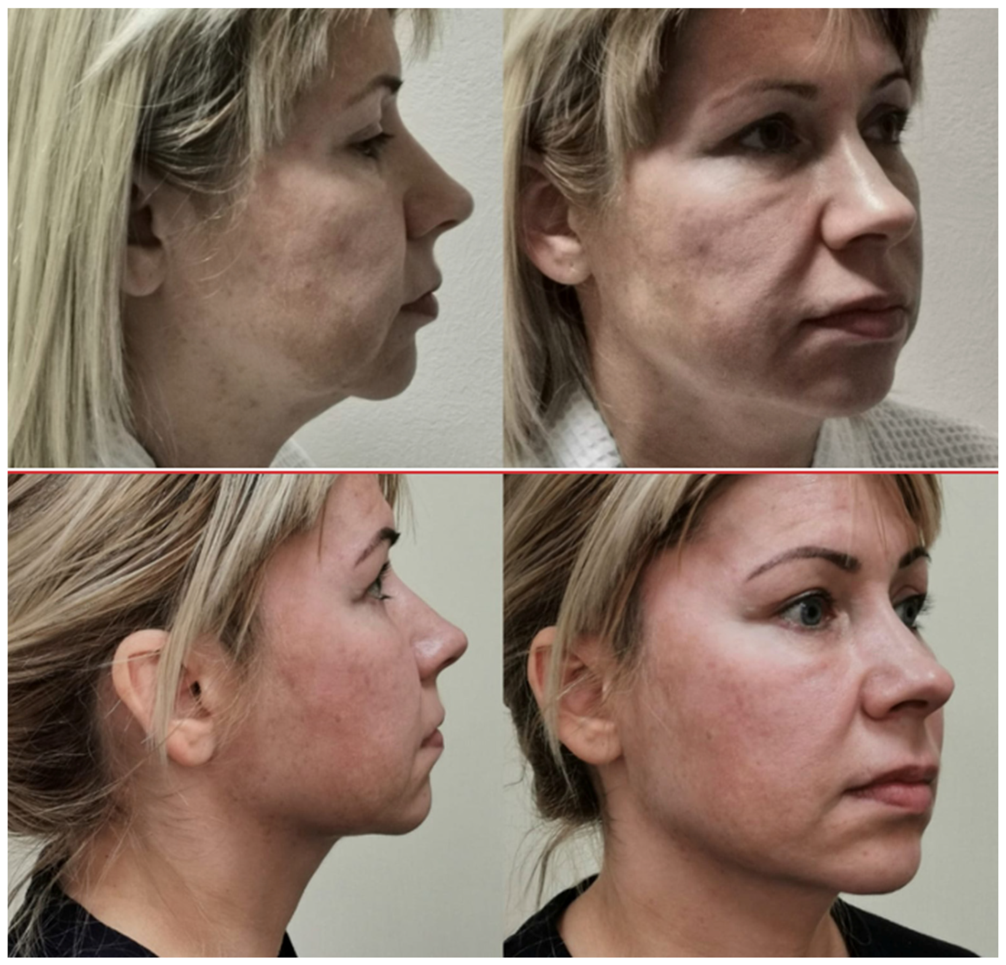
Alterations in adipose tissue volume, reduced skin elasticity, and soft-tissue ptosis contribute to changes in facial contours and the oval shape of the face as well as the deepening of the nasolabial and tear trough folds [2,3,4]. The desire to restore youthful facial contours and volume without leaving visible surgical scars has led plastic surgeons to explore minimally invasive yet effective methods for addressing age-related changes [5,6,7,8]. Women in the early stages of facial ptosis, having exhausted non-surgical options, often seek what they refer to as a “small” lift: “Just like this—a little!” (Figure 1) [9]. Should less aggressive, yet still effective, techniques be considered?
Upon detailed examination of the patient shown in Figure 1, it becomes evident that her face does not exhibit a typical zonal pattern of soft-tissue descent. Based on clinical observations and published data, early manifestations of facial soft-tissue prolapse are characterized by a combination of the following features: deepening of the nasolabial folds, skeletonization of the infraorbital region with protrusion of orbital fat, formation of the tear trough deformity, increased cutaneous mobility, and more pronounced contouring of the zygomatic fat pad [10].
Facelift techniques based on mobilization of soft tissues as a single unit using a subperiosteal plane may limit the ability to achieve optimal aesthetic correction in such cases. This limitation is related to the relatively preserved relationship between the periosteum and the overlying skin, which restricts the effective redistribution and repositioning of superficial soft tissues required for harmonious facial rejuvenation.
Concomitantly, these changes are often accompanied by a decline in skin turgor, the appearance of jowls, and progressive skin laxity in the submental and cervical regions, reflecting the multidirectional nature of facial aging [11].
Although soft-tissue descent and increased skin mobility occur across all facial zones simultaneously, the degree of expression varies. Patients most commonly complain about the development of jowls and sagging in the cervicomental area, often overlooking ptosis in the lower malar region. However, they tend to express concern about the sudden appearance of infraorbital fat protrusion [12].
In all cases, we adopt an individualized, comprehensive approach, assessing the face holistically rather than focusing solely on isolated zones.
The introduction of endoscopic techniques has marked a significant advancement in minimally invasive facial rejuvenation surgery.
Facial plastic surgery has widely adopted endoscopic techniques. Between 1996 and 2025, a series of monographs and clinical studies have confirmed the safety and efficacy of endoscopic facelift procedures [13,14,15,16,17,18,19].
Although endoscopic techniques have significantly expanded the options for facial rejuvenation, subperiosteal and temporal endoscopic approaches have certain limitations, particularly in correcting the lower face and neck. In addition, subperiosteal dissection may be associated with prolonged edema and sensory disturbances. These limitations prompted the development of the transtemporal endoscopic subcutaneous lifting technique, designed to enable targeted repositioning of superficial soft tissues with a favorable safety profile.
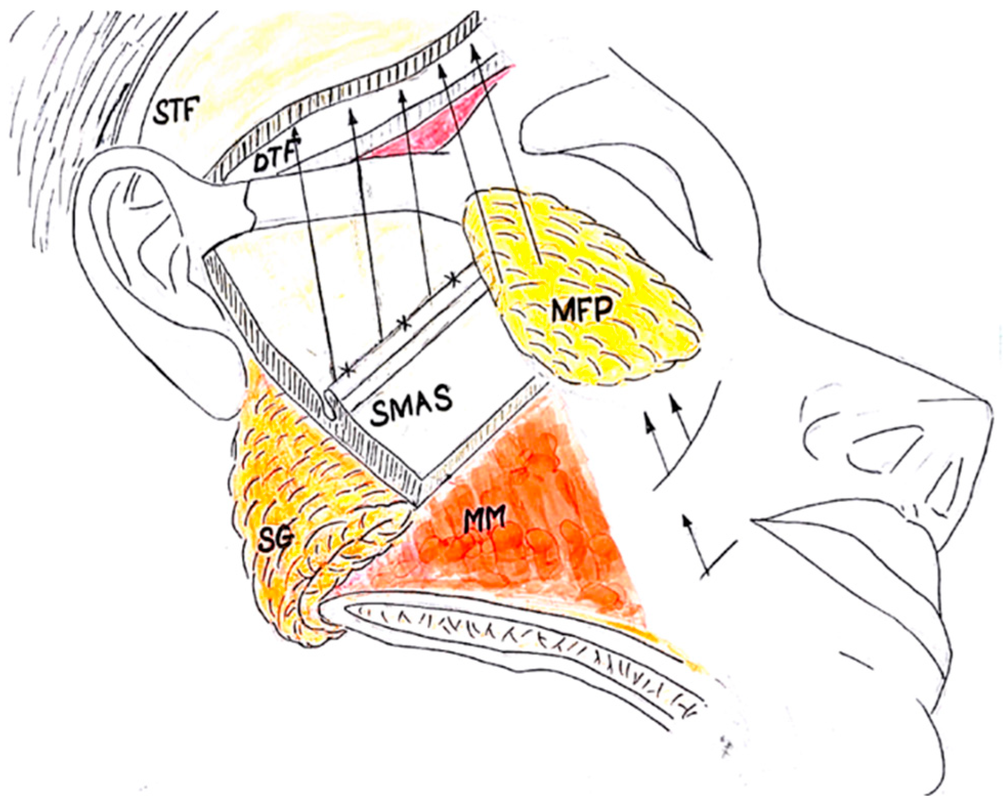
Unlike the classic endoscopic subperiosteal dissection performed via the transtemporal approach, our technique involves subcutaneous dissection above the superficial temporal fascia and the superficial musculoaponeurotic system (SMAS), utilizing endoscopic assistance to ensure optimal visualization. During the procedure, osteocutaneous and myocutaneous retaining ligaments are released to allow for sufficient mobility of the skin–fat flap—similar to what is achieved in deep plane facelifts, but within the subcutaneous plane. Based on our analysis of facial soft-tissue descent vectors, we consider vertical SMAS plication to be more physiologically appropriate (Figure 2).
Transtemporal endoscopic subcutaneous dissection enables redraping of the skin–fat layer along an upper-medial vector, reducing the risk of skin folding in the preauricular region and eliminating the need for a preauricular incision.
The purpose of this study was to evaluate clinical outcomes, complication rates, and patient-reported satisfaction following transtemporal endoscopic subcutaneous lifting over an 18-year period. A total of 612 procedures were performed, for which 201 consecutive female patients aged 30–45 years met the inclusion criteria and were included in an observational analysis of a prospectively maintained surgical database. Patient-reported satisfaction was assessed using a Likert scale (2006–2017) and the validated FACE-Q instrument (2018–2024), and postoperative complications were systematically recorded. The results demonstrated high patient satisfaction and a low complication rate.

2. Materials and Methods

2.1. Study Design

Of 612 TESL procedures performed during the study period, 201 female patients met the predefined inclusion criteria and were enrolled in an observational analysis of a prospectively maintained surgical database conducted between 2006 and 2024. Consecutive eligible patients were included. Patients with incomplete follow-up data at specific time points were excluded from the respective statistical analyses but remained part of the overall cohort description. The sample size was determined by the total number of eligible patients treated within the specified timeframe. Patient demographic data, surgical technique specifics, and postoperative outcomes were collected prospectively.
The study was conducted in accordance with the principles of the Declaration of Helsinki. Ethical approval was obtained from the Local Ethics Committee of Sechenov University (Protocol No. 24-23, dated 7 December 2023). All participants provided written informed consent prior to inclusion in the study, including consent for publication of clinical data and photographic images.
Standardized two-dimensional photographs were obtained preoperatively and at 3, 6, and 12 months and 3 years postoperatively to objectively assess aesthetic outcomes. Pre- and postoperative images were analyzed using consistent pixel-per-inch resolution, and proportional measurements were compared across time points.
Patient satisfaction rates and complication frequencies were statistically analyzed using IBM SPSS Statistics (version 28.0; IBM Corp., Armonk, NY, USA). Descriptive statistics were used to summarize patient satisfaction scores and complication rates. No inferential comparisons between time points were planned.
To minimize selection bias, consecutive eligible patients were included. Standardized photographic documentation and validated patient-reported outcome measures (FACE-Q and Likert scale) were used to reduce measurement bias.
Inclusion criteria were adult women aged 30–45 years presenting with early-to-moderate facial soft-tissue ptosis, malar fat pad descent, and early manifestations of jowling. Candidates demonstrated minimal to moderate skin redundancy, preserved skin quality and elasticity, and no indication for traditional rhytidectomy with preauricular incisions. All patients sought correction of early facial aging changes without generalized facial skin laxity.
Exclusion criteria included generalized or advanced facial skin laxity, significant excess skin requiring formal skin excision through preauricular approaches, previous sub-SMAS or deep-plane facelifts, severe platysmal banding requiring open cervical correction, and significant chronic comorbid conditions contraindicating elective surgery.
All procedures were performed using Karl Storz endoscopic equipment (KARL STORZ SE & Co. KG, Tuttlingen, Germany).

2.2. Surgical Technique

All surgical procedures were performed by a single senior surgeon with extensive experience in endoscopic facial surgery.
Preoperative preparation includes marking the incision lines in the temporal region of the scalp. The marking process involves identifying key anatomical landmarks: the zygomatic arch and its transition to the zygomatic bone, the area designated for dissection of the skin–fat flap, and the SMAS plication line. Additionally, zones requiring correction are delineated, including deep nasolabial folds, tear trough deformities, and jowls. Areas of excess adipose tissue deposition are also identified.
Under general or local anesthesia combined with intravenous sedation, a subcutaneous infiltration of the temporal region, cheek, chin, and neck is performed using a solution volume of 200–250 mL (containing 1 mL of epinephrine and 2 mL of 10% lidocaine per 1000 mL of Ringer’s solution).
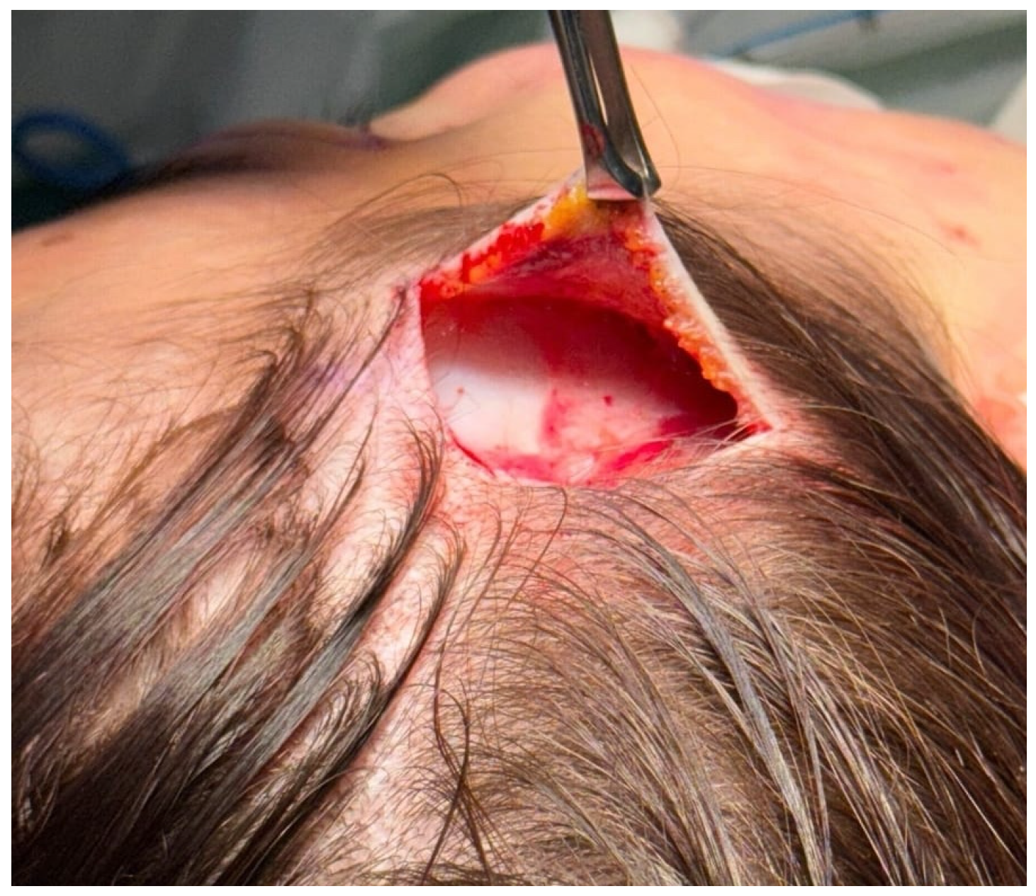
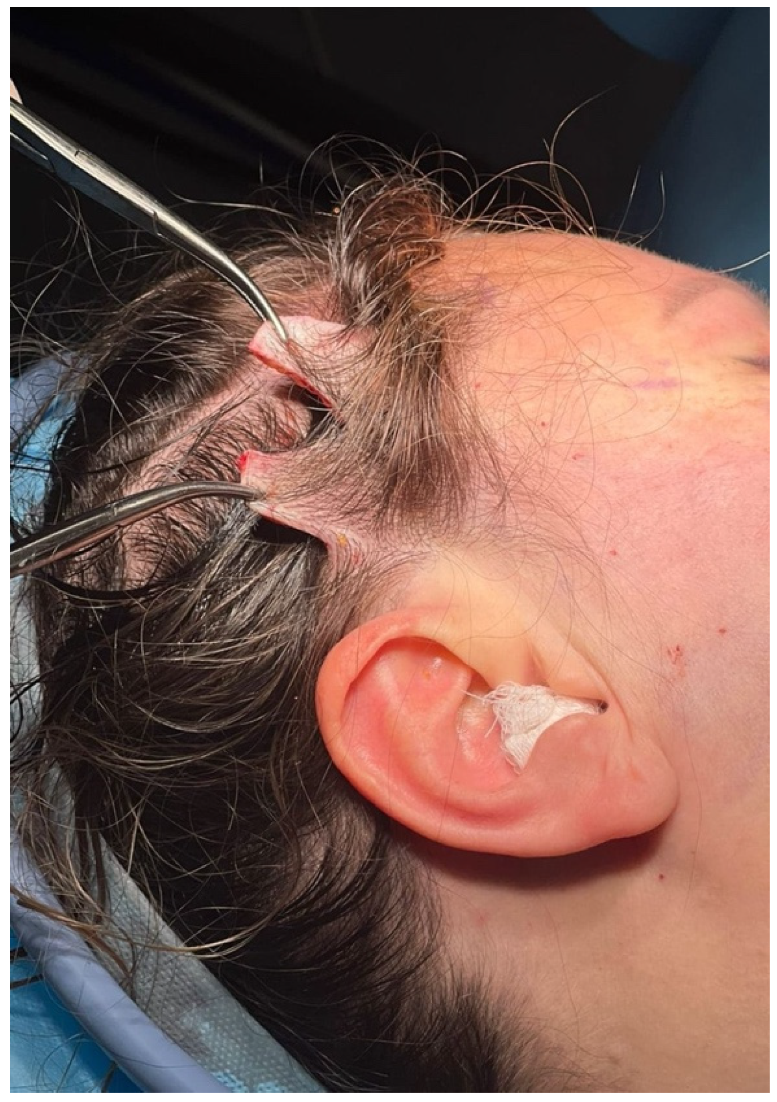
Next, a 4 cm skin incision is made in the temporal scalp area down to the superficial temporal fascia (Figure 3).
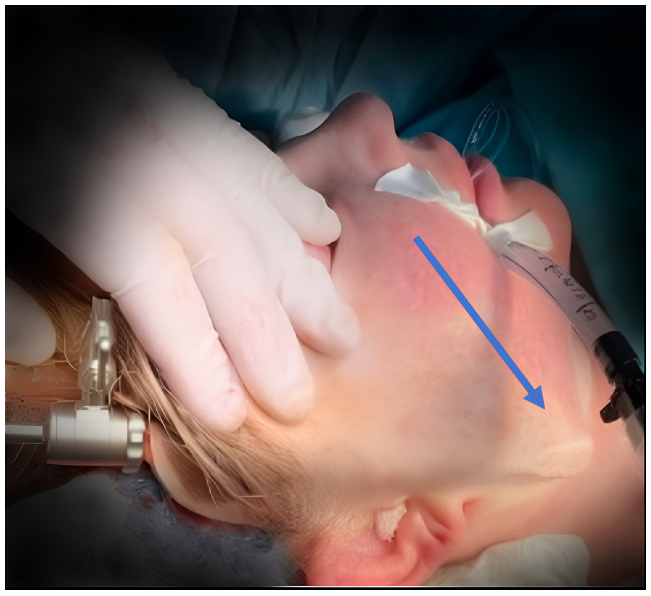
The subsequent step involves extensive subcutaneous dissection of the skin–fat flap overlying the SMAS in the temporal and malar regions under endoscopic visualization (Figure 4). Complete mobilization of the cheek skin–fat flap is achieved by elevating it above the orbicularis oculi muscle, releasing the malar fat pad, and extending the dissection medially up to the nasolabial fold. A critical component of this procedure is the careful division of osteocutaneous and myocutaneous retaining ligaments within the subcutaneous plane, which facilitates unrestricted mobilization of the skin–fat flap.
This dissection continues inferiorly to include the submental and cervical areas, thereby maximizing flap mobility. The neck is approached through the transtemporal access in the subcutaneous plane superficial to the SMAS (Figure 4). If present, excess subcutaneous fat in the cervicomental region is aspirated via a small stab incision within the chin crease using a 3 mm spatula-type cannula, preserving approximately 0.5 cm of subcutaneous fat over the skin flap to maintain vascularity.
Following liposuction, further flap mobilization is performed through the same access using a “swallowtail” dissector under endoscopic guidance.
During the elevation of the malar fat pad, it is secured to the deep temporal fascia at the lateral orbital rim. This results in an essentially vertical vector of traction on the malar fat pad, which helps to smooth the contour of the nasolabial fold (Figure 2).
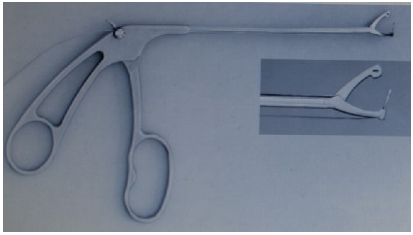
Due to the limited working space within the subcutaneous plane, the use of conventional needle holders for SMAS suturing is challenging and sometimes impossible. Therefore, we employ instruments borrowed from endoscopic surgery, often adapted from arthroscopy. One such tool used for passing sutures through the SMAS is the Acufex Suture Punch (Smith & Nephew, Andover, MA, USA) (Figure 5). Hemostasis is achieved using an endoscopic coagulator.
During SMAS plication, hydrodissection of the sub-SMAS plane with 20–30 mL of solution is performed to avoid inadvertent capture of the facial nerve branches. The retaining ligaments are released at the supra-SMAS level, and muscular manipulation is performed as a single unit during the SMAS plication step. The described technique does not involve subperiosteal dissection of the zygomatic region, as the intended vector of tissue repositioning is achieved entirely within the subcutaneous and sub-SMAS planes.
Two types of sutures are employed in our technique: plicational (interrupted) sutures on the SMAS, followed by additional long suspension sutures securing the SMAS to the deep temporal fascia. We use Supramid 3/0 (polyamide) and Maxbraid 2/0 (polyethylene) sutures. For SMAS suturing, Ethibond 2/0 sutures with a specially designed needle compatible with the Acufex Suture Punch are also utilized.
In cases where these instruments cannot be used, a percutaneous suture technique with 10 cm straight needles ((Richard-Allan®, Aspen Surgical, Caledonia, MI, USA) is applied. Knot tying during SMAS plication is performed using a dedicated knot-pusher instrument (KARL STORZ SE & Co. KG, Tuttlingen, Germany) borrowed from endoscopic surgery.
SMAS plication is carried out along a line connecting the angle of the mandible to the lateral canthus of the eye in a superior–lateral direction to correct SMAS sagging. This plication effectively recontours the facial profile, eliminating jowls. Subsequently, long suspension sutures are placed and anchored to the deep temporal aponeurosis in a vertical vector. Elevation and fixation of the platysma contribute to the improvement of the cervicomental angle.
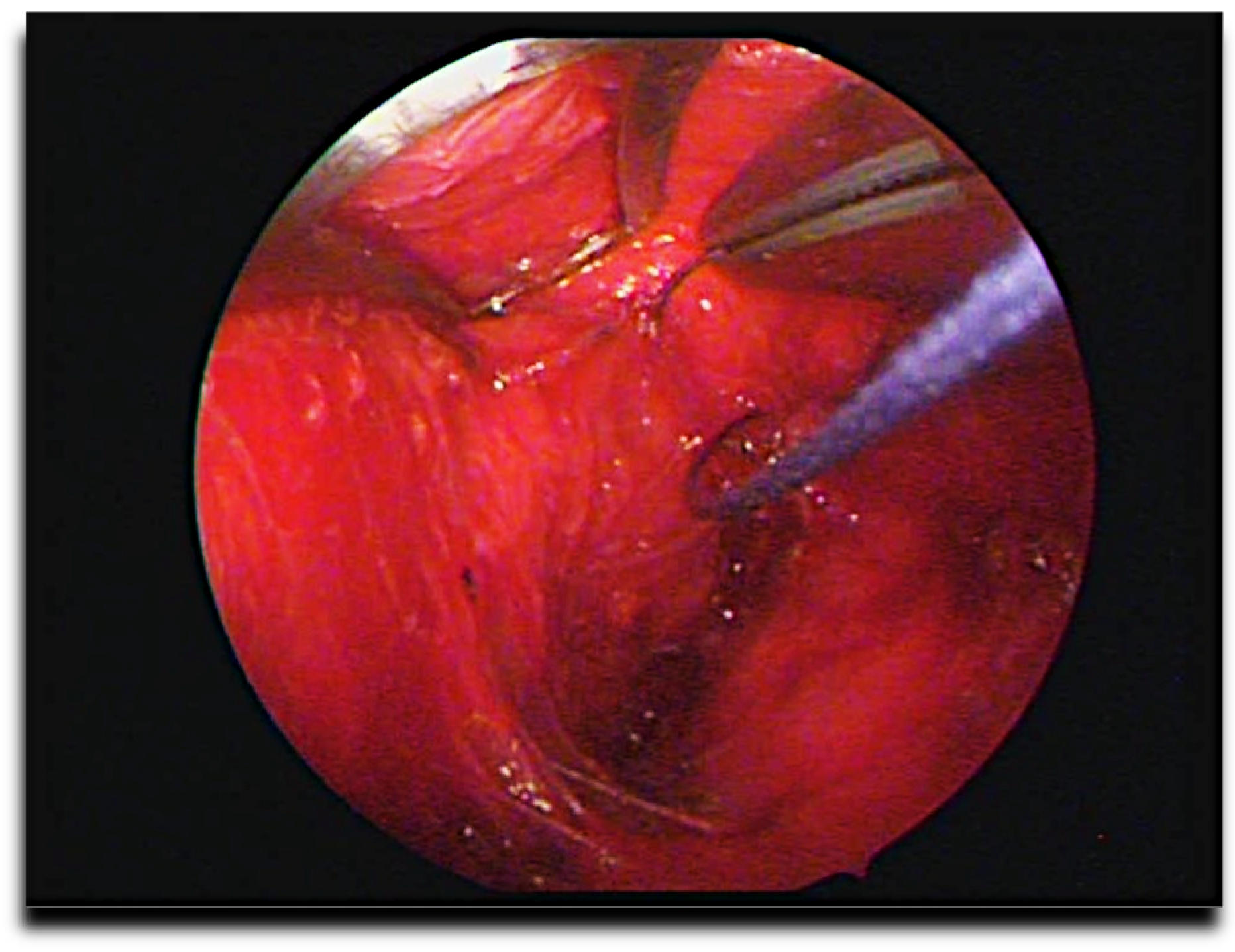
Using endoscopic instruments (KARL STORZ SE & Co. KG, Tuttlingen, Germany), the malar fat pad is sutured and elevated (Figure 6), followed by fixation to the temporal aponeurosis.
The final stage involves redistribution of the skin–fat flap with vertical or vertical–medial displacement. This vector of flap repositioning is crucial, as it aligns with the physiological vectors of tissue descent and helps to prevent skin folding anterior to the auricle.
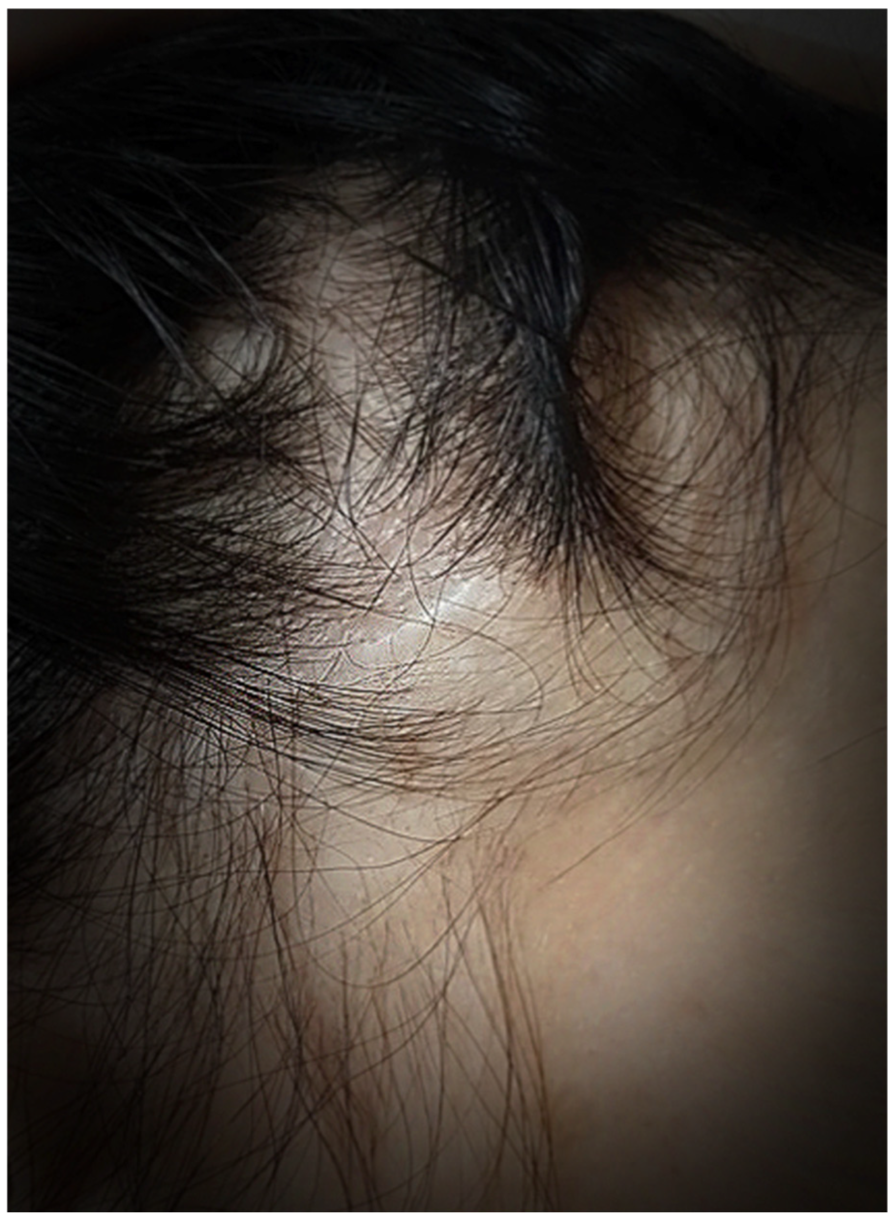
Excess soft tissues in the transtemporal access zone (Figure 7) are excised with minimal tension. Sutures are placed using non-absorbable suture material and anchored to the deep temporal aponeurosis. Skin closure is performed using surgical staples, and at 6 months postoperatively, scars are minimally visible (Figure 8).
The lifting effect in TESL is achieved primarily through extensive subcutaneous mobilization, vertical SMAS plication, and vector-oriented repositioning of the mobilized soft tissues. Cutaneous excision is minimal and individualized, performed only to achieve tension-free closure. The technique does not rely on wide skin resection as the principal lifting mechanism.
Quantitative measurement of excised skin was not systematically performed, as skin removal was minimal and tailored to individual tissue characteristics.

2.3. Patient Satisfaction Analysis

An analysis of patient satisfaction with surgical outcomes was conducted in a cohort of 201 patients aged 30 to 45 years who underwent the procedure described above. From 2006 to 2017, satisfaction was assessed in 138 patients using the Likert scale. Between 2018 and 2024, the Face-Q Score was used as the primary assessment tool.
Patients were divided into two age groups:
  • 2006–2017: 30–35 years (n = 50), 36–45 years (n = 88);
  • 2018–2024: 30–35 years (n = 22), 36–45 years (n = 41).
Age distribution and complication rates were calculated using IBM SPSS Statistics (version 28.0).
Patient satisfaction was assessed at 3, 6, and 12 months postoperatively using the FACE-Q® (aesthetics scales: Satisfaction with Facial Appearance, Satisfaction with Outcome, and Social Function (Q-Portfolio; Memorial Sloan Kettering Cancer Center, New York, NY, USA)) or the Likert scale. Statistical analysis was performed using IBM SPSS Statistics (version 28.0; IBM Corp., Armonk, NY, USA). Patients with missing follow-up data were excluded from the respective analyses. The mean Likert scale scores were reported for each age group (ranging from 1 = “completely dissatisfied” to 5 = “very satisfied”). Mean FACE-Q scores were converted to Rasch-transformed scores, with values of 75–100 indicating high satisfaction.
Patient satisfaction was assessed using a 5-point Likert scale from 2006 to 2017. Mean Likert scores were calculated for each age group and follow-up time point (3, 6, and 12 months). Overall mean satisfaction scores were calculated as weighted means based on subgroup sample sizes. Missing values were not imputed, and analyses were performed using available follow-up data only.
Patient satisfaction was assessed using different instruments during the study period. From 2006 to 2017, satisfaction was evaluated using a 5-point Likert scale, which provided a single global satisfaction score. From 2018 to 2024, satisfaction was assessed using the FACE-Q instrument, a validated patient-reported outcome measure comprising three subscales: Satisfaction with Facial Appearance, Satisfaction with Outcome, and Social Function.
Postoperative complications were recorded prospectively during follow-up visits and classified according to standard clinical practice.

3. Results

3.1. Pre- and Postoperative Photographs of Patients Undergoing Endoscopic Subcutaneous Facelifts

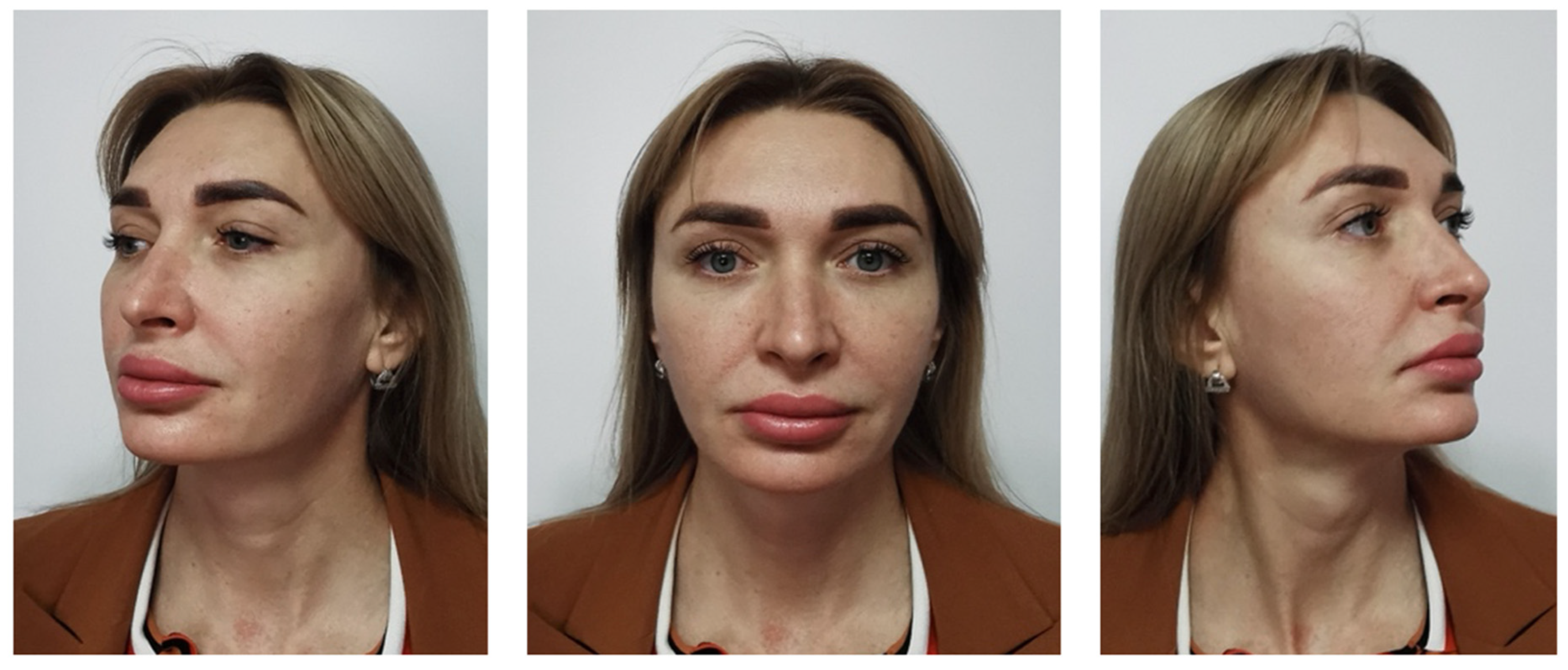
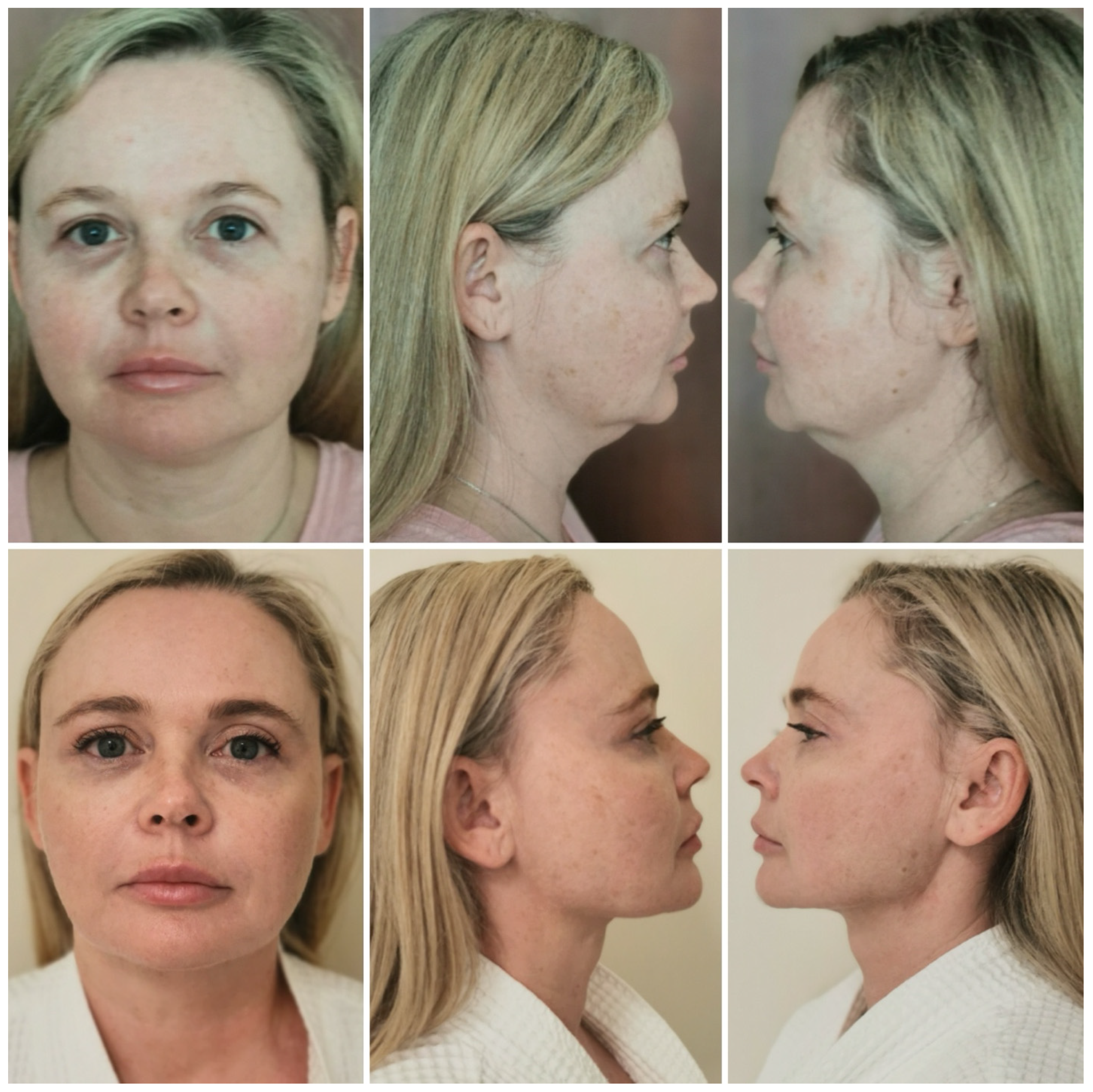
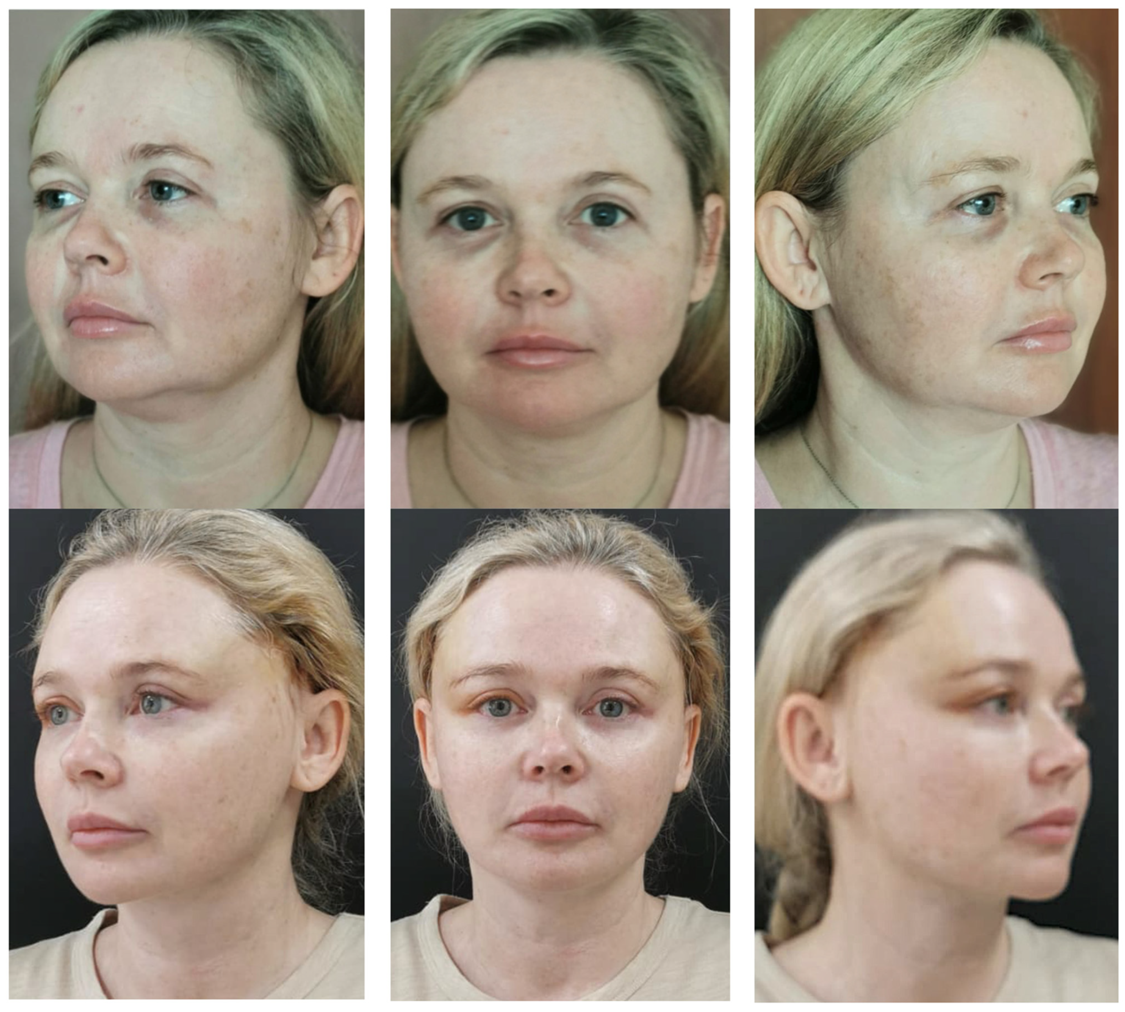
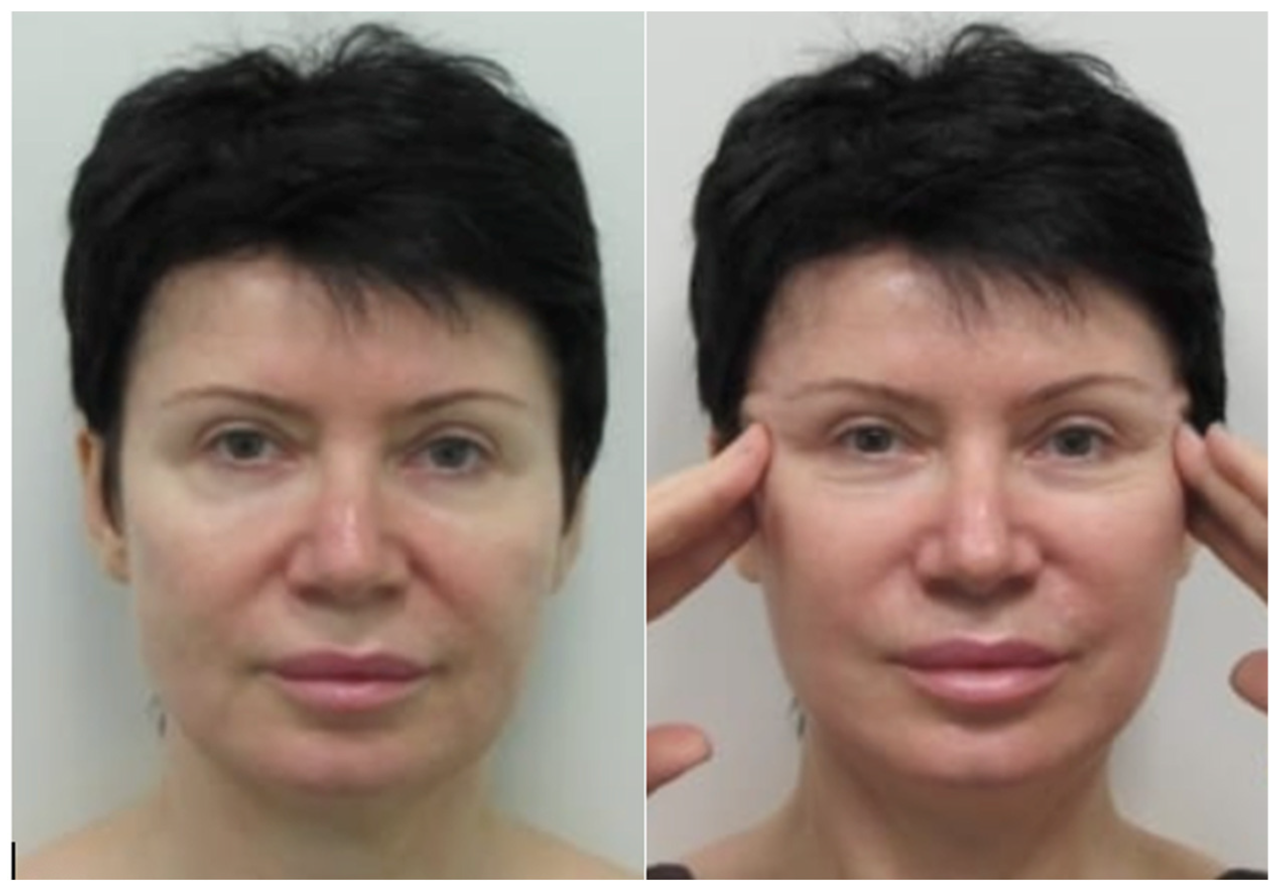
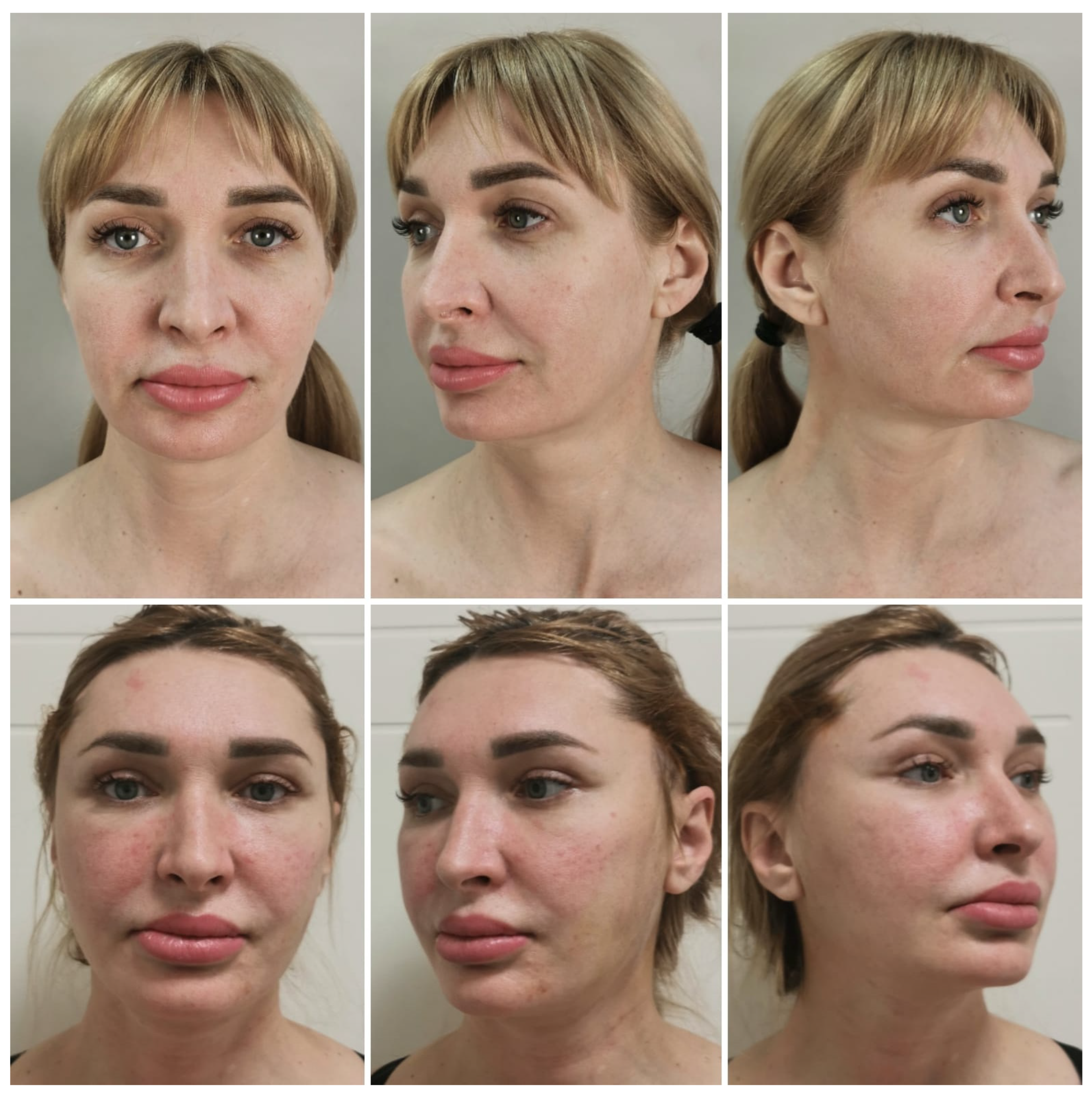
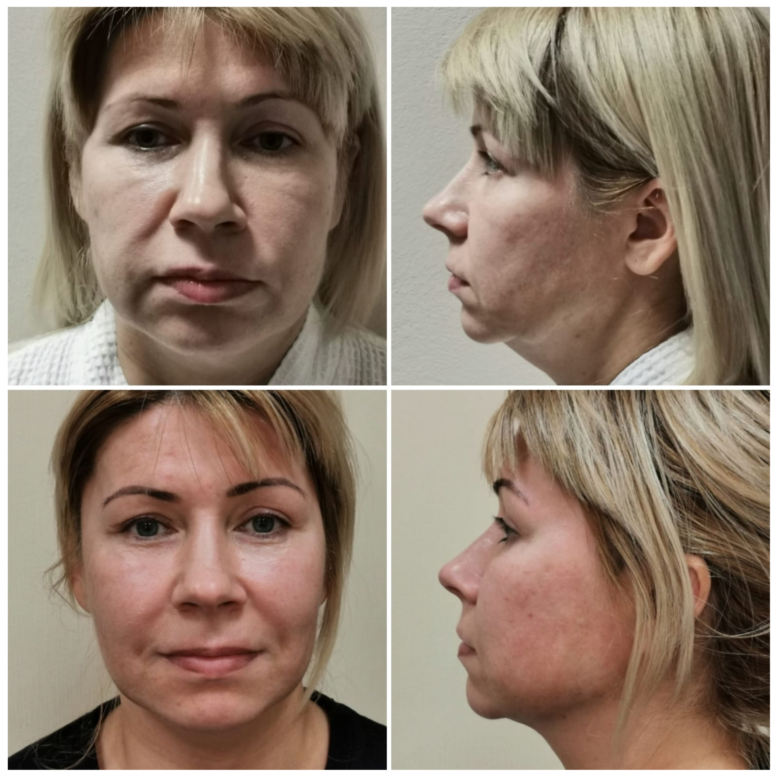
Photographs of patients before and after undergoing endoscopic subcutaneous midface lifting are presented in Figure 9, Figure 10, Figure 11, Figure 12, Figure 13 and Figure 14. These images demonstrate improvement in midfacial volume restoration and elevation of descended soft tissues, with reduction of jowling and refinement of the cervicomental angle. The postoperative photographs illustrate redraping of the skin–fat layer along an upper-medial vector, resulting in improved facial contour and lower face definition. The transtemporal approach allowed correction of facial ptosis without visible preauricular scars, contributing to favorable aesthetic outcomes with minimal cutaneous stigmata of surgery.
The patients shown in these figures were selected as representative clinical examples of the study cohort. Selection was not based on aesthetic outcome but on the availability of standardized pre- and postoperative photographs and complete follow-up data.
This technique was associated with favorable outcomes in restoring the position and volume of midfacial soft tissues through controlled repositioning of superficial fat compartments and the SMAS layer along a vertical vector. Extension of the subcutaneous dissection to the cervicomental region allows elevation of the platysma and lower facial soft tissues, contributing to improvement of jowling and refinement of the cervicomental angle. The transtemporal approach allows wide soft-tissue mobilization with minimal visible scarring, as no incisions are made anterior to the auricle.

3.2. Patient Satisfaction

Between 2006 and 2017, patient satisfaction was assessed using a 5-point Likert scale. This group included 138 patients, of whom 50 (36%) were in the 30–35 age group and 88 (64%) in the 36–45 age group.
From 2018 to 2024, satisfaction was evaluated using the FACE-Q scale. This part of the study included 63 patients, with 22 (34.92%) aged 30–35 years and 41 (65.08%) aged 36–45 years.
Mean satisfaction scores for patients operated on from 2006 to 2017 are presented in Table 1. Satisfaction levels remained consistently high across both age groups throughout the follow-up period. The overall mean Likert satisfaction score across all follow-up time points was 4.62 out of 5, indicating a high level of patient satisfaction with the surgical outcomes.
Patient satisfaction was assessed using different instruments during the study period. From 2006 to 2017, satisfaction was evaluated using a 5-point Likert scale, which provided a single global satisfaction score. From 2018 to 2024, satisfaction was assessed using the FACE-Q instrument, a validated patient-reported outcome measure comprising three subscales: Satisfaction with Facial Appearance, Satisfaction with Outcome, and Social Function. Thus, the FACE-Q provided a multidimensional assessment of patient-reported outcomes, unlike the single-item Likert scale used in the earlier cohort.
Because the Likert scale and FACE-Q represent different outcome measures, the results from the two cohorts are presented in separate tables and were not directly compared.
Satisfaction data collected via the FACE-Q scale (2018–2024) also showed consistently high patient satisfaction at all follow-up points (3, 6, and 12 months) (Table 2, Table 3 and Table 4).

3.3. Complications

The complications presented in Table 5 were selected because they represent the clinically relevant adverse events observed in the study cohort. These included cicatricial alopecia, postoperative hematoma, and skin pleating in the preauricular region, which required surgical revision. No postoperative inflammatory or neurological complications were observed. No additional complications, including wound infection, seroma, facial nerve injury, skin necrosis, or delayed wound healing, were recorded during the follow-up period.

4. Discussion

The creation of a composite sub-SMAS flap does not always adequately address skin laxity in the central facial oval. In such cases, subcutaneous dissection with division of the osteocutaneous and myocutaneous ligaments allows precise repositioning and correction of ptotic superficial soft tissues. Apparently, the ideal approach involves a two-plane dissection combining both subperiosteal and subcutaneous layers.
  • Endoscopically assisted facelift performed via remote temporal access with subcutaneous dissection and SMAS plication represents a promising method for correcting facial skin folds and redistributing the overlying tissues compared to purely subperiosteal dissection.
  • We reviewed studies addressing age-related changes in the anthropometric characteristics of the facial skeleton associated with continuous cranial bone growth [20], as well as the “movement” of the facial skeleton or bone resorption [21]. The latter process is typically related to tooth loss [22].
  • We consider the bony structures to be stable support elements for soft-tissue fixation. Changes in these structures in patients aged 30–45 years are not critical.
  • During our analysis, facial tissues were categorized according to their stability.
  • In our view, stable structures include the zygomatic bone, zygomatic arch, and cranial vault bones.
The following are conditionally stable facial structures:
Osteocutaneous ligaments are subject to minimal stretching and serve as suspension points for the overlying soft tissues, which are more prone to significant elongation, resulting in skin folds.
Myocutaneous ligaments exhibit greater mobility due to the elasticity of the underlying muscles [23].
The superficial musculoaponeurotic system (SMAS) is also involved in this dynamic [24].
Unstable structures include the skin and subcutaneous fat. The entire soft-tissue complex of the face and neck, under the influence of gravity, is subject not only to stretching but also to proliferative processes, i.e., direct tissue growth.
Because the skin volume actually increases, merely repositioning the tissues to their original location is insufficient; the excess tissue must also be excised to restore the original facial contours.
Over several years of performing this type of surgery, we have refined the technique, developing a vector-divergent lifting approach. Elevation of the malar fat pad is performed using long sutures anchored to the deep temporal fascia near the lateral orbital rim, which allows smoothing of the nasolabial fold contour and lifting of the central facial oval.
SMAS plication is carried out in the superior–lateral direction, effectively correcting SMAS ptosis with smoothing of the vertical perioral folds, associated with a sad expression, and the elimination of jowls.
The next step involves the placement of long suspension sutures anchored to the deep temporal aponeurosis in a vertical vector. Elevation and fixation of the platysma contribute to the improvement of the cervicomental contour and reinforce the platysma’s position within the SMAS complex.
Uniform redistribution of the skin–fat flap predominantly in a vertical–medial direction requires extensive subcutaneous dissection of the face and corresponds to the physiological vectors of tissue descent, thereby reducing the likelihood of skin folding anterior to the auricle.
While the upper-medial vector of skin–fat flap redistribution aims to reduce the likelihood of preauricular skin pleating, minor folding in this region was observed in 11 patients (5.47%) and required secondary revision. These revisions were minor contour-optimizing procedures and did not affect the overall aesthetic outcome.
Our vector-divergent facelift technique has improved surgical outcomes and postoperative tissue stability.
One of the notable postoperative challenges associated with the transtemporal approach is localized alopecia. In our series, alopecic patches did not exceed 1 cm in width and were generally visually inconspicuous in women with long hair. When patients expressed aesthetic concern, the alopecic area was excised, and direct closure of the hair-bearing flap was performed without elevation of the hairline.
Additionally, partial de-epithelialization of the scar allowed it to be inset beneath adjacent healthy hair-bearing tissue, with proximal fixation to the deep temporal fascia. This maneuver reinforced temporal support and contributed to the stability of the lifting result.
Based on these considerations, TESL is indicated primarily in patients with early-to-moderate facial ptosis and minimal skin redundancy. In cases of generalized facial skin laxity requiring substantial skin excision, conventional rhytidectomy techniques may be more appropriate.

Limitations

This study has several limitations. The observational design requires cautious interpretation of the findings, as no direct comparative analysis with alternative facelift techniques was performed. Only female patients aged 30–45 years were included, which limits the generalizability of the results to male patients and other age groups.
Furthermore, the extended study period (2006–2024) may have been associated with gradual refinements in surgical technique, patient selection, and perioperative management, potentially introducing temporal heterogeneity.
Accordingly, the findings are primarily applicable to women with early-stage facial soft-tissue ptosis treated in specialized surgical settings. Extrapolation beyond this patient population should be undertaken with caution.
TESL is not intended for patients with advanced generalized facial laxity or significant skin redundancy. In such cases, conventional rhytidectomy techniques involving wider skin excision and preauricular approaches may be more appropriate. Therefore, TESL should be regarded as a targeted intervention for early-to-moderate facial ptosis rather than a universal substitute for traditional facelift procedures.

5. Conclusions

The endoscopically assisted subcutaneous facelift performed through a temporal approach provides endoscopic visualization of the surgical field and allows controlled mobilization and repositioning of superficial soft tissues. In the present study, this technique was associated with favorable clinical outcomes and a low rate of postoperative complications. The absence of preauricular incisions results in minimal visible scarring, which may be advantageous for patients presenting with early signs of facial soft-tissue ptosis.

Author Contributions

Conceptualization, A.E.A. and V.I.S.; methodology, A.E.A., V.I.S. and A.S.B. (Anastasiya S. Borisenko); software, A.S.B. (Anna S. Bairamova); validation, A.E.A., V.I.S. and A.S.B. (Anastasiya S. Borisenko); formal analysis, A.S.B. (Anastasiya S. Borisenko); investigation, A.S.B. (Anna S. Bairamova) and A.S.B. (Anastasiya S. Borisenko); resources, A.E.A. and V.I.S.; data curation, A.S.B. (Anastasiya S. Borisenko); writing—original draft preparation, A.S.B. (Anna S. Bairamova); writing—review and editing, A.E.A., V.I.S. and A.S.B. (Anastasiya S. Borisenko); visualization, A.S.B. (Anna S. Bairamova); supervision, A.E.A. and V.I.S.; project administration, A.E.A.; funding acquisition, A.E.A. All authors have read and agreed to the published version of the manuscript.

Funding

This research received no external funding. The publication was funded by the authors.

Institutional Review Board Statement

The study was conducted in accordance with the principles of the Declaration of Helsinki. Ethical approval was obtained from the Local Ethics Committee of Sechenov University (Protocol No. 24-23, dated 7 December 2023).

Informed Consent Statement

Informed consent was obtained from all subjects involved in the study. Written informed consent has been obtained from the patient(s) to publish this paper.

Data Availability Statement

De-identified data supporting the findings of this study are available from the corresponding author upon reasonable request. Individual patient data are not publicly available due to privacy and ethical considerations.

Conflicts of Interest

The authors declare no conflicts of interest.

References

  1. Manturova, N.E.; Talolin, N.P.; Andryushchenko, O.A. The use of algorithms in aesthetic facial surgery. Plast. Surg. Aesthetic Med. 2023, 2, 67–75. (In Russian) [Google Scholar] [CrossRef]
  2. Quatela, V.; Azzi, J.P.; Antunes, M. Endoscopic-Assisted Facelifting. Facial Plast. Surg. 2014, 30, 413–421. [Google Scholar] [CrossRef] [PubMed]
  3. Verbo, E.V.; Manturova, N.E.; Orlova, Y.M. The evolution of facial rejuvenation surgery techniques. Plast. Surg. Aesthetic Med. 2022, 4, 66–76. (In English) [Google Scholar] [CrossRef]
  4. Dayan, S.; Rivkin, A.; Sykes, J.M.; Teller, C.F.; Weinkle, S.H.; Shumate, G.T.; Gallagher, C.J. Aesthetic Treatment Positively Impacts Social Perception: Analysis of Subjects From the HARMONY Study. Aesthetic Surg. J. 2019, 39, 1380–1389. [Google Scholar] [CrossRef]
  5. Nellis, J.C.; Ishii, M.; Byrne, P.J.; Boahene, K.D.O.; Dey, J.K.; Ishii, L.E. Association Among Facial Paralysis, Depression, and Quality of Life in Facial Plastic Surgery Patients. JAMA Facial Plast. Surg. 2017, 19, 190–196. [Google Scholar] [CrossRef] [PubMed] [PubMed Central]
  6. Wu, L.; Hua, Z.; Tong, D.; Zhu, S.; Zhang, C.; Wang, X.; Wang, J.J. Guided suturing technique for midface lift through minimal temporal incision. Plast. Reconstr. Aesthetic Surg. 2021, 74, 3108–3113. [Google Scholar] [CrossRef] [PubMed]
  7. Bellity, P.; Bellity, J. Facial Rejuvenation Enhancing Cheek Lift. Arch. Plast. Surg. 2017, 44, 559–563. [Google Scholar] [CrossRef] [PubMed] [PubMed Central]
  8. Ugradar, S.; Kim, J.S.; Massry, G. A Review of Midface Aging. Ophthalmic Plast. Reconstr. Surg. 2023, 39, 123–131. [Google Scholar] [CrossRef] [PubMed]
  9. Kappos, E.A.; Temp, M.; Schaefer, D.J.; Haug, M.; Kalbermatten, D.F.; Toth, B.A. Validating Facial Aesthetic Surgery Results with the FACE-Q. Plast. Reconstr. Surg. 2017, 139, 839–845. [Google Scholar] [CrossRef]
  10. Kaya, K.S.; Cakmak, O. Facelift Techniques: An Overview. Facial Plast. Surg. 2022, 38, 540–545. [Google Scholar] [CrossRef] [PubMed]
  11. Mendelson, B.C.; O’Brien, J.X. The Aging Face. In Plastic Surgery; Elsevier: Amsterdam, The Netherlands, 2018; Section VIII, Chapter 61. [Google Scholar]
  12. Fedok, F.G.; Chaikhoutdinov, I.; Garritano, F. The difficult neck in facelifting. Facial Plast. Surg. 2014, 30, 438–450. [Google Scholar] [CrossRef]
  13. Botti, G.; Botti, C. Midface Lift: Our Current Approaches. Handchir. Mikrochir. Plast. Chir. 2014, 46, 224–233. [Google Scholar] [CrossRef] [PubMed]
  14. Pascali, M.; Botti, C.; Cervelli, V.; Botti, G. Critical Analysis of Midface Reconstruction Results. Plast. Reconstr. Surg. 2015, 135, 1305–1316. [Google Scholar] [CrossRef] [PubMed]
  15. Gencer, M.; Kerem, M.; Sağlam, Y.; Tatar, B.E. Dual-Plane Midface Lift Through Transoral and Transtemporal Approach. Aesthetic Plast. Surg. 2024, 48, 4609–4618. [Google Scholar] [CrossRef] [PubMed]
  16. Saltz, R.; Ohana, B. Thirteen Years of Experience with the Endoscopic Midface Lift. Aesthetic Surg. J. 2012, 32, 927–936. [Google Scholar] [CrossRef]
  17. Alimova, S.M.; Sharobaro, V.I.; Shamanaeva, L.S.; Penaeva, S.A. Psychometric scales in the assessment of satisfaction with the results of aesthetic correction of the cervicofacial region. Plast. Surg. Aesthetic Med. 2021, 4, 77–82. [Google Scholar] [CrossRef]
  18. Pessa, J.E.; Chen, Y. Curve analysis of the aging orbital aperture. Plast. Reconstr. Surg. 2002, 109, 751–755. [Google Scholar] [CrossRef]
  19. Pessa, J.E. An algorithm of facial aging: Verification of Lambros’s theory by three-dimensional stereolithography, with reference to the pathogenesis of midfacial aging, scleral show, and the lateral suborbital trough deformity. Plast. Reconstr. Surg. 2000, 106, 479–488. [Google Scholar] [CrossRef]
  20. Kul, Z.; Eryilmaz, E.; Özer, E. Transtemporal Endoscopic Deep Plane Face Lift. Plast. Reconstr. Surg. Glob. Open 2025, 13, e6461. [Google Scholar] [CrossRef]
  21. Firat, M. Endoscopic Deep Plane Facelift: A Classified Approach. Aesthetic Surg. J. 2025. [Google Scholar] [CrossRef]
  22. Mendelson, B.C.; Hartley, W.; Scott, M.; McNab, A.; Granzow, J.W. Age-related changes of the orbit and midcheek and the implications for facial rejuvenation. Aesthetic Plast. Surg. 2007, 31, 419–423. [Google Scholar] [CrossRef] [PubMed]
  23. Charafeddine, A.H.; Drake, R.; McBride, J.; Zins, J.E. Facelift: History and Anatomy. Clin. Plast. Surg. 2019, 46, 505–513. [Google Scholar] [CrossRef] [PubMed]
  24. Freeman, M.S. Rejuvenation of the midface. Facial Plast. Surg. 2003, 19, 223–236. [Google Scholar] [CrossRef] [PubMed]
Figure 1. Preoperative clinical photograph of a patient with early-stage facial ptosis demonstrating the desired extent of surgical rejuvenation.
Cosmetics 13 00081 g001
Figure 2. Vertical plication of the SMAS allows for redefinition of the jawline and effective correction of jowls, restoring a youthful facial contour. The arrows indicate the direction of plication.
Cosmetics 13 00081 g002
Figure 3. Incision in the temporal region of the scalp.
Cosmetics 13 00081 g003
Figure 4. Subcutaneous dissection extending to the chin and neck regions is performed under endoscopic visualization. The arrow indicates the extent of the extended dissection achievable with this technique, allowing correction of the jawline and submental region.
Cosmetics 13 00081 g004
Figure 5. Acufex Suture Punch.
Cosmetics 13 00081 g005
Figure 6. Suturing and elevation of the malar fat pad.
Cosmetics 13 00081 g006
Figure 7. Excision and redistribution of the excess skin–fat flap with fixation to the deep temporal aponeurosis using non-absorbable suture material.
Cosmetics 13 00081 g007
Figure 8. Scar condition 6 months after the surgical procedure.
Cosmetics 13 00081 g008
Figure 9. Preoperative view and 3 months after endoscopic subcutaneous midface lift.
Cosmetics 13 00081 g009
Figure 10. Preoperative view and 6 months after endoscopic subcutaneous midface lift.
Cosmetics 13 00081 g010
Figure 11. Preoperative view and 1 year after endoscopic subcutaneous midface lift.
Cosmetics 13 00081 g011
Figure 12. Preoperative view and 3 years after endoscopic subcutaneous midface lift.
Cosmetics 13 00081 g012
Figure 13. Preoperative view and 3 years after endoscopic subcutaneous midface lift.
Cosmetics 13 00081 g013
Figure 14. Preoperative view and 3 years after endoscopic subcutaneous midface lift.
Cosmetics 13 00081 g014
Table 1. Patient satisfaction among those operated on from 2006 to 2017 (Likert scale).
Follow-Up Period30–35 Years36–45 YearsOverall Mean
3 months4.684.794.75
6 months4.204.574.43
12 months4.524.864.69
Overall (2006–2017)4.62
Table 2. FACE-Q™—Satisfaction with Facial Appearance.
Follow-Up PeriodAge Group 30–35 Years
(22 Patients)
Age Group 36–45 Years
(41 Patients)
3 months95 FACE-Q points97.3 FACE-Q points
6 months74.3 FACE-Q points95 FACE-Q points
12 months86.9 FACE-Q points98.8 FACE-Q points
Table 3. FACE-Q™—Satisfaction with Outcome.
Follow-Up PeriodAge Group 30–35 Years
(22 Patients)
Age Group 36–45 Years
(41 Patients)
3 months94.8 FACE-Q™ points95 FACE-Q™ points
6 months85.2 FACE-Q™ points93.1 FACE-Q™ points
12 months97.6 FACE-Q™ points98 FACE-Q™ points
Table 4. FACE-Q™—Social Function.
Follow-Up PeriodAge Group 30–35 Years (22 Patients)Age Group 36–45 Years (41 Patients)
3 months91 FACE-Q™ points95.7 FACE-Q™ points
6 months85.8 FACE-Q™ points89.2 FACE-Q™ points
12 months94.5 FACE-Q™ points96.7 FACE-Q™ points
Table 5. Postoperative complications stratified by age group.
ComplicationAge Group 30–35 Years (n = 72)Age Group 36–45 Years (n = 129)Total (n = 201)
Cicatricial alopecia6 patients (8.33%)7 patients (5.43%)13 patients (6.47%)
Postoperative hematoma1 patient (1.39%)3 patients (2.33%)4 patients (1.99%)
Skin pleating in the preauricular area4 patients (5.56%)7 patients (5.43%)11 patients (5.47%)
Inflammation0 patients (0%)0 patients (0%)0 patients (0%)
Values are presented as number of patients (%).
Disclaimer/Publisher’s Note: The statements, opinions and data contained in all publications are solely those of the individual author(s) and contributor(s) and not of MDPI and/or the editor(s). MDPI and/or the editor(s) disclaim responsibility for any injury to people or property resulting from any ideas, methods, instructions or products referred to in the content.