Article

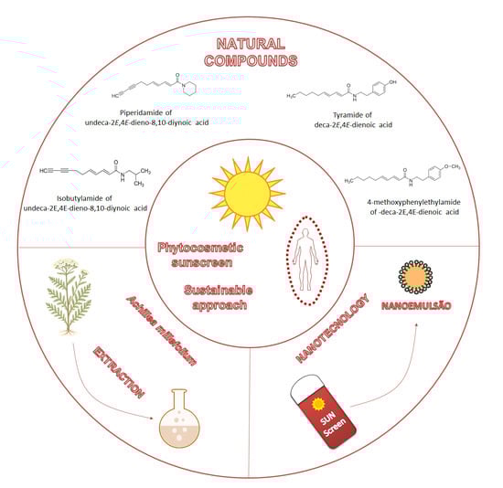
Development of a Multifunctional Phytocosmetic Nanoemulsion Containing Achillea millefolium: A Sustainable Approach

by
Thais Silva Christiani
1,
Luciana Pereira Rangel
2,
Andressa Souto Ramalho Soares
3,
Anne Caroline Candido Gomes
4,
Ariely Costa dos Santos
2,
Mariana Sato S. B. Monteiro
3,
Naomi Kato Simas
1 and
Eduardo Ricci-Junior
3,*
1
Laboratório de Produtos Naturais e Ensaios Biológicos, Faculdade de Farmácia, Universidade Federal do Rio de Janeiro, Rio de Janeiro 21941-902, RJ, Brazil
2
Laboratório de Bioquímica Tumoral, Faculdade de Farmácia, Universidade Federal do Rio de Janeiro, Rio de Janeiro 21941-902, RJ, Brazil
3
Laboratório de Desenvolvimento Galênico (LADEG), Faculdade de Farmácia, Universidade Federal do Rio de Janeiro, Rio de Janeiro 21941-902, RJ, Brazil
4
Instituto Federal de Educação, Ciência e Tecnologia, Campus Realengo, Rio de Janeiro 21715-000, RJ, Brazil
*
Author to whom correspondence should be addressed.
Cosmetics 2025, 12(6), 255; https://doi.org/10.3390/cosmetics12060255
Submission received: 18 September 2025 / Revised: 17 October 2025 / Accepted: 28 October 2025 / Published: 11 November 2025

Abstract

Skin aging, including photoaging, is primarily triggered by chronic exposure to solar radiation, which induces free radical formation, cellular deoxyribonucleic acid (DNA) damage, and structural skin alterations. Achillea millefolium L. (Asteraceae) is rich in phenolic compounds and alkamides, substances known for their antioxidant activity. This study aimed to develop and characterize a photoprotective phytocosmetic nanoemulsion containing crude root extract of A. millefolium. The extract exhibited a total phenolic content of 3.067 ± 1.911 µg GAE/mL, potent antioxidant activity (EC50 = 69.11 ± 8.899 µg/mL), moderate tyrosinase inhibition (19 ± 1.8%), and no cytotoxicity in keratinocytes. The extract was incorporated into nanoemulsions at concentrations of 0.1%, 0.5%, and 1%. The resulting droplets showed mean diameters of 217 to 230 nm, with a significant increase in the polydispersity index (PDI) after extract addition (p < 0.05). Transmission electron microscopy (TEM) confirmed the spherical morphology of the droplets. The in vitro sun protection factor (SPF) was 14 ± 0.9 in the control formulation and increased to 15 ± 2.0 (0.1%), 22 ± 5.2 (0.5%), and 17 ± 1.0 (1%), suggesting a synergistic effect between the extract and chemical filters. All formulations demonstrated UVA/UVB ratio > 0.6, a pH near to 5, occlusive properties, and good spreadability. The results indicate that A. millefolium extract holds potential for safe photoprotective formulations, offering a valuable antioxidant and depigmenting activity in addition to enhancing the SPF. This position is an innovative alternative to phytocosmetic development.

Graphical Abstract

1. Introduction

The increase in life expectancy has driven the demand for beauty and personal care products [1]. This is largely because advancing age is accompanied by biochemical and metabolic changes that directly affect the skin, leading to cutaneous aging. This complex process results from two distinct mechanisms: chronological aging (an intrinsic factor) and extrinsic aging, primarily associated with environmental factors [2,3,4]. Intrinsic aging is progressive, resulting from the body`s natural senescence, and is influenced by genetic factors, hormonal changes, and oxidative stress [5]. Extrinsic aging, on the other hand, is associated with environmental factors and lifestyle, such as solar radiation, poor diet, alcohol consumption, and stress [5]. The cumulative effects of these extrinsic factors compound intrinsic aging [6], manifesting as wrinkles, loss of firmness, dryness, hyperpigmentation, erythema, telangiectasia, and, in severe cases, skin cancer [3,4].
The global population has become increasingly concerned about the prevention and reversal of these signs of skin aging [6,7]. The anti-aging concept involves two main strategies: prevention through measures that minimize aging signs, and treatment, aimed at reversing established clinical manifestations [4]. In this context, multifunctional cosmetics represent one of the most effective strategies for both prevention and treating skin aging [6,8].
As noted, ultraviolet (UV) radiation is the main driven of photoaging, a degenerative process characterized by structural and functional alterations of the skin [3,6,9,10]. Beyond UV, visible light and infrared radiation also contribute to dermal damage [3]. While organic UV filters are widely used in photoprotective formulations, several have been linked to health and environmental concerns, including allergic reactions, irritation, toxicity, and significant ecological impacts, such as coral bleaching [11]. This combined concern for human safety and environmental sustainability has pushed the cosmetic industry toward investing in natural formulations that combine efficacy with reduced environmental impact [12,13].
Considering that conventional cosmetics may generate residues harmful to human health and the environment [12,14], the use of plant-derived actives ingredients with antioxidants, moisturizing, and depigmenting properties has emerged as a promising alternative. Previous studies have demonstrated that incorporating natural extracts, such as those from red propolis and sugarcane, into photoprotective formulas can successfully increase the in vitro Sun Protection Factor (SPF), enhance antioxidant activity, and improve overall skin protection, all while promoting sustainability [15,16].
Among plant species of cosmetic interest, Achillea millefolium L. (Asteraceae) is particularly relevant. This perennial plant, native to Europe and western Asia, is widely distributed globally and found in regions of Brazil’s Atlantic Forest [17,18]. The aerial parts of this species possess recognized antioxidant potential, mainly due to their high content of phenolic compounds [19,20,21,22]. Furthermore, a clinical study demonstrated a significant reduction in the appearance of wrinkles and pores after two months of using a cosmetic formulation containing a 2% A. millefolium extract [23]. Key bioactive compounds include flavonoids like apigenin, luteolin, quercetin and their glycosylated derivatives, as well as phenolic acids such as caffeoylquinic acid [21,24,25,26,27,28,29,30]. Furthermore, alkamides, present in over 100 plant species, including A. millefolium, exhibit antimicrobial, immunomodulatory, anti-inflammatory, and anti-aging activities, often used in cosmetics to smooth wrinkles and produce a lifting effect [17,31,32,33]. An example is spilanthol, found in Acmella oleracea, which is employed in commercial products (Gatuline®, SYN-COLL®, and ChroNOline™), for reducing subcutaneous facial contractions without cytotoxicity and enhanced skin permeation [34,35,36].
The roots of A. millefolium have been shown to contain several alkamides, in-clud-ing deca-2E,4E,6Z-trienoic acid-2,3-dehydropiperidinamide; de-ca-2E,4E,6Z,8Z-tetraenoic acid-2,3-dehydropiperidinamide; de-ca-2E,4E,6E,8Z-tetraenoic acid-2,3-dehydropiperidinamide; and deca-2E,4E-dienoic acid-4-methoxyphenylethylamide [37,38]. Our research group has also identified iso-butylamide of undeca-2E,4E-dieno-8,10-diynoic acid, tyramine amid of de-ca-2E,4E-dieno-9,10-diynoic acid, pellitorine, and homospilanthol [39].
In this context, the present study aimed to develop a multifunctional phytocosmetic photoprotective nanoemulsion combining two organic filters, octyl methoxycinnamate (OMC) and diethylamino hydroxybenzoyl hexyl benzoate (DHHB), with the crude root extract of A. millefolium. The novelty of this study lies in presenting, for the first time, a stable nanoemulsion incorporating a crude extract specifically derived from Achillea millefolium roots. The root is a little-explored source whose distinct phytochemical profile, potentially rich in alkamides and sinapaldehyde, is leveraged here as a multifunctional anti-photoaging phytochemical. The nanoemulsion platform serves as an advanced delivery system that significantly enhances photoprotection (evidenced by increased and broader coverage). The work is distinguished by its comprehensive approach, which integrates: Thorough physicochemical characterization, Evaluation of primary anti-aging activities (antioxidant and depigmenting potential), Direct correlation of these activities with the observed gains in sun protection performance. Taken together, this research systematically demonstrates the potential of the A. millefolium root extract as a multifunctional active delivered via nanoemulsion, simultaneously addressing multiple pathways of photoprotection and anti-aging.

2. Materials and Methods

2.1. Materials

The roots of A. millefolium were donated by the Vita Oliva Garden company. The voucher specimen was registered and deposited at the Herbarium Prof. Jorge Pedro Pereira Carauta under the code HUNI 6894. The solvent Ethanol P.A. was purchased from Neon Comercial Reagentes Analíticos Ltda (São Paulo, Brazil). High-Performance Liquid Chromatography (HPLC)-grade acetonitrile was obtained from Tedia High Purity Solvents (Rio de Janeiro, Brazil). The organic filters octyl methoxycinnamate (OMC) and diethylamino hydroxybenzoyl hexyl benzoate (DHHB) were obtained from Merck (São Paulo, Brazil) and Univar (São Paulo, Brazil), respectively. Polysorbate 80 (Tween™80) and the preservative methylparaben were purchased from Fagron (São Paulo, Brasil). The triblock copolymer of ethylene oxide and propylene oxide (Pluronic® F-127) was obtained from Sigma-Aldrich (São Paulo, Brazil).

2.2. Crude Extract Preparation

The crude extract of the roots was obtained via cold hydroalcoholic maceration for fifteen days using 70% ethanol. The resulting liquid extract was concentrated under reduced pressure using a rotary evaporator (IKA RV 10 digital, Campinas, Brazil). The final aqueous residue was subsequently lyophilized (K120 Liotop freeze dryer, São Carlos, Brazil) to yield the dry crude extract.

2.3. Analysis of the Extract by High-Performance Liquid Chromatography Coupled to Mass Spectrometry (HPLC-ESI/MS)

The hydroalcoholic root extract was monitored for the presence of alkamides and phenolic compounds using High-Performance Liquid Chromatography (Dionex Ultimate 3000, São Paulo, Brazil) coupled to Mass Spectrometry (HPLC-ESI/MS, Ion Trap 3D LCQ feet, Thermo Fisher Sci., São Paulo, Brazil), with electrospray ionization in positive mode. Gradient elution was performed using 1% acetic acid in H2O (A) and acetonitrile (B) at the following proportions: t = 0 min: A:B (80:20, v/v), t = 0–150 min: A:B (10:90, v/v), t = 150–151 min: A:B (80:20, v/v), and t = 151–166 min: A:B (80:20, v/v) [39]. For the analysis of phenolic compounds, a gradient composed of 1% formic acid in H2O (A) and 1% formic acid in methanol (B) was used, applied as follows: t = 0 min: A:B (90:10, v/v), t = 30–34 min: A:B (10:90, v/v), and t = 34–37 min: A:B (90:10, v/v) [40].

2.4. Determination of Total Phenolic Content

The evaluation of phenolic compounds was performed using the Folin–Ciocalteu method [41]. Sample solutions were prepared by solubilizing 3 mg of the crude extract of A millefolium in 3 mL of methanol, Folin–Ciocalteu reagent solution diluted 1:10 in methanol, and sodium carbonate solution obtained by dissolving 3.75 g in 50 mL of distilled water. In a 96-well plate, 30 µL of the sample was added to 150 µL of Folin–Ciocalteu reagent. After 5 minutes, 120 µL of sodium carbonate solution was added. Methanol was used as the blank. After 90 minutes of reaction in the absence of light, the absorbance was measured at 760 nm using a microplate reader (SpectraMax Plus384, San Jose, CA, USA). All analyses were performed in triplicate. A calibration curve was prepared using Gallic acid as standard at concentrations of 100, 75, 50, 37.5, 25, 12.5, and 5 µg/mL, following the same method described above. The total phenolic content was expressed as micrograms of Gallic acid equivalents per milliliter (µg GAE/mL).

2.5. Evaluation of Antioxidant Activity

Antioxidant activity was assessed by the 2,2-diphenyl-1-picrylhydrazyl (DPPH) radical scavenging method [42]. Sample and standard solutions were prepared at concentrations of 700, 350, 280, 140, 70, 28, and 14 µg/mL, corresponding to final assay concentrations of 500, 250, 200, 100, 50, 25, and 5 µg/mL. For this, 3.5 mg of the crude extract of Achillea millefolium was dissolved in methanol in a 5 mL volumetric flask; subsequent dilutions in methanol were prepared from this stock solution to a final volume of 3 mL. A methanolic DPPH solution at 300 µM (120 µg/mL) was prepared by dissolving 1.2 mg of DPPH in methanol in a 10 mL volumetric flask. The assay was performed on a 96-well plate. Methanol was used as the solvent control, while the reaction control consisted of 125 µL of methanol and 50 µL of DPPH. The sample blank consisted of 125 µL of the initial extract solution diluted in methanol and 50 µL of DPPH. The remaining test solutions at the concentrations described above were analyzed by mixing 125 µL of sample with 50 µL of DPPH. The plate was then incubated in the dark for 30 minutes, after which the absorbance was measured at 518 nm using a microplate reader (SpectraMax Plus384, San Jose, CA, USA). All analyses were performed in triplicate.
Quercetin was used as a positive control for the DPPH assay. The concentration of quercetin used was 1 to 50 µg/mL for IC50 determination.

2.6. Tyrosinase Inhibition Assay

The tyrosinase inhibition assay was performed according to the methodology described by Khatib et al. (2005) [43], using tyrosinase enzyme (Sigma-Aldrich, São Paulo, Brazil). In a flat-bottom 96-well microplate, the reaction mixture was prepared with 70 µL of potassium phosphate buffer (pH 6.5), 20 µL of tyrosinase solution (100 U/mL), and 70 µL of Achillea millefolium extract solution in water at defined concentrations (16 µg/mL, 40 µg/mL, and 80 µg/mL) [44]. The plate was incubated at room temperature. After 5 minutes, 60 µL of L-tyrosine (2 mM) was added and the mixture was further incubated according to the manufacturer’s specifications. Kojic acid was used as the positive control for tyrosinase inhibition at a concentration of 5 µg/mL. This concentration was selected based on reported data indicating that the EC50 is 2.99 µg/mL after 120 of reaction [44]. The sample control was prepared without the inhibitor, by adding buffer solution instead, and this was designated as the blank. The optical density of the samples was measured at 492 nm relative to the control containing the inhibitor, at the following points: 0 h, 30 min, 1 h, 2 h, and 3 h. All experiments were carried out in triplicate. The percentage of enzymatic inhibition was calculated according to Equation (1):
%   I n i b i t i o n = A B S b l a n k A B S s a m p l e A B S b l a n k × 100

2.7. Cell Viability Assay

Cell viability was evaluated using the methodology described by Marques et al. (2018) [45], employing human skin cells (immortalized human keratinocyte line, HaCaT) and measured by the colorimetric tetrazolium salt reduction (MTT) assay. HaCaT cells were cultured in 96-well plates using RPMI medium and treated with different concentrations of the A. millefolium crude extract (0.25% and 0.1% from a 250 mg/mL stock solution). The experiments were performed in triplicate. Dimethyl sulfoxide (DMSO) (0.1% concentration) was used as the solvent control for comparison purposes. Plates were incubated at 37 °C in a 5% CO2 atmosphere. Mean values and standard deviations were obtained from n = 3 determinations.
After 72 hours of treatment, cell viability was assessed by the MTT assay, which involves the reduction of MTT salt to formazan crystals by mitochondria of viable cells. MTT was added to each well and incubated for 2 hours. The resulting formazan crystals were solubilized with 100 µL/well of DMSO, and absorbance was measured at 570 nm and 650 nm using a microplate reader (SpectraMax Paradigm Molecular Devices, San Jose, CA, USA). Statistical analysis was performed using paired Student’s t-testing Sigma Plot software, version 14.0, with p < 0.05 considered statistically significant.

2.8. Development of Nanoemulsions

The nanoemulsions were developed following the melt-emulsification method. The components concentration was selected according to Teixeira et al. (2019) [46]. Initially, a 12.5% Pluronic® F-127 solution was prepared in deionized water. The crude extract of A. millefolium (at concentrations of 0.1%, 0.5%, and 1%, w/w) was then incorporated into the 12.5% Pluronic® F-127 solution and homogenized in an ultrasonic bath (7Lab) for 5 minutes at 25 °C. The selection of the extract concentration (%) for the sunscreen nanoformulation was based on findings from previous studies conducted by our research group [47]. The oil phase was prepared by combining the chemical filters was prepared using DHHB (8%, w/w) and OMC (10%, w/w), the nonionic surfactant Tween™ 80 (10%, w/w), and the preservative methylparaben (0.1%, w/w). These components were homogenized on a stirring plate (IKA C-MAG HS7) and heated to 70 °C until a homogeneous mixture was obtained. The heated oil phase and the aqueous phase were then combined and homogenized on a stirring plate until a homogeneous pre-emulsion mixture was obtained. Subsequently, the mixture was subjected to ultrasonic processing (Sonics Vibra Cell) for 5 minutes at 100% amplitude and 5 °C, with the aid of a cold bath to maintain temperature during formulation processing [47]. Four final formulations were obtained: nanoemulsion containing 0.1% A. millefolium, nanoemulsion containing 0.5% A. millefolium, nanoemulsion containing 1% A. millefolium, and a blank nanoemulsion without A. millefolium extract (Table 1).

2.9. Characterization of Nanoemulsions

2.9.1. Macroscopic Evaluation

The nanoemulsions were evaluated macroscopically at predefined time after preparation (time 0, 24 h, 48 h, 15 days, and 30 days) to identify potential modifications or signs of instabilities. Color and physical changes, such as creaming, coalescence, and/or phase separation, were analyzed by visual inspection [47].

2.9.2. pH Determination

The pH of the nanoemulsions was determined by direct measurement of the samples using a portable pH meter (pHep Hanna, Toledo, São Paulo, Brazil). All measurements were performed in triplicate.

2.9.3. Evaluation of Droplet Means Diameter and Polydispersity Index

The nanoemulsions were evaluated for their droplet means diameter and polydispersity index (PDI) using a dynamic light scattering (DLS) instrument (Zetasizer Nano ZS, Malvern Instruments, Malvern, United Kingdom). Measurements were performed on the samples after processing, at room temperature (25 °C), using a 5 mL quartz cuvette. Before the analysis, the nanoemulsions were diluted in deionized water (1:30 v/v) to prevent multi-scattering effects. A volume of 1 mL of the diluted nanoformulation (1:10, v/v) was added to the cuvette, maintaining room temperature (25 °C). All measurements were performed in triplicate (n = 3 determinations). The final values are presented as mean ± standard deviation (SD) of three measurements for each formulation. The PDI calculated by the equipment reflects the homogeneity profile of the particle diameter distribution [47].

2.9.4. Transmission Electron Microscopy (TEM)

The morphology of nanoemulsions was evaluated by transmission electron microscopy (TEM). The blank nanoemulsion (without crude extract) and the nanoemulsion containing 0.5% crude extract of A. millefolium were selected for analysis. Samples were prepared by diluting one drop of the nanoemulsion into 30 mL of distilled water, at room temperature (25 °C). The mixture was gently stirred to form a homogeneous dispersion of the nanoformulation. A drop of this solution was placed onto a copper grid. The grid was placed in a desiccator (temperature of 25°C) to eliminate water. After water evaporation, the samples were analyzed using a TEM (Tecnai G2 Spirit Bio TWIN, Hillsboro, Oregon, USA) at 80 K magnification and 80 kV. The micrographs of the nanoemulsions were recorded [48].

2.9.5. Viscosity

Rheological analyses of the nanoemulsions were performed using a rotational rheometer (AR 200, TA Instruments, São Paulo, Brazil) equipped with parallel plate geometry (25 cm diameter, 1° angle, and 1 mm gap). Viscosity curves were obtained in controlled shear rate mode, with a programmed shear rate rape varying from 1 to 200 s−1 and returning to 1 s−1, at temperature of 25 °C. Thixotropy of the formulations was evaluated using the complete shear rate cycle (ascending and descending curves), and was quantified by the area of the hysteresis loop generated between the two curves, which indicates the recovery of viscosity after the application of stress. To classify the flow behavior, the viscosity versus shear rate data were fitted to the Power Law Model [16].

2.9.6. Spreadability

The spreadability of the formulations was evaluated using a Texture Analyzer (TA.XTPlus, Itatiba, SP, Brazil) by assessing the parameters firmness and work shear. Approximately 10 g of each formulation were placed in a conical sample holder, ensuring the absence of air bubbles, and allowed to rest for 30 minutes. The calibration height was set at 25 mm, and a reference weight of 5000 g was used to calibrate the equipment balance. The analysis was performed in compression mode at a speed of 3 mm/s, increasing to 10 mm/s after the test, with the trigger type set to the bottom option. All analyses were carried out in triplicate, and results were expressed as mean ± standard deviation.

2.9.7. Occlusion Factor

The occlusion factor analysis of the formulations was carried out according to Monteiro et al. (2020) [49]. Glass beakers (40 mL) containing 30 mL of distilled water were covered with filter paper (cellulose filter, 90 mm, Whatman, cut-off size 3 μm) (São Paulo, Brazil) and fixed with rubber bands. 220 mg of each formulation was evenly spread on the filter paper covering the respective beakers. For the controls, only distilled water was used to moisten the filter paper. Each beaker was weighed on an analytical balance (Bel HPBG-2285Di-ION, Piracicaba, SP, Brazil). The beakers were then stored in an oven at 40 °C for 48 h, and their weights were recorded at 0 h, 8 h, 24 h, and 48 h. After calculating the mean values from the triplicates, the occlusion factor (F) of each formulation was determined based on Equation (2):
F = A B A × 100
A: Mean amount of water loss from the blank (in grams); B: Mean amount of water loss from the test formulation (in grams).

2.9.8. In Vitro Analysis of SPF, UVA/UVB Ratio, and Critical Wavelength

To evaluate the photoprotective properties of the nanoemulsions, the following parameters were determined in vitro using a transmittance analyzer with an integrating sphere (North Sutton, New Hampshire, USA): in vitro (Sun Protection Factor) SPF value determined by the ratio between the transmittance of the formulations and the standard, UVA/UVB ratio, indicative of the breadth of protection against ultraviolet radiation, and critical wavelength (λc), related to the extent of protection in the UVA region. Quartz plates (25 cm2) were used as support, with one side covered by semipermeable adhesive tape (Transpore™, São Paulo, Brazil) to simulate the scattering and absorption properties of the skin. Using a micropipette, 0.75 mg/cm2 of each formulation was applied to the support and manually spread in circular motions until a uniform film was obtained. Glycerin was spread on the support as the reference for 100% transmission. The SPF value, UVA/UVB ratio, and λc of the nanoemulsions were determined in triplicate [16,50,51,52].

2.10. Statistical Analysis

Data were expressed as mean±standard deviation. Statistical analysis was performed using analysis of variance (ANOVA) in GraphPad Prism 5.0 software (Boston, Massachusetts, USA), with p < 0.05 considered statistically significant.
Cytotoxicity assays were conducted in triplicate, and the results were expressed as mean ± standard deviation (n = 3). Statistical analysis was performed using paired Student’s t-test in SigmaPlot software (version 14.0), with a significance level set at p < 0.05.

3. Results and Discussion

3.1. Characterization of the Crude Extract of A. millefolium

3.1.1. HPLC-ESI/MS Analysis of the Extract

HPLC-ESI/MS analysis confirmed the presence of both alkamides and phenolics in the crude extract of A. millefolium. The chromatogram obtained using the method optimized for phenolics is shown in Figure 1, while the chromatogram corresponding to the alkamides method is presented in Figure 2. The proposed structures of the identified alkamides, which are summarized in Table 2, were established by comparison with results reported in the literature [39]. Full HPLC-ESI/MS Analysis of the Extract can be seen in Figure S1 (Supplementary Material).
Upon analyzing the chromatogram obtained using the phenolic method (Figure 1), a characteristic compound, identified as sinapaldehyde, was detected at the retention time of 19.57 minutes. This compound belongs to the phenolic class, well known for its antioxidant activity. Due to their chemical structure, phenolics can scavenge free radicals and chelating metals, thereby inhibiting oxidative reactions that negatively affect cells [53,54]. When applied topically, these compounds can protect the skin against free radical–induced damage, help delay cellular aging, and provide additional anti-inflammatory effects, as well as protection against oxidative stress and pathogenic agents [21,22,54]. In comparison with the literature, no other phenolic compounds were identified in the analyzed chromatogram, highlighting the necessity for further investigation to fully characterize the complete phenolic profile present in the extract.
The identified alkamides were fragmented by mass spectrometry, and the resultant MS/MS spectra were used for comparison with the literature and for the proposed structural elucidation of the detected fragments. All the alkamides identified have previously been reported in A. millefolium [38,39] and exhibit characteristic absorption peaks in the ultraviolet region, ranging from 260 to 266 nm. Furthermore, it was observed that alkamides possessing terminal alkynes eluted first. This behavior is attributed to the higher acidity of these groups, which consequently increases the compound polarity [39]. Alkamides constitute a group of bioactive metabolites recognized for their antimicrobial, immunomodulatory, anti-inflammatory, and antinociceptive activities, among others [17,31]. They have also been successfully employed in topical cosmetic formulations to smooth wrinkles, owing to their anti-aging properties and lifting effect [32,33].

3.1.2. Evaluation of Total Phenolic Content

The quantification of total phenolics in the crude root extract of A. millefolium resulted in 3.067 ± 1.911 µg GAE/mL. Phenolic content was measured using the Folin–Ciocalteu reagent. The results were derived from a calibration curve (y = 0.0102x + 0.674, R2 = 0.9942) of gallic acid. The results were reported in µg of gallic acid equivalents (GAE) per mL [42]. According to the phytochemical profile shown through LC-DAD-MS, the extract of roots from A. millefoilium has alkamides as major compounds and not phenolics, which may explain the low value of total phenolic content. No studies were found in the literature reporting the determination of total phenolics specifically in extracts obtained from the roots of this species. However, studies conducted with the aerial parts of the plant indicate higher concentrations, ranging from 16.41 µg GAE/mg to 69.25 µg GAE/mL [11,55]. These findings confirm the presence of phenolic compounds in the crude root extract of A. millefolium, although at lower concentrations compared to the aerial parts. A comparative study using the crude extract of the aerial parts of five Achillea species (A. millefolium, A. clypeolata, A. asplenifolia, A. nobilis subsp. nelreichii, and A. crithmifolia) observed that the crude extract of A. millefolium showed the highest total phenolic content (93.63 ± 1.01 mg GAE/g) when obtained by subcritical water extraction [56]. Several studies highlight natural phenolic compounds as potential cosmetic agents due to their antioxidant and anti-aging properties. These compounds contribute to photoprotection, skin whitening, and the delay of skin aging through the neutralization of free radicals, metal ion chelation, reduction of inflammatory processes, and modulation of cellular signaling pathways [57,58,59,60,61]. A positive correlation has been observed between total phenolic content and antioxidant activity in topical formulations, highlighting the importance of these compounds in skin protection [62]. Natural phenolics are increasingly considered in herbal formulations due to their potential photoprotective properties against UV radiation, as their valuable topical application is considered effective in inhibiting erythema [63].

3.1.3. Evaluation of Antioxidant Activity

The crude root extract of A. millefolium presented an EC50 of 69.11 ± 8.899 µg/mL for antioxidant activity. Literature reports EC50 values for extracts obtained from the aerial parts of A. millefolium ranging from 18 to 185 µg/mL. This activity mainly attributed to phenolic compounds such as luteolin, chlorogenic acid, and rutin [20,64,65,66]. The similarity between the results obtained in this study and those described for the aerial parts suggests that the roots also contain relevant bioactive metabolites contributing to antioxidant potential.
It is noteworthy that in a previous study conducted by our group, the dichloromethane fraction of A. millefolium roots exhibited significantly higher antioxidant activity, with an EC50 of 6.9 µg/mL [40]. This may be attributed to the fact that the crude extract contains a complex mixture of metabolites, while the dichloromethane fraction successfully concentrates lipophilic compounds with high biological activity.
It is important to highlight that no studies were found in the literature regarding the antioxidant activity of extracts specifically obtained from the roots of A. millefolium, which lends a novel and original character to this work. The literature recognizes alkamides as synergistic compounds that enhance the antioxidant activity of phenolics, even though they are not considered antioxidant agents per se [67]. Therefore, it is reasonable to assume that the simultaneous presence of phenolics and alkamides in the roots may result in synergistic effects, thereby increasing the observed antioxidant potential of the crude extract.
The DPPH analytical curve for quercetin (positive control) is shown in Figure S2 (Supplementary Material). The concentration of quercetin used was 1 to 50 µg/mL and IC50 for quercetin was 12.41 µg/mL.

3.1.4. Tyrosinase Inhibition Assay

Tyrosinase is a key enzyme in the melanin synthesis process, and its inhibition is widely used as a parameter to evaluate the depigmenting activity of compounds and cosmetic formulations [68,69]. In this study, tyrosinase inhibitory activity was evaluated over time by comparing kojic acid, an established positive control widely recognized for its potent enzyme inhibition, with the crude root extract of Achillea millefolium, which constituted the primary focus of the investigation. The evaluation focused on comparing the temporal inhibition profiles of both samples, allowing the results to be interpreted across three key aspects: initial inhibitory efficacy, stability of inhibition over time, and overall variation trend throughout the assay period.
Figure 3 shows the results of the tyrosinase inhibition assay. It was observed only the highest concentration (80 µg/mL) of the crude root extract of A. millefolium exhibited tyrosinase inhibitory activity, while the lower concentrations (16 µg/mL and 40 µg/mL) showed no inhibition.
Regarding initial efficacy, kojic acid showed high tyrosinase inhibition at time zero (50 ± 3.4%), confirming its role as a robust positive control. In contrast, the crude extract of A. millefolium demonstrated lower initial inhibition (19 ± 1.8%) compared to kojic acid (p < 0.05), indicating a moderate initial effect. Both compounds exhibited a decrease in inhibitory activity over time; however, this reduction was more pronounced for kojic acid (p < 0.05). Thus, although kojic acid stands out for its initial potency, it demonstrated less temporal stability. Conversely, A. millefolium extract exhibited a weaker immediate inhibitory effect but maintained a residual effect for up to 3 hours, suggesting potential as an adjuvant or stabilizing agent in topical depigmenting formulations.
It is worth noting that the extract concentration used in this assay (80 µg/mL) was considerably lower than the concentrations expected in final formulations (1000 µg/mL, 5000 µg/mL, and 10,000 µg/mL). This suggests that the extract’s inhibitory activity could be far more significant under actual cosmetic use conditions. No studies were found in the literature evaluating the tyrosinase inhibition activity for A. millefolium. However, a study involving jambu (Acmella oleracea), a species from the same botanical family (Asteraceae), reported 39% enzyme inhibition at a concentration of 0.16 mg/mL, providing a relevant comparative data point within the family [70].
There are currently no records regarding the effect of alkamides identified in our A. millefolium extract on tyrosinase inhibition. Nevertheless, relevant findings exist for other species within the family. In the case of jambu (A. oleracea), the observed inhibitory effect was specifically attributed to spilanthol [70], an alkamide present in the aerial parts of that species [71]. Given this evidence from a related species, it is a valid proposition that the inhibitory activity observed in the present study for the A. millefolium root extract is also significantly related to the alkamides present in its composition.

3.1.5. Cell Viability Assay

Evaluating the toxicity of natural products is essential to ensure their safety and efficacy, particularly in cosmetics, which are applied directly to the skin. Although natural ingredients are often considered safe, their chemical complexity and inherent variability require careful attention. Studies have demonstrated that natural products may contain substances with allergenic, phototoxic, or irritant potential [72].
Since the developed formulations were intended for topical application, cytotoxicity was assessed in skin cells. For this purpose, the HaCaT cell line, composed of immortalized human keratinocytes, was used. These cells exhibit complete epidermal differentiation capacity and adequately represent the outermost layer of the skin, making them widely employed in safety studies for dermatological and cosmetic products [73].
The cell viability assay with HaCaT cells was performed to predict the safety of the A. millefolium extract. The results of this essay are presented in Figure 4.
HaCaT cells were treated for 72 hours with crude extract of A. millefolium at concentrations of 0.1% and 0.25%. Cell viability was assessed by the MTT assay and expressed as a percentage relative to the untreated control (0.1% DMSO). Data represents the mean ± standard deviation of three independent experiments. The control group exhibited 100% ± 9.4 cell viability, while cells exposed to the crude extract of A. millefolium at 0.1% and 0.25% exhibited 89.57% ± 6.31 and 74.11% ± 2.27 viability, respectively (Figure 4). Statistical analysis indicated no significant differences between groups (p > 0.05). According to ISO 10993-5:2009, the MTT assay establishes that a material is considered to have cytotoxic potential when cell viability falls below 70% relative to the blank control. Conversely, if cell viability remains above this threshold, the material is classified as non-cytotoxic [74].
The MTT assay was performed with lower concentrations of A. millefolium extract (0.1% and 0.25%) as a preliminary screening to ensure safety in the HaCaT keratinocyte model, which is highly sensitive to cytotoxic effects. These concentrations were selected to minimize nonspecific in vitro toxicity and to establish an initial safety margin. However, for the development of topical formulations, higher concentrations of the extract (0.5% and 1%) were employed. This decision was based on the need to achieve the desired cosmetic and therapeutic effects on the skin, considering that the stratum corneum represents a significant barrier to permeability. Additionally, the excipients present in the formulations may modulate both skin penetration and local tolerability, further supporting the use of higher extract concentrations. The results showed that, at the tested concentrations, the crude extract of A. millefolium exhibited low toxicity to the keratinocyte cell line, reinforcing its potential for topical use and indicating application safety. This finding is consistent with a study that evaluated plant extracts obtained from isolated flowers or from a combination of flowers, leaves, and stems, confirming their safety for cosmetic use at the concentrations and forms currently employed [75]. Nevertheless, it should be noted that additional assays, such as skin irritation tests and ex vivo or in vivo models, are necessary to confirm the full safety profile of the extract at the concentrations used in the final formulations

3.2. Characterization of Nanoemulsions

3.2.1. Macroscopic Evaluation

The appearance of the developed nanoemulsions can be observed in Figure 5. All formulations appeared homogeneous, fluid, and odorless. The blank nanoemulsion (without the crude extract of A. millefolium) exhibited a white color. In contrast, the nanoemulsions containing the crude extract of A. millefolium presented a slightly yellowish coloration, which became more intense as the extract concentration in the formulation increased. Crucially, all nanoemulsions maintained their color and stability throughout the entire analysis period.

3.2.2. Evaluation of Droplet Means Diameter and Polydispersity Index

The nanoemulsions were characterized by droplet means diameter and polydispersity index (PDI), with results presented in Table 3. The PDI is a dimensionless measure of the width of the size distribution, ranging from ranges from 0 to 1 [76]. The blank nanoemulsion (without the A. millefolium extract) showed an average size of 217 ± 4.65 nm and a PDI of 0.265 ± 0.008. The nanoemulsions containing A. millefolium crude extract at concentrations of 0.1%, 0.5%, and 1% exhibited an average size of 230 ± 6.88 nm, 222 ± 0.56 nm, and 230 ± 3.35 nm, respectively, and PDI values of 0.419 ± 0.033, 0.373 ± 0.015, and 0.383 ± 0.015, respectively.
All formulations were within the nanometric scale, consistently measuring around 200 nm. This size range is highly desirable for topical sunscreens, as it ensures the formulations remain confined to the epidermis to effectively perform their photoprotective function [77]. Statistical analysis showed no significant difference in the mean droplet diameter among the nanoemulsions containing A. millefolium extract at different concentrations (p > 0.05). However, a statistically significant difference was observed between the mean droplet diameter of the blank formulation and those containing A. millefolium at concentrations of 0.1% and 1.0% (p < 0.05), indicating a measurable effect of the extract on droplet size.
All formulations exhibited a PDI value above 0.2, which indicates a polydisperse droplet size distribution [76,78]. There was no statistical difference observed in the PDI values among the nanoemulsions containing A. millefolium extract (p > 0.05). Nonetheless, a statistically significant difference was found between the of the blank formulation and all formulations containing the extract (p < 0.05). This key finding demonstrates that the incorporation of the crude A. millefolium extract into the nanoemulsion matrix led to a distinct increase in particle polydispersity compared to the blank system. This finding suggests that the extract’s complex phytochemical composition, likely including phenolic compounds and alkamides, interfered with the optimal organization of the surfactant layer at the oil-water interface [76,78]. Such competition or alteration of interfacial tension can impair emulsification of efficiency, leading to a wider distribution of droplet sizes (i.e., higher polydispersity).

3.2.3. Transmission Electron Microscopy (TEM)

The morphology of nanoemulsion was analyzed by TEM, assessing both the blank formulation (Figure 6A) and the formulation containing 0.5% A. millefolium crude extract (Figure 6B). The TEM images provide a qualitative overview of the systems, showing a homogeneous dispersion of the nanoemulsion droplets [48]. At higher magnifications, the droplets appeared as distinct rounded structures with a dark gray coloration, uniformly distributed over the grid surface. The visually observed droplet sizes ranged from 100 to 300 nm, which successfully confirms the nanometric scale previously determined by Dynamic Light Scattering (DLS) measurements. A notable observation was that the incorporation of the crude extract into the nanoemulsion matrix appeared to make the droplets more susceptible to structural changes (more fragile) after the drying process required for TEM analysis.

3.2.4. pH Determination

The pH of all developed nanoemulsions remained around 5 (blank: 5.08 ± 0.07; A. millefolium 0.1%: 5.15 ± 0.02; A. millefolium 0.5%: 5.16 ± 0.06; A. millefolium 1%: 5.18 ± 0.02). This slightly acidic range is considered ideal for topical formulations, as it is compatible with the skin’s natural physiological [79,80]. Therefore, these nanoemulsions are expected to be compatible with the skin, minimizing the potential for irritation or disruption of the skin barrier function.

3.2.5. Viscosity

The viscosity of the nanoemulsions containing crude A. millefolium extract would alter the rheology of the formulations. Figure 7 shows the viscosity curve of nanoemulsions. It was observed that the formulations with A. millefolium at concentrations of and exhibited similar viscosity values. In contrast, the formulation containing extract showed a measurably higher viscosity compared to the other two concentrations. This suggests that the extract acts as a thickening agent only when incorporated at higher concentrations.
Viscosity is a fundamental rheological parameter that expresses a fluid’s resistance to flow — the higher the viscosity, the greater the resistance. Fluids are classified as Newtonian or non-Newtonian, with the latter further subdivided based on their deformation characteristics (e.g., pseudoplastic, plastic, and dilatant fluids) [81]. In the analyzed formulations, the viscosity graphs showed a hyperbolic profile characteristic of pseudoplastic non-Newtonian fluids. This indicates desirable shear-thinning behavior, where viscosity reduces with an increasing shear rate, facilitating ease of application onto the skin. Studies indicate that the incorporation of plant extracts into formulations can significantly alter viscosity, either increasing or decreasing it depending on the extract’s physicochemical properties [82,83]. In the present study, it was observed that the viscosity of the nanoemulsion increased proportionally with the concentration of the crude A. millefolium root extract. Specifically, the 1.0% extract formulation exhibited a measurably higher viscosity than the 0.1% and 0.5% formulations.
This concentration-dependent increase in viscosity can be correlated with the phytochemical composition of the crude extract. The identification of sinapaldehyde (a phenol with well-known antioxidant activity [53,54]) supports this hypothesis. Phenolic compounds contain hydroxyl groups capable of forming multiple hydrogen bonds with water molecules in the aqueous phase. This effect increases intermolecular cohesion and, consequently, the fluid’s resistance to flow, contributing directly to the observed increase in viscosity. Furthermore, the presence of alkamides may also impact the nanoemulsion’s structural organization. These metabolites are known to interact with both the lipid and interfacial phases, potentially altering the arrangement of the droplets or the packing of the surfactant layer [39]. Thus, the synergy between phenolics and alkamides may explain not only the enhanced antioxidant potential of the extract but also the rheological modification observed in the nanoemulsions. The modified viscosity may enhance the retention time and overall stability of the final topical product.
Although the increase in viscosity and PDI are distinct phenomena (one rheological and the other related to size distribution), the correlation suggests that the same set of intermolecular interactions caused by the complex phytochemical composition of the extract is responsible for increasing the internal cohesion of the system (elevating viscosity) and for disrupting the homogeneous formation of the interfacial layer (elevating the PDI).

3.2.6. Spreadability

The spreadability of the nanoemulsions containing crude A. millefolium extract was evaluated to determine whether the incorporation of the active ingredient at different concentrations altered the physical and application properties of the final formulation. This analysis involves measuring two key parameters: firmness and shear work (or work of shear), with the results displayed in Figure 8. These metrics directly correlate with the consumer’s perception of ease of application and rub-in feel.
Firmness is a measure of a material’s resistance to deformation, reflecting how much it resists compression or changes in shape. A higher firmness indicates a more solid and consistent product [84]. In the analysis of values obtained for the different nanoemulsion concentrations (0.1%, 0.5%, and 1%), presented in Figure 8A, no statistically significant difference was observed (p > 0.05). This result indicates that varying the extract concentration did not measurably affect the overall firmness of the formulation.
Shear work corresponds to the amount of energy required to deform the product to a certain point and is often associated with resistance to stretching or compression during application [85]. As shown in Figure 8B, the nanoemulsion with 1% A. millefolium showed the highest average shear work value (758.50 ± 11.63 G.s), indicating a greater force required for deformation and suggesting greater resistance to spreading. This result is consistent with the viscosity data, which also showed higher values for this specific concentration. The formulation with 0.1% showed a shear work of 720.17 ± 58.69 G.s, which was higher than that observed for the 0.5% concentration (636.46 ± 30.83 G.s). In the statistical analysis, no significant differences were found between the 0.1% and 0.5% formulations, or between the 0.1% and 1% formulations (p > 0.05). However, a significant difference was observed between the 0.5% and 1% concentrations (p < 0.05). This indicates that while the concentration is statistically distinct from the formulation regarding the effort required for spreading, the concentration falls within a statistically overlapping range with both the 0.5% and 1% formulations.
The results of the spreadability test are critical for determining the ideal concentration of A. millefolium in the nanoemulsion, given that parameters such as uniformity, firmness, and resistance are crucial for final product quality [86,87]. Furthermore, formulations exhibiting lower variability (lower standard deviation) tend to be more suitable for applications where texture stability and product uniformity are priorities. Based on the results presented, the 0.1% A. millefolium nanoemulsion showed the most advantageous physical profile for topical application. This formulation exhibited the highest average firmness coupled with the lowest standard deviation, a combination that strongly indicates superior consistency and uniformity—qualities highly desirable for products requiring stable texture. In contrast, the 1% A. millefolium nanoemulsion required the most energy for deformation (highest shear work), suggesting greater structural resistance (aligning with its higher viscosity). However, it displayed lower average firmness than the 0.1% nanoemulsion. This profile suggests a highly cohesive system that is difficult to spread but less rigid upon initial compression [84,85].
The 0.5% A. millefolium nanoemulsion proved to be the least robust option, showing the lowest value for both shear work and firmness, along with the highest variability (standard deviation). This indicates the lowest overall structural integrity and consistency among the tested concentrations. Ultimately, the concentration provides the optimal balance, offering a stable, predictable texture crucial for a high-quality cosmetic product.
The analysis of the mechanical properties of nanoemulsions containing crude A. millefolium extract confirms that the firmness and shear work parameters are directly related to the extract’s phytochemical composition and its interactions with the colloidal matrix. Although no statistically significant differences were observed in firmness values among the tested concentrations (p > 0.05), the 0.1% nanoemulsion presented the highest average firmness and the lowest variability. This behavior suggests greater structural uniformity and stability. This optimal arrangement is likely associated with the lower concentration of metabolites, which allows the emulsion’s primary stabilizers to establish a more stable and homogeneous droplet arrangement without excessive interference [86,87].
On the other hand, the higher shear work observed in the 1% extract formulation suggests that at higher concentrations, phytochemical compounds such as phenolics (e.g., sinapaldehyde) and alkamides intensify intermolecular interactions within the emulsion. Phenolics, due to their ability to form multiple hydrogen bonds, phenolics increase cohesion among molecules in the aqueous phase, leading to higher viscosity. Alkamides with their amphiphilic character interact at the oil/water interface, further enhancing structural resistance. These strong interactions explain the greater energy required to deform the system, which is consistent with the higher viscosity values found for 1% concentration.
The results show that the phytochemistry of the extract plays a crucial role not only in bioactivity but also in the sensory and technological properties of nanoemulsions. The 0.1% concentration appears more suitable for ensuring firmness and textural uniformity, while the 1% concentration provides greater structural resistance associated with the rheological effects of the metabolites.

3.2.7. Occlusion Factor

Occlusion refers to a semi-solid formulation’s ability to form a film over the skin, which reduces or prevents water evaporation and, consequently, promotes better tissue hydration. Its effectiveness is influenced by factors such as droplet size and the formulation’s lipid content, among others [49]. The results of the occlusion test are presented in Figure 9. No statistically significant differences were observed in the occlusion values among the different nanoemulsions (at concentrations of 0.1%, 0.5% and 1%) at the measured time points of 8, 24, and 48 hours (p > 0.05). This finding indicates that the crude A. millefolium root extract, when incorporated into the nanoemulsions, did not contribute measurable additional moisturizing activity via the occlusive mechanism. Therefore, it is impossible to correlate the presence of the crude extract with an enhanced occlusive effect or this specific type of moisturizing function.
This result can be explained considering the phytochemical composition of the crude A. millefolium root extract. The identified phenolic compounds, such as sinapaldehyde, are primarily known for their antioxidant and anti-inflammatory activities. However, they do not possess sufficient lipophilic character to significantly contribute to the formation of a continuous, semi-occlusive film over the skin that effectively reduces transepidermal water loss (TEWL) [49]. Occlusion is typically mediated by highly lipophilic substances like waxes, heavy oils, or petrolatum, which are absent in the extract. Similarly, although alkamides contain apolar regions capable of interacting with lipid phases, they are generally present in relatively low concentrations in the crude extract. Therefore, they are not primarily responsible for forming thick, structured films necessary to achieve a measurable occlusive effect. In essence, the extract’s components interact primarily within the nanoemulsion’s aqueous or interfacial phases (as seen by their effect on viscosity and PDI), but they lack the necessary bulk lipophilicity to establish a physical barrier on the skin surface to enhance hydration via occlusion.

3.2.8. In Vitro SPF Analysis, UVA/UVB Ratio, and Critical Wavelength

Transmission spectroscopy using an integrating sphere allows the estimation of in vitro SPF and offers advantages such as being non-invasive, having low cost compared to in vivo tests, and being exempt from ethics committee approval [16,88].
The in vitro SPF, UVA/UVB ratio, and critical wavelength (λc) results are summarized in Table 4. No statistically significant differences in SPF were detected between nanoemulsions containing 0.1% or 1.0% A. millefolium extract and the blank formulation (p > 0.05). In contrast, the 0.5% extract formulation showed a significantly higher SPF than all other formulations, including the blank (p < 0.05). Figure S3 (Supplementary Material) present the presents the data from the statistical analysis of the In vitro SPF analysis.
In relation to the UVA/UVB ratio, significant differences were observed between the blank and the 0.1, 0.5 and 1.0% extract formulations (p < 0.05). No significant difference with p > 0.05 between the 0.1, 0.5 and 1.0% A. millefolium extract formulations (p > 0.05). The incorporation of the extract into the sunscreen formulations resulted in a noticeable shift of the absorption profile toward the UVA region compared with the UVB range. This spectral broadening indicates an enhanced capacity to absorb longer wavelengths, which is a key characteristic for achieving broad-spectrum photoprotection and improving overall efficacy against photoaging and UVA-induced oxidative damage.
The critical wavelength, defined as the wavelength below which 90% of the total UV absorbance occurs, is a key metric. Regulatory guidelines require a λc value of ≥370 nm for a product classified as broad-spectrum [88]. Significant differences in λc were found between the Blank and 0.1 and 0.5% extract formulations (p < 0.05), whereas the 1% extract did not statistically differ from the blank (p > 0.05).
The 0.1 and 0.5% formulations also differed from the 1.0% formulations (p < 0.05), while no difference was observed between 0.1 and 0.5% (p > 0.05). Overall, incorporation of 0.1 and 0.5% extract consistently shifted absorption toward longer UVA wavelengths compared with the blank and 1% formulations. This shift confirms that the extract, when used at 0.1 or 0.5%% concentration, effectively contributes to the UVA protection range, thereby enhancing the broad-spectrum capability of the nanoemulsion.
Incorporation of the crude A. millefolium root extract modulated the photoprotection parameters across all tested concentrations. The 0.5% formulation was particularly effective, achieving the highest SPF value (22 ± 5.2). This enhancement is attributed to the extract’s phytochemical profile. Phenolic compounds (e.g., sinapaldehyde) and flavonoids, both identified in the extract, are known UVB absorbers [21,24,25,26,27,28,29,30] and likely contribute to the observed SPF increase. Beyond direct UV absorption, these metabolites can scavenge UV-induced free radicals, potentially mitigate oxidative damage and offer secondary protection against photoaging.
Alkamides, another relevant class present in the extract, exhibit UV absorption and may interact at the oil–water interface of the nanoemulsion, thereby promoting a more uniform distribution of UV-active components. This dual action -UB absorption combined with interfacial stabilization may underline the improved UVA/UVB ratios observed at 0.1 to 1.0% extract formulations relative to the blank. Finally, the superior performance at 0.5% suggests an optimal balance between direct UV filtering by phenolics/flavonoids and interfacial effects attributed to alkamides [25,26,27,28,89].
Consistent with COLIPA (2009) [88] and FDA (2011) [90] criteria, all formulations reached λc values at or near 370 nm, qualifying them as broad-spectrum products and indicating effective protection against both UVA and UVB radiation. Collectively, the data support that the phytochemistry of A. millefolium—rich in phenolics, flavonoids, and alkamides—contributes to broad-spectrum photoprotection. The extract enhances the system by combining direct UV absorption with antioxidant activity.
Thus, the results demonstrate that the phytochemistry of A. millefolium extract, rich in phenolics, flavonoids, and alkamides, is directly correlated with its photoprotective capacity. It acts both as natural UV filters and as antioxidant agents that enhance the formulation’s stability and effectiveness against photoaging.

4. Conclusions

This study successfully obtained and characterized hydroalcoholic extracts from Achillea millefolium roots, confirming the presence of relevant bioactive compounds, such as alkamides and phenolics, including sinapaldehyde. These constituents were directly associated with the significant antioxidant activity observed in vitro assays, as evidenced by DPPH radical scavenging, and with depigmenting potential through tyrosinase inhibition. These findings reinforce their suitability for topical cosmetic formulations aimed at preventing photoaging.
The nanoemulsions developed with different concentrations of the extract exhibited physicochemical characteristics suitable for dermocosmetic use, including nanometric droplet sizes, spherical morphology confirmed by TEM, skin-compatible pH, and stable rheological profile (viscosity). The formulations also demonstrated desirable functional properties, such as appropriate spreadability, which favors uniform application.
Regarding photoprotection, an increase in SPF values was observed in the extract containing formulations compared to the blank nanoemulsion, suggesting a synergistic effect between the plant’s bioactive compounds and the chemical UV filters. Additionally, an increase in the UVA/UVB ratio was observed, indicating a broader protection spectrum. Finally, the sunscreen formulation containing 0.5% extract demonstrated the most favorable characteristics, showing the highest SPF value, optimal UVA/UVB ratio, and the most desirable critical wavelength.
The absence of cytotoxicity in keratinocytes, combined with antioxidant potential and mild tyrosinase inhibition, supports the safety and multifunctionality of the extract for anti-aging formulations. Thus, the results support the feasibility of using A. millefolium in nanoemulsions with photoprotective purposes, pointing to promising perspectives for the development of innovative phytocosmetics. However, further research is recommended through in vivo studies, extended stability testing, and sensory analyses to confirm the efficacy, safety, and consumer acceptability of the formulations under real-use conditions.

Supplementary Materials

The following supporting information can be downloaded at: https://www.mdpi.com/article/10.3390/cosmetics12060255/s1, Figure S1. Chromatograms obtained by HPLC-ESI/MS analysis of the crude root extract of A. millefolium using the phenolics method and alkamides method; Figure S2. Analytical curve of Quercetin (Positive Control); Figure S3. Statistical analysis of Sun Protection Factor (SPF), UVA/UVB Ratio and Critical Wavelength.

Author Contributions

Conceptualization, T.S.C., A.C.C.G., M.S.S.B.M., N.K.S. and E.R.-J.; Methodology, T.S.C., A.C.C.G., M.S.S.B.M., N.K.S., E.R.-J., L.P.R., A.C.d.S. and A.S.R.S.; software, T.S.C. and A.C.d.S.; Formal analysis, T.S.C., A.C.C.G., M.S.S.B.M., N.K.S., E.R.-J., L.P.R., A.C.d.S. and A.S.R.S.; Investigation, T.S.C., A.C.C.G., M.S.S.B.M., N.K.S., E.R.-J., L.P.R., A.C.d.S. and A.S.R.S.; Resources, M.S.S.B.M. and E.R.-J.; Data curation, T.S.C.; Writing—original draft preparation, T.S.C.; Writing—review and editing, T.S.C., A.C.C.G., M.S.S.B.M., N.K.S. and E.R.-J.; Supervision, A.C.C.G., M.S.S.B.M., N.K.S. and E.R.-J.; Project administration, M.S.S.B.M., N.K.S. and E.R.-J.; Funding acquisition, M.S.S.B.M. and E.R.-J. All authors have read and agreed to the published version of the manuscript.

Funding

This article was supported by the following institutions: Carlos Chagas Filho Foundation for Research Support of the State of Rio de Janeiro (FAPERJ)—”Cientista do Nosso Estado” Program (Grant numbers: E-26/203.921/2024; E-26/200.278/2024; E-26/200.279/2024); National Council for Scientific and Technological Development (CNPq) – Productivity Grant in Research PQ (Grant number: 309790/2023-9); “E_10/2024—Apoio Às Cientistas Mães Com Vínculo Em Icts Do Estado Do Rio De Janeiro—2024” (Grant number: E-26/210.002/2025); E_21/2023—Programa Jovem Cientista Do Nosso Estado—2023 (Grant number: E-26/204.441/2024); E_13/2023—Auxílio Básico À Pesquisa (APQ1)—2023 (Grant number: E-26/210.303/2024).

Institutional Review Board Statement

Not applicable.

Informed Consent Statement

Not applicable.

Data Availability Statement

The original contributions presented in this study are included in the article/Supplementary Material. Further inquiries can be directed to the corresponding author(s).

Acknowledgments

The authors would like to thank the “Farmácia Universitária” of the Federal University of Rio de Janeiro (UFRJ) for its support in the development of this research project. During the preparation of this manuscript/study, the author(s) used ChatGPT-5, GPT5 for the purposes of assisting in text translation and in responding to reviewers, as well as in grammatical and spelling correction. The authors have reviewed and edited the output and take full responsibility for the content of this publication.

Conflicts of Interest

The authors declare no conflict of interest.

References

  1. National Programmes for Age-Friendly Cities and Communities: A Guide. Available online: https://www.who.int/publications/i/item/9789240068698 (accessed on 4 March 2025).
  2. Bonifant, H.; Holloway, S. A review of the effects of ageing on skin integrity and wound healing. Br. J. Community Nurs. 2019, 24, 28–33. [Google Scholar] [CrossRef] [PubMed]
  3. Mesa-Arango, A.C.; Flórez-Muñoz, S.V.; Sanclemente, G. Mechanisms of skin aging. Iatreia 2017, 30, 160–170. [Google Scholar] [CrossRef]
  4. Oliveira, A.Z.M.D. Desenvolvimento de Formulações Cosméticas com Ácido Hialurónico. Master’s Thesis, University of Porto, Porto, Portugal, December 2009. [Google Scholar]
  5. Veloso, L.M.; Santana, B.; Colombo, C.R.; Vargas, E.J.C.; Vieira, L.R.; Valadão, M.F.; Valadão, M.F.; Bizzi, L.A. O uso de bioestimuladores injetáveis de colágeno para controle de sinais de envelhecimento facial. Recima21 2023, 4, 1–11. [Google Scholar] [CrossRef]
  6. Fernandes, I.N.; Silva, M.J.A.; Sampaio, L.H.F. Avaliação dos efeitos de um emissor de ondas ultrassônicas no tratamento do envelhecimento facial. Braz. J. Health Rev. 2022, 5, 2127–2139. [Google Scholar] [CrossRef]
  7. Xu, T.; Zhao, K.; Guo, X.; Tu, J.; Zhang, D.; Sun, W.; Kong, X. Low-intensity pulsed ultrasound inhibits adipogenic differentiation via HDAC1 signalling in rat visceral preadipocytes. Adipocyte 2019, 8, 292–303. [Google Scholar] [CrossRef]
  8. Bannach, A.B.; Rangel, K.C.; Abreu, C.S.; Malés, F.C.; Morais, D.C.; Oliveira, G.P.; Zung, S.P. Avaliação dos efeitos antienvelhecimento por métodos in vitro e ex vivo de produto cosmético multifuncional. Braz. J. Health Rev. 2022, 5, 8136–8146. [Google Scholar] [CrossRef]
  9. Montagner, S.; Costa, A. Bases biomoleculares do fotoenvelhecimento. An. Bras. Dermatol. 2009, 84, 263–269. [Google Scholar] [CrossRef] [PubMed]
  10. Pinto, J.E.S.; Mickos, T.B.; Silva, K.F.; Sartor, C.F.P.; Felipe, D.F. Estudo da atividade fotoprotetora de diferentes extratos vegetais e desenvolvimento de formulação de filtro solar. In Proceedings of the VIII Encontro Internacional de Produção Científica Cesumar, Maringá, Brazil, 22–25 October 2013. [Google Scholar]
  11. Silva, A.C.P.; Paiva, J.P.; Diniz, R.R.; Dos Anjos, V.M.; Silva, A.B.S.M.; Pinto, A.V.; Dos Santos, E.P.; Leitão, A.C.; Cabral, L.M.; Rodrigues, C.R.; et al. Photoprotection assessment of olive (Olea europaea L.) leaves extract standardized to oleuropein: In vitro and in silico approach for improved sunscreens. J. Photochem. Photobiol. 2019, 193, 162–171. [Google Scholar] [CrossRef]
  12. Nunes, K.C.; Alves, B.L.; dos Santos, R.S.; de Araújo, L.A.; Bergamasco, R.; Bruschi, M.L.; Ueda-Nakamura, T.; Lautenschlager, S.d.O.S.; Nakamura, C.V. Exploring Skin Biometrics, Sensory Profiles, and Rheology of Two Photoprotective Formulations with Natural Extracts: A Commercial Product Versus a Vegan Test Formulation. Cosmetics 2025, 12, 112. [Google Scholar] [CrossRef]
  13. Flor, J.; Mazin, M.R.; Ferreira, L.A. Cosméticos Naturais, Orgânicos e Veganos. Cosmet. Toilet. 2019, 31, 30–36. [Google Scholar]
  14. Silva, A.L.F.; Silva, W.G.; Silva, J.M.L. A influência dos cosméticos naturais e orgânicos na saúde da pele: Benefícios e desafios. Ciência Atual 2024, 21, 737–747. [Google Scholar]
  15. Almeida, W.A.S.; Antunes, A.S.; Penido, R.G.; Correa, H.S.G.; Nascimento, A.M.; Andrade, A.L.; Santos, V.R.; Cazati, T.; Amparo, T.R.; Souza, G.H.B.; et al. Photoprotective activity and increase of SPF in sunscreen formulation using lyophilized red propolis extracts from Alagoas. Rev. Bras. Farm. 2019, 29, 373–380. [Google Scholar] [CrossRef]
  16. Alves, P.E.; Gomes, A.C.C.; Gomes, A.K.C.; Nigro, F.; Kuster, R.M.; Freitas, Z.M.F.; Coutinho, C.S.C.; Monteiro, M.S.S.B.; Santos, E.P.; Simas, N.K. Development and Characterization of Phytocosmetic Formulations with Saccharum officinarum. Rev. Bras. Farm. 2020, 30, 406–415. [Google Scholar] [CrossRef]
  17. Molina-Torres, J.; Garcia Chávez, A. Alcamidas en plantas: Distribución e importancia. Av. Perspect. 2001, 20, 377–387. [Google Scholar]
  18. Resolução RDC Nº 10, de 9 de Março de 2010. Available online: http://bvsms.saude.gov.br/bvs/saudelegis/anvisa/2010/res0010_09_03_2010.html (accessed on 7 February 2021).
  19. Kintzios, S.; Papageorgiou, K.; Yiakoumettis, I.; Baričevič, D.; Kušar, A. Evaluation of the antioxidants activities of four Slovene medicinal plant species by traditional and novel biosensory assays. J. Pharm. Biomed. Anal. 2010, 53, 773–776. [Google Scholar] [CrossRef]
  20. Trumbeckaite, S.; Benetis, R.; Bumblauskiene, L.; Burdulis, D.; Janulis, V.; Toleikis, A.; Viškelis, P.; Jakštas, V. Achillea millefolium L. s.l. herb extract: Antioxidant activity and effect on the rat heart mitochondrial functions. Food Chem. 2011, 127, 1540–1548. [Google Scholar] [CrossRef]
  21. Vitalini, S.; Beretta, G.; Iriti, M.; Orsenigo, S.; Basilico, N.; Dall’Acqua, S.; Iorizzi, M.; Fico, G. Phenolic compounds from Achillea millefolium L. and their bioactivity. Acta Biochim. Pol. 2011, 58, 203–209. [Google Scholar] [CrossRef] [PubMed]
  22. Dalili, A.; Milani, S.E.; Kamali, N.; Mohammadi, S.; Pakbaz, M.; Jamalnia, S.; Sadeghi, M. Beneficial effects of Achillea millefolium on skin injuries; a literature review. J. Essent. Oil Res. 2022, 34, 479–489. [Google Scholar] [CrossRef]
  23. Pain, S.; Altobelli, C.; Boher, A.; Cittadini, L.; Favre-Mercuret, M.; Gaillard, C.; Sohm, B.; Vogelgesang, B.; André-Frei, V. Surface rejuvenating effect of Achillea millefolium extract. Int. J. Cosmet. Sci. 2011, 33, 535–542. [Google Scholar] [CrossRef] [PubMed]
  24. Kocevar, N.; Glavac, I.; Injac, R.; Kreft, S. Comparison of capillary electrophoresis and high performance liquid chromatography for determination of flavonoids in Achillea millefolium. J. Pharm. Biomed. Anal. 2008, 46, 609–614. [Google Scholar] [CrossRef]
  25. Elliott, A.J.; Scheiber, S.A.; Thomas, C.; Pardini, R.S. Inhibition of glutathione reductase by flavonoids. Biochem. Pharmacol. 1992, 44, 1603–1608. [Google Scholar] [CrossRef] [PubMed]
  26. Shirley, B.W. Flavonoid biosynthesis: ‘new’ functions for an ‘old’ pathway. Trends Plant Sci. 1996, 1, 377–382. [Google Scholar] [CrossRef]
  27. Ferrali, M.; Signorini, C.; Caciotti, B.; Sugherini, L.; Ciccoli, L.; Giachetti, D.; Comporti, M. Protection against oxidative damage of erythrocyte membranes by the flavonoid quercetin and its relation to iron chelating activity. FEBS Lett. 1997, 416, 123–129. [Google Scholar] [CrossRef] [PubMed]
  28. Cos, P.; Ying, L.; Calomme, M.; Hu, J.P.; Cimanga, K.; Van Poel, B.; Pieters, L.; Vlietinck, A.J.; Vanden Berghe, D. Structure-activity relationship and classification of flavonoids as inhibitors of xanthine oxidase and superoxide scavengers. J. Nat. Prod. 1998, 61, 71–76. [Google Scholar] [CrossRef]
  29. Hirano, R.; Sasamoto, W.; Matsumoto, A.; Itakura, H.; Igarashi, O.; Kondo, K. Antioxidant ability of various flavonoids against DPPH radicals and LDL oxidation. J. Nutr. Sci. Vitaminol. 2001, 47, 357–362. [Google Scholar] [CrossRef] [PubMed]
  30. Saewan, N.; Jimtaisong, A. Photoprotection of natural flavonoids. J. Appl. Pharm. Sci. 2013, 3, 129–141. [Google Scholar] [CrossRef]
  31. Boonen, J.; Bronselaer, A.; Nielandt, J.; Veryser, L.; De Tré, G.; De Spiegeleer, B. Alkamid database: Chemistry, occurrence and functionality of plant N-alkylamides. J. Ethnopharmacol. 2012, 142, 563–590. [Google Scholar] [CrossRef]
  32. Artaria, C.; Maramaldi, G.; Bonfigli, A.; Rigano, L.; Appendino, G. Lifting properties of the alkamide fraction from the fruit husks of Zanthoxylum bungeanum. Int. J. Cosmet. Sci. 2011, 33, 328–333. [Google Scholar] [CrossRef]
  33. Veryser, L. Analytical, Pharmacokinetic and Regulatory Characterisation of Selected Plant n-alkylamides. Ph.D. Thesis, Universiteit Gent, Ghent, Belgium, 4 October 2016. [Google Scholar]
  34. Demarne, M.F.; Passaro, V.G. Use of an Acmella oleracea Extract for the Botulinum Toxin-Like Effect Thereof in an Ant-Wrinkle Cosmetic Composition. US 7,531,193 B2, 12 May 2009. [Google Scholar]
  35. Barbosa, A.F.; Carvalho, M.G.; Smith, R.E.; Sabaa-Srur, A.U.O. Spilanthol: Occurrence, extraction, chemistry and biological activities. Rev. Bras. Farm. 2016, 26, 128–133. [Google Scholar] [CrossRef]
  36. De Spiegeleer, B.; Boonen, J.; Malysheva, S.V.; Mavungu, J.D.; De Saeger, S.; Roche, N.; Blondeel, P.; Taevernier, L.; Veryser, L. Skin penetration enhancing properties of the plant N-alkylamide spilanthol. J. Ethnopharmacol. 2013, 148, 117–125. [Google Scholar] [CrossRef]
  37. Greger, H.; Hofer, O. Polyenoic acid piperideides and other alkamides from Achillea millefolium. Phytochemistry 1989, 28, 2363–2368. [Google Scholar] [CrossRef]
  38. Veryser, L.; Taevernier, L.; Wynendaele, E.; Verheust, Y.; Dumoulin, A.; De Spiegeleer, B. N-alkylamide profiling of Achillea ptarmica and Achillea millefolium extracts by liquid and gas chromatography–mass spectrometry. J. Pharm. Anal. 2017, 7, 34–47. [Google Scholar] [CrossRef]
  39. Irie, M.F.; Pacheco, M.M.; Gomes, A.K.C.; Simas, N.K.; Gomes, A.C.C. Raízes de Achillea millefolium: Estudo fitoquímico e avaliação do potencial antioxidante. In Proceedings of the IX Encontro da Saúde do IFRJ/Campus Realengo, Rio de Janeiro, Brazil, 11 February 2022. [Google Scholar] [CrossRef]
  40. Mekinić, I.G.; Skroza, D.; Ljubenkov, I.; Krstulović, L.; Možina, S.S.; Katalinić, V. Phenolic Acids Profile, Antioxidant and Antibacterial Activity of Chamomile, Common Yarrow and Immortelle (Asteraceae). Nat. Prod. Commun. 2014, 9, 1745–1748. [Google Scholar] [CrossRef]
  41. Singleton, V.L.; Orthofer, R.; Lamuela-Raventós, R.M. Analysis of total phenols and other oxidation substrates and antioxidants by means of Folin-Ciocalteu reagent. Methods Enzymol. 1999, 299, 152–178. [Google Scholar] [CrossRef]
  42. Brand-Williams, W.; Cuvelier, M.E.; Berset, C. Use of free radical method to evaluate antioxidant activity. Lebensm-Wiss. Techonol 1995, 28, 25–30. [Google Scholar] [CrossRef]
  43. Khatib, S.; Nerya, O.; Musa, R.; Shmuel, M.; Tamir, S.; Vaya, J. Chalcones as potent tyrosinase inhibitors: The importance of a 2, 4-substituted resorcinol moiety. Bioorg Med. Chem. 2005, 13, 433–441. [Google Scholar] [CrossRef]
  44. Macrini, D.J.; Suffredini, I.B.; Varella, A.D.; Younes, R.N.; Ohara, M.T. Extracts from Amazonian plants have inhibitory activity against tyrosinase: An in vitro evaluation. Braz. J. Pharm. Sci. 2009, 45, 715–721. [Google Scholar] [CrossRef]
  45. Marques, T.Z.S.; Santos-Oliveira, R.; Siqueira, L.B.O.; Cardoso, V.D.S.; de Freitas, Z.M.F.; Barros, R.C.D.S.A.; Villa, A.L.V.; Monteiro, M.S.S.B.; Dos Santos, E.P.; Ricci-Junior, E. Development and characterization of a nanoemulsion containing propranolol for topical delivery. Int. J. Nanomed. 2018, 13, 2827–2837. [Google Scholar] [CrossRef] [PubMed]
  46. Teixeira, J.; de Freitas, Z.M.F.; Monteiro, M.S.S.B.; Vermelho, A.B.; Ricci Junior, E.; dos Santos, E.P. Development and characterization of photoprotective formulations containing keratin particles. Braz. J. Pharm. Sci. 2019, 55, 1–14. [Google Scholar] [CrossRef]
  47. Morgado, C.S.; Simas, N.K.; Gomes, A.C.C.; Luz, D.A.; Kuster, R.M.; Monteiro, M.S.S.B.; Ricci-Júnior, E. Development of Phytocosmetic Photoprotective Nanoemulsion Containing Winery Industry Waste. Braz. J Pharmacog. 2025, 35, 1. [Google Scholar] [CrossRef]
  48. Siqueira, L.B.O.; Santos, A.P.M.; Silva, M.R.M.; Pinto, S.R.; Santos-Oliveira, R.; Ricci Júnior, E. Pharmaceutical nanotechnology applied to phthalocyanines for the promotion of antimicrobial photodynamic therapy: A literature review. Photodiagnosis Photodyn. Ther. 2022, 39, 102896. [Google Scholar] [CrossRef]
  49. Monteiro, M.S.S.B.; dos Santos, T.M.; Oliveira, C.A.; Freitas, Z.M.F.; Santos, E.P. Desenvolvimento e avaliação de hidrogeis de carboximetilcelulose para o tratamento de feridas. Infarma Ciências Farm. 2020, 32, 41–55. [Google Scholar] [CrossRef]
  50. Hojerová, J.; Medovcíková, A.; Mikula, M. Photoprotective efficacy and photostability of fifteen sunscreen products having the same label SPF subjected to natural sunlight. Int. J. Pharm. 2011, 15, 27–38. [Google Scholar] [CrossRef] [PubMed]
  51. Velasco, M.V.R.; Balogh, T.S.; Pedriali, C.A.; Sarruf, F.D.; Pinto, C.A.S.O.; Kaneko, T.M.; Baby, A.R. Novas metodologias para avaliação da eficácia fotoprotetora (in vitro)—Revisão. Rev. Ciênc Farm. Básica 2011, 32, 27–38. [Google Scholar]
  52. Ruvolo-Junior, E.; Kollias, N.; Cole, C. New noninvasive approach assessing in vivo sun protection factor (SPF) using diffuse reflectance spectroscopy (DRS) and in vitro transmission. Photodermatol. Photoimmunol. Photomed. 2014, 30, 202–211. [Google Scholar] [CrossRef]
  53. Rahman, M.M.; Rahaman, M.S.; Islam, M.R.; Rahman, F.; Mithi, F.M.; Alqahtani, T.; Almikhlafi, M.A.; Alghamdi, S.Q.; Alruwaili, A.S.; Hossain, M.S.; et al. Role of Phenolic Compounds in Human Disease: Current Knowledge and Future Prospects. Molecules 2021, 27, 233. [Google Scholar] [CrossRef]
  54. Gomez-Molina, M.; Albaladejo-Marico, L.; Yepes-Molina, L.; Nicolas-Espinosa, J.; Navarro-León, E.; Garcia-Ibañez, P.; Carvajal, M. Exploring Phenolic Compounds in Crop By-Products for Cosmetic Efficacy. Int. J. Mol. Sci. 2024, 25, 5884. [Google Scholar] [CrossRef] [PubMed]
  55. Hayat, S.; Miana, G.A.; Kanwal, M.; Ahsan, Z.; Tariq, M.J. Determination of Total Flavonoid Content and Phenolic Content, Antioxidant Assay, and Antiepileptic Activity of Achillea millefolium Extract. Nat. Prod. Commun. 2025, 20, 1–14. [Google Scholar] [CrossRef]
  56. Radovanović, K.; Gavarić, N.; Švarc-Gajić, J.; Brezo-Borjan, T.; Zlatković, B.; Lončar, B.; Aćimović, M. Subcritical Water Extraction as an Effective Technique for the Isolation of Phenolic Compounds of Achillea Species. Processes 2023, 11, 86. [Google Scholar] [CrossRef]
  57. Campanini, M.Z.; Custódio, D.L.; Ivan, A.L.M.; Martins, S.M.; Paranzini, M.J.R.; Martinez, R.M.; Verri, W.A., Jr.; Vicentini, F.T.M.C.; Arakawa, N.S.; Faria, T.J.; et al. Topical Formulations Containing Pimenta pseudocaryophyllus Extract: In Vitro Antioxidant Activity and In Vivo Efficacy Against UV-B-Induced Oxidative Stress. AAPS PharmSciTech 2014, 15, 86–95. [Google Scholar] [CrossRef][Green Version]
  58. Petruk, G.; Del Giudice, R.; Rigano, M.M.; Monti, D.M. Antioxidants from Plants Protect against Skin Photoaging. Oxid. Med. Cell Longev. 2018, 2018, 1454936. [Google Scholar] [CrossRef]
  59. Jeon, J.; Sung, J.; Lee, H.; Kim, Y.; Jeong, H.S.; Lee, J. Protective activity of caffeic acid and sinapic acid against UVB-induced photoaging in human fibroblasts. J. Food Biochem. 2019, 43, 12701. [Google Scholar] [CrossRef]
  60. Wang, L.; Lee, W.; Jayawardena, T.U.; Cha, S.H.; Jeon, Y.J. Dieckol, an algae-derived phenolic compound, suppresses airborne particulate matter-induced skin aging by inhibiting the expressions of pro-inflammatory cytokines and matrix metalloproteinases through regulating NF-κB, AP-1, and MAPKs signaling pathways. Food Chem. Toxicol. 2020, 146, 111823. [Google Scholar] [CrossRef]
  61. Merecz-Sadowska, A.; Sitarek, P.; Kucharska, E.; Kowalczyk, T.; Zajdel, K.; Cegliński, T.; Zajdel, R. Antioxidant Properties of Plant-Derived Phenolic Compounds and Their Effect on Skin Fibroblast Cells. Antioxidants 2021, 10, 726. [Google Scholar] [CrossRef] [PubMed]
  62. Mapoung, S.; Semmarath, W.; Arjsri, P.; Umsumarng, S.; Srisawad, K.; Thippraphan, P.; Yodkeeree, S.; Limtrakul, P. Determination Of Phenolic Content, Antioxidant Activity, And Tyrosinase Inhibitory Effects of Functional Cosmetic Creams Available on the Thailand Market. Plants 2021, 10, 1383. [Google Scholar] [CrossRef] [PubMed]
  63. Soto, M.L.; Falqué, E.; Domínguez, H. Relevance of Natural Phenolics from Grape and Derivative Products in the Formulation of Cosmetics. Cosmetics 2015, 2, 259–276. [Google Scholar] [CrossRef]
  64. Dolwitsch, C.B.; Pires, F.B.; Loose, R.F.; Dal Prá, V.; Schneider, V.M.; Schmidt, M.E.P.; Monego, D.L.; Carvalho, C.A.; Fernandes, A.A.; Mazutti, M.A.; et al. Atividade Antioxidante (ROO•, O2•- e DPPH) e Compostos Fenólicos Majoritários para folha, flor, ramo e inflorescência da Achillea (celastraceae). Ciência Nat. 2016, 38, 1487–1495. [Google Scholar] [CrossRef]
  65. Guz, L.; Adaszek, Ł.; Wawrzykowski, J.; Ziętek, J.; Winiarczyk, S. In vitro antioxidant and antibabesial activities of the extracts of Achillea millefolium. Pol. J. Vet. Sci. 2019, 22, 369–376. [Google Scholar] [CrossRef]
  66. Zahiruddin, S.; Parveen, A.; Khan, W.; Parveen, R.; Ahmad, S. TLC-Based Metabolite Profiling and Bioactivity-Based Scientific Validation for Use of Water Extracts in AYUSH Formulations. Evid. Based Complement. Alternat Med. 2021, 2021, 2847440. [Google Scholar] [CrossRef]
  67. Manayi, A.; Vazirian, M.; Saeidnia, S. Echinacea purpurea: Pharmacology, phytochemistry and analysis methods. Pharmacogn. Rev. 2015, 9, 63–72. [Google Scholar] [CrossRef]
  68. Lim, T.Y.; Lim, Y.Y.; Yule, C.M. Evaluation of antioxidant, antibacterial andanti-tyrosinase activities of four macaranga species. Food Chem. 2009, 114, 594–599. [Google Scholar] [CrossRef]
  69. Karim, A.A.; Azlan, A.; Ismail, A.; Hashim, P.; Gani, S.S.A.; Zainudin, B.H.; Abdul-lah, N.A. Phenolic composition, antioxidant, anti-wrinkles and tyrosinaseinhibitory activities of cocoa pod extract. BMC Complement. Altern. Med. 2014, 14, 381. [Google Scholar] [CrossRef]
  70. Barbosa, A.F.; Silva, K.C.B.; de Oliveira, M.C.C.; de Carvalho, M.G.; Sabaa Srur, A.U.O. Effects of Acmella oleracea methanolic extract and fractions on the tyrosinase enzyme. Rev. Bras. Farm. 2016, 26, 321–325. [Google Scholar] [CrossRef]
  71. Cheng, Y.-B.; Liu, R.H.; Ho, M.-C.; Wu, T.-Y.; Chen, C.-Y.; Lo, I.-W.; Hou, M.-F.; Yuan, S.-S.; Wu, Y.-C.; Chang, F.-R. Alkylamides of Acmella oleracea. Molecules 2015, 20, 6970–6977. [Google Scholar] [CrossRef] [PubMed]
  72. Gaston, T.E.; Mendrick, D.N.; Paine, M.F.; Roe, A.L.; Yeung, C.K. “Natural” is not synonymous with “Safe”: Toxicity of natural products alone and in combination with pharmaceutical agents. Regul. Toxicol. Pharmacol. 2020, 113, 104642. [Google Scholar] [CrossRef] [PubMed]
  73. Alnuqaydan, A.M.; Sanderson, B.J. Toxicity and Genotoxicity of Beauty Products on Human Skin Cells In Vitro. J. Clin. Toxicol. 2016, 6, 1–9. [Google Scholar] [CrossRef]
  74. ISO 10993-5:2009; Biological Evaluation of Medical Devices—Part 5: Tests for In Vitro Cytotoxicity. International Organization for Standardization: Geneva, Switzerland, 2009.
  75. Becker, L.C.; Bergfeld, W.F.; Belsito, D.V.; Hill, R.A.; Klaassen, C.D.; Liebler, D.C.; Marks, J.G., Jr.; Shank, R.C.; Slaga, T.J.; Snyder, P.W.; et al. Safety Assessment of Achillea millefolium as Used in Cosmetics. Int. J. Toxicol. 2016, 35, 5S–15S. [Google Scholar] [CrossRef]
  76. McClements, D.J. Nanoemulsions versus microemulsions: Terminology, differences, and similarities. Soft Matter 2012, 8, 1719–1729. [Google Scholar] [CrossRef]
  77. Villa Verde, F.R.; von der Weid, I.; Santos, P.R. Rio de Janeiro: Instituto Nacional da Propriedade Industrial—INPI, Diretoria de Patentes—DIRPA, Coordenação-Geral de Estudos, Projetos e Disseminação de Informação Tecnológica—CEPIT, Divisão de Estudos e Projetos—DIESP. 2017; Radar Tecnológico—n. 14; 17 f. Available online: https://www.gov.br/inpi/pt-br/assuntos/informacao/copy_of_14RadarTecnologiconanocosmtico29_11_2017.pdf (accessed on 13 August 2025).
  78. Carvalho, I.P.S. Desenvolvimento de Nanopartículas Lipídicas Sólidas no Carreamento de Extrato Alcaloídico de Solanum lycocarpum e Avaliação Biológica In Vitro em Células de Câncer de Bexiga. Master’s Thesis, University of São Paulo, São Paulo, Brazil, 2016. [Google Scholar]
  79. Lambers, H.; Piessens, S.; Bloem, A.; Pronk, H.; Finkel, P. Natural skin surface pH is on average below 5, which is beneficial for its resident flora. Int. J. Cosmet. Sci. 2006, 28, 359–370. [Google Scholar] [CrossRef]
  80. Souza, I.; Tavares, R.S.N.; Bravo, M.G.L.; Gaspar, L.R. Biologia, histologia e fisiologia da pele. Cosmet. Toilet. Bras. 2020, 32, 14–21. [Google Scholar]
  81. Lahoud, M.H.; Campos, R. Aspectos teóricos relacionados à reologia farmacêutica. Visão Acadêmica 2010, 11, 65–73. [Google Scholar] [CrossRef]
  82. Carvalho, T.V.; Costa, R.A.; Guimarães, A.B.M.; Sousa, I.L.L.; Gonçalves, K.G.; Nascimento, W.M.C. Desenvolvimento de formas farmacêuticas semissólidas contendo o extrato aquoso obtido das cascas do Anacardium occidentale L. e realização do estudo de estabilidade acelerado. Rev. Nurs. 2019, 22, 3150–3154. [Google Scholar] [CrossRef]
  83. Kotowy, J.; Marcondes, F.B.; Silva, T.F.B.X.; Lubi, N.C. Avaliação da estabilidade de emulsão desenvolvida com extratos de Aloe vera (Aloe vera L.), Calêndula (Calendula officinalis L.), Camomila (Matricaria chamomilla L.) e Centella asiática (Centella asiática L.). Sci. Electron. Arch. 2020, 13, 97–101. [Google Scholar] [CrossRef]
  84. Tai, A.; Bianchini, R.; Jachowicz, J. Texture analysis of cosmetic/pharmaceutical raw materials and formulations. Int. J. Cosmet. Sci. 2014, 36, 291–304. [Google Scholar] [CrossRef] [PubMed]
  85. Calixto, L.S.; Infante, V.H.P.; Maia Campos, P.M.B.G. Design and Characterization of Topical Formulations: Correlations Between Instrumental and Sensorial Measurements. AAPS PharmSciTech 2018, 19, 1512–1519. [Google Scholar] [CrossRef] [PubMed]
  86. Gore, E.; Picard, C.; Savary, G. Spreading behavior of cosmetic emulsions: Impact of the oil phase. Biotribology 2018, 16, 17–24. [Google Scholar] [CrossRef]
  87. Costa, G.M.D. Desenvolvimento e Eficácia Clínica de Formulações Dermocosméticas para o Controle de Alterações Cutâneas Relacionadas ao Fotoenvelhecimento. Ph.D. Thesis, University of São Paulo, São Paulo, Brazil, 2023. [Google Scholar]
  88. The European Cosmetics Association. Colipa Guidelines—Method for the In Vitro Determination of UVA Protection. Available online: https://downloads.regulations.gov/FDA-1978-N-0018-0698/attachment_70.pdf (accessed on 25 May 2025).
  89. Kale, S.; Gaikwad, M.; Bhandare, S. Determination and comparison of in vitro SPF of topical formulation containing Lutein ester from Tagetes erecta L. flowers, Moringa oleifera Lam seed oil and Moringa oleifera Lam seed oil containing Lutein ester. Int. J. Pharm. Biomed. Res. 2011, 2, 1220–1224. [Google Scholar]
  90. FDA Rules and Regulations—Labeling and Effectiveness Testing; Sunscreen Drug Products for Over-the-Counter Human Use. Federal Register, Vol. 76, No. 117, Friday; 17 June 2011. Available online: https://www.govinfo.gov/content/pkg/FR-2011-06-17/pdf/2011-14766.pdf (accessed on 25 May 2025).
Figure 1. Chromatogram obtained by HPLC-ESI/MS analysis of the crude root extract of A. millefolium using the phenolics method.
Cosmetics 12 00255 g001
Figure 2. Chromatogram obtained by HPLC-ESI/MS analysis of the crude root extract of A. millefolium using the alkamides method.
Cosmetics 12 00255 g002
Figure 3. Tyrosinase inhibition by the crude extract of A. millefolium in comparison to the control (kojic acid).
Cosmetics 12 00255 g003
Figure 4. Cell viability of HaCaT keratinocytes exposed to the crude extract of A. millefolium. Cytotoxicity assays were conducted in triplicate, and the results were expressed as mean ± standard deviation (n = 3 determinations).
Cosmetics 12 00255 g004
Figure 5. Physical characteristics of nanoemulsions. (A) blank; (B) A. millefolium 0.1%; (C) A. millefolium 0.5%; (D) A. millefolium 1%.
Cosmetics 12 00255 g005
Figure 6. Transmission electron microscopy of nanoemulsions. (A) Blank; (B) A. millefolium 0.5%.
Cosmetics 12 00255 g006
Figure 7. Viscosity curve of nanoemulsions containing crude A. millefolium extract at 0.1%, 0.5%, and 1%.
Cosmetics 12 00255 g007
Figure 8. Firmness (A) and shear work (B) results of nanoemulsions containing crude A. millefolium extract at concentrations of 0.1%, 0.5%, and 1%.
Cosmetics 12 00255 g008
Figure 9. Occlusion results of nanoemulsions containing crude A. millefolium extract.
Cosmetics 12 00255 g009
Table 1. Composition of the blank nanoemulsion and nanoemulsions containing crude extract of A. millefolium at concentrations of 0.1%, 0.5%, and 1%.
ComponentsNanoemulsions
Blank
% (w/w)
A
% (w/w)
B
% (w/w)
C
% (w/w)
Achillea millefolium-0.10.51
Diethylamino hydroxybenzoyl hexyl benzoate (DHHB)8888
Octyl Methoxycinnamate (OMC)10101010
Polysorbate 80 (Tween™ 80)10101010
Methylparaben0.10.10.10.1
Solution of Pluronic® F-127 (12.5%) * q.s. 10 gq.s. 10 gq.s. 10 gq.s. 10 g
q.s. quantity sufficient; * Solution of triblock copolymer of poly(ethylene oxide)-poly(propylene oxide)-poly(ethylene oxide) (PEO–PPO–PEO) (Pluronic® F-127) 12.5%.
Table 2. Alkamides identified in the crude root extract of A. millefolium.
Retention Time (min) UVmax (nm)[M+H]Molecular FormulaName
19.57260207.02C11H12O4sinapaldehyde
37.59260230.11C15H19NOIsobutylamide of undeca-2E,4E-dieno-8,10-diynoic acid
41.91264242.15C16H19NOPiperidamide of undeca-2E,4E-dieno-8,10-diynoic acid
47.13262222.14C14H23NOIsobutylamide of deca-2E,4E,8Z-trienoic acid
49.89264288.18C18H25NO2Tyramide of deca-2E,4E-dienoic acid
52.73266234.19C15H23NOPiperidamide of deca-2E,4E,8Z-trienoic acid
58.05262224.21C14H25NOIsobutylamide of deca-2E,4E-dienoic acid (pellitorine)
60.24266270.18C18H23NOIsobutylamide of tetradeca-2E,4E,12Z-trieno-8,10-diynoic acid
64.14266236.26C15H25NOPiperidamide of deca-2E,4E-dienoic acid
66.28262302.20C19H27NO24-methoxyphenylethylamide of -deca-2E,4E-dienoic acid
Table 3. Results of the evaluation of the mean droplet diameter (nm) and polydispersity index (PDI) of blank nanoemulsions and those with A. millefolium extract at 0.1%, 0.5%, and 1%.
FormulationBlankA. millefolium 0.1%A. millefolium 0.5%A. millefolium 1%
Droplet means diameter (nm)217 ± 4.65230 ± 6.88222 ± 0.56230 ± 3.35
Polydispersity index (PDI)0.265 ± 0.0080.419 ± 0.0330.373 ± 0.0150.383 ± 0.015
Table 4. Results of in vitro Sun Protection Factor (SPF) analysis, UVA/UVB ratio, and critical wavelength (λc) of A. millefolium nanoemulsions.
FormulationBlankA. millefolium
0.1%
A. millefolium
0.5%
A. millefolium
1%
Sun Protection Factor (SPF)14.4 ± 0.9 a,b15.3 ± 2.0 a,b22.7 ± 5.2 a17.0 ± 1.0 a,b
UVA/UVB ratio0.638 ± 0.005 c0.652 ± 0.010 b,c0.648 ± 0.008 b,c0.648 ± 0.005 b,c
Critical wavelength (λc)369.9 ± 0.3 b,d371.1 ± 0.5 d,e370.6 ± 0.5 d,e370.0 ± 0.0 b,e
a Significant difference with p < 0.05 (One-way ANOVA with Tukey’s Multiple Comparative Test). Extract 0.5% presents statistical difference in relation to the other samples: Blank, Ext. 0.1% and Ext. 1%; b No significant difference with p > 0.05 (One-way ANOVA with Tukey’s Multiple Comparative Test); c Significant difference with p < 0.05 (One-way ANOVA with Tukey’s Multiple Comparative Test). Blank presents statistical difference in relation to the other samples: Ext. 0.1%, Ext. 0.5% and Ext. 1%; d Significant difference with p < 0.05 (One-way ANOVA with Tukey’s Multiple Comparative Test). Blank presents statistical difference in relation to the other samples: Ext. 0.1% and Ext. 0.5%; e Significant difference with p < 0.05 (One-way ANOVA with Tukey’s Multiple Comparative Test). Ext. 1% presents statistical difference in relation to Ext. 0.1% and 0.5%. Results expressed as mean ± standard deviation of n = 9 determinations.
Disclaimer/Publisher’s Note: The statements, opinions and data contained in all publications are solely those of the individual author(s) and contributor(s) and not of MDPI and/or the editor(s). MDPI and/or the editor(s) disclaim responsibility for any injury to people or property resulting from any ideas, methods, instructions or products referred to in the content.

