Article

Total Flavonoid Extraction from Baihao Yinzhen Utilizing Ultrasound-Assisted Deep Eutectic Solvent: Optimization of Conditions, Anti-Inflammatory, and Molecular Docking Analysis

1
Key Laboratory of Synthetic and Biological Colloids, Ministry of Education, School of Chemical and Material Engineering, Jiangnan University, Wuxi 214122, China
2
Shanghai Chunzhibao Biotechnology Co., Ltd., Shanghai 201100, China
*
Authors to whom correspondence should be addressed.
These authors contributed equally to this work.
Cosmetics 2025, 12(6), 245; https://doi.org/10.3390/cosmetics12060245
Submission received: 26 September 2025 / Revised: 28 October 2025 / Accepted: 28 October 2025 / Published: 5 November 2025
(This article belongs to the Section Cosmetic Formulations)

Abstract

Background: Despite extensive phytochemical research on white tea varieties, flavonoid profiling in Baihao Yinzhen remains scarce. The development of green and efficient extraction methods is essential to facilitate its potential application in cosmetic formulations. Methods: A deep eutectic solvent-based ultrasound-assisted extraction (DES-UAE) was developed for Baihao Yinzhen flavonoids. After screening of 14 DESs and optimizing the conditions via single-factor and response surface methodology, the extracts were analyzed by UPLC-MS. Anti-inflammatory activity was assessed in LPS-induced RAW264.7 cells by measuring TNF-α and IL-6 levels, with molecular docking simulating flavonoid–cytokine interactions; Results: Among 14 tested deep eutectic solvents, hydroxypropyl-β-cyclodextrin/lactic acid (HP-β-CD/La) was identified as the most effective solvent for flavonoid extraction. Under optimized conditions (HBD/HBA mass ratio 3:1, temperature 60 °C, water content 40%, solid–liquid ratio 1:19, extraction time 62 min), the maximum flavonoid yield reached 108.72 mg RE/g DW. The DES extract (2.5 μg/mL) significantly suppressed TNF-α and IL-6 secretion in LPS-stimulated RAW264.7 cells compared to the water extract. UPLC-MS identified five major flavonoid glycosides, and molecular docking revealed their strong binding affinities with TNF-α and IL-6 proteins. Conclusions: DES-UAE provides an efficient green method for flavonoid extraction. The extract demonstrates significant anti-inflammatory activity, supporting its potential as a natural cosmetic ingredient. This study aimed to develop an efficient and green DES-UAE method for the extraction of flavonoids from Baihao Yinzhen, in order to evaluate the antioxidant and anti-inflammatory activities of the extract and to explore the potential interaction mechanisms of key flavonoids with inflammatory targets via molecular docking.

1. Introduction

Baihao Yinzhen (commonly known as “Silver Needle”; Camellia sinensis (L.) Kuntze), a premium white tea variety, is primarily cultivated in the Fujian Province of China, notably in the Fuding and Zhenghe regions. Recognized as one of China’s Ten Great Teas, this rare bud-derived tea exhibits pronounced bioactive properties, including antioxidant, anti-inflammatory, and anti-aging effects [1,2,3]. These health benefits stem from its rich array of bioactive constituents, particularly flavonoids and associated phenolic compounds [4,5,6,7], such as apigenin, quercetin, kaempferol, and their glycosides (e.g., apigenin-6,8-di-C-glucoside, quercitrin), alongside catechin derivatives (e.g., epigallocatechin gallate, EGCG) and flavonol glycosides [8,9,10]. These compounds underlie both its distinctive flavor and health-promoting properties. Mechanistic studies reveal that apigenin glycosides extend Caenorhabditis elegans lifespan by 23.9% via ROS reduction and longevity-associated gene modulation [11]. Quercetin glycosides scavenge H2O2 and OH radicals through phenolic hydroxyl groups (C-3′ and C-4′) and inhibit inflammatory proteases [12]. Kaempferol glycosides suppress NOX4-mediated ferroptosis in renal cells, demonstrating nephroprotective potential [13]. Notably, kaempferol glycosides exhibit potent DPPH/ABTS radical scavenging activity and dose-dependent anti-proliferative effects on SMMC-7721 cells (IC50 = 0.38 μM, p < 0.05), confirming dual antioxidant and antitumor functions [14].
The efficient extraction of flavonoids presents persistent challenges involving optimization of process parameters, methodology scalability, and compound stability [15]. Established isolation strategies include solvent-based extraction, thermal reflux distillation, and Soxhlet methodology [16]. Deep eutectic solvents (DESs), first reported by Abbott et al. (2003) [17] comprise hydrogen bond donors (HBDs) and acceptors (HBAs) mixed at defined molar ratios. Adjusting HBD/HBA combinations and stoichiometries tailors DES polarity and hydrogen-bonding capacity, enabling efficient solubilization of polar to moderately polar phytochemicals [18,19]. DESs enhance extraction by permeating cellular matrices and disrupting cell wall integrity [20,21], establishing them as sustainable replacements for conventional organic extractants in bioactive isolation [22,23]. In addition, DES-based ultrasound-assisted extraction (DES-UAE) demonstrates superior efficiency through reduced extraction time, increased yields, lower solvent consumption, and decreased energy use. Peng et al. achieved flavonoid yields of 54.69 ± 0.19 mg RE/g DW using betaine-urea NADES with ultrasound (UAN), representing a 1.7-fold enhancement over conventional solvent-based UAE (UATS) [24].
Despite extensive phytochemical research on white tea varieties, flavonoid profiling in Baihao Yinzhen remains scarce [6]. To address this gap and leverage the pharmacological significance of flavonoids from Baihao Yinzhen (abbreviated as SNF), this study was designed to develop and optimize a tailored DES-UAE method, followed by a multi-faceted evaluation of the resulting extract encompassing its antioxidant and anti-inflammatory efficacy in vitro, the identification of its major constituents via UHPLC-Q-TOF-MS, and finally, an in silico investigation into the molecular interactions of key flavonoids with the inflammatory cytokines TNF-α and IL-6.

2. Materials and Methods

2.1. Plant Materials

The dried leaves of Baihao Yinzhen were provided by Shanghai Chunzhibao Biotechnology Co., Ltd. (Shanghai, China). The leaves were ground into a fine powder using a grinder, thoroughly blended to ensure homogeneity, and sieved through a 40-mesh screen. This uniform powder was used as the sole starting material for all extractions and subsequent analyses to ensure comparability of results.

2.2. Chemicals and Reagents

Reagents including betaine, proline, glycine, glucose, β-cyclodextrin, hydroxypropyl-β-cyclodextrin, lactic acid, malic acid, citric acid, acetylacetone, pyruvic acid, glycerol, ethylene glycol, ascorbic acid, potassium persulfate, sodium hydroxide, 2,2-azino-bis-(3-methylbenzothiothiuranol-6-sulfonate) (ABTS), and 1,1-diphenyl-2-pyrrolidinohydrazide (DPPH) were sourced from Mac Ltd. (Shanghai, China); rutin, aluminum nitrate, and sodium nitrate from Sinopharm Chemical Reagent Co., Ltd.; lipopolysaccharide (LPS), DMEM, penicillin–streptomycin, fetal bovine serum (FBS), and MTT from Beyotime Biotechnology (Shanghai, China); and Mouse TNF-α/IL-6 ELISA kits from Ebioscience Biotechnology (Shanghai, China). RAW 264.7 murine macrophages were procured from Xinrun Biotechnology (Wuxi, China).

2.3. Preparation of DESs

DESs were synthesized using the established stirring–heating method [25]. Briefly, HBAs and HBDs were mixed at specified molar ratios and stirred at 80 °C for 30–60 min with a magnetic stirrer until a clear liquid formed. Then 20% (w/w) of water was added. Homogeneous and clear DESs were obtained after stirring for 30 min. The molar compositions of the prepared DESs and their corresponding HBA/HBD ratios are detailed in Table 1.

2.4. Extraction Procedure

An amount of 0.5 g of Baihao Yinzhen powder was mixed with 10 mL of DES in centrifuge tubes. Ultrasound-assisted extraction was performed at 480 W and 60 °C for 60 min using an ultrasonic cleaner (SUNNE, SN-QX-65D, Shanghai, China). After centrifugation (12,000 rpm, 10 min), supernatants were collected for analysis. Water control extractions used identical parameters (480 W, 60 °C, 60 min).
The HP-β-CD/Lac DES showing optimal extraction efficiency was selected for solvent screening. We evaluated the impact of HBA:HBD mass ratios (2:3, 1:2, 1:3, 1:4, and 1:5) and water content (0, 20, 40, 60, and 80%) on extraction yield. Ultrasound power and centrifugation parameters followed established protocols. All extractions were performed in triplicate.

2.5. Determination of Total Flavonoids

Total flavonoids were quantified via colorimetry (Chen et al., 2020) [26]. To validate the method in DES matrix, a standard addition approach was performed. Serial dilutions of rutin standard (40–120 μg) were prepared both in pure solvent and in DES matrix. The DES matrix showed consistent negative interference with an average recovery of 72.3%. Therefore, all total flavonoid content (TFC) values reported subsequently are apparent yields and have not been corrected for this matrix suppression effect, which should be considered when making direct comparisons with extracts obtained using other solvents. Briefly, 200 μL of 10-fold diluted extract was mixed with 150 μL NaNO2 (5%, w/v) and 150 μL Al(NO3)3 (10%, w/v), followed by 2 mL NaOH (1 mol/L). Absorbance at 510 nm was measured against a rutin standard curve (Y = 0.00135X + 0.07045, R2 = 0.998, n = 5). The SNF yield was calculated as follows:
SNF   extraction   rate   mg   RE / g   DW = mean   masses   of   rutin   equivalent   in   the   extract   mg mean   mass   of   Baihao   Yinzhen   powder   ×   100 %

2.6. Optimization of SNF Extraction

Parameter optimization used sequential experimental design: one-way testing defined operating ranges for extraction temperature (30, 40, 50, 60, 70 °C), time (30, 40, 50, 60, 70 min), and liquid–solid ratio (10, 20, 30, 40, 50 mL/g). Box–Behnken design then optimized DES-UAE for maximal SNF yield with three coded variables: X1 (time, min), X2 (temperature, °C), and X3 (liquid–solid ratio, mL/g) at levels −1, 0, +1. Seventeen randomized runs (including five central replicates) were generated and analyzed in Design-Expert 13.0 (Stat-Ease Inc., Minneapolis, MN, USA), using SNF yield (mg RE/g DW) as response (Y). Model validity was confirmed by standard error analysis comparing the experimental vs. the predicted value.

2.7. Determination of Antioxidant Activity

2.7.1. DPPH Radical Scavenging Activity

The DPPH+ scavenging activity of SNF post-DES-UAE extraction was determined by Jiang’s method [27] with modifications. Serial dilutions (0.2–180.0 mg/mL) were prepared. To account for potential matrix effects, blank controls containing only DES solvent (at equivalent concentrations present in the extracts) were included in parallel. Aliquots (100 μL) or blank were mixed with 100 μL 0.2 mmol/L DPPH+ in 96-well plates, incubated dark (30 min), and measured at 517 nm. Vitamin C served as positive control. Scavenging percentages and IC50 values were calculated using the following formula:
Scavenging   percentage   %   =   ( 1 A sample A blank A control )
Asample = Absorbance of SNF solution with DPPH chromogenic agent; Ablank = Absorbance of sample matrix without DPPH; Acontrol = Absorbance of DPPH solution control.

2.7.2. ABTS Radical-Scavenging Activity

ABTS radical scavenging activity was assessed following Dong et al. [28] with modifications. ABTS radicals were generated by reacting 7.0 mmol/L ABTS and 2.45 mmol/L K2S2O8 at 25 °C in the dark for 13 h ± 1 h, then diluted in ethanol to A734nm ≈ 0.70 ± 0.02. For 11 SNF concentrations (0.2–180.0 mg/mL), 40 μL sample was mixed with 160 μL ABTS solution, and DES solvent blanks were included at equivalent concentrations. After incubation in the dark (25 °C, 6 min), absorbance was measured at 734 nm. Vitamin C served as positive control. ABTS radical scavenging activity was calculated using Equation (2) with appropriate blank corrections.

2.8. Cell Culture

RAW264.7 murine macrophages (Xinrun Biotechnology, Wuxi, China) were cultured in DMEM containing 10% FBS and 1% penicillin–streptomycin at 37 °C under 5% CO2 with a relative humidity of 90%.

2.9. Cell Viability Assay

The cytotoxicity of optimized HP-β-CD/Lac DES and its SNF extract on RAW264.7 macrophages was assessed via MTT assay. Cells were seeded in 96-well plates (1 × 10⁴ cells/well), treated with DES (1.25–20 μg/mL) for 24 h, then incubated with MTT solution (0.5 mg/mL, 4 h). After removing supernatant, formazan crystals were dissolved in DMSO (100 μL) and the absorbance measured at 490 nm using a microplate reader. Cell viability was calculated using the following formula:
Cell   viability = ( OD sample OD pc ) OD nc OD pc
Five replicates were performed per condition. Medium-only wells served as positive controls (PC), and untreated cells as negative controls (NC).

2.10. Liquid Chromatography–Mass Spectrometry Analysis

Chromatographic separation was conducted using a Shimadzu LC-30A(Shimadzu, Kyoto, Japan) ultra-high performance liquid chromatography system, which included a diode array detector and an ACQUITY UPLC® BEH column (1.7 μm particle size, 2.1 × 50 mm) from Waters Corporation. The primary and secondary mass spectrometry data of the chemical constituents were obtained using ultra-high performance liquid chromatography–quadrupole time-of-flight high-resolution mass spectrometry (UHPLC-QTOF-MS) with a high-resolution mass spectrometer from AB SCIEX (Framingham, MA, USA). A sample solution of 1 mg/mL was prepared in a 40% methanol solution and subjected to ultrasonication to ensure complete dissolution. Quadrupole time-of-flight mass spectrometry (Q-TOFMS) analysis was conducted using a mobile phase composed of methanol (A) and pure water (B). The gradient elution procedure was conducted as outlined below: 0–1.5 min: 5% A; 1.5–2.5 min: 10% A; 2.5–14 min: 10–40% A; 14–30 min: 40–95% A; 30–33 min: 95% A; 33–38 min: 95–5% A; 38–41 min: 5% A. Putative annotation of compounds was performed by comparing the accurate mass measurements (mass error < 5 ppm) and MS/MS fragmentation patterns with the OTCML database within the TraceFinder software(version 4.0). It is important to note that these identifications are tentative, as they were not conclusively confirmed by co-injection with authentic chemical standards. However, the high mass accuracy and MS/MS spectral matching provide a high level of confidence in the annotations.

2.11. Enzyme-Linked Immunosorbent Assay (ELISA)

Anti-inflammatory activity of DES extracts was evaluated in LPS-stimulated RAW 264.7 macrophages. Cells were seeded at 2 × 10⁵ cells/well in 24-well plates for 24 h. Experimental groups included the following: (1) Blank (DMEM only); (2) LPS control (1 μg/mL LPS); (3) DES control (2.5 μg/mL HP-β-CD/Lac + LPS); (4) DES-SNF (0.625/1.25/5 μg/mL extract + LPS); (5) Water-SNF (0.625/1.25/2.5 μg/mL extract + LPS). Preparation of treatment solutions: To ensure accurate and reproducible dosing of the viscous blank DES and extract solutions, a mass-based dilution protocol was employed. Briefly, a precise mass of the original blank DES, DES extract, or water extract solution was dissolved in dimethyl sulfoxide (DMSO) to prepare intermediate stock solutions. These stock solutions were then serially diluted with DMEM to achieve the final treatment concentrations as indicated. The concentrations reported (e.g., 2.5 μg/mL) refer to the final mass concentration of the original blank DES solution or extract solution in the cell culture medium. The final concentration of DMSO in all groups, including the vehicle control, was rigorously kept below 0.1% (v/v), which was confirmed to have no effect on cell viability. The final pH and osmolality of the culture medium containing the highest concentration of DES (2.5 μg/mL) were measured to be within the physiological ranges of 7.2–7.4 and 280–320 mOsm/kg, respectively, indicating no significant deviation from standard cell culture conditions. Following 24 h co-incubation, TNF-α and IL-6 levels in supernatants were quantified by ELISA kits.

2.12. Molecular Docking

Crystal structures of TNF-α (PDB:1TNF, 2.60 Å), TNFR1 extracellular domain (PDB:1EXT, 1.85 Å), IL-6 (PDB:1ALU, 1.90 Å), and IL-6R extracellular domain (PDB:1N26, 2.40 Å) were retrieved from the Protein Data Bank. TNF-α-TNFR1 (HADDOCK ID:491970) and IL-6–IL-6R (HADDOCK ID:492539) complexes were generated via protein–protein docking. Molecular docking employed AutoDock Vina 1.1.2 with the following protocol: (1) Energy-minimized removal of heteroatoms and crystallographic waters. The protonation states of protein residues were set at pH 7.4 using AutoDock Tools. (2) Construction of 40 × 40 × 40 Å3 grid boxes encompassing active sites. The center of the grid box for TNF-α was set at (x = −0.206, y = 32.638, z = −4.687), for TNFR1 at (x = 19.968, y = 49.675, z = 39.93), for IL-6 at (x = 2.669, y = −19.932, z = 8.838), and for IL-6R at (x = 26.832, y = 44.417, z = 70.739). (3) Conformation/orientation screening to identify optimal ligand binding poses by minimal binding energy, and (4) Visualization of docked complexes in PyMOL v0.99.

2.13. Statistical Analysis

All results were expressed as mean ± standard deviation (SD). IC50 values were determined by nonlinear regression analysis of concentration–response curves using GraphPad Prism version 9.0 (GraphPad Software, San Diego, CA, USA). Comparisons of IC50 values between DES extracts and water extracts were performed using unpaired Student’s t-test with Welch’s correction for unequal variances (n = 3 independent experiments). The differences among the three groups were analyzed using one-way analysis of variance (ANOVA) followed by Tukey’s test. The significance levels were indicated as follows: * p < 0.05, considered to be statistically significant.

3. Results

3.1. Screening of DES

In this study, fourteen DES formulations were systematically prepared and evaluated for their effectiveness in extracting SNF. When betaine (Bet) or proline (Pro) were used as HBAs, the polyol-based DESs demonstrated higher extraction capabilities for flavonoids compared to the organic acid-based counterparts. Notably, sugar-derived DES systems utilizing lactic acid as HBD exhibited superior performance. The HP-β-CD/Lac combination achieved the optimal apparent extraction yield of 83.36 ± 0.97 mg RE/g DW (Figure 1). Comparative analysis revealed that the HP-β-CD/Lac system significantly outperformed conventional water extraction methods in SNF recovery. Based on these findings, the HP-β-CD/Lac formulation was selected for subsequent optimization studies.

3.2. Effect of Molar Ratio and Water Content in DES on Extraction Efficiency

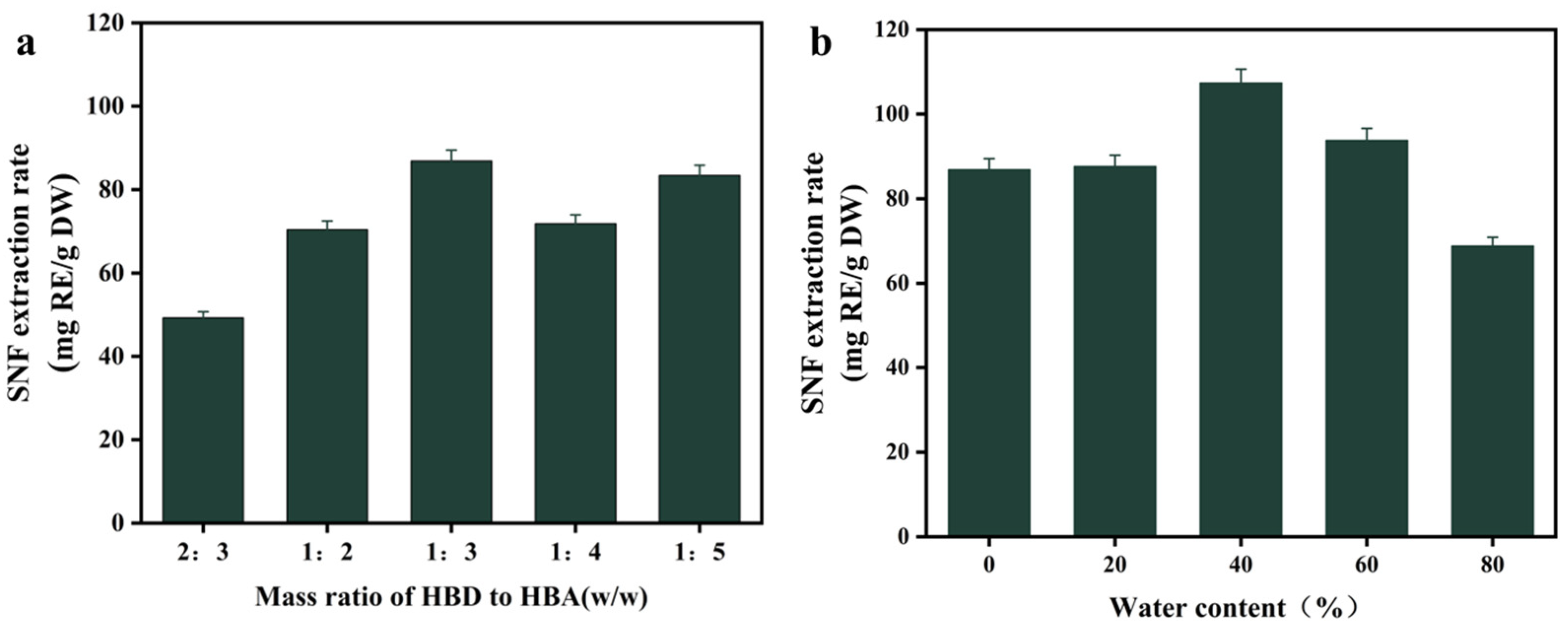
The optimization of the HP-β-CD/Lac-based DES for SNF extraction was systematically investigated by modulating the HBA (HP-β-CD) to HBD (Lac) mass ratio. As depicted in Figure 2a, the SNF extraction efficiency displayed a non-linear dependence on the HBA/HBD ratio. The maximum apparent extraction efficiency (86.87 ± 2.61 mg RE/g DW) was achieved at a 1:3 HP-β-CD/Lac mass ratio. Subsequent optimization of water content (Figure 2b) revealed that 40% content yielded the highest SNF recovery. Exceeding 40% water content led to a 12.66% decrease in extraction efficiency. The finalized DES formulation (1:3 HBA/HBD ratio, 40% water) demonstrated a 2.3-fold higher apparent SNF extraction efficiency than conventional water extraction (45.99 ± 0.53 mg RE/g DW).

3.3. Single-Factor Experiments

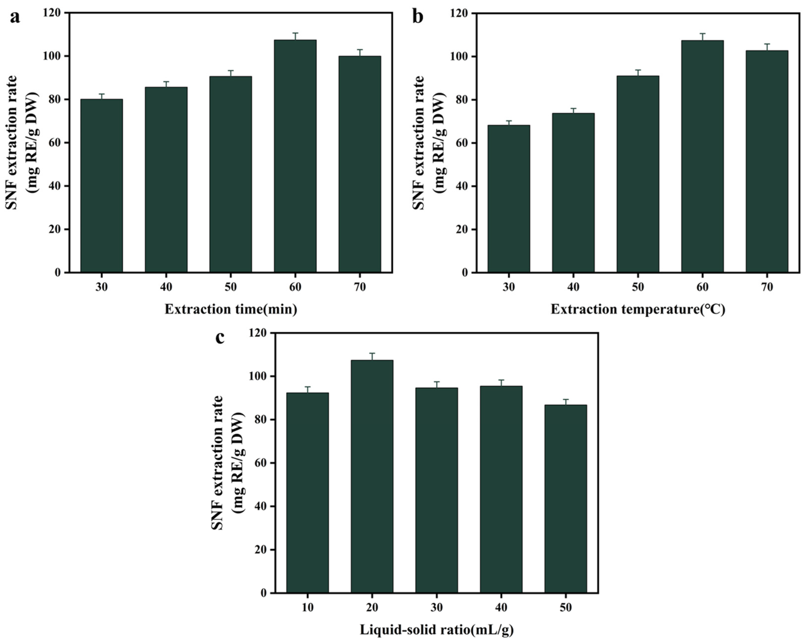
Multiple physicochemical parameters influence flavonoid extraction yields [29]. To evaluate their effects, we systematically investigated three critical extraction parameters—duration, temperature, and solid-to-solvent ratio—on SNF recovery. As shown in Figure 3a, SNF yields increased with extraction time up to 60 min, reaching a maximum apparent yield of 107.41 ± 3.22 mg RE/g DW, beyond which efficiency decreased by 6.95%. Regarding temperature, SNF yields increased by 57.50% from 30 °C to 60 °C, then decreased by 4.56% at 70 °C, exhibiting a bell-shaped profile (Figure 3b). Optimization of the solid-to-solvent ratio revealed that the yield reached equilibrium saturation at 20 mL/g (Figure 3c). Further increases beyond this ratio resulted in diminishing returns.

3.4. Optimization of Extraction Conditions by Box–Behnken Design

A Box–Behnken design was implemented with extraction time (X1), temperature (X2), and liquid–solid ratio (X3) as independent variables, and SNF yield as the response. Seventeen randomized experiments (Design-Expert v13.0, Stat-Ease Inc., Minneapolis, MN, USA) revealed significant variable interactions (Table 2). SNF extraction yields spanned 84.74–109.50 mg RE/g DW, confirming substantial parameter influence. Response surface methodology generated the following second-order polynomial model (coded units):
SNF   yield   ( mg   RE / g   DW ) = 107.91 + 1.95 X 1 + 0.74 X 2 2.21 X 3 6.17 X 1 X 2 + 2.16 X 1 X 3 + 1.32 X 2 X 3 5.15 X 1 2 8.40 X 2 2 9.64 X 3 2
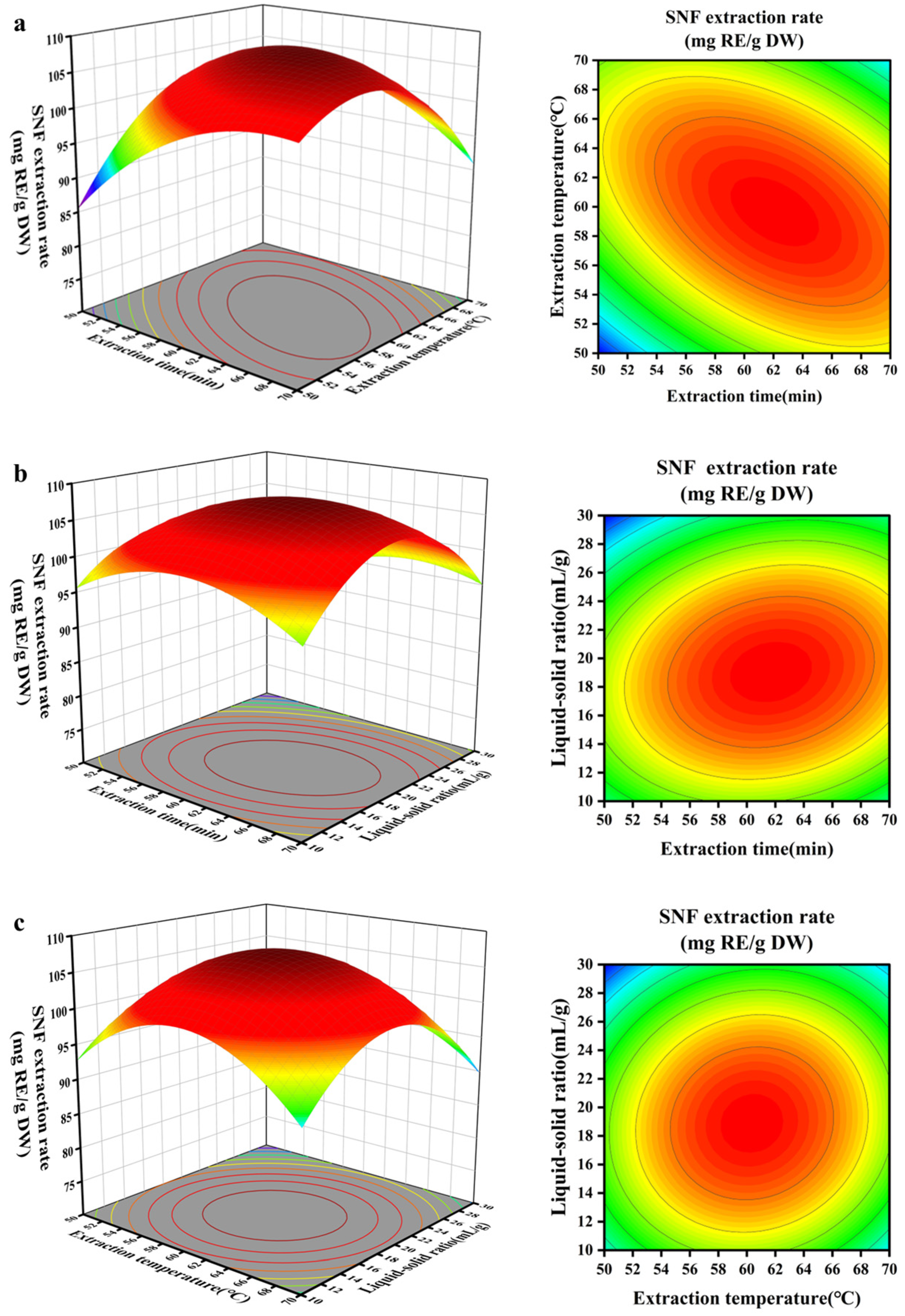
ANOVA confirmed the quadratic model’s robustness for predicting SNF extraction (p < 0.0001, R2 = 0.9904), with a non-significant lack-of-fit (p > 0.05). The analysis of linear coefficients revealed that extraction time (X1) exhibited a significant positive influence (p < 0.01), while solid–liquid ratio (X3) exerted a significant negative impact (p < 0.01). The quadratic terms (X12, X22, X32) and the X1X2 interaction term demonstrated highly significant effects (p < 0.0001). The parameter hierarchy, based on the magnitude of the effects, was ranked as X3 > X1 > X2 (Table S1). The 2D and 3D response surface plots (Figure 4) showed distinct maxima within the experimental domains. Figure 4a–c indicated that the interaction between extraction temperature and time (X1X2) exhibited the steepest response surface gradient, with the hierarchy of interaction influences quantified as: X1X2 > X2X3 > X1X3.
The RSM optimization predicted the following theoretical optima: temperature = 59.67 °C, duration = 61.89 min, and solid–liquid ratio = 1:19.04 mL/g, with a predicted apparent yield of 108.20 mg RE/g DW. For experimental verification, parameters were adjusted to 60 °C, 62 min, and 1:19 mL/g. Subsequent validation trials achieved an apparent yield of 108.72 ± 2.17 mg RE/g DW (Table 3), representing a 0.48% deviation from the predicted value.

3.5. Antioxidant Activity

The antioxidant capacity of SNF extracts obtained by DES-UAE and conventional water extraction was evaluated using DPPH and ABTS radical scavenging assays. Control experiments confirmed that the blank DES solvent itself contributed negligible radical scavenging activity (Figure S1), ensuring that the observed antioxidant effects were attributable to the extracted compounds rather than matrix interference.
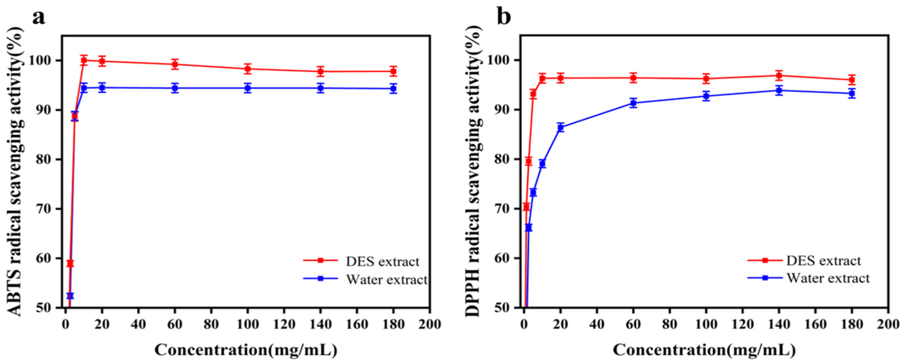
Flavonoid antioxidant activity was evaluated via standard free radical scavenging assays (DPPH+ and ABTS+), with SNF extracts prepared using both water extraction and the optimized DES-UAE method. As shown in Figure 5a,b, both extracts exhibited concentration-dependent radical scavenging activity (0–10 mg/mL). The antioxidant capacity was quantified by IC50 values [30]. The DES extract demonstrated superior activity to water extract against both radicals: IC50 = 1.346 ± 0.15 mg/mL (DPPH+) versus 1.702 ± 0.28 mg/mL, and 2.031 ± 0.36 mg/mL (ABTS+) versus 2.366 ± 0.33 mg/mL.

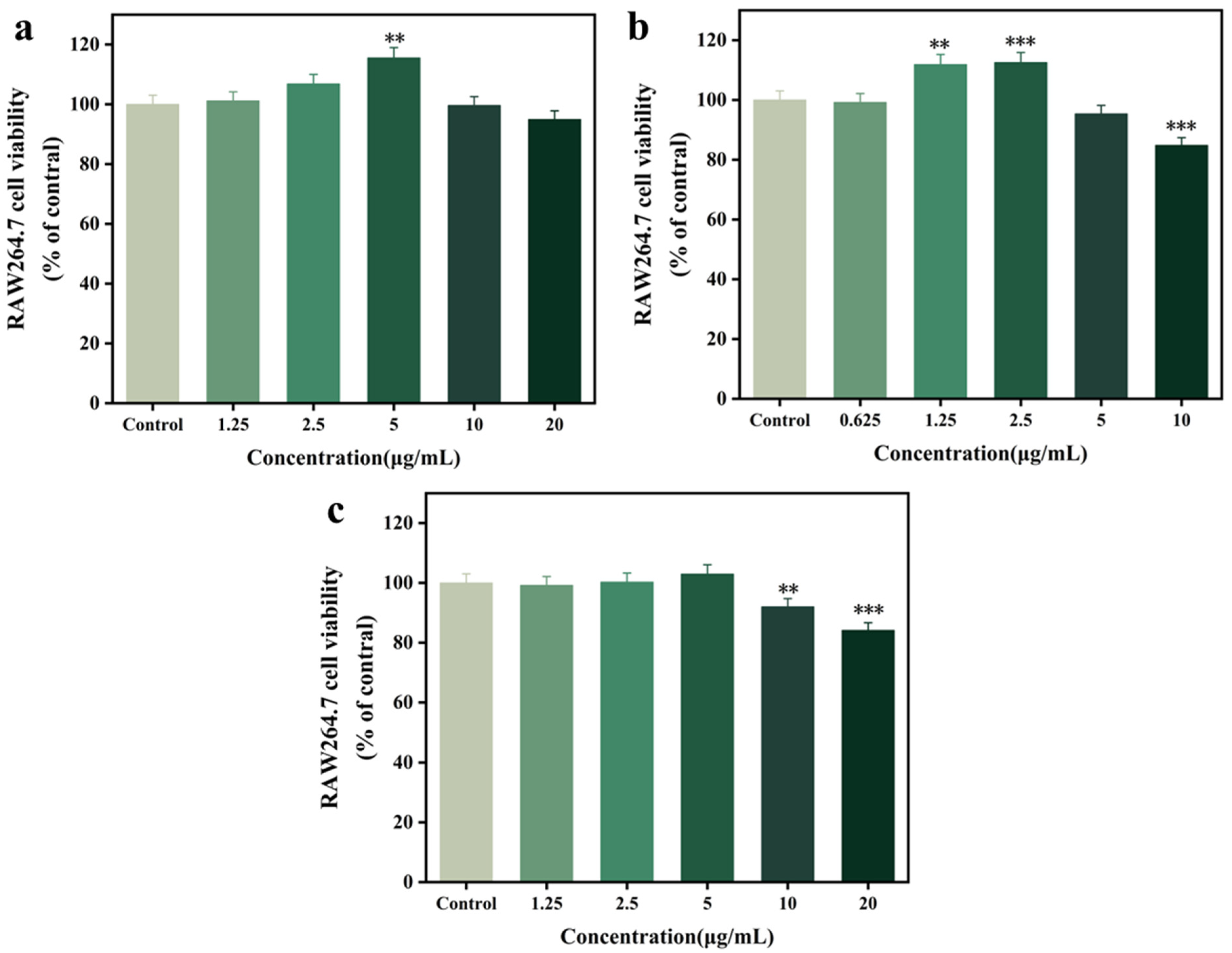
3.6. Cytotoxicity Assay of RAW 264.7 Cells

Macrophages play a critical role in host defense against pathogens by secreting key immunomodulatory mediators, including interleukins, tumor necrosis factor, and nitric oxide [31]. To evaluate anti-inflammatory activity, this study quantified two principal pro-inflammatory cytokines: TNF-α and IL-6. Cytotoxicities of optimized DES extracts, water extracts, and blank DES solvents were evaluated in murine RAW 264.7 macrophages using MTT assays. As shown in Figure 6a–c, water extracts at 1.25–5 μg/mL exhibited concentration-dependent proliferative effects after 24 h, reaching peak viability enhancement (102.96 ± 3.08%) at 5 μg/mL. DES extracts demonstrated significantly stronger proliferative responses at lower concentrations (0.625–2.5 μg/mL), achieving maximum viability (112.52 ± 3.37%) at 2.5 μg/mL (p < 0.05 vs. water control). Notably, blank DES solvents exhibited no cytotoxicity at concentrations ≤5 μg/mL. The aforementioned results served as the preferred concentration for the ensuing cell-related investigations.

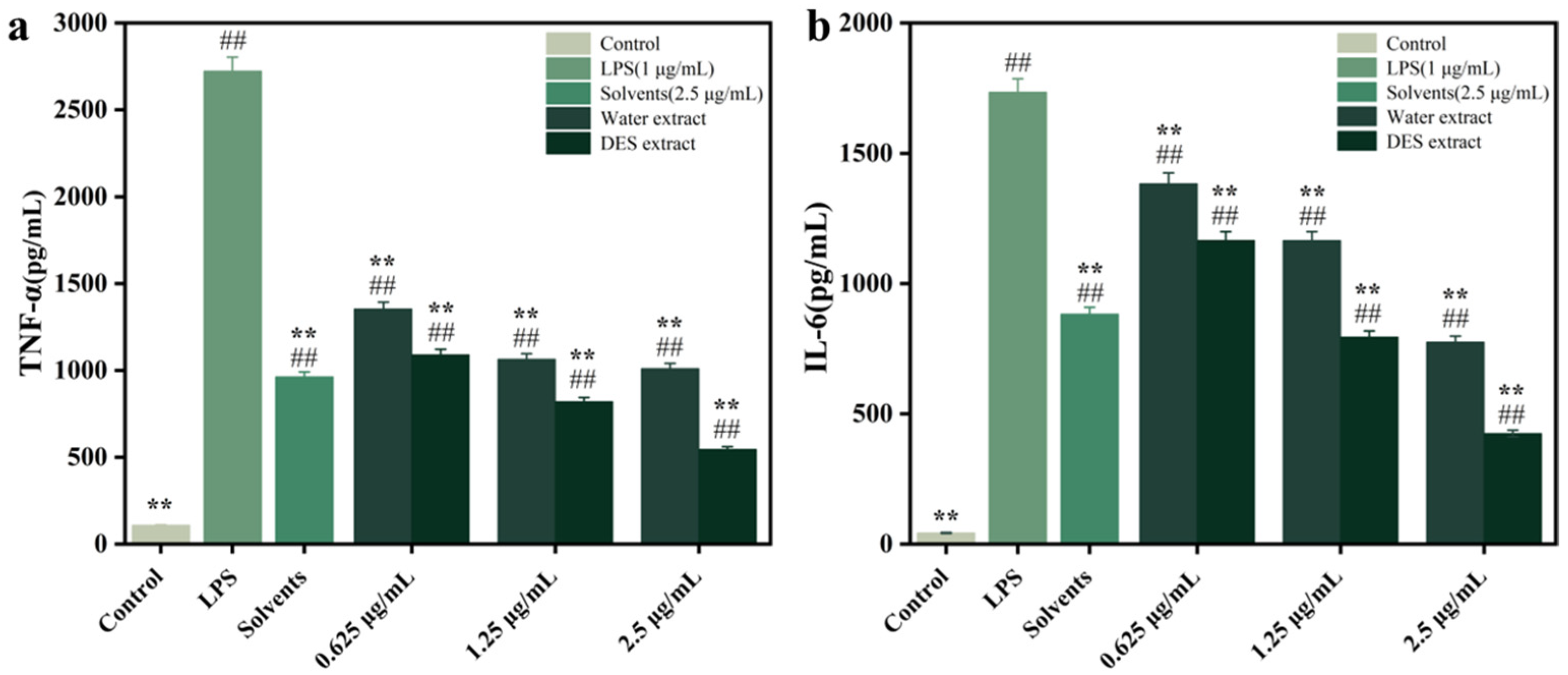
3.7. Inhibiting RAW264.7 Cell Inflammatory Factor Release

We further investigated the inhibitory effects of SNF extracts on LPS-induced inflammatory cytokine secretion (IL-6 and TNF-α) in RAW 264.7 macrophages (Figure 7a,b). LPS stimulation significantly elevated IL-6 and TNF-α levels compared to untreated controls (p < 0.01). Both DES-processed and water SNF extracts demonstrated dose-dependent suppression of these cytokines across low, medium, and high concentrations (0.625–2.5 μg/mL), with significant inhibition (p < 0.01) observed at all doses. The DES extracts exhibited 15.8–45.2% greater inhibition of IL-6 than water extracts at equivalent concentrations. This superior efficacy extended to TNF-α suppression (19.5–38.4% enhancement, p < 0.05). Further analysis revealed that the blank DES solvent reduced TNF-α by 64.6 ± 1.9% and IL-6 by 49.1 ± 1.5% at 2.5 μg/mL.

3.8. Components Analysis of SNF by UHPLC-Q-TOF-MS Anlysis

Following the bioactivity assessments, compositional analysis of SNF extracts obtained under optimized conditions (60 °C, 62 min, solid–liquid ratio 1:19 mL/g) was performed using UPLC-ESI-QTOF-MS/MS (Table S2). The analysis led to the putative annotation of 23 bioactive constituents: 10 amino acids (e.g., L-threonine, L-glutamic acid), 5 flavonoids (including apigenin-6,8-di-C-glucoside and quercetin 3-O-β-d-glucuronide), 5 phenolic acids (e.g., protocatechuic acid), 2 alkaloids (including caffeine and guanine), and 1 phenylpropanoid compound.

3.9. Molecular Docking Analysis

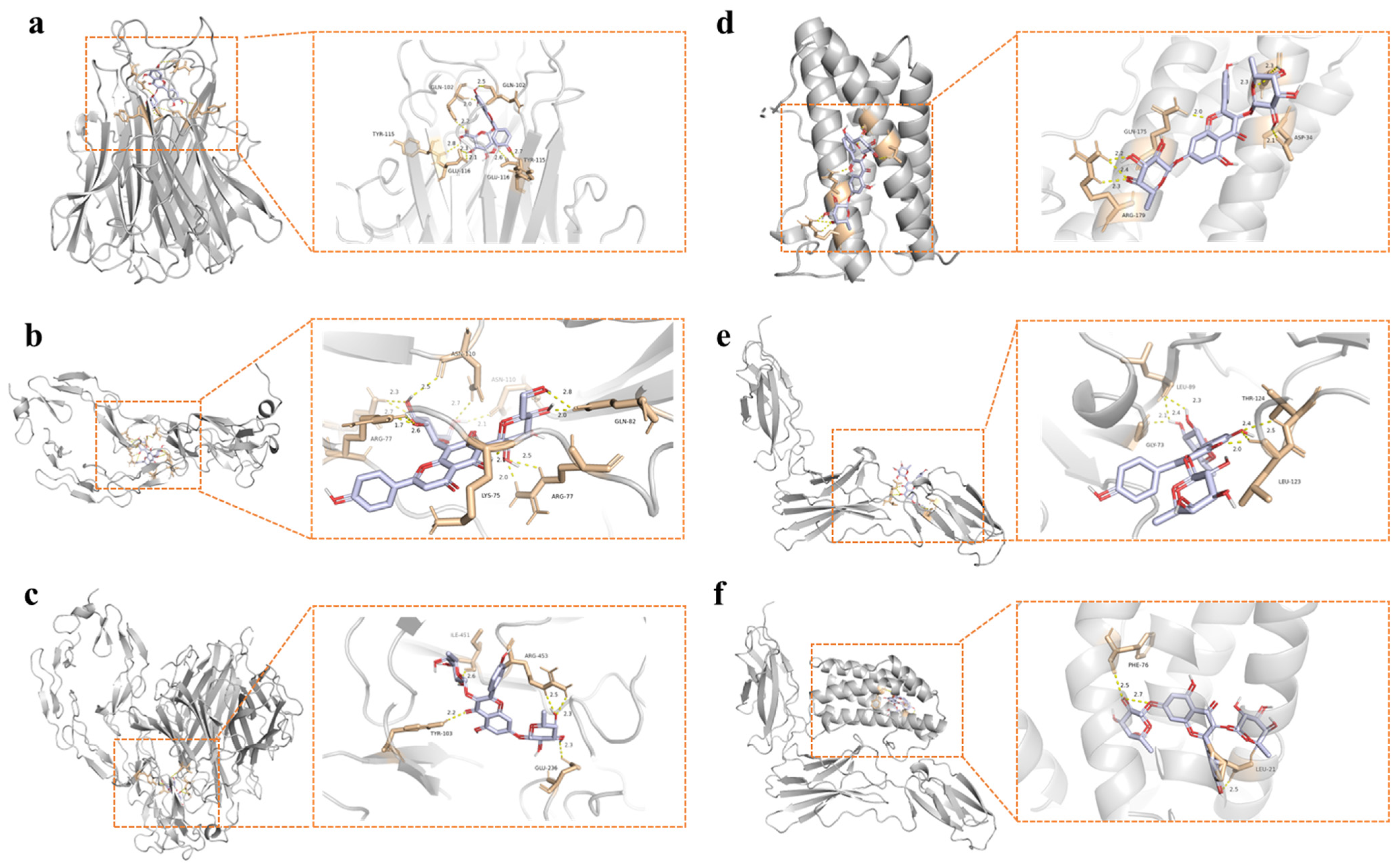
Molecular docking analysis assessed interactions between the 23 identified bioactive compounds and key inflammatory targets: IL-6, IL-6R, TNF-α, and TNF-R1. Docking simulations focused specifically on ligand binding to the IL-6/IL-6R and TNF-α/TNF-R1 signaling complexes. Table 4 presents the binding energies for all ligand-target combinations, while Figure 8 showcases representative binding modes for the lowest-energy interactions. Notably, quercetin 3-O-β-d-glucuronide exhibited a binding affinity with TNF-α (ΔG = −11.1 kcal/mol). Molecular visualization predicted that this compound forms three hydrogen bonds with TNF-α residues: GLN102 (average bond length: 2.2 Å), TYR115 (2.7 Å), and GLU116 (2.2 Å) (Figure 8a).
Apigenin-6,8-C-diglucoside demonstrated strong binding affinity to TNF-R1 (ΔG = −10.2 kcal/mol; Figure 8b), forming hydrogen bonds with LYS75, ARG77, GLN82, and ASN110 residues. Similarly, kaempferol exhibited enhanced binding to the TNF-α/TNF-R1 complex (ΔG = −11.0 kcal/mol), establishing four hydrogen bonds with residues TYR-103, GLU-236, ARG-453, and ILE-451.
Molecular docking analysis also demonstrated kaempferitrin’s significant binding affinity to IL-6 (ΔG = −7.6 kcal/mol), IL-6R (ΔG = −7.4 kcal/mol), and the IL-6/IL-6R complex (ΔG = −8.6 kcal/mol). Hydrogen bonding interactions were observed at key residues: ASP34, GLN175, and ARG179 for IL-6; LEU89, GLY73, LEU123, and THR124 for IL-6R; and the shared interface residues LEU21, PHE76 for the complex (Figure 8d–f).

4. Discussion

The growing demand for natural anti-inflammatory agents in cosmetics underscores the need for sustainable extraction methods and well-characterized bioactive ingredients. This study demonstrates that a tailored DES-UAE efficiently recovers flavonoids from Baihao Yinzhen tea, yielding an extract with notable anti-inflammatory activity suitable for cosmetic applications.
As shown in Figure 1, screening of 14 DES formulations revealed that the HP-β-CD/Lac system achieved the highest extraction yield. DES performance is closely linked to its physicochemical properties, including viscosity, polarity, and hydrogen-bonding capacity [32]. The superior efficiency of HP-β-CD/Lac can be attributed to its three-dimensional hydrogen-bonding network, which enhances molecular interactions with flavonoids [25]. Further optimization of the HBA/HBD mass ratio (Figure 2a) demonstrated a nonlinear relationship between composition and extraction yield, reflecting the sensitivity of DES properties to component stoichiometry [33]. A mass ratio of 1:3 was identified as optimal. Water content also critically influenced extraction efficiency (Figure 2b), with 40% content maximizing flavonoid recovery, while higher levels led to a 12.66% decrease in yield, consistent with disruption of the DES supramolecular network beyond the hydration threshold [34].
Single-factor experiments (Figure 3) revealed how extraction parameters influence yield through mass transfer and stability mechanisms. Time-dependent increases up to 60 min reflect matrix disruption and compound solubilization [35], while longer durations induced degradation, likely due to ultrasonic cavitation and thermal effects [36]. Temperature exhibited a bell-shaped profile, balancing improved mass transfer with thermal degradation beyond 60 °C [37]. Solid–solvent ratio optimization indicated saturation at 20 mL/g, with further increases yielding diminishing returns due to solvent saturation [36].
Response surface methodology (Figure 4, Table 2) confirmed these trends and enabled process optimization. The highly significant model (p < 0.0001, R2 = 0.9904) and close agreement between predicted (108.20 mg RE/g DW) and experimental (108.72 mg RE/g DW) values validated the model’s robustness. The negative coefficients for quadratic and interaction terms corroborated the existence of optimal parameter ranges. The strong temperature–time interaction highlighted the synergistic effect of thermal and temporal factors on extraction efficiency.
The DES extract showed significantly stronger antioxidant activity than the water extract (Figure 5), with lower IC50 values against both DPPH+ and ABTS+ radicals, indicating more efficient extraction of antioxidant flavonoids. In LPS-stimulated RAW 264.7 macrophages, the DES extract exhibited dose-dependent suppression of TNF-α and IL-6 (Figure 7), outperforming the water extract by 15.8–45.2% in IL-6 inhibition and 19.5–38.4% in TNF-α suppression. Notably, the blank DES solvent itself showed intrinsic anti-inflammatory activity, suggesting a synergistic role in enhancing flavonoid bioactivity.
UPLC-ESI-QTOF-MS/MS analysis tentatively identified 23 bioactive constituents, including five major flavonoid glycosides such as apigenin-6,8-di-C-glucoside and quercetin 3-O-β-d-glucuronide, which are recognized as putative anti-inflammatory agents [38]. Molecular docking (Table 4, Figure 8) revealed high-affinity binding between these flavonoids and key inflammatory targets. Quercetin 3-O-β-d-glucuronide showed strong binding to TNF-α (ΔG = −11.1 kcal/mol), forming hydrogen bonds with GLN102, TYR115, and GLU116, suggesting competitive inhibition of TNF-α/TNFR1 complex formation [39]. Similarly, apigenin-6,8-C-diglucoside and kaempferol bound strongly to TNFR1 and the TNF-α/TNFR1 complex, respectively, supporting flavonoid-mediated interference with TNF signaling [40,41]. In the IL-6 pathway, kaempferitrin exhibited significant affinity for IL-6, IL-6R, and their complex, with conserved interactions at Arg179—a key residue in IL-6 signaling [42]. These results align with reported mechanisms of Camellia sinensis flavonoids in modulating JAK/STAT and NF-κB pathways [43,44].
Notably, our investigation revealed that the blank DES solvent itself exhibited intrinsic anti-inflammatory activity. This observation can be plausibly attributed to the lactic acid component, which has been reported to suppress pro-inflammatory macrophage activation through specific metabolic and epigenetic regulations [45]. While this indicates that the overall anti-inflammatory effect of the DES extract may result from a potential synergy between the flavonoids and the DES medium, this hypothesis requires further validation with component-control experiments. The significantly greater suppression of TNF-α and IL-6 by the DES extract compared to the blank DES unequivocally highlights the critical and enhanced role of the flavonoids extracted and potentially delivered by this green solvent system—a highly desirable property for topical applications.
Complementing this functional advantage, the safety and applicability of this ready-to-use DES–extract formulation for cosmetics are underpinned by the well-established biocompatibility of its constituent solvents. Lactic acid is a widely used cosmetic ingredient, recognized for its moisturizing, exfoliating, and skin-brightening properties [46]. Similarly, HP-β-CD is a safe and approved excipient commonly employed to enhance the solubility, stability, and dermal delivery of bioactive compounds [47]. Therefore, the final formulation leverages the intrinsic safety and complementary functions of its constituents, positioning it as a potent, self-formulated bioactive ingredient particularly suited for cosmetic products aimed at soothing inflammation and promoting skin health.
The high extraction yield (108.72 mg RE/g DW), coupled with the intrinsic bioactivity of the HP-β-CD/Lac system, positions this DES as both an efficient extractant and a potential bioactive carrier for cosmetic formulations [48]. The identified flavonoids contribute to the observed anti-inflammatory effects via direct interaction with pro-inflammatory cytokines, supporting a molecular mechanism for the extract’s efficacy. This targeted anti-cytokine activity distinguishes the extract from general antioxidant tea polyphenols, highlighting its potential for sensitive or reactive skin applications.
To fully realize this potential, future work should focus on assessing the topical efficacy and safety in skin-relevant models, such as human keratinocytes (HaCaT) or reconstructed human epidermis, using endpoints like IL-8 and COX-2. Furthermore, while the DES components are well-established as safe for topical use, formulation studies are warranted to optimize skin feel, stability, and compatibility with other cosmetic ingredients, which may include pH adjustment to the skin’s natural range (4.5–5.5) for leave-on products. Ultimately, clinical trials would be essential to confirm the efficacy and consumer tolerance in human subjects.
From a green chemistry perspective, the environmental profile of our DES-UAE process was evaluated (Table S3). The method aligns with green principles through its use of a biodegradable, biobased DES (HP-β-CD/Lac) to replace volatile organic solvents. A key advantage of this approach is that it directly yields a ready-to-use extract without the need for energy-intensive solvent removal steps [49] Crucially, the final cosmetic extract contains no residues of conventional volatile organic solvents, as none were employed in the extraction process. The use of methanol was strictly confined to the analytical UHPLC-MS stage for component identification and is distinct from the green extraction process itself. While the ability to use the extract without DES removal is an inherent advantage, future work will focus on further greening the process, such as enabling DES solvent recycling for multiple extraction cycles and transitioning from batch to continuous operation mode to improve energy and material efficiency.
The energy consumption was calculated to be 992 kWh per kg of dry biomass. More relevant for assessing the process efficiency to produce the final ready-to-use formulation, the energy input was 49.6 kWh per kg of the final DES–extract complex. While the former value is characteristic of a laboratory-scale ultrasonic bath, the latter provides a more accurate and favorable perspective on the process’s energy profile. This efficiency stems from the direct use of the extract without energy-intensive solvent removal or purification steps. When benchmarked against conventional extraction, which often requires subsequent concentration and solvent recovery—processes that are notoriously energy-intensive—our DES-UAE method presents a compelling advantage in terms of overall energy integration and simplicity.
This recognized energy intensity at the laboratory scale, however, underscores the inherent scalability challenges of the UAE process itself. For industrial implementation, hydrodynamic cavitation (HC) presents a robust alternative, as it has been successfully demonstrated in continuous, pilot-scale operations for plant extraction [50]. HC offers superior energy efficiency and linear scalability compared to UAE. Therefore, future work will focus on adapting our optimized DES extraction to a continuous HC system, integrating DES solvent recovery and heat exchange to directly address this energy intensity and enhance the overall sustainability and economic viability of the process.
In conclusion, this study establishes HP-β-CD/Lac-based DES-UAE as a green and efficient method for obtaining anti-inflammatory flavonoids from Baihao Yinzhen. The extract demonstrates dual antioxidant and anti-inflammatory properties, supported by mechanistic molecular docking insights. These findings underscore its potential as a natural bioactive ingredient for cosmetic products aimed at soothing inflammation and promoting skin health.

5. Conclusions

This study established an eco-friendly DES-UAE platform for efficient recovery of SNF. HP-β-CD/Lac (1:3 HBA/HBD mass ratio) emerged as the optimal solvent system, achieving maximal apparent SNF yield (108.72 mg RE/g DW) under parameterized conditions: 40% water modulation, 60 °C for 62 min, and 1:19 solid–liquid ratio. DES-UAE enhanced extraction efficiency by 0.7-fold relative to conventional methods. The DES extract demonstrated significant bioactivity: (1) antioxidant capacity (IC50 = 1.346 and 2.031 mg/mL against DPPH+ and ABTS+); (2) dose-dependent anti-inflammatory effects (75.5% IL-6 and 80.0% TNF-α suppression in LPS-stimulated RAW 264.7 macrophages at 2.5 μg/mL, p < 0.01). The five flavonoids discovered among the 23 compounds via UHPLC-Q-TOF-MS were the probable pharmacologically active constituents. Molecular docking provided in silico evidence for potential high-affinity binding (ΔG ≤ −7.7 kcal/mol) between these flavonoids and inflammatory targets, offering a structural basis for the observed bioactivity. Collectively, this work demonstrates HP-β-CD/Lac-based UAE as a sustainable, high-efficiency platform for valorizing total flavonoids in Baihao Yizhen—with optimized extracts holding significant promise for nutraceutical and pharmacological applications.

Supplementary Materials

The following supporting information can be downloaded at: https://www.mdpi.com/article/10.3390/cosmetics12060245/s1: Table S1—The results of analysis of variance (ANOVA); Table S2—Identification of components of SNF by UHPLC-Q-TOF-MS analysis; Figure S1—Radical scavenging activity of the blank HP-β-CD/Lac DES solvent; Figure S2—MS/MS fragmentation spectra for the five major flavonoid glycosides; Table S3—Sustainability Assessment of the DES-UAE Method Based on Path2Green 12 Principles.

Author Contributions

Conceptualization, L.D., S.H. and Y.C.; Methodology, Z.Z., Y.C., W.H., H.C. and L.D.; Validation, Z.Z., Y.C., W.H. and D.K.; Formal analysis, Z.Z. and H.C.; Investigation, D.K.; Resources, Y.C., S.H. and L.D.; Data curation, Y.C. and L.D.; Writing—original draft, Z.Z.; Writing—review and editing, L.D.; Visualization, Z.Z.; Software, Z.Z.; Supervision, S.H. and L.D.; Project administration, S.H. and L.D.; Funding acquisition, S.H. All authors have read and agreed to the published version of the manuscript.

Funding

This research was funded by the Shanghai Chunzhibao Biotechnology Co., Ltd.

Institutional Review Board Statement

Not applicable.

Informed Consent Statement

Not applicable.

Data Availability Statement

The data used to support the findings of this study are available from the corresponding author upon request.

Conflicts of Interest

Funding and co-author affiliations include a commercial entity, Shanghai Chunzhibao Biotechnology Co., Ltd., which provided materials and financial support. The authors declare that the sponsor had no role in the design, execution, interpretation, or writing of the study. All other authors declare no competing interests.

References

  1. Somavanshi, D.B.; Kamble, P.R.; Patil, C.D.; Jadhav, S.P.; Sonawane, G.B. A Review on the Antioxidant and Antiaging Properties of White Tea. JPRI 2021, 33, 129–136. [Google Scholar] [CrossRef]
  2. Gabr, G.A.; Hassan, H.M.M.; Seshadri, V.D.; Hassan, N.M.M. Comparative Study of Phenolic Profile, Antioxidant and Antimicrobial Activities of Aqueous Extract of White and Green Tea. Z. Naturforschung C 2022, 77, 483–492. [Google Scholar] [CrossRef]
  3. Dai, W.; Xie, D.; Lu, M.; Li, P.; Lv, H.; Yang, C.; Peng, Q.; Zhu, Y.; Guo, L.; Zhang, Y.; et al. Characterization of White Tea Metabolome: Comparison against Green and Black Tea by a Nontargeted Metabolomics Approach. Food Res. Int. 2017, 96, 40–45. [Google Scholar] [CrossRef]
  4. Zhao, F.; Wang, X.; Liu, H.; Qian, J. In Vitro and in Vivo Anti-Inflammatory Models Demonstrate Oligopeptides Play a Significant Role in Anti-Inflammatory Properties of White Tea. J. Funct. Foods 2024, 112, 105983. [Google Scholar] [CrossRef]
  5. Gao, J.; Chen, D.; Xie, D.; Peng, J.; Hu, Z.; Lin, Z.; Dai, W. Investigations of the Highly Efficient Processing Technique, Chemical Constituents, and Anti-Inflammatory Effect of N-Ethyl-2-Pyrrolidinone-Substituted Flavan-3-Ol (EPSF)-Enriched White Tea. Food Chem. 2024, 450, 139328. [Google Scholar] [CrossRef] [PubMed]
  6. Yang, Y.; Liang, Q.; Zhang, B.; Zhang, J.; Fan, L.; Kang, J.; Lin, Y.; Huang, Y.; Tan, T.-C.; Ho, L.-H. Adsorption and Desorption Characteristics of Flavonoids from White Tea Using Macroporous Adsorption Resin. J. Chromatogr. A 2024, 1715, 464621. [Google Scholar] [CrossRef]
  7. Wang, R.; Yang, Z.; Zhang, J.; Mu, J.; Zhou, X.; Zhao, X. Liver Injury Induced by Carbon Tetrachloride in Mice Is Prevented by the Antioxidant Capacity of Anji White Tea Polyphenols. Antioxidants 2019, 8, 64. [Google Scholar] [CrossRef] [PubMed]
  8. Selim, D.A.; Shawky, E.; Abu El-Khair, R.M. Identification of the Discriminatory Chemical Markers of Different Grades of Sri Lankan White, Green and Black Tea (Camellia sinenesis L.) via Metabolomics Combined to Chemometrics. J. Food Compos. Anal. 2022, 109, 104473. [Google Scholar] [CrossRef]
  9. Liu, X.; Zhou, F.; Wen, M.; Jiang, S.; Long, P.; Ke, J.-P.; Han, Z.; Zhu, M.; Zhou, Y.; Zhang, L. LC-MS and GC–MS Based Metabolomics Analysis Revealed the Impact of Tea Trichomes on the Chemical and Flavor Characteristics of White Tea. Food Res. Int. 2024, 191, 114740. [Google Scholar] [CrossRef] [PubMed]
  10. Zhao, Y.; Li, F.; Zhou, H.; Wang, Y.; Bian, J.; Sun, Y.; Du, X. Utilizing Nontargeted Metabolomics Integrated with Quality Component Quantitation via Differential Analysis to Reveal the Taste Differences between and Metabolite Characteristics of Hawk Black Tea and Hawk White Tea. Food Res. Int. 2024, 197, 115216. [Google Scholar] [CrossRef]
  11. Elkhedir, A.E.; Iqbal, A.; Zogona, D.; Mohammed, H.H.; Murtaza, A.; Xu, X. Apigenin Glycosides from Green Pepper Enhance Longevity and Stress Resistance in Caenorhabditis Elegans. Nutr. Res. 2022, 102, 23–34. [Google Scholar] [CrossRef] [PubMed]
  12. Bojilov, D.; Manolov, S.; Nacheva, A.; Dagnon, S.; Ivanov, I. Characterization of Polyphenols from Chenopodium Botrys after Fractionation with Different Solvents and Study of Their In Vitro Biological Activity. Molecules 2023, 28, 4816. [Google Scholar] [CrossRef]
  13. Li, J.; Yang, J.; Xian, Q.; Su, H.; Ni, Y.; Wang, L. Kaempferitrin Attenuates Unilateral Ureteral Obstruction-induced Renal Inflammation and Fibrosis in Mice by Inhibiting NOX4 -mediated Tubular Ferroptosis. Phytother. Res. 2024, 38, 2656–2668. [Google Scholar] [CrossRef]
  14. Li, J.; Si, C.; Hong, W.; Xia, C.; Yang, Y.; He, Y.; Su, M.; Long, X.; Zhang, H. Identification of the Chemical Components of Ethanol Extract of Chenopodium Ambrosioides and Evaluation of Their in Vitro Antioxidant and Anti Tumor Activities. Trop. J. Pharm. Res. 2022, 21, 1689–1697. [Google Scholar] [CrossRef]
  15. De Rijke, E.; Out, P.; Niessen, W.M.A.; Ariese, F.; Gooijer, C.; Brinkman, U.A.T. Analytical Separation and Detection Methods for Flavonoids. J. Chromatogr. A 2006, 1112, 31–63. [Google Scholar] [CrossRef]
  16. Hao, Y.; Pei, F.; Huang, J.; Li, G.; Zhong, C. Application of Deep Eutectic Solvents on Extraction of Flavonoids. J. Sep. Sci. 2024, 47, 2300925. [Google Scholar] [CrossRef]
  17. Abbott, A.P.; Capper, G.; Davies, D.L.; Rasheed, R.K.; Tambyrajah, V. Novel Solvent Properties of Choline Chloride/Urea mixtures. Chem. Commun. 2003, 70–71. [Google Scholar] [CrossRef]
  18. Kyriakoudi, A.; Radojčić Redovniković, I.; Vidović, S.; Radošević, K.; Andreou, T.; Mourtzinos, I.; Cvjetko Bubalo, M. Coupling Deep Eutectic Solvents with Innovative Extraction Techniques towards Plant Derived Bioactive Compositions. RSC Sustain. 2024, 2, 1675–1691. [Google Scholar] [CrossRef]
  19. Chen, J.; Li, Y.; Wang, X.; Liu, W. Application of Deep Eutectic Solvents in Food Analysis: A Review. Molecules 2019, 24, 4594. [Google Scholar] [CrossRef] [PubMed]
  20. Tang, X.; Xiong, Y.; Wei, X.; Zhou, J.; Feng, C. Extraction Efficiency of Loganic Acid and Gentiopicroside from Gentianae Radix et Rhizoma by Deep Eutectic Solvent-Based Ultrasound-Assisted Extraction. Results Chem. 2024, 10, 101740. [Google Scholar] [CrossRef]
  21. Jiao, P.; He, X.; Ma, S.; Wang, S.; Niu, Q. Ultrasonic-Assisted Extraction of Antioxidants from Perilla Frutescens Leaves Based on Tailor-Made Deep Eutectic Solvents: Optimization and Antioxidant Activity. Molecules 2023, 28, 7554. [Google Scholar] [CrossRef]
  22. Zhang, M.; Zhang, Z.; Gul, Z.; Tian, M.; Wang, J.; Zheng, K.; Zhao, C.; Li, C. Advances of Responsive Deep Eutectic Solvents and Application in Extraction and Separation of Bioactive Compounds. J. Sep. Sci. 2023, 46, 2300098. [Google Scholar] [CrossRef]
  23. Morozova, O.V.; Vasil’eva, I.S.; Shumakovich, G.P.; Zaitseva, E.A.; Yaropolov, A.I. Deep Eutectic Solvents for Biotechnology Applications. Biochem. Mosc. 2023, 88, S150–S175. [Google Scholar] [CrossRef]
  24. Peng, W.; Wang, X.; Wang, W.; Wang, Y.; Huang, J.; Zhou, R.; Bo, R.; Liu, M.; Yin, S.; Li, J. Comparison, Optimization and Antioxidant Activity of Ultrasound-Assisted Natural Deep Eutectic Solvents Extraction and Traditional Method: A Greener Route for Extraction of Flavonoid from Moringa Oleifera Lam. Leaves. Ultrason. Sonochem. 2024, 109, 107003. [Google Scholar] [CrossRef]
  25. Huang, D.; Chen, L.; Chen, X.; Huang, X.; Yang, Y.; Liu, J.; Lin, Y.; Liu, Y.; Li, H. Supramolecular Deep Eutectic Solvents as Green Media for Efficient Extraction of Tea Polyphenols and Its Application in Bio-Active Film. Food Chem. 2025, 465, 141904. [Google Scholar] [CrossRef] [PubMed]
  26. Chen, S.; Li, X.; Liu, X.; Wang, N.; An, Q.; Ye, X.M.; Zhao, Z.T.; Zhao, M.; Han, Y.; Ouyang, K.H.; et al. Investigation of Chemical Composition, Antioxidant Activity, and the Effects of Alfalfa Flavonoids on Growth Performance. Oxidative Med. Cell. Longev. 2020, 2020, 8569237. [Google Scholar] [CrossRef] [PubMed]
  27. Jiang, Y.; Li, D.; Ma, X.; Jiang, F.; He, Q.; Qiu, S.; Li, Y.; Wang, G. Ionic Liquid–Ultrasound-Based Extraction of Biflavonoids from Selaginella Helvetica and Investigation of Their Antioxidant Activity. Molecules 2018, 23, 3284. [Google Scholar] [CrossRef]
  28. Dong, J.; Dong, Z.; Zhao, L.; Yang, D.; Bo, Y.; Zhang, X.; Wu, G.; An, M. The Evaluation of Five Bioactive Compounds Content and in Vitro Antioxidant of Caryophylli Flos Extracts Obtained by Natural Deep Eutectic Solvents. Sustain. Chem. Pharm. 2022, 30, 100838. [Google Scholar] [CrossRef]
  29. Yang, Y.; Huang, Y.; Lu, B.; Alrosan, M.; Easa, A.M.; Alu’datt, M.H.; Tan, T.C. Flavonoids Extracts from Zhenghe White Tea. Food Res. 2024, 8, 140–147. [Google Scholar] [CrossRef]
  30. Wang, C.-Y.; Chen, Y.-W.; Hou, C.-Y. Antioxidant and Antibacterial Activity of Seven Predominant Terpenoids. Int. J. Food Prop. 2019, 22, 230–238. [Google Scholar] [CrossRef]
  31. Li, M.; Rao, C.; Wang, C.; Cui, X.; Xiong, Y. Natural Deep Eutectic Solvents-Based Selective Extraction of Saponins from Panax Notoginseng: Process Optimization, Chemical Profiling, and Bioactivities Evaluation. Sep. Purif. Technol. 2024, 341, 126882. [Google Scholar] [CrossRef]
  32. Xing, C.; Cui, W.-Q.; Zhang, Y.; Zou, X.-S.; Hao, J.-Y.; Zheng, S.-D.; Wang, T.-T.; Wang, X.-Z.; Wu, T.; Liu, Y.-Y.; et al. Ultrasound-Assisted Deep Eutectic Solvents Extraction of Glabridin and Isoliquiritigenin from Glycyrrhiza Glabra: Optimization, Extraction Mechanism and in Vitro Bioactivities. Ultrason. Sonochem. 2022, 83, 105946. [Google Scholar] [CrossRef] [PubMed]
  33. Liu, C.; Qiao, L.; Gao, Q.; Zhang, F.; Zhang, X.; Lei, J.; Ren, M.; Xiao, S.; Kuang, J.; Deng, S.; et al. Total Biflavonoids Extraction from Selaginella Chaetoloma Utilizing Ultrasound-Assisted Deep Eutectic Solvent: Optimization of Conditions, Extraction Mechanism, and Biological Activity in Vitro. Ultrason. Sonochem. 2023, 98, 106491. [Google Scholar] [CrossRef]
  34. Jiang, Z.-M.; Wang, F.-F.; Liu, J.; Huang, T.-Q.; Zhang, H.; Liu, E.-H. Deep Eutectic Solvents in the Efficient Detoxification and Extraction of Alkaloids of Aconiti Lateralis Radix Praeparata. ACS Sustain. Chem. Eng. 2022, 10, 5674–5682. [Google Scholar] [CrossRef]
  35. Liu, Y.; Zhe, W.; Zhang, R.; Peng, Z.; Wang, Y.; Gao, H.; Guo, Z.; Xiao, J. Ultrasonic-Assisted Extraction of Polyphenolic Compounds from Paederia scandens (Lour.) Merr. Using Deep Eutectic Solvent: Optimization, Identification, and Comparison with Traditional Methods. Ultrason. Sonochem. 2022, 86, 106005. [Google Scholar] [CrossRef]
  36. Ma, P.; Li, Z.; Jin, Y.; Zuo, J.; Zhang, Y.; Dong, A.; Xiao, D.; Burenjargal, M. Green and Efficient Extraction Process of Flavonoids from Sea Buckthorn Fruits by Natural Deep Eutectic Solvents Aided with Ultrasound. Microchem. J. 2024, 205, 111265. [Google Scholar] [CrossRef]
  37. Gao, J.; Xie, L.; Peng, Y.; Li, M.; Li, J.; Ni, Y.; Wen, X. Deep Eutectic Solvents as New Extraction Media for Flavonoids in Mung Bean. Foods 2024, 13, 777. [Google Scholar] [CrossRef] [PubMed]
  38. Li, C.; He, J.; Yang, Y.; Gou, Y.; Wang, Z.; Chen, H.; Zhao, X. White Tip Silver Needle (Slightly Fermented White Tea) Flavonoids Help Prevent Aging via Antioxidative and Anti-Inflammatory Effects. Drug Des. Dev. Ther. 2021, 15, 1441–1457. [Google Scholar] [CrossRef]
  39. Wang, Y.; Ye, R.; Fan, L.; Zhao, X.; Li, L.; Zheng, H.; Qiu, Y.; He, X.; Lu, Y. A TNF-α Blocking Peptide That Reduces NF-κB and MAPK Activity for Attenuating Inflammation. Bioorg. Med. Chem. 2023, 92, 117420. [Google Scholar] [CrossRef] [PubMed]
  40. Kim, O.T.P.; Le, M.D.; Trinh, H.X.; Nong, H.V. In Silico Studies for the Interaction of Tumor Necrosis Factor-Alpha (TNF-α) with Different Saponins from Vietnamese Ginseng (Panax vietnamesis). Biophysics 2016, 13, 173–180. [Google Scholar] [CrossRef]
  41. Damergi, B.; Essid, R.; Fares, N.; Khadraoui, N.; Ageitos, L.; Ben Alaya, A.; Gharbi, D.; Abid, I.; Rashed Alothman, M.; Limam, F.; et al. Datura stramonium Flowers as a Potential Natural Resource of Bioactive Molecules: Identification of Anti-Inflammatory Agents and Molecular Docking Analysis. Molecules 2023, 28, 5195. [Google Scholar] [CrossRef]
  42. Kang, S.; Kishimoto, T. Interplay between Interleukin-6 Signaling and the Vascular Endothelium in Cytokine Storms. Exp. Mol. Med. 2021, 53, 1116–1123. [Google Scholar] [CrossRef]
  43. Wang, Y.; Jin, X.; Fan, Q.; Li, C.; Zhang, M.; Wang, Y.; Wu, Q.; Li, J.; Liu, X.; Wang, S.; et al. Deciphering the Active Compounds and Mechanisms of HSBDF for Treating ALI via Integrating Chemical Bioinformatics Analysis. Front. Pharmacol. 2022, 13, 879268. [Google Scholar] [CrossRef]
  44. Bakrim, S.; Fessikh, M.E.; Elhrech, H.; Omari, N.E.; Amanullah, M.; Ming, L.C.; Moshawih, S.; Bouyahya, A. Targeting Inflammation in Cancer Therapy: From Mechanistic Insights to Emerging Therapeutic Approaches. J. Transl. Med. 2025, 23, 588. [Google Scholar] [CrossRef]
  45. Wang, J.; Yang, P.; Yu, T.; Gao, M.; Liu, D.; Zhang, J.; Lu, C.; Chen, X.; Zhang, X.; Liu, Y. Lactylation of PKM2 Suppresses Inflammatory Metabolic Adaptation in Pro-Inflammatory Macrophages. Int. J. Biol. Sci 2022, 18, 6210–6225. [Google Scholar] [CrossRef]
  46. Theodorou, I.M.; Kapoukranidou, D.; Theodorou, M.; Tsetis, J.K.; Menni, A.E.; Tzikos, G.; Bareka, S.; Shrewsbury, A.; Stavrou, G.; Kotzampassi, K. Cosmeceuticals: A Review of Clinical Studies Claiming to Contain Specific, Well-Characterized Strains of Probiotics or Postbiotics. Nutrients 2024, 16, 2526. [Google Scholar] [CrossRef]
  47. Sharma, R.; Hastings, C.; Staretz-Chacham, O.; Raiman, J.; Paucar, M.; Spiegel, R.; Murray, B.; Hurst, B.; Liu, B.; Kjems, L.; et al. Long-Term Administration of Intravenous Trappsol® CycloTM (HP-β-CD) Results in Clinical Benefits and Stabilization or Slowing of Disease Progression in Patients with Niemann-Pick Disease Type C1: Results of an International 48-Week Phase I/II Trial. Mol. Genet. Metab. Rep. 2023, 36, 100988. [Google Scholar]
  48. Sahu, S.; Kumari, D.; Kusam; Kuila, A.; Gurjar, R.S.; Sharma, K.; Verma, R. Deep Eutectic Solvent Extraction of Polyphenol from Plant Materials: Current Status and Future Prospects in Food Applications. Food Chem. 2025, 482, 144125. [Google Scholar] [CrossRef]
  49. De Souza Mesquita, L.M.; Contieri, L.S.; e Silva, F.A.; Bagini, R.H.; Bragagnolo, F.S.; Strieder, M.M.; Sosa, F.H.B.; Schaeffer, N.; Freire, M.G.; Ventura, S.P.M.; et al. Path2Green: Introducing 12 Green Extraction Principles and a Novel Metric for Assessing Sustainability in Biomass Valorization. Green Chem. 2024, 26, 10087–10106. [Google Scholar] [CrossRef]
  50. Tagliavento, L.; Nardin, T.; Chini, J.; Vighi, N.; Lovatti, L.; Testai, L.; Meneguzzo, F.; Larcher, R.; Zabini, F. Sustainable Exploitation of Apple By-Products: A Retrospective Analysis of Pilot-Scale Extraction Tests Using Hydrodynamic Cavitation. Foods 2025, 14, 1915. [Google Scholar] [CrossRef]
Figure 1. Apparent extraction rate of SNF by DESs.
Cosmetics 12 00245 g001
Figure 2. Effect of different DES molar ratios (a), water contents (b) on the apparent extraction yields of SNF.
Cosmetics 12 00245 g002
Figure 3. Effect of different extraction time (a), extraction temperature (b) and liquid-to-solid ratio (c) on the apparent extraction yields of SNF.
Cosmetics 12 00245 g003
Figure 4. 3D/2D response surface diagram of SNF. (a) Varying extraction time and extraction extraction temperature, (b) Varying extraction time and liquid–solid ratio, (c) Varying extraction temperature and liquid–solid ratio.
Cosmetics 12 00245 g004
Figure 5. Antioxidant activity of SNF DPPH: (a) ABTS, (b) radical scavenging activity.
Cosmetics 12 00245 g005
Figure 6. Different concentrations of DES (a), DES extract (b), water extract (c) of SNF on the cell viability of RAW264.7. Statistical analysis was performed by ANOVA. Error bars indicate the standard deviation of three independent tests in triplicate. (**, *** indicate p < 0.01, 0.001, vs. Control group).
Cosmetics 12 00245 g006
Figure 7. Inflammatory factor levels TNF-α (a), IL-6 (b) in RAW264.7 cells induced by LPS. Each value represents means ± SD, n = 3. and ## p < 0.01 refer to significant difference and highly significant difference, respectively, compared to the control group. and ** p < 0.01 refer to significant difference and highly significant difference, respectively, compared to the LPS group.
Cosmetics 12 00245 g007
Figure 8. 3D binding conformation schematic of TNF-α with quercetin 3-O-β-d-glucuronide (a), TNFR1 with apigenin-6,8-di-C-glucoside (b), TNF-α-TNFR1, IL-6, IL-6R, and IL-6-IL-6R complexes with kaempferitrin (cf). The compounds are presented mainly as pale purple coarser stick structure and situated in the catalytic core domain of their respective target proteins. The beige stick models represent catalytic core residues of the target proteins. The dashed yellow line represents hydrogen bonding.
Cosmetics 12 00245 g008
Table 1. Synthesis of DESs with different components and molar ratios.
Solvent AbbreviationHBAHBDMolar/Mass Ratio
Bet/LacBetaineLactic acid1:2
Bet/MAMalic acid1:2
Bet/CitCitric acid1:1
Bet/GlGlycerol1:2
Pro/GlProlineGlycerol1:2
Pro/EGGlycol1:2
Pro/LacLactic acid1:2
Pro/HacLevulinic acid1:2
Glu/LacGlucoseLactic acid1:6
Glu/Cit Citric acid1:2
Glu/Gl Glycerol1:2
Gly/LacGlycineLactic acid1:2
β-CD/Pacβ-cyclodextrinPyruvate acid1:5 (w/w)
HP-β-CD/LacHydroxypropyl-β-cyclodextrinLactic acid1:5 (w/w)
Table 2. Independent variables, their levels for the Box–Behnken design, and the responses obtained.
RunX1: Tim
(min)
X2: Temperature
(°C)
X3: Liquid–Solid Ratio
(mL/g)
Y: Apparent SNF
(mg RE/g DW)
150.00 (−1)50.00 (−1)20.00 (0)85.81 ± 0.37
270.00 (+1)50.00 (−1)20.00 (0)101.71 ± 0.50
350.00 (−1)70.00 (+1)20.00 (0)99.36 ± 0.23
470.00 (+1)70.00 (+1)20.00 (0)90.59 ± 0.64
550.00 (−1)60.00 (0)10.00 (−1)94.64 ± 0.38
670.00 (+1)60.00 (0)10.00 (−1)94.57 ± 0.25
750.00 (−1)60.00 (0)30.00 (+1)87.37 ± 0.25
870.00 (+1)60.00 (0)30.00 (+1)95.93 ± 0.44
960.00 (0)50.00 (−1)10.00 (−1)93.25 ± 0.77
1060.00 (0)70.00 (+1)10.00 (−1)92.38 ± 0.70
1160.00 (0)50.00 (−1)30.00 (+1)84.74 ± 0.21
1260.00 (0)70.00 (+1)30.00 (+1)89.13 ± 0.19
1360.00 (0)60.00 (0)20.00 (0)109.46 ± 0.50
1460.00 (0)60.00 (0)20.00 (0)108.00 ± 0.56
1560.00 (0)60.00 (0)20.00 (0)108.59 ± 0.66
1660.00 (0)60.00 (0)20.00 (0)106.10 ± 0.61
1760.00 (0)60.00 (0)20.00 (0)107.41 ± 0.19
Table 3. Extraction results of different extraction methods.
SolventTemperature
(°C)
Time
(min)
Liquid–Solid Ratio
(g/mL)
SNF Extraction Rate
(mg RE/g DW)
DES-UAEDES60621:19108.72 ± 2.17
Water-UAEWater60621:1963.677 ± 1.91
Table 4. Binding energy between potential active components and target proteins.
Binding Energy (kcal/mol)
IL-6IL-6RIL-6-IL-6RTNF-αTNFR1TNF-α-TNFR1
Quercetin−7.1−7.2−8.1−9.2−8.1−8.8
L-Threonine−3.9−3.7−4.1−5.1−3.9−4.7
L-Glutamic acid−4.5−4.4−4.6−5.5−4.4−5.1
L-Proline−4.6−4.7−4.2−5.2−4.2−5.0
L-Valine−4.4−3.9−4.0−4.8−4.0−5.0
Quinic acid−5.7−5.1−5.5−7.5−6.3−7.1
Theanine−4.6−3.6−4.5−5.2−4.8−5.9
L-Leucine−4.5−3.9−4.3−5.3−4.4−5.3
L-Tyrosine−5.2−4.8−5.5−6.0−5.0−5.9
Phenylalanine−4.8−4.5−5.5−5.8−4.8−6.5
Guanine−5.2−5.2−5.2−6.8−5.5−6.6
L-Tryptophan−5.5−4.9−5.8−6.7−4.4−7.4
Methyl gallate−5.6−5.1−5.6−6.1−5.8−6.8
Lysine−4.1−3.4−4.1−4.8−3.9−4.7
Kaempferol-3-glucoside−7.6−7.4−8.6−9.1−7.7−11.0
Protocatechuic acid−5.8−5.1−6.2−6.8−5.5−6.6
Benzaldehyde−5.1−4.3−5.8−5.8−4.5−5.8
Protocatechualdehyde−5.5−5.1−5.7−6.1−5.0−5.9
Caffeine−5.2−5.0−5.7−6.2−5.8−6.5
Quercetin 3-O-β-d-glucuronide−7.2−7.1−7.9−11.1−7.5−9.5
Ferulic acid−5.8−4.9−5.1−6.2−5.8−6.5
Apigenin-6,8-di-C-glucoside−6.6−7.1−7.7−8.4−10.2−9.7
Epicatechin−6.6−6.9−8.3−9.0−8.0−8.7
Quercetin−7.1−7.2−8.1−9.2−8.1−8.8
L-Threonine−3.9−3.7−4.1−5.1−3.9−4.7
Disclaimer/Publisher’s Note: The statements, opinions and data contained in all publications are solely those of the individual author(s) and contributor(s) and not of MDPI and/or the editor(s). MDPI and/or the editor(s) disclaim responsibility for any injury to people or property resulting from any ideas, methods, instructions or products referred to in the content.