Communication

Glutathione Trisulphide Improves Skin Brightness with Anti-Melanogenesis Effects and Maintains Intracellular Persulphide Levels

Research and Development Division, Mitsubishi Corporation Life Sciences Limited, 1-1-3 Yurakucho, Chiyoda-ku, Tokyo 100-0006, Japan
*
Author to whom correspondence should be addressed.
Cosmetics 2025, 12(5), 234; https://doi.org/10.3390/cosmetics12050234
Submission received: 1 September 2025 / Revised: 3 October 2025 / Accepted: 18 October 2025 / Published: 21 October 2025
(This article belongs to the Section Cosmetic Dermatology)

Abstract

Melanin, the principal component of skin pigmentation, is produced through tyrosinase activity. Recently, supersulphides have been identified in human cells and found to play a role in maintaining cellular activities. Glutathione trisulphide (GSSSG), a supersulphide with antioxidant properties, has various biological effects. However, the influence of melanin production on intracellular persulphide levels remains unclear, and no studies have reported the anti-melanogenic effects of GSSSG. Therefore, we aimed to elucidate the impact of melanogenesis on intracellular persulphide abundance and synthesis, as well as the efficacy of GSSSG in inhibiting melanogenesis in melanoma cells. In this study, B16-F0 cells were used to evaluate the melanin and intracellular persulphide levels by alpha-melanocyte-stimulating hormone (α-MSH). Moreover, the effects of GSSSG on melanin production were studied. The results revealed that α-MSH-induced melanogenesis significantly increased melanin production and decreased intracellular persulphide levels. Furthermore, the expression of persulphide synthesis genes, including Cars2 and Cbs, was significantly downregulated by α-MSH. In contrast, GSSSG significantly suppressed α-MSH-induced melanin production. Notably, GSSSG restored the intracellular persulphide levels reduced by α-MSH and upregulated Cars2 and Cbs expression. These findings suggest that GSSSG exerts anti-melanogenesis effects, maintains intracellular persulphide levels, and improves skin brightness.

1. Introduction

Melanin is a key component of skin pigmentation, and inhibiting melanogenesis is a common strategy for achieving a lighter skin tone [1,2]. Melanogenesis can be suppressed through various mechanisms, including the inhibition of reactive oxygen species (ROS) production. ROS are linked to the initiation of melanoma, downregulation of microphthalmia-associated transcription factor (MITF), a key transcription factor in melanin synthesis, and the direct inhibition of tyrosinase, a critical enzyme in melanogenesis [3,4]. Numerous compounds that target these pathways have shown promise as skin-lightening agents and are widely used in the cosmetic industry [5,6].
Supersulphides, which have a polysulphide structure with multiple sulphur atoms linked together, exhibit unique nucleophilic and electrophilic properties [7]. They exist in human cells and play diverse roles, such as antioxidant defence and energy metabolism [8]. Moreover, supersulphide also contributes to a variety of functions, such as bone regeneration and differentiation [9,10]. Therefore, it is an essential component of human health. Supersulphides are synthesised in the human body by enzymes, such as cysteinyl-tRNA synthetase 2 (CARS2) and cystathionine β-synthase (CBS) [10]. Given the role of supersulphide in regulating intracellular oxidative systems, melanogenesis triggered by ROS, as previously discussed, is maybe regulated by supersulphides in human cells. However, the effects of supersulphides on melanin production and their synthetic pathways remain unexplored.
In addition, glutathione trisulphide (GSSSG) is one of the supersulphides found in human cells [10]. GSSSG exerts a wide range of effects, including neuroprotective and anti-inflammatory properties [9,11,12]. Moreover, GSSSG treatment has been shown to enhance intracellular polysulphide levels [8], suggesting its potential as a novel anti-ageing compound. Recently, it has been reported that albumin persulphide has anti-melanogenic effects [13]. However, no studies have investigated the potential anti-melanogenic effect of GSSSG. This finding prompted us to explore the potential whitening properties of GSSSG.
Therefore, we aimed to investigate the relationship between melanin production and intracellular supersulphide levels and evaluate whether GSSSG suppresses melanogenesis or not. In the present study, we used B16-F0 cell line and α-MSH (a widely used stimulant for melanin production) to perform the melanin assay [14,15]. To determine the intracellular persulphide levels, we used Sulfane Sulfur Probe 4 (SSP4), which can detect the total persulphide levels [8].

2. Materials and Methods

2.1. Cell Culture and Treatment

B16-F0 cells were obtained from the American Type Culture Collection (ATCC; Manassas, VA, USA). Cells were maintained in Dulbecco’s Modified Eagle’s Medium (Thermo Fisher Scientific, Waltham, MA, USA), supplemented with 10% foetal bovine serum (Thermo Fisher Scientific) and 1% penicillin/streptomycin (Wako, Tokyo, Japan). The cultures were incubated in a humidified incubator at 37 °C with 5% CO2. For assay, the foetal bovine serum volume was reduced to 5%.

2.2. Cell Viability Assay

The cell viability was measured by the 3-4,5-di-methylthiazol-2-yl-2,5-diphenyltetrazolium bromide, yellow tetrazole (MTT) assay, as described in a previous study [16]. The cells were seeded at 8 × 10³ cells/well in 96-wells plate. After seeding, plate was incubated at 37 °C 5% CO2. After 16 h, cells were pretreated with GSSSG for 30 min, followed by a treatment with α-MSH (Sigma-Aldrich, Burlington, MA, USA) for 24 h at 37 °C 5% CO2. After incubation, supernatant was discarded and 100 μL fresh medium with 0.5 mg/mL MTT solution was added into each well and incubated for 4 h at 37 °C in the dark. Sodium dodecyl sulphate (10%; Wako. C12H25NaSO4) was added, and the plates were incubated for 16 h at 37 °C. The absorbance was measured at 570 nm by Varioskan LUX microplate reader (Thermo Fisher Scientific).

2.3. Determination of Melanin Content and Cellular Tyrosinase Activity

To determine the abundance of cellular melanin, melanin content in α-MSH-induced B16-F0 cells was measured following a previously described method [17]. Briefly, cells were seeded at a density of 1.0 × 10⁵ cells/well in 6-wells plates with 2 mL medium and incubated in a humidified incubator at 37 °C with 5% CO2. After 016 h incubation, the medium was replaced with fresh medium, and GSSSG was added at concentrations of 25 or 50 μM. After 30 min, 400 nM α-MSH was added to induce melanogenesis, and the cells were incubated for 24 h. At the end of the treatment, the cells were washed twice with phosphate-buffered saline (PBS; Wako) and collected into 1.5 mL tubes. The cells were centrifuged at 10,000× g for 5 min, and the resulting pellets were washed twice with PBS and centrifuged again.
To determine melanin content, the cell pellets were dried in a thermal cycler (Eppendorf, Hamburg, Germany) at 75 °C for 30 min. After drying, the pellets were lysed with 1 N NaOH and incubated at 90 °C for 30 min to dissolve the melanin. The dissolved melanin was then transferred to a 96-wells plate, and the absorbance was measured at 405 nm using a microplate reader (Thermo Fisher Scientific).
To determine the cellular tyrosinase activity, the cell pellets were dissolved in RIPA buffer (Wako). Subsequently, the mixture was centrifuged at 10,000× g for 15 min at 4 °C, and the supernatant was collected. The supernatant was transferred to a 96-wells plate and mixed with 0.8 mg/mL L-DOPA (Sigma-Aldrich). Subsequently, the mixture was incubated at 37 °C for 1 h, and the absorbance was measured at 490 nm. The results were corrected for protein content, which was determined using a protein assay kit (Thermo Fisher Scientific).

2.4. RNA Extraction, cDNA Synthesis, and Quantitative Real-Time PCR (RT-qPCR)

Total RNA was extracted using the RNeasy Mini Kit (Qiagen, Hilden, Germany) according to the manufacturer’s instructions. Briefly, RNA was dissolved in RNase-free water (Wako) and quantified using a NanoDrop 2000 spectrophotometer (Thermo Fisher Scientific). cDNA was synthesised using the High-Capacity cDNA Reverse Transcription Kit (Thermo Fisher Scientific) according to the manufacturer’s protocol. Real-time PCR was performed using the SYBR Green Supermix (Bio-Rad, Hercules, CA, USA) according to the manufacturer’s protocol. The thermal cycling conditions were as follows: an initial denaturation step at 95 °C for 10 min, followed by 60 cycles at 95 °C for 15 s and 60 °C for 1 min. The designed primers used for RT-qPCR were purchased from Bio-Rad.

2.5. Detection of Intracellular Persulphide Levels

To detect the intracellular persulphide levels, Sulfane Sulfur Probe 4 (SSP4; Dojindo, Kumamoto, Japan) was used followed by the manufacturer’s instructions. The cells were seeded in black 96 wells plate (Thermo Fisher Scientific) at 1 × 10⁴ cells/well and incubated in a humidified incubator at 37 °C with 5% CO2. After 16 h incubation, GSSSG and α-MSH were treated as mentioned previously. After 24 h, cells were washed with serum-free DMEM. And then, cells were treated with 20 μM SSP4 working solution and incubated at 37 °C and 5% CO2 for 15 min. After that, PBS was added to wash the cells twice and fluorescence was measured at Ex/Em = 482/515 nm by plate reader (Thermo Fisher Scientific).

2.6. 2,2-Diphenyl-1-picrylhydrazyl (DPPH) Antioxidant Assay

The antioxidant activity of GSSSG was evaluated by DPPH assay. The kit was purchased from DOJINDO, and the procedure was followed according to the manufacturer’s instructions. The DPPH solution, dissolved in ethanol, was mixed with GSSSG at indicated concentrations (0.1~2 mM) and transplanted into a 96-wells plate. The plate was then incubated at 25 °C for 30 min. After incubation, the absorbance was measured at 517 nm using a plate reader (Thermo Fisher Scientific).

2.7. GSSSG Preparation

The GSSSG was manufactured by Mitsubishi Corporation Life Sciences Limited (Tokyo, Japan). Its purity was determined to be 96–97% based on a high-performance liquid chromatography analysis.

3. Results

3.1. Cell Viability

An MTT assay was performed to determine the non-cytotoxic concentrations of α-MSH and GSSSG (Figure 1). As a result, α-MSH and 25~100 µM GSSSG did not show the decrease in viability. Although not statistically significant, the 100 μM GSSSG treatment group exhibited a modest reduction in cell viability. Therefore, 25 and 50 µM GSSSG were determined as treatment concentrations for the experiments described below.

3.2. α-MSH Activates Melanogenesis and Suppresses Intracellular Persulphide Levels and Synthesis-Related Genes

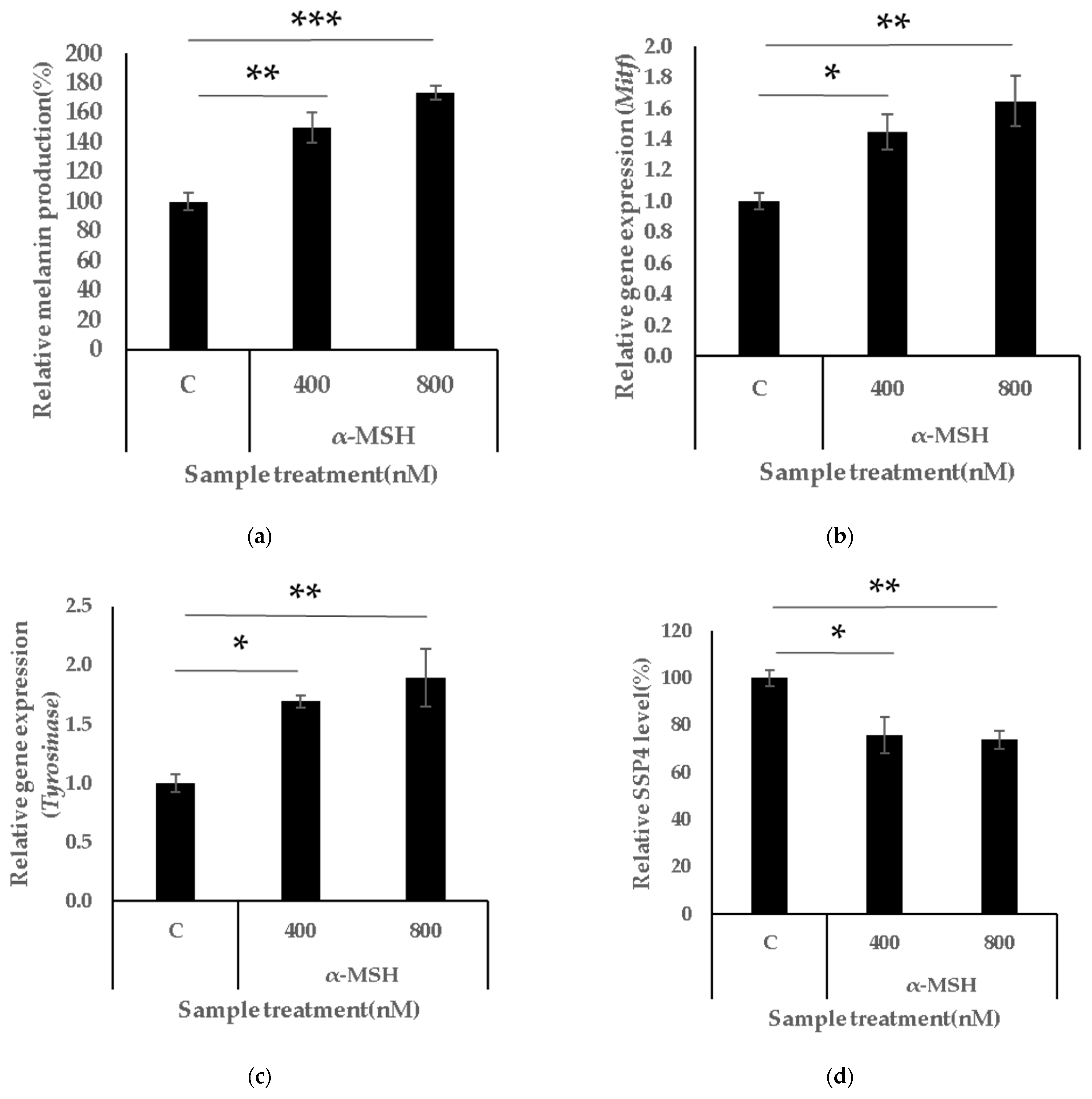
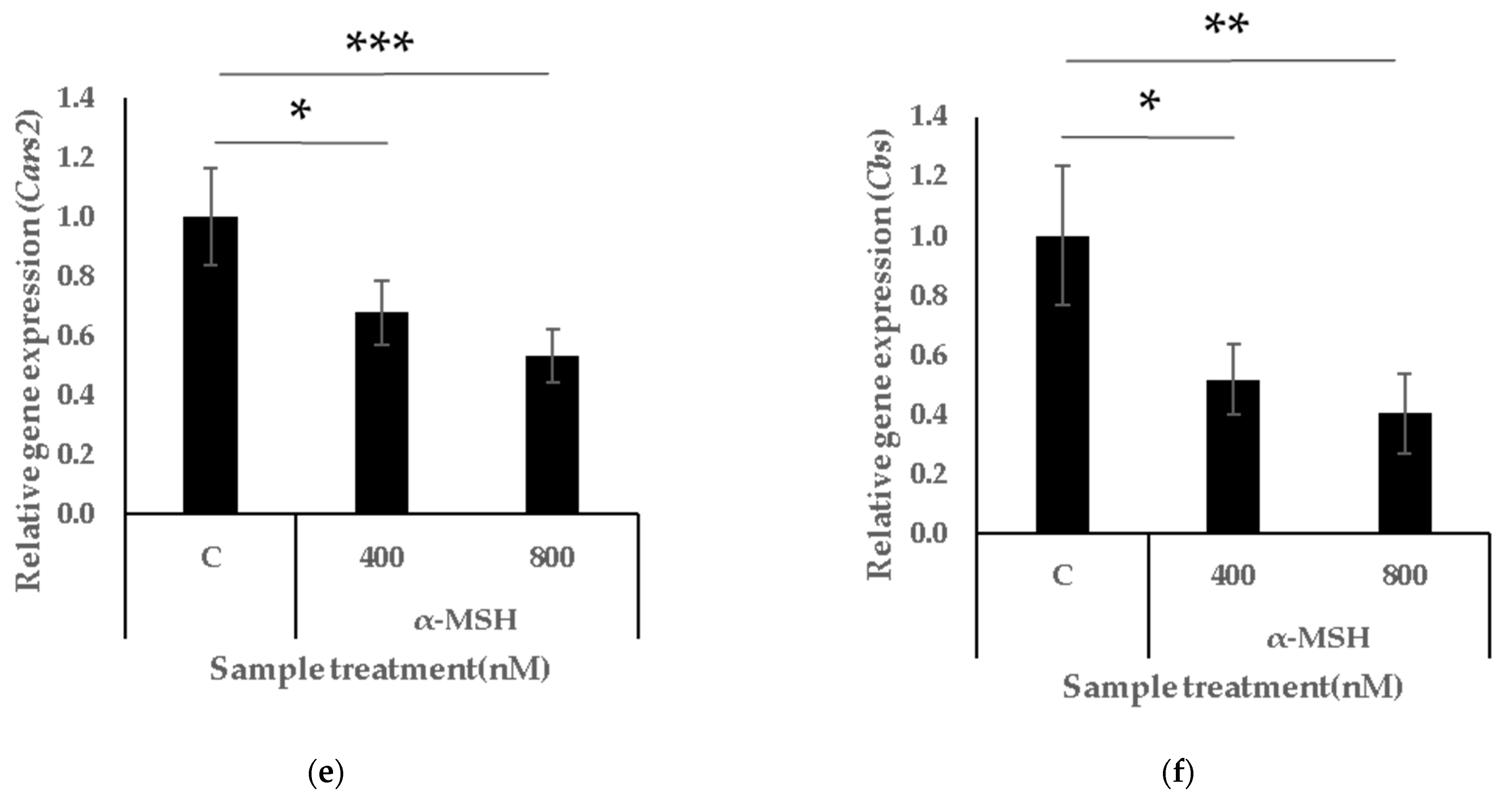
Consistent with previous studies, α-MSH exposure significantly increased melanin content and the expression of melanin-related genes, such as Mitf and tyrosinase (Tyr), in a dose-dependent manner compared to the control (Figure 2a,c) [18,19]. In contrast, intracellular persulphide content was significantly reduced by α-MSH in a dose-dependent manner (Figure 2d). Moreover, the expression levels of persulphide synthesis enzymes, such as Cars2 and Cbs, were significantly downregulated (Figure 2e,f). These results suggest that melanogenesis and persulphide synthesis exhibit opposing trends.

3.3. GSSSG Inhibits α-MSH-Induced Melanogenesis

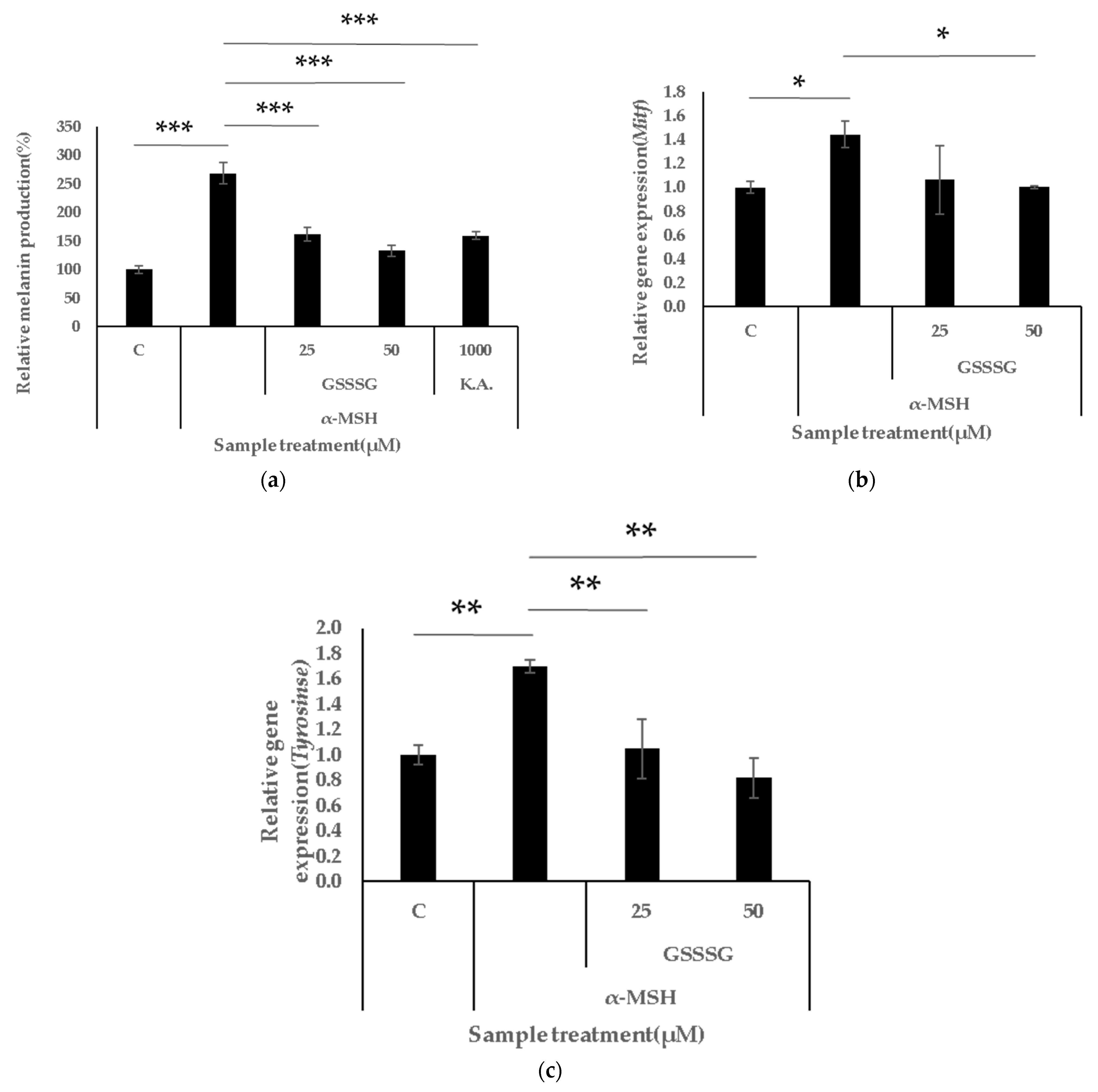
Next, we investigated the anti-melanogenic effects of GSSSG in B16-F0 cells. Melanin content was significantly reduced by GSSSG treatment (Figure 3a), with 25 μM GSSSG supressing melanin production as efficiently as 1000 μM kojic acid (positive control). Furthermore, GSSSG downregulated melanin-related genes, such as MITF and TYR in a dose-dependent manner (Figure 3b,c). These results suggested that GSSSG has anti-melanogenic activity through downregulation of MITF and TYR.

3.4. GSSSG Restores Intracellular Persulphide Levels

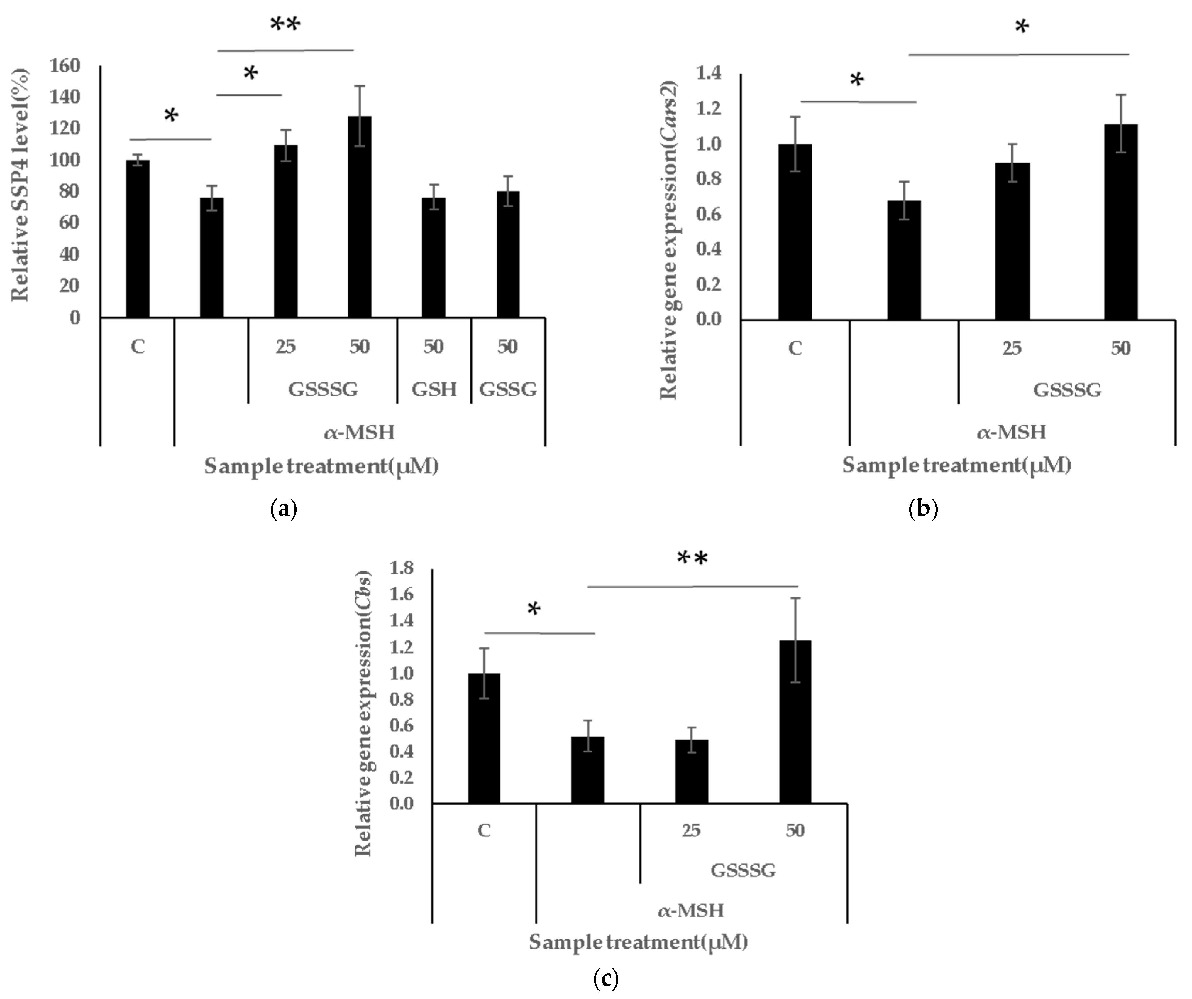
Intracellular persulphide level was also measured in GSSSG- and α-MSH-treated cells. As a result, intracellular persulphide level was significantly restored by GSSSG (Figure 4a). In this study, GSH and GSSG were also evaluated as derivatives of GSSSG; however, neither GSH nor GSSG showed intracellular supersulphide restoration. Moreover, GSSSG significantly upregulated the expression of supersulphide synthesis enzymes (Figure 4b,c). These results suggest that GSSSG prevents α-MSH-induced decrease in supersulphide abundance and synthesis-related gene expression.

3.5. GSSSG Inactivates Intracellular Tyrosinase Activity and Exhibits Antioxidant Effects

To further investigate the anti-melanogenic mechanism of GSSSG, we assessed its effects on intracellular tyrosinase activity and antioxidant capacity using a DPPH assay. Intracellular tyrosinase activity was significantly inactivated by GSSSG compared with that in the control group (Figure 5a). Additionally, GSSSG exhibited radical-scavenging activity, indicating its antioxidant properties (Figure 5b). These findings suggest that GSSSG exerts anti-melanogenic effects through conventional mechanisms, including the inhibition and antioxidation of the tyrosinase enzyme.

3.6. Statistical Analysis

Statistical analyses were performed using EZR version 4.1.2 (Saitama Medical Centre, Saitama, Japan). All data are presented as mean ± SD. Comparisons between the α-MSH group and other groups were conducted using one-way analysis of variance (ANOVA), followed by Dunnett’s test. Statistical significance was defined as * p < 0.05, ** p < 0.01, and *** p < 0.001.

4. Discussion

In this study, we investigated the effects of melanogenesis on intracellular supersulphide levels and their associated synthesis genes. Additionally, we examined the anti-melanogenic and skin-brightening effects of GSSSG, as well as its role in restoring intracellular supersulphide levels.
Melanin is the principal component of skin pigmentation [20]. Skin damage caused by UV radiation induces oxidative stress, which causes tyrosinase activation [21,22]. This leads to melanin production [23]. These are the well-established mechanisms of melanogenesis.
Recent studies have reported the presence of supersulphides in human cells, where they play diverse roles, including antioxidant activity and involvement in energy metabolism [24,25]. Supersulphides are synthesised in human cells by several enzymes, such as CARS2 and CBS [26]. Furthermore, GSSSG, a supersulphide, has been shown to exert anti-inflammatory and neuroprotective effects [27,28]. Notably, albumin persulphide has demonstrated anti-melanogenic properties [13]. These previous findings motivated us to investigate the relationship between melanin production and intracellular supersulphides, as well as the potential inhibitory effects of GSSSG on melanogenesis.
In our study, α-MSH-induced melanogenesis in melanoma cells resulted in a significant increase in melanin production, with upregulation of Mitf and Tyr expression (Figure 3a–c). Simultaneously, intracellular supersulphide levels were significantly decreased, with a corresponding downregulation of Cars2 and Cbs expression (Figure 4a–c). A previous study demonstrated that MITF expression was significantly upregulated, whereas Cars2 was downregulated in cutaneous melanoma [29]. Given that GSSSG and albumin persulphide have anti-melanogenic effects, it is likely that intracellular persulphides play a role in regulating melanin production in human cells. Previous reports on albumin persulphide have demonstrated its inhibitory effect on melanin production through the suppression of ROS and NO. In the present study, we further investigated the effects of GSSSG on key melanogenesis-related factors such as Tyr and Mitf. Although further studies are needed to clarify whether there are differences in the effects between albumin persulphide and GSSSG, our findings suggest that supersulphide compounds may exert these biological effects. Our findings suggest that excessive melanin production depletes intracellular supersulphide levels, and that melanogenesis downregulates supersulphide synthesis gene expression, thereby reducing the likelihood of new supersulphide formation. To the best of our knowledge, the transcription factors regulating Cars2 remain unidentified. However, elucidating the mechanism by which the expression of this supersulphide biosynthetic enzyme is downregulated during melanogenesis will be a key focus and motivation for our future research.
In this context, we demonstrated that GSSSG treatment significantly suppresses melanin production, restores intracellular supersulphide levels, and upregulates Cars2 and Cbs expression (Figure 4). The increase in intracellular persulphide levels was unique to GSSSG, as previously reported [30]. Whether the increase in intracellular persulphide levels induced by GSSSG is primarily due to direct supplementation or modulation of synthetic enzymes remains unclear. Clarifying this mechanism is a key motivation for our future research. Furthermore, GSSSG exhibited conventional anti-melanogenic mechanisms, such as ROS scavenging activity and tyrosinase inhibition (Figure 5). These results indicate that GSSSG comprehensively suppresses melanogenesis via ordinary mechanisms, such as tyrosinase activity, and maintains intracellular supersulphide levels. α-MSH is produced in human skin cells, with UV exposure being one of its key triggers. Moreover, supersulphides are degraded upon UV irradiation [31]. Several studies have suggested that supersulphides play important roles in human health [32]. Given their susceptibility to degradation, maintaining optimal supersulphide levels in the body may be beneficial and could be supported by GSSSG supplementation.
Several published studies have reported that GSH or GSSG can reduce melanogenesis or melanin content in the skin [5]. In our study, we focused on GSSSG and its potential anti-melanogenic effects. In previous study, the melanin-suppressing effects of GSH were observed at concentrations in the mM range, whereas in our study, GSSSG showed significant inhibitory effects even at several tens of μM levels [33]. This difference may be attributed to distinct mechanisms by which intracellular supersulphide levels are regulated, suggesting a unique role of GSSSG in modulating melanogenesis not found in GSH and GSSG. Furthermore, our study revealed that GSSSG suppresses tyrosinase enzymatic activity and exhibits antioxidant effects (Figure 5). These findings suggest that GSSSG may exert higher anti-melanogenic activity than conventional whitening agents such as GSH or K.A, possibly due to its ability to comprehensively activate multiple mechanisms involved in melanogenesis suppression (Figure 3a).

5. Conclusions

Overall, our findings reveal an inverse correlation between melanin production, intracellular supersulphide levels, and supersulphide metabolism. Additionally, GSSSG exhibits anti-melanogenic and skin-brightening effects, while maintaining intracellular supersulphide levels. These results indicate that intracellular supersulphides play a regulatory role in melanin production and could serve as novel targets for the inhibition of melanogenesis. Furthermore, GSSSG may serve as a novel skin-brightening agent that comprehensively regulates conventional and supersulphide-related pathways involved in melanin regulation.

Author Contributions

Conceptualisation, Y.U.; methodology, Y.U.; software, Y.U.; validation, Y.U.; formal analysis, Y.U.; investigation, Y.U.; resources, Y.U.; data curation, Y.U.; writing—original draft preparation, Y.U.; writing—review and editing, Y.U.; visualisation, Y.U.; supervision, T.S.; project administration, T.S. All authors have read and agreed to the published version of the manuscript.

Funding

This research was funded by the Mitsubishi Corporation Life Sciences Co., Ltd. No financial support was received from other companies or external funding sources.

Institutional Review Board Statement

Not applicable.

Informed Consent Statement

Not applicable.

Data Availability Statement

The data presented in this study are available upon request from the corresponding author.

Acknowledgments

We thank our colleagues at the Mitsubishi Corporation Life Sciences Co., Ltd. for their helpful discussions.

Conflicts of Interest

Y.U. and T.S. are employed by Mitsubishi Corporation Life Sciences Co., Ltd. The funders had no role in the design of the study; in the collection, analyses or interpretation of data; in the writing of the manuscript; or in the decision to publish the results.

Abbreviations

The following abbreviations are used in this manuscript:
CARS2cysteinyl-tRNA synthetase 2
Cars2cysteinyl-tRNA synthetase 2 gene
CBScystathionine β-synthase
Cbscystathionine β-synthase gene
GSHglutathione
GSSHglutathione disulphide
MITFmicrophthalmia-associated transcription factor
MITFmicrophthalmia-associated transcription factor gene
ROSreactive oxygen species
SDstandard deviation
TYRtyrosinase gene
α-MSHalpha-melanocyte-stimulating hormone

References

  1. Gromkowska-Kępka, K.J.; Puścion-Jakubik, A.; Markiewicz-Żukowska, R.; Socha, K. The impact of ultraviolet radiation on skin photoaging—review of in vitro studies. J. Cosmet. Dermatol. 2021, 20, 3427–3431. [Google Scholar] [CrossRef]
  2. Zhao, W.; Yang, A.; Wang, J.; Huang, D.; Deng, Y.; Zhang, X.; Qu, Q.; Ma, W.; Xiong, R.; Zhu, M.; et al. Potential application of natural bioactive compounds as skin-whitening agents: A review. J. Cosmet. Dermatol. 2022, 21, 6669–6687. [Google Scholar] [CrossRef]
  3. Ferreira, S.M.; Gomes, S.M.; Santos, L. A novel approach in skin care: By-product extracts as natural UV filters and an alternative to synthetic ones. Molecules 2023, 28, 2037. [Google Scholar] [CrossRef]
  4. Merecz-Sadowska, A.; Sitarek, P.; Kowalczyk, T.; Zajdel, K.; Kucharska, E.; Zajdel, R. The modulation of melanogenesis in B16 cells upon treatment with plant extracts and isolated plant compounds. Molecules 2022, 27, 4360. [Google Scholar] [CrossRef]
  5. Uchida, Y.; Kaneda, T.; Ono, M.; Matsuoka, M.; Nakamura, U.; Ishida, A.; Yamasaki, Y.; Takeo, H.; Sakurai, T. The effect of cysteine peptide ingestion on skin brightness, a randomized, double-blind, placebo-controlled, parallel-group human clinical trial. Cosmetics 2023, 10, 72. [Google Scholar] [CrossRef]
  6. Barayeu, U.; Sawa, T.; Nishida, M.; Wei, F.-Y.; Motohashi, H.; Akaike, T. Supersulfide biology and translational medicine for disease control. Br. J. Pharmacol. 2023; early view. [Google Scholar] [CrossRef]
  7. Akaike, T.; Morita, M.; Ogata, S.; Yoshitake, J.; Jung, M.; Sekine, H.; Motohashi, H.; Barayeu, U.; Matsunaga, T. New aspects of redox signaling mediated by supersulfides in health and disease. Free Radic. Biol. Med. 2024, 222, 539–551. [Google Scholar] [CrossRef] [PubMed]
  8. Alam, M.M.; Kishino, A.; Sung, E.; Sekine, H.; Abe, T.; Murakami, S.; Akaike, T.; Motohashi, H. Contribution of NRF2 to sulfur metabolism and mitochondrial activity. Redox Biol. 2023, 60, 102624. [Google Scholar] [CrossRef] [PubMed]
  9. Maemura, M.; Morita, M.; Ogata, S.; Miyamoto, Y.; Ida, T.; Saito, T.; Yoshitake, J.; Matsunaga, T.; Akaike, T.; Yano, F. Supersulfides contribute to joint homeostasis and bone regeneration. Redox Biol. 2025, 81, 103545. [Google Scholar] [CrossRef]
  10. Sasama, Y.; Yoshimura, K.; Hoshino, M.; Sasa, K.; Akaike, T.; Morita, M.; Miyamoto, Y. Supersulfides support bone growth by promoting chondrocyte proliferation in the growth plates. J. Oral Biosci. 2024, 66, 76–81. [Google Scholar] [CrossRef]
  11. Sasaki, Y.; Numakura, T.; Yamada, M.; Sugiura, H.; Matsunaga, T.; Ida, T.; Morita, M.; Suzuki, A.; Matsumoto, S.; Kawaguchi, M.; et al. Glutathione supersulphide regulates T-cell receptor signalling. bioRxiv 2024. bioRxiv: 2024.04.30.591985. [Google Scholar] [CrossRef]
  12. Ezaka, M.; Marutani, E.; Miyazaki, Y.; Kanemaru, E.; Selig, M.K.; Boerboom, S.L.; Ostrom, K.F.; Stemmer-Rachamimov, A.; Bloch, D.B.; Brenner, G.J.; et al. Oral administration of glutathione trisulfide increases reactive sulfur levels in dorsal root ganglion and ameliorates paclitaxel-induced peripheral neuropathy in mice. Antioxidants 2022, 11, 2122. [Google Scholar] [CrossRef]
  13. Ikeda, M.; Ishima, Y.; Kinoshita, R.; Chuang, V.T.G.; Tasaka, N.; Matsuo, N.; Watanabe, H.; Shimizu, T.; Ishida, T.; Otagiri, M.; et al. A novel S-sulfhydrated human serum albumin preparation suppresses melanin synthesis. Redox Biol. 2018, 14, 354–360. [Google Scholar] [CrossRef]
  14. Kienzler, C.; Contreras, M.; Treger, J.; Liau, M.; Owens, C.; Prins, M. Transcriptome analysis of novel B16 melanoma metastatic variants generated by serial intracarotid artery injection. Acta Neuropathol. Commun. 2025, 13, 10. [Google Scholar] [CrossRef]
  15. Zheng, S.; Deng, R.; Xie, S.; Huang, G.; Ou, Z.; Shen, Z. Typha pollen extract inhibit melanogenesis via α-MSH/MC1R signaling pathway in B16 and melasma mouse model. Arch. Dermatol. Res. 2025, 317, 321. [Google Scholar] [CrossRef] [PubMed]
  16. Uchida, Y.; Ferdousi, F.; Takahashi, S.; Isoda, H. Comprehensive transcriptome profiling of antioxidant activities by glutathione in human HepG2 cells. Molecules 2024, 29, 1090. [Google Scholar] [CrossRef] [PubMed]
  17. Zhou, S.; Riadh, D.; Sakamoto, K. Grape extract promoted α-MSH-induced melanogenesis in B16F10 melanoma cells, which was inverse to resveratrol. Molecules 2021, 26, 5959. [Google Scholar] [CrossRef] [PubMed]
  18. Ha, S.Y.; Jung, J.Y.; Yang, J.-K. Camellia japonica essential oil inhibits α -MSH-induced melanin production and tyrosinase activity in B16F10 melanoma cells. Evid. Based Complement. Alternat. Med. 2021, 2021, 6328767. [Google Scholar] [CrossRef]
  19. Solano, F. Photoprotection and skin pigmentation: Melanin-related molecules and some other new agents obtained from natural sources. Molecules 2020, 25, 1537. [Google Scholar] [CrossRef]
  20. Snyman, M.; Walsdorf, R.E.; Wix, S.N.; Gill, J.G. The metabolism of melanin synthesis—From melanocytes to melanoma. Pigment. Cell Melanoma Res. 2024, 37, 438–452. [Google Scholar] [CrossRef]
  21. Netcharoensirisuk, P.; Umehara, K.; De-Eknamkul, W.; Chaotham, C. Cajanin suppresses melanin synthesis through modulating MITF in human melanin-producing cells. Molecules 2021, 26, 6040. [Google Scholar] [CrossRef]
  22. Chen, Q.; Tao, W.; Wang, J.; Li, J.; Zheng, M.; Liu, Y.; Fang, Z. Inhibitive mechanism of loquat flower isolate on tyrosinase activity and melanin synthesis in mouse melanoma B16 cells. Biomolecules 2024, 14, 895. [Google Scholar] [CrossRef]
  23. Nishimura, A.; Yoon, S.; Matsunaga, T.; Ida, T.; Jung, M.; Ogata, S.; Morita, M.; Yoshitake, J.; Unno, Y.; Barayeu, U.; et al. Longevity control by supersulfide-mediated mitochondrial respiration and regulation of protein quality. Redox Biol. 2024, 69, 103018. [Google Scholar] [CrossRef]
  24. Zhou, L.; Nishimura, A.; Umezawa, K.; Kato, Y.; Mi, X.; Ito, T.; Urano, Y.; Akaike, T.; Nishida, M. Supersulfide catabolism participates in maladaptive remodeling of cardiac cells. J. Pharmacol. Sci. 2024, 155, 121–130. [Google Scholar] [CrossRef]
  25. Matsunaga, T.; Sano, H.; Takita, K.; Morita, M.; Yamanaka, S.; Ichikawa, T.; Numakura, T.; Ida, T.; Jung, M.; Ogata, S.; et al. Supersulphides provide airway protection in viral and chronic lung diseases. Nat. Commun. 2023, 14, 4476. [Google Scholar] [CrossRef] [PubMed]
  26. Khodade, V.S.; Toscano, J.P. Reactive sulfur species in biology and medicine. Antioxidants 2023, 12, 1759. [Google Scholar] [CrossRef] [PubMed]
  27. Zhang, T.; Pan, Y.; Sawa, T.; Akaike, T.; Matsunaga, T. Supersulfide donors and their therapeutic targets in inflammatory diseases. Front. Immunol. 2025, 16, 1581385. [Google Scholar] [CrossRef] [PubMed]
  28. Mansouri, A.; Reiner, Ž.; Ruscica, M.; Tedeschi-Reiner, E.; Radbakhsh, S.; Ekta, M.B.; Sahebkar, A. Antioxidant effects of statins by modulating Nrf2 and Nrf2/HO-1 signaling in different diseases. J. Clin. Med. 2022, 11, 1313. [Google Scholar] [CrossRef]
  29. Iciek, M.; Bilska-Wilkosz, A.; Kozdrowicki, M.; Górny, M. Reactive sulfur species and their significance in health and disease. Biosci. Rep. 2022, 42, BSR20221006. [Google Scholar] [CrossRef]
  30. Switzer, C.H. How super is supersulfide?: Reconsidering persulfide reactivity in cellular biology. Redox Biol. 2023, 67, 102899. [Google Scholar] [CrossRef]
  31. Lee, S.-G.; Hwang, J.-W.; Kang, H. Antioxidant and skin-whitening efficacy of a novel decapeptide (DP, KGYSSYICDK) derived from fish by-products. Mar. Drugs 2024, 22, 374. [Google Scholar] [CrossRef]
  32. Pan, Y.; Matsunaga, T.; Zhang, T.; Akaike, T. The Therapeutic Potential of Supersulfides in Oxidative Stress-Related Diseases. Biomolecules 2025, 15, 172. [Google Scholar] [CrossRef] [PubMed]
  33. Lee, Y.; Kumar, S.; Kim, S.H.; Seong, K.-Y.; Lee, H.; Kim, C.; Jung, Y.-S.; Yang, S.Y. Odorless glutathione microneedle patches for skin whitening. Pharmaceutics 2020, 12, 100. [Google Scholar] [CrossRef] [PubMed]
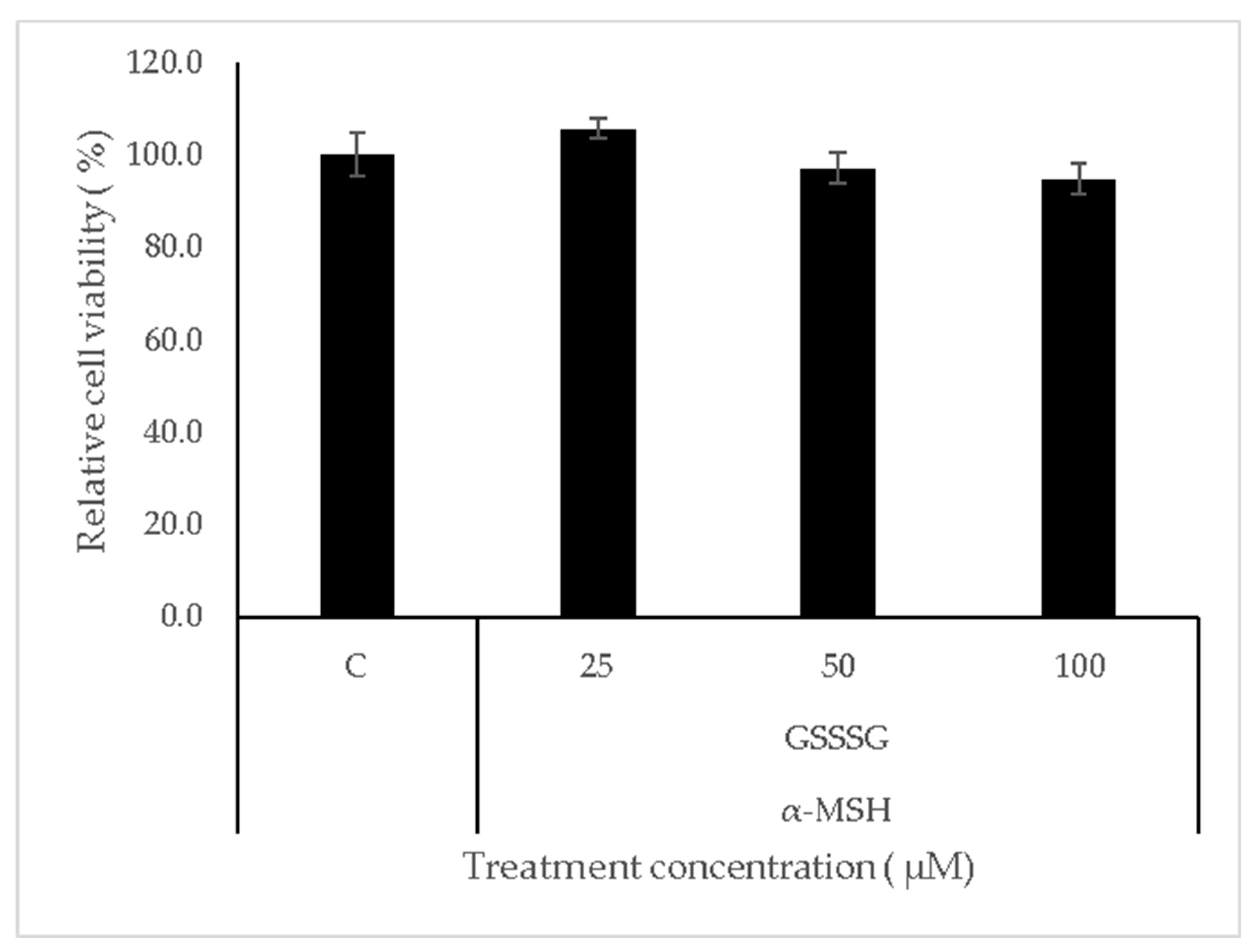
Figure 1. Cell viability of α-MSH- and GSSSG-treated cells. B16-F0 cells were treated with 400 nM α-MSH and GSSSG for 24 h. Cell viability was measured by MTT assay. All data are presented as the mean ± standard deviation (SD). Comparisons with the control were performed using Dunnett’s test.
Cosmetics 12 00234 g001
Figure 2. The effect of α-MSH on melanogenesis and persulphide synthesis in B16-F0 cells. B16-F0 cells were treated with α-MSH for 24 h. All data are presented as the mean ± standard deviation (SD). Comparisons with the control were performed using Dunnett’s test (* p < 0.05, ** p < 0.01, *** p < 0.001). (a) Melanin content analysis. (b,c) Relative expression levels of melanin-synthesis-related genes. (d) Detection of intracellular persulphides using SSP4 fluorescence. (e,f) Relative expression levels of persulphide-synthesis-related genes. α-MSH, alpha-melanocyte-stimulating hormone; Cars2, cysteinyl-tRNA synthetase 2; Cbs, cystathionine β-synthase; C, control.
Cosmetics 12 00234 g002aCosmetics 12 00234 g002b
Figure 3. Effect of GSSSG on α-MSH-induced melanogenesis in B16-F0 cells. B16-F0 cells were pretreated with GSSSG for 30 min, followed by treatment with 400 nM α-MSH for 24 h. Data are presented as the mean ± standard deviation (SD). Comparisons with the control were performed using Dunnett’s test (* p < 0.05, ** p < 0.01, *** p < 0.001). (a) Melanin content analysis. K.A. is kojic acid. (b,c) Relative expression levels of melanin synthesis-related genes.
Cosmetics 12 00234 g003
Figure 4. Effect of GSSSG for intracellular supersulphide on α-MSH-treated B16-F0 cells. B16-F0 cells were pretreated with GSSSG for 30 min, followed by treatment with 400 nM α-MSH for 24 h. Data are presented as the mean ± standard deviation (SD). Comparisons with the control were performed using Dunnett’s test (* p < 0.05, ** p < 0.01). (a) Determination of intracellular persulphide levels content by SSP4 analysis. (b,c) Relative expression levels of supersulphide-synthesis-related genes. GSSSG, glutathione trisulphide; GSH, glutathione; GSSG, glutathione disulphide; Cars2, cysteinyl-tRNA synthetase 2; Cbs, cystathionine β-synthase; C, control.
Cosmetics 12 00234 g004
Figure 5. Effect of GSSSG on intracellular tyrosinase activity and antioxidant capacity. B16-F0 cells were pretreated with GSSSG for 30 min, followed by treatment with 400 nM α-MSH for 24 h. Data are presented as the mean ± standard deviation (SD). Comparisons with the control were performed using a t-test (* p < 0.05). (a) Intracellular tyrosinase activity. (b) DPPH assay results. GSSSG, glutathione trisulphide; α-MSH; alpha-melanocyte-stimulating hormone.
Cosmetics 12 00234 g005
Disclaimer/Publisher’s Note: The statements, opinions and data contained in all publications are solely those of the individual author(s) and contributor(s) and not of MDPI and/or the editor(s). MDPI and/or the editor(s) disclaim responsibility for any injury to people or property resulting from any ideas, methods, instructions or products referred to in the content.