Review

Genetic Insights into Acne, Androgenetic Alopecia, and Alopecia Areata: Implications for Mechanisms and Precision Dermatology

by
Gustavo Torres de Souza
1,2
1
Fagron Genomics, 08226 Barcelona, Spain
2
Human Genome and Stem Cell Research Center, São Paulo University, São Paulo 05508-000, Brazil
Cosmetics 2025, 12(5), 228; https://doi.org/10.3390/cosmetics12050228
Submission received: 4 September 2025 / Revised: 4 October 2025 / Accepted: 13 October 2025 / Published: 15 October 2025
(This article belongs to the Special Issue Feature Papers in Cosmetics in 2025)

Abstract

Chronic dermatological conditions such as acne vulgaris, androgenetic alopecia (AGA), and alopecia areata (AA) affect hundreds of millions worldwide and contribute substantially to quality-of-life impairment. Despite the availability of systemic retinoids, anti-androgens, and JAK inhibitors, therapeutic responses remain heterogeneous and relapse is common, underscoring the need for biologically grounded stratification. Over the past decade, large genome-wide association studies and functional analyses have clarified disease-specific and cross-cutting mechanisms. In AA, multiple independent HLA class II signals and immune-regulatory loci such as BCL2L11 and LRRC32 establish antigen presentation and interferon-γ/JAK–STAT signalling as central drivers, consistent with clinical responses to JAK inhibition. AGA is driven by variation at the androgen receptor and 5-α-reductase genes alongside WNT/TGF-β regulators (WNT10A, LGR4, RSPO2, DKK2), explaining follicular miniaturisation and enabling polygenic risk prediction. Acne genetics highlight an immune–morphogenesis–lipid triad, with loci in TGFB2, WNT10A, LGR6, FASN, and FADS2 linking follicle repair, innate sensing, and sebocyte lipid metabolism. Barrier modulators such as FLG and OVOL1, first described in atopic dermatitis, further shape inflammatory thresholds across acne and related phenotypes. Together, these findings position genetics not as an abstract catalogue of risk alleles but as a map of tractable biological pathways. They provide the substrate for patient-stratified interventions ranging from JAK inhibitors in AA, to endocrine versus morphogenesis-targeted strategies in AGA, to lipid- and barrier-directed therapies in acne, while also informing cosmetic practices focused on barrier repair, sebaceous balance, and follicle health.

1. Why Use Genetics to Approach Dermatology?

Chronic dermatological disorders such as acne vulgaris, androgenetic alopecia (AGA), and alopecia areata (AA) are among the most prevalent non-communicable conditions worldwide, contributing significantly to both physical and psychosocial burden. According to the most recent Global Burden of Disease (GBD) estimates, acne alone affects approximately 231 million individuals at any given time, ranking among the leading causes of disability-adjusted life years (DALYs) in adolescents and young adults [1,2]. AGA, the most common form of hair loss, exhibits a striking age- and sex-related prevalence, affecting up to 50% of men by the no age of 50 and approaching 80% lifetime prevalence in European male populations [3,4]. While typically non-scarring, AGA has a sustained psychosocial impact and is associated with significant cosmetic distress. AA, by contrast, is an autoimmune form of hair loss with unpredictable course and frequent relapse; recent global analyses indicate that its contribution to DALYs is increasing, largely driven by population growth and ageing [5,6]. Collectively, these conditions illustrate the scale of the dermatologic burden and highlight the need for mechanistically informed, personalised treatments.
Despite advances in therapeutics, current management remains limited by incomplete efficacy, tolerability issues, and high variability in individual response. In acne, systemic retinoids such as isotretinoin remain the most effective treatment, yet their use is constrained by teratogenicity, mucocutaneous side-effects, and psychiatric concerns, restricting them to severe cases. Quality-of-life and mental-health impacts of both the disease and treatment burden are consistently reported [7]. For AGA, 5-α-reductase inhibitors (finasteride and dutasteride) reduce dihydrotestosterone levels and achieve meaningful regrowth in subsets of patients; however, real-world data reveal marked heterogeneity in treatment response, and concerns persist regarding sexual and neuropsychiatric adverse effects. Dutasteride is often reported to be more effective than finasteride, but this comes with an ongoing debate over long-term safety and tolerability [8,9,10,11]. In AA, the approval of Janus kinase (JAK) inhibitors represents a milestone, with robust regrowth observed during active therapy. Nevertheless, randomised withdrawal studies demonstrate high relapse rates after discontinuation, underscoring the need for chronic disease framing and maintenance strategies [5,12,13]. These examples illustrate that conventional trial data often fail to predict individual outcomes, emphasising the importance of genetic and mechanistic stratification.
Human genetics offers a framework to dissect this variability, clarify underlying mechanisms, and inform precision-oriented interventions. In acne, large meta-analyses comprising more than 20,000 cases identified 46 independent risk loci, including genes regulating follicle morphogenesis (TGFB2, WNT10A, LGR6) and wound-healing pathways, while polygenic scores explain up to 5.6% of phenotypic variance and correlate with severity [7]. AGA is now recognised as highly polygenic: a landmark GWAS identified 71 independent loci explaining ~38% of SNP-heritability, highlighting the androgen receptor, WNT, and TGF-β pathways as key axes [3]. More recent predictors leveraging over 100 SNPs already stratify risk with clinically meaningful discrimination between no, moderate, and severe hair loss categories [8]. For AA, genome-wide meta-analysis pinpointed HLA-DR within the MHC as the dominant risk determinant, with multiple independent allelic effects, and identified non-HLA regulators including BCL2L11 and LRRC32, placing antigen presentation and interferon signalling at the centre of disease biology [5,14]. These findings have been reinforced in East Asian cohorts, where Taiwanese GWAS highlighted HLA-DQA1/DQB1 haplotypes and IFN-γ–linked networks as key drivers, supporting the generalisability of this model across ancestries [6].
Beyond disease-specific insights, genetics has revealed convergence across conditions. Immune and antigen-presentation pathways are dominant in AA but also exert modulatory roles in acne subsets. Morphogenetic pathways centred on WNT/TGF-β signalling emerge as common denominators between acne and AGA, both of which rely on follicle cycling and repair. Lipid biosynthesis pathways are particularly prominent in acne, with genes such as FASN and FADS2 modulating sebaceous activity, but have contextual relevance in AGA where sebaceous enlargement and altered lipid milieu are well documented. Finally, barrier and keratinisation regulators such as FLG and OVOL1, initially highlighted in atopic dermatitis genetics, are emerging as modulators of acne phenotypes, exemplifying how barrier integrity may tune inflammatory thresholds. Thus, GWAS and network analyses collectively underscore that while each disease has distinct entry points, they converge on shared immune, barrier, and morphogenetic axes, which positions genetics as a bridge between mechanism and personalised therapy [15,16,17,18].

2. Acne Vulgaris—Immune—Morphogenesis—Lipid Triad

2.1. Innate Sensing & Inflammatory Tone

Acne vulgaris is now understood as an inflammatory disorder in which innate immune sensing in the pilosebaceous unit sets the stage for disease expression. Toll-like receptor 2 (TLR2) and Toll-like receptor 4 (TLR4) are expressed in keratinocytes and sebocytes and activate canonical NF-κB pathways upon stimulation by microbial and environmental cues, leading to release of IL-1β, IL-6, IL-8, and TNF-α [19,20]. Evidence from lesion biopsies and in vitro models shows that Cutibacterium acnes can activate TLR4–NF-κB signalling and that particulate matter can amplify this response, pointing to TLR4 as an upstream hub that integrates microbiota and exposome inputs into pro-inflammatory cytokine signalling [19,20,21,22,23]. Lesional profiling consistently finds elevated TNF-α accompanied by increased IL-10, suggesting a composite cytokine milieu that both promotes inflammation and engages regulatory T-cell circuits, with IL-10 likely acting to contain excessive tissue damage while not fully abrogating inflammation [7]. Figure 1 summarises the TLR4 signaling interaction in the pathogenesis of acne.
Importantly, innate sensing intersects with barrier physiology. C. acnes may disrupt keratinocyte tight junctions and alter barrier-related gene expression, which lowers the activation threshold for subsequent inflammatory triggers and facilitates cytokine diffusion within the follicular unit [7]. This creates a feed-forward loop where microbial stimuli and barrier damage reinforce each other. Within this frame, TLR4 serves as a nexus that links microbial recognition to cytokine output and to barrier state, and therefore acts as a tractable upstream node in the acne network [19,20].
Mechanistically adjacent players connect the innate hub to barrier and lipid axes. FLG-AS1, a non-coding transcript in the filaggrin locus, and OVOL1, a keratinocyte differentiation regulator, provide routes by which innate triggers may alter epidermal differentiation and barrier proteins; on the lipid side, genes that shape sebocyte lipid handling can modulate TLR-driven outputs through lipid-mediated signalling and membrane composition effects, providing a rationale for considering PNPLA3, APOE, and SOAT1 as pathway-level modifiers even if they are not established acne GWAS loci [24,25].

2.2. Morphogenesis & Stem-Cell

Genetic studies converge on follicle morphogenesis and stem-cell governance as central to acne susceptibility. Early severe-acne GWAS highlighted TGFB2 and OVOL1, both linked to epidermal differentiation and hair-follicle development, and thus pointed to morphogenetic control as an aetiologic layer beyond simple “infection and sebum” models [24,26]. A subsequent severe-acne meta-analysis broadened this theme and connected risk to wound-healing and tissue-repair pathways of the follicular unit [27].
The largest meta-GWAS to date identified 46 independent signals, with fine-mapping implicating causal genes involved in hair-follicle development and extracellular-matrix remodelling, consistent with the concept that aberrant repair and junctional-zone dynamics shape lesion propensity and evolution [24]. Further expansion added loci within the WNT axis, including LGR5 and the ZNRF3–KREMEN1 module, and complemented previously reported signals at LGR6 and WNT10A [24,26]. LGR5 and LGR6 act as receptors for R-spondins and mark cycling stem-cell compartments, while ZNRF3 negatively regulates WNT receptor abundance and KREMEN1 partners with DKK1 to inhibit WNT signalling. Together these loci map to the control knobs of stem-cell fate decisions in the junctional zone and sebaceous gland, providing a genetic substrate for the clinical observation that comedogenesis, inflammation, and repair are tightly interwoven processes [24,25,26].
Functionally this implies that risk alleles can shift the follicle’s regenerative set-point toward states that favour micro-comedone formation, aberrant keratinisation, and maladaptive wound-healing, especially under pro-inflammatory conditions. It also rationalises why retinoids, which reprogram keratinocyte differentiation and junctional-zone architecture, have broad efficacy across acne phenotypes even though their use is constrained by safety considerations [7].

2.3. Lipid Biosynthesis & Sebocyte Biology

Sebaceous glands are central to acne pathophysiology because they generate the lipid milieu that fuels microbial growth and modulates local immunity. Human sebocytes engage in de novo lipogenesis (DNL), producing fatty acids and complex lipids that accumulate in sebum. DNL is significantly upregulated in acne, with isotretinoin treatment shown to reduce sebaceous lipid output by inducing sebocyte apoptosis and repressing transcriptional pathways controlling lipid metabolism [27]. Pharmacological inhibition of acetyl-CoA carboxylase, the rate-limiting enzyme for DNL, reduces sebum secretion in vivo, highlighting a tractable therapeutic axis [28,29].
Genetics supports the centrality of lipid pathways. The recent European meta-GWAS identified FASN and FADS2 as risk loci [25]. FASN encodes fatty acid synthase, a hallmark of mid-stage sebocyte differentiation, whereas FADS2 catalyses Δ6-desaturation of palmitic acid to generate sapienic acid, the major monounsaturated fatty acid in human sebum and a lipid implicated in C. acnes colonisation. In addition, FADS2 also desaturates linoleic acid to γ-linolenic acid, linking sebaceous lipid metabolism to broader PUFA pathways. Variants in these loci provide a mechanistic link between sebocyte lipid output and acne liability. Proteome-wide Mendelian randomisation also implicated FASN protein levels as inversely associated with acne risk, strengthening the causal inference [30,31].
WNT–lipid interactions provide an additional mechanistic bridge. ZNRF3 and KREMEN1, both associated with acne, act as negative regulators of WNT receptor abundance and co-receptors for DKK1, respectively. These genes are expressed in sebocytes and keratinocytes, and their disruption could shift the balance of WNT signalling toward altered sebocyte fate and lipogenesis. The integration of lipid metabolism with morphogenetic pathways may explain why sebum hypersecretion and follicular cycling abnormalities often co-occur in acne [32,33].
Beyond GWAS-validated loci, pathway candidates such as PNPLA3, APOE, and SOAT1 merit consideration. PNPLA3 and APOE are key regulators of systemic lipid handling and have been implicated in liver steatosis and lipoprotein metabolism. While not reproducibly associated with acne in GWAS, their expression in sebaceous tissues and ability to modulate lipid droplet turnover and inflammation suggest plausible modifying effects. SOAT1, involved in cholesterol esterification, is enriched in sebaceous glands and altered in meibomian gland models [34,35]. Its role in controlling the cholesterol/ester balance of sebaceous lipids makes it a candidate for influencing sebum fluidity and comedogenesis, even if genetic evidence is not yet definitive. Together, these data emphasise that sebaceous lipid metabolism is both genetically and mechanistically coupled to acne, providing avenues for therapy through modulation of fatty acid synthesis, desaturation, and esterification [36,37].

2.4. Insights from Large Studies

Large-scale human genetics has reframed acne as a three-axis system rather than a single-axis “sebum disease.” A meta-GWAS that aggregated hundreds of thousands of individuals identified 46 independent signals, of which 29 were novel, and fine-mapping tied many of these to hair-follicle development and wound-healing pathways. The derived polygenic risk score (PRS) explained up to 5.6 percent of liability, which is notable for a common and environmentally sensitive trait, and importantly the PRS tracked with clinical severity [7,25].
Subsequent reports have shown additional loci, including FASN, LGR5, ZNRF3–KREMEN1, and FADS2, and demonstrated tangible risk stratification in the population: individuals in the top 5 percent of PRS carried approximately 1.6-fold higher risk of acne compared to the average, with the enrichment of WNT and lipid biosynthesis categories underscoring the dual importance of morphogenesis and metabolis [24]. Complementary analyses focused on severity suggest that susceptibility and severity are only partially overlapping genetic spaces, nominating additional pathways involved in scarring, fibrosis, and chronicity; this separation implies that prediction of onset and prediction of progression will likely require distinct marker sets and that treatment selection may need to be staged by disease phase rather than solely by baseline risk [26].
From a translational perspective, these studies provide two immediate applications. First, they highlight targetable biological pathways, such as WNT regulators and lipid enzymes, which can guide mechanism-matched interventions and complementary cosmetic strategies. Second, they enable quantitative stratification with PRS that can be combined with clinical features to enrich trials, prioritise early interventions for high-burden adolescents, and generate realistic expectations of response under different therapeutic mechanisms [7,26,38].

2.5. Barrier as a Modulator

Barrier integrity is increasingly recognised as a modifier of acne risk and severity. The pilosebaceous unit sits at the interface of innate immune activation and keratinocyte differentiation, where barrier dysfunction can amplify inflammatory signalling. Genetic studies support this connection. Variants at the OVOL1 locus, a transcription factor involved in keratinocyte terminal differentiation, have been associated with acne risk in genome-wide analyses, although OVOL1 dysregulation is more widely known for its role in atopic dermatitis and related barrier disorders. [25]. OVOL1 regulates filaggrin (FLG) expression through an IL-13–OVOL1–FLG axis that is well described in atopic dermatitis (AD) [23,38,39,40]. Its presence in acne GWAS underscores a shared barrier biology between acne and AD.
FLG itself, a cornerstone barrier protein, has been linked to acne phenotype through cross-disease analyses. In a Bangladeshi AD cohort, FLG loss-of-function variants were unexpectedly associated with reduced lifetime prevalence of acne [41,42]. This suggests that barrier deficiency may shift follicular milieu in ways that reduce comedogenesis, although ancestry-specific effects and environmental context likely modulate the direction of association. Such findings exemplify the complexity of barrier–acne interplay.
Mechanistic studies show that C. acnes can disrupt keratinocyte tight junctions and alter barrier proteins, creating a feedback loop where microbial sensing via TLR2/TLR4 and downstream cytokines increases transepidermal water loss (TEWL) and further compromises barrier resilience. Clinical data support this: TEWL is elevated in acne patients, and both systemic and topical therapies (e.g., retinoids) can exacerbate barrier fragility. Adjunctive barrier repair strategies (ceramide-dominant moisturisers, niacinamide) have been shown to mitigate irritation and improve outcomes, demonstrating translational relevance of barrier genetics and biology [43,44,45,46,47].
Thus, the barrier axis represents not only a mechanistic bridge between acne and AD but also a therapeutic opportunity. Genetic modulators such as OVOL1 and FLG provide insight into individual variability, while functional data reinforce the role of keratinocyte junctions in determining inflammatory thresholds [41,48,49].

3. Alopecia Areata—Antigen Presentation and Immune Privilege Collapse

Alopecia areata (AA) is a relapsing, non-scarring hair-loss disorder in which cytotoxic T cells target the hair follicle after the collapse of its normal immune-privileged state. Human genetics places HLA class II at the centre of disease risk, with multiple independent signals that fine-map to HLA-DR amino-acid positions and regulatory variants that alter antigen presentation [50,51]. Non-HLA loci implicate apoptosis control, regulatory T cell biology, and cytokine circuits that signal through JAK/STAT. Studies in East-Asian cohorts reproduce the HLA-centric architecture and emphasise interferon-γ (IFN-γ) as a driver of HLA upregulation in follicular epithelium, consistent with immune-privilege failure and sustained autoreactive loops [5,6,52,53,54,55].

3.1. HLA Class II as the Primary Hub

Across genome-wide studies, the most robust and largest-effect signals reside in the major histocompatibility complex (MHC), resolving to HLA class II and, in particular, to HLA-DR [5,52]. Fine-mapping analyses identify multiple independent effects within the region, including substitutions at DRB1 residues that shape the peptide-binding groove. These variants are predicted to alter the spectrum and stability of follicle-derived peptide presentation to CD4+ T cells, thereby changing activation thresholds once immune privilege is breached [5].
Mechanistically, the hair bulb normally exhibits low MHC expression and a local milieu enriched for immunoregulatory mediators, i.e., TGF-β, α-MSH, and neuropeptides that maintain a state of relative immune invisibility. Under inflammatory pressure, antigen-presenting cells and even follicular epithelial cells can express HLA-DR, permitting the display of follicular peptides that are now more efficiently accommodated by risk HLA-DR molecules. This provides a route by which common HLA variation, rather than a single autoantigen, broadens the potential autoreactive repertoire and licenses a cytotoxic response in genetically primed individuals [5,53,56,57].
The centrality of HLA class II also aligns with histopathology and immunophenotyping. Active AA lesions often show perifollicular infiltrates enriched for CD4+ and CD8+ T cells, increased MHC expression on outer-root-sheath keratinocytes, and local chemokine gradients that favour T-cell retention. These observations are consistent with a model in which HLA-DR-dependent activation of helper T cells supports and amplifies cytotoxic effector function in situ [52,53].

3.2. Non-HLA Immune Regulators

Non-HLA loci add biological resolution by highlighting immune checkpoints that modulate initiation and persistence of disease. BCL2L11 encodes BIM, a pro-apoptotic BH3-only protein that governs apoptosis in lymphocytes and also influences catagen biology in the follicle. Risk alleles at BCL2L11 suggest that dysregulated apoptosis can facilitate survival of autoreactive T cells and perturb the follicle’s cycle dynamics at the immune–epithelial interface [5,58,59]. This fits the clinicopathological observation that anagen hairs abruptly enter catagen/telogen in active AA, consistent with apoptosis-linked regression.
LRRC32, which encodes GARP, is essential for the activation of latent TGF-β on the surface of regulatory T cells. Associations at LRRC32 implicate impaired Treg function and insufficient activation of TGF-β, a cytokine that ordinarily contributes to the maintenance of immune privilege around the bulb. Reduced Treg potency would be expected to lower the threshold for effector activation and to lessen the local induction of tolerogenic signals in the follicle microenvironment [5,53,60,61].
Signals at SH2B3/ATXN2 extend the picture to adaptor-mediated cytokine signalling with well-documented pleiotropy across autoimmune diseases. Variation at SH2B3 may tune responses to multiple cytokines and growth factors, thereby influencing both T-cell activation and tissue responses downstream of inflammatory cues [5].
Functionally, these genomic findings converge with a core effector loop that is IFN-γ/IL-15 and JAK/STAT dependent. In active AA, cytotoxic CD8+ T cells bearing NKG2D produce IFN-γ, which in turn upregulates MHC expression in follicular epithelium and induces chemokines such as CXCL9 and CXCL10 that reinforce recruitment. Follicular keratinocytes and dermal papilla cells can produce IL-15, which signals through JAK1/JAK3 and STAT5 to support survival and proliferation of autoreactive T cells. Together, these circuits sustain disease as long as antigen presentation remains high and regulatory brakes remain weak [53,54,55,62,63].
An integrated interpretation is that HLA-DR sets the antigen-presentation landscape, BCL2L11 sets apoptotic thresholds in key compartments, LRRC32 sets the strength of Treg-mediated immune control, and SH2B3 tunes cytokine responsiveness across the infiltrate. The net effect is a follicular niche that is unusually permissive for the ignition and maintenance of CD8+ T-cell cytotoxicity once triggered [5,54,58,60,61].

3.3. East-Asian Cohorts and the Immune Theme

Genetic studies in East-Asian populations, including a Taiwanese genome-wide association analysis, confirm the primacy of antigen presentation and lymphocyte-activation networks, with reproducible associations at HLA-DQA1/DQB1 and related class II haplotypes [6,56,57]. Network and pathway analyses in these cohorts emphasise antigen presentation, T-cell activation, and cytokine signalling categories, indicating that the HLA-centric architecture is not limited to European ancestry. This cross-ancestry concordance strengthens the inference that antigen presentation is the universal hub of AA risk [6,56,57].
Mechanistically, the East-Asian data are consistent with an IFN-γ–first sequence of events at the follicle. Activated CD8+/NKG2D+ T cells produce IFN-γ, which induces class I and class II MHC expression on follicular keratinocytes and dermal papilla cells. The resulting increase in HLA-DR expression amplifies peptide display, enhances CD4+ T-cell help, and stabilises a cytotoxic niche, while IL-15 produced by follicular epithelium sustains the effector pool through JAK/STAT. This is precisely the immunologic loop that JAK inhibitors disrupt clinically, and it explains why disease often relapses when therapy is withdrawn and IFN-γ production returns [6,54,55].
The East-Asian echo also helps to separate universal from context-specific features. While HLA class II effects and JAK-dependent loops appear universal, the strength of individual non-HLA signals may vary by ancestry due to allele frequency differences and local linkage structure. This has practical implications for designing genetic risk scores or biomarker panels that generalise across populations [6].

3.4. Immune-Privilege Loss: Integrating Genetics and Follicular Biology

The hair follicle is among the few mammalian tissues that exhibit a defined immune-privileged state during anagen. This state includes reduced expression of HLA class I and class II, active local production of immunosuppressive mediators such as TGF-β and α-MSH, and expression of apoptotic ligands that eliminate infiltrating lymphocytes. Structural features of the anagen bulb, including a basement-membrane barrier and specialised extracellular-matrix composition, further limit immune access [53,56,57,58,64].
Genetics anchors these features in specific molecular circuits. HLA-DR polymorphisms determine how robustly follicular peptides can be presented once MHC is induced. IL-15 and TGF-β2 have emerged as critical guardians of privilege; dysregulation of either, in the context of heightened IFN-γ signalling, dismantles the barrier and promotes sustained presentation to autoreactive T cells [53,65,66]. BCL2L11 provides a mechanism for abrupt transitions in hair-cycle state, since increased pro-apoptotic drive can synchronise catagen entry and expose antigens in the setting of inflammation. LRRC32 variation points to insufficient Treg-mediated activation of latent TGF-β, reducing local immunoregulation exactly where it is most needed. SH2B3 pleiotropy suggests that cytokine set-points across multiple lineages can be shifted by common variants, potentially explaining heterogeneous treatment responses even among patients with similar clinical severity [5,54].
From this integrated perspective, AA is best framed as a two-step process. First, triggers that increase IFN-γ tone, i.e., infections, trauma, or unknown endogenous cues, induce HLA expression in the follicle and recruit effector cells. Second, genetically set thresholds in antigen presentation, apoptosis, and regulatory control determine whether immune privilege collapses and remains collapsed. This framing clarifies why AA often exhibits sudden onset, patchy distribution, and high relapse rates, and why therapies that transiently reduce signalling through JAK/STAT can produce regrowth without fully resetting the underlying predisposition [53,54,55,62].

3.5. Clinical Implications and Translational Outlook

The dominance of antigen presentation and JAK-linked cytokine loops explains current therapeutic successes and limitations. JAK inhibitors reduce IFN-γ signalling and IL-15-dependent survival cues, leading to hair regrowth in a substantial subset of patients. However, withdrawal leads to frequent relapse, which is consistent with genetics pointing to a persistent liability in antigen presentation and immune set-points that pharmacologic suppression does not erase [54,55]. This argues for long-term disease framing and maintenance strategies rather than finite courses.
Several translational paths follow directly from the genetic map. First, HLA-informed stratification could identify individuals at higher risk of chronic or relapsing disease and may eventually guide antigen-specific tolerance strategies. Second, biomarkers of IFN-γ/IL-15 activity, i.e., blood or lesional expression of interferon-stimulated genes or soluble mediators, could be used to titrate JAK inhibitor dosing, identify impending relapse, and design rational tapering protocols. Third, Treg-supportive interventions that enhance LRRC32-dependent activation of latent TGF-β merit exploration, particularly as adjuncts to JAK blockade. Fourth, apoptosis-modulating strategies informed by BCL2L11 biology could be studied for their ability to stabilise anagen in responders. Finally, ancestry-aware models that incorporate East-Asian data will be important for building portable polygenic scores and for testing whether specific non-HLA variants predict response or relapse risk across populations [5,6,60,61,62,63,67].
In summary, AA genetics defines a coherent disease architecture with HLA-DR–centred antigen presentation at its core, modulated by apoptosis, regulatory tolerance, and JAK-linked cytokine loops. The same architecture explains why therapies that target the IFN-γ/IL-15 axis produce robust, but often reversible, remissions. Embedding these insights into trial design and clinical decision pathways should enable more durable control through maintenance strategies and mechanism-matched combinations, while ongoing gene discovery and cross-ancestry work will refine risk prediction and therapeutic targeting [5,6,53,54,55].

4. Androgenetic Alopecia—AR–WNT Cross-Talk and Polygenic Prediction

4.1. Genetic Architecture

Androgenetic alopecia (AGA), or male-pattern baldness, is one of the most heritable dermatological traits, with twin and family studies estimating heritability up to 80%. Early candidate studies consistently implicated the androgen receptor (AR) locus on the X chromosome, but the scale of polygenicity only became clear after large genome-wide association studies (GWAS) [55,68]. A landmark analysis in over 70,000 men identified 71 independent loci, explaining approximately 38% of SNP-based heritability and mapping to 219 protein-coding genes distributed across androgen metabolism, WNT signalling, apoptosis, and TGF-β pathways [3,69]. Within this framework, AR and its neighbouring gene EDA2R represented the strongest single association, with multiple independent signals observed. Near SRD5A2, five distinct hits were identified, underscoring the importance of 5-α-reductase activity in dihydrotestosterone (DHT) generation. Together, these findings provided a quantitative scaffold for AGA risk and clarified that relatively few loci can explain a large fraction of genetic variance compared to other complex diseases [70].
Subsequent UK Biobank analyses expanded the picture to more than 200,000 men, cataloguing over 600 near-independent loci and confirming a SNP-heritability of about 0.39 with pedigree estimates at 0.62 [71]. This underscores AGA’s highly polygenic nature while simultaneously validating the robustness of the original 71-locus architecture. More recent integrative reviews consolidate signals to about 389 non-redundant loci, still explaining ~39% of phenotypic variance, with enrichment in androgenic, WNT, and morphogenetic pathways [14,72]. These datasets collectively highlight two mechanistic poles: androgen metabolism (AR, SRD5A1/2, CYP19A1) and follicle cycling/repair modules governed by WNT and TGF-β cross-talk. In particular, functional alleles that decrease WNT10A expression or perturb LGR4/RSPO2–DKK2 signalling provide causal links from variant to diminished WNT tone and shortened anagen, aligning with the miniaturisation phenotype of AGA follicles [73,74].
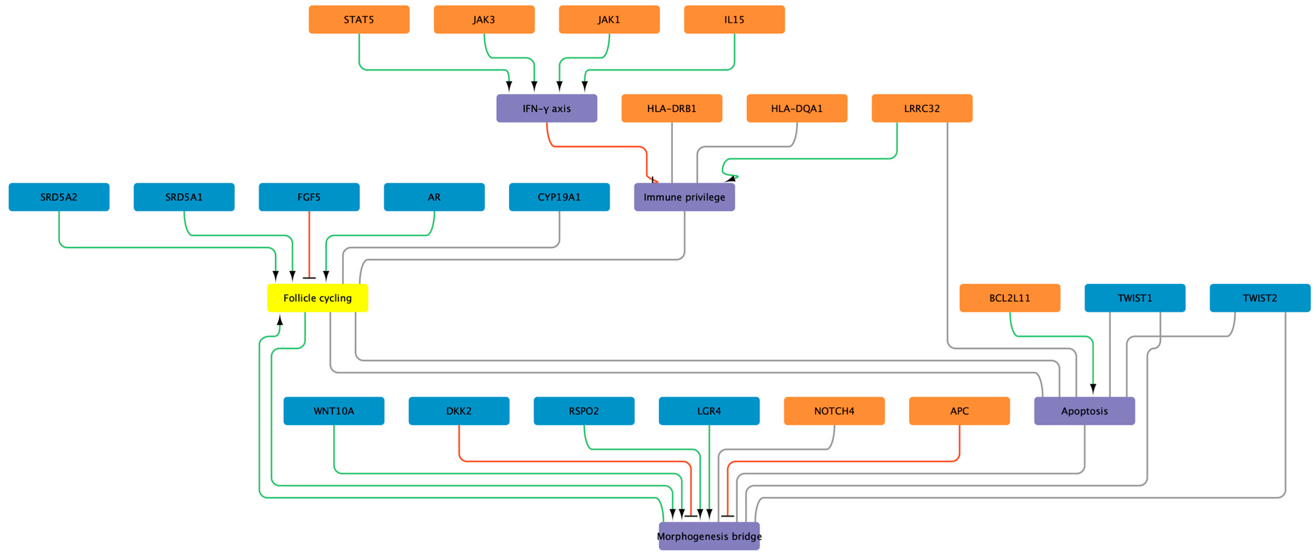
Importantly, these mechanistic axes do not exist in isolation. The Taiwanese GWAS of alopecia areata (AA) reported antigen presentation and IFN-γ–linked pathways as dominant [6], but also uncovered loci in APC and NOTCH4 that intersect directly with WNT signalling (APC regulating β-catenin stability, NOTCH modulating follicle stem-cell fate). When juxtaposed with AGA loci, these findings point to a shared morphogenetic layer across otherwise distinct diseases. This integrative perspective is summarised in Figure 2, which positions AGA’s androgen–WNT–TGF-β architecture alongside AA’s HLA-IFN–APC/NOTCH signals, illustrating how morphogenesis acts as a common bridge despite divergent upstream triggers.

4.2. Translation Snapshot

Polygenic and pathway-anchored insights in androgenetic alopecia (AGA) are beginning to support decisions that matter in clinic and trials. First, polygenic classifiers distinguish men with severe versus absent hair loss at useful levels, with performance improving as discovery scales and as models are pruned and externally validated [8,75]. This enables early counselling and surveillance for individuals with high genetic burden, i.e., earlier photographic follow up, discussion of adherence hurdles, and selection of therapies that match the dominant biological axis, androgen versus morphogenesis. Second, the architecture points directly to targetable modules. Signals at AR and SRD5A2 rationalise 5-α-reductase inhibition, while WNT and TGF-β burden aligns with anagen support and regenerative strategies, for example approaches that enhance β-catenin tone or buffer TGF-β restraint, noting that direct WNT agonism remains investigational [14,72,73].
A complementary track is pharmacogenetics. In a large retrospective dataset of 26,607 individuals, our analysis associated eight variants with AGA diagnosis across androgen metabolism, vasodilation and prostaglandin biology, and extracellular matrix components, including PTGES2, SRD5A2, COL1A1, ACE, PTGFR, PTGDR2, and CRABP2 [70]. This pattern explains much of the heterogeneous response observed in routine practice, since minoxidil activation depends on sulfation capacity and local haemodynamics, and 5-α-reductase inhibition depends on isoenzyme context. Building on that foundation, we performed a focused 26-SNP panel in 252 participants showed that genotype-informed prescribing improved overall response rates across minoxidil, finasteride, dutasteride, and adjuncts, while identifying robust predictors of poor response to specific drugs, for example SULT1A1 rs1042028 for minoxidil and SRD5A1 rs39848 for dutasteride. Network interrogation of SNP–SNP interactions in that panel linked prostaglandin receptors with mucin and fucosyltransferase pathways, suggesting that lipid mediators and epithelial interface genes modulate drug benefit beyond androgen metabolism alone. Although retrospective and single-centre designs introduce limitations, these datasets demonstrate that small, pathway-chosen panels can already prevent ineffective treatment courses and shorten the time to a working regimen.
A practical translational framework follows. At the first visit in a young man with family history, a compact PRS can flag high near-term progression risk, and a minimal pharmacogenetic set can guide first-line choice—for example, minoxidil versus early 5α-reductase inhibition, or the need for upfront combination therapy. During follow-up, state markers of follicle cycling, such as expression of WNT targets in plucked hair or dermal papilla signatures from micro-biopsies, can be layered onto genotype to titrate dose and to trigger an early switch if response is unlikely. In clinical trials, PRS and pharmacogenetic panels can enrich for mechanistically matched subgroups—for example, AR- and SRD5A2-heavy genotypes for endocrine studies, or WNT- and TGF-β-heavy genotypes for anagen support or regenerative interventions—thereby reducing heterogeneity and increasing statistical power. Importantly, the portability of these approaches across ancestries remains an open question, since most AGA loci were discovered in European cohorts, and sex-specific modifiers such as CYP19A1 and tissue aromatase expression are particularly relevant in women with pattern hair loss [3,72].
Our current line of work extends this logic in two directions. The first is treatment-specific association testing, where variants are analysed within exposure strata, for example minoxidil users versus finasteride users, with interaction terms that separate disease susceptibility from pharmacologic response. The second is epistasis-aware modelling, where modest interaction effects, for example PTGFR with MUC1 or GPR44 with FUT2, are retained if they improve calibration and decision utility, not just discrimination. These steps should produce compact, interpretable algorithms that can be validated prospectively and updated as new loci are added, similar to the way cardiovascular scores evolved.
The cross-disease view is clinically useful. Alopecia areata (AA) is driven by antigen presentation and an interferon-γ and interleukin-15 loop, while AGA is driven by endocrine and morphogenetic pathways. The shared ground lies in morphogenesis and apoptosis control, for example TGF-β and BCL2L11 biology, which is precisely where anagen-supporting strategies and anti-regression approaches operate. Figure 2 summarises this connection, placing AGA hubs, for example AR, SRD5A2, WNT10A, LGR4 and RSPO2, DKK2, TGFB2, alongside AA hubs, for example HLA-DRB1, BCL2L11, LRRC32, SH2B3, and highlighting morphogenesis and apoptosis as the bridge between endocrine-driven miniaturisation and immune-privilege collapse [3,5,6,53,70,72]. The implication is straightforward. Biomarkers for AGA should prioritise AR activity and WNT or TGF-β tone, while cross-cutting markers that report on apoptosis or matrix remodelling can complement both AGA and AA pathways in longitudinal monitoring.
In summary, translational movement in AGA now operates on two coordinated tracks. Polygenic risk stratifies who progresses and when, while pharmacogenetics informs what to start and what to avoid for a given individual. Recent studies provide concrete levers for both tracks, illustrating how small, biologically motivated panels can deliver immediate clinical value, while larger discovery efforts continue to raise the ceiling on prediction [70,76].

5. Cross-Disease Synthesis: Where Networks Meet

Acne vulgaris, alopecia areata, and androgenetic alopecia enter disease through different pathways, yet they repeatedly converge on a some shared biological axes. In alopecia areata, human genetics centres risk within HLA class II, with multiple independent signals that map to HLA-DR peptide-binding residues and to immune-regulatory nodes that modulate the quality and persistence of T-cell responses [5,52]. That architecture is consistent with a follicular environment susceptible to interferon-γ–driven up-regulation of antigen presentation and chemokines, which sustains a cytotoxic niche when immune privilege fails. East-Asian data echo the same theme and add specificity by nominating HLA-DQA1/DQB1 haplotypes and networks enriched for antigen presentation and lymphocyte activation, together with signals that regulate Notch and WNT control points that influence follicle fate [69,77,78]. By contrast, acne does not show dominant HLA peaks in genome-wide scans, but lesional biology demonstrates robust innate sensing via TLR2/TLR4 with downstream NF-κB and an interferon tone in subsets, placing interferon activity as a modulator rather than the primary driver in that disease [19,20]. Androgenetic alopecia is anchored by endocrine and morphogenetic pathways, with the X-chromosome androgen receptor and 5-α-reductase loci at one pole and WNT/TGF-β communities at the other, a configuration that explains miniaturisation through shortened anagen and altered repair [3,24].
One clear point of contact across conditions is WNT/TGF-β morphogenesis. Acne meta-analyses identify risk variants near genes that steer follicle development and junctional-zone repair, including TGFB2 and WNT module components such as WNT10A, LGR5/LGR6, and negative regulators of receptor abundance like ZNRF3 and KREMEN1 [79,80,81]. These loci make biological sense in a tissue where repeat cycles of micro-injury and repair define outcome, and where junctional-zone decisions bias toward comedogenesis when morphogenesis is off-centre. In androgenetic alopecia, WNT/TGF-β sits beside androgen signalling in pathway enrichments from large discovery efforts, with risk alleles and expression data pointing to reduced WNT tone and increased anagen restraint as proximate mechanisms for miniaturisation; signals across LGR4, RSPO2, WNT10A and DKK2 are recurrent examples [3,79,81,82]. The Taiwanese alopecia areata study adds a complementary angle by nominating APC and NOTCH4 among associated or network-linked loci, tying a classically immune-mediated disease back to follicle fate control via β-catenin stability and stem-cell governance [78,79,82,83]. Taken together, these findings argue that morphogenesis is not disease-exclusive. It is a shared layer that conditions how inflammation, endocrine tone, or environmental triggers are translated into tissue outcomes.
A second axis is antigen presentation and interferon signalling. In alopecia areata, convergent genetics and immunopathology support a loop where CD8+/NKG2D+ cells produce interferon-γ, follicular epithelium up-regulates MHC class I and class II, and helper T cells are more effectively licensed via risk HLA-DR molecules, while IL-15 from follicular cells sustains effector survival through JAK/STAT [5,54]. This loop explains the clinical performance of JAK inhibitors and the frequent relapse after withdrawal. Acne sits differently on this axis [24,54,62]. Here, interferon pathways are detectable but usually downstream of bacterial products and pollutant exposure that engage TLRs and cGAS–STING, producing a composite cytokine milieu in which TNF and IL-1 family members dominate and interferon functions as an amplifier rather than the lead signal [40]. The contrast clarifies why antigen-specific tolerance concepts feel natural in alopecia areata but less so in acne, where upstream microbial sensing and sebocyte biology are stronger levers [20,54,84,85].
Lipid metabolism–inflammation coupling provides a third shared thread with different weights. In acne, genetic and functional evidence converge on sebocyte lipogenesis and desaturation, with FASN and FADS2 providing concrete links between risk variation, sapienate-rich lipid profiles, and microbe–innate crosstalk [86,87,88,89]. These pathways modulate membrane composition, ligand availability for pattern recognition, and barrier lipids that set inflammatory thresholds. In androgenetic alopecia, the lipid theme is less prominent in GWAS but is biologically relevant because androgens drive sebaceous enlargement and sebum changes in balding scalp, modifying the local milieu in which follicles cycle. This helps explain why interventions that change androgen tone often alter sebaceous biology in parallel, and why lipid-directed adjuncts may sometimes improve cosmetic endpoints even when hair counts change modestly [3]. Alopecia areata again differs: lipids are not a leading genetic category, but apoptosis control at the follicle–immune interface, typified by BCL2L11, intersects with lipid-modulated danger signalling in ways that may influence relapse or chronicity [5,59,90].
Barrier and keratinisation are a fourth bridge, clearest between acne and atopic dermatitis but with broader implications. The IL-13–OVOL1–FLG axis established in atopic dermatitis underscores how epithelial differentiation pathways tune barrier proteins and tight-junction resilience. Acne cohorts show increased transepidermal water loss, treatment-related barrier fragility, and emerging signals that filaggrin-linked transcriptional pathways are altered in acne skin, which together support a model where barrier state sets the activation threshold for microbial and environmental triggers [6,48,49]. In practical terms, barrier-first adjuncts reduce irritancy from retinoids and may blunt flares by pushing the threshold upward. While barrier genetics are not front-and-centre in androgenetic alopecia or alopecia areata discovery scans, the same logic applies: any shift that lowers epithelial resilience will favour amplification of upstream drivers, whether those drivers are TLR ligands in acne or interferon cues in areata. The Taiwanese network analysis reinforces this by highlighting pathways of antigen processing and phagosome maturation that are sensitive to epithelial context and junctional integrity [3].
The synthesis is that these conditions differ in entry points but end up negotiating the same few corridors: antigen presentation and interferon signalling, WNT/TGF-β morphogenesis, lipid metabolism–inflammation, and barrier/keratinisation. This explains shared clinical patterns, for example why scarring and fibrosis phenotypes track with sustained repair pressure across diseases, or why sebaceous changes accompany both acne and androgenetic alopecia even as upstream triggers diverge [27,33,69,77]. It also suggests concrete translational moves. In alopecia areata, the antigen-presentation and JAK/STAT core argues for long-term control strategies keyed to interferon and IL-15 activity, with attention to HLA background when designing biomarkers or tolerance approaches [5,52]. In acne, lipid and morphogenesis axes support combining sebocyte-directed therapy with agents that improve repair quality and barrier robustness, and they explain why isotretinoin, which resets differentiation and lipogenesis, sits uniquely upstream [24,75]. In androgenetic alopecia, endocrine and WNT/TGF-β burdens can be read together to guide mechanism-matched therapy and to frame polygenic stratification for early intervention, while acknowledging that sebaceous balance and barrier context modulate the visible phenotype [6].
Across all three, the practical message is to treat the disease-specific trigger while measuring and, where possible, correcting the shared axes that determine tissue response. Doing so should improve durability of control in alopecia areata, reduce relapse and irritation in acne, and increase the odds of stabilising miniaturisation in androgenetic alopecia. The genetics do not collapse these conditions into one, but they do allow a good insight into the space of mechanisms that have to be understood to manage the diseases therapeutically. The bridges outlined here are assembled from convergent lines of evidence that include GWAS proximity, pathway enrichments, and lesional biology, rather than direct causal experimentation. Effect sizes for many acne loci and for polygenic scores are modest, and discovery for AGA has been dominated by European cohorts, while the strength and composition of non-HLA signals in AA vary by ancestry. Lipid–apoptosis intersections in AA are biologically plausible but are less genetically anchored than the HLA core. These points mean the network view should be treated as hypothesis-generating and used to prioritise mechanism-matched interventions and biomarkers, rather than as a deterministic map.

6. Conclusions—from Genes to Precision Dermatology & Cosmetics

Genetic discoveries across acne, alopecia areata (AA), and androgenetic alopecia (AGA) have moved dermatology beyond descriptive pathology into stratified frameworks that can guide therapy and prognosis. In AA, genome-wide studies consistently highlight HLA class II variation and interferon-driven circuits, providing the rationale for JAK inhibition and now supporting biomarker development for relapse prediction and long-term maintenance [5,6,52]. In AGA, large-scale GWAS mapped an architecture that juxtaposes androgen metabolism with WNT/TGF-β morphogenesis, enabling polygenic scores that already discriminate between severe and absent hair loss in population cohorts and suggest pathway-matched choices between endocrine and regenerative strategies [14,54]. Acne genetics, in parallel, reveal a triad of innate immunity, WNT-linked morphogenesis, and sebaceous lipid metabolism, with loci such as TGFB2, WNT10A, and FASN connecting microbial sensing and sebum biology to lesion formation and isotretinoin response [24,26]. Together, these findings provide a mechanistic substrate for moving from one-size-fits-all interventions to patient-stratified care.
The translational impact of this convergence is visible across therapeutic domains. For AA, immune-profiling of interferon-responsive gene sets in blood or lesional biopsies could identify patients at risk of relapse and titrate JAK inhibitor dosing over chronic courses [50,91,92]. For AGA, androgen- versus WNT-driven profiles derived from PRS or pharmacogenetic panels may rationalize first-line therapy—finasteride or dutasteride when AR/SRD5A burden is high, regenerative or WNT-supportive strategies when morphogenetic signals dominate [3]. In acne, sebocyte-lipid modules and innate-immune tone open avenues for targeted lipid modulation, sebum-directed inhibitors, and microbiome-inflammation interventions that complement isotretinoin, while barrier fragility linked to FLG/OVOL1 argues for barrier-first adjuncts [38,39,41,93,94]. Importantly, these disease-specific insights extend to dermato-cosmetic practice: lipid re-balancing agents, barrier repair formulations, and follicle-health maintenance can be guided by the same pathways that underpin disease risk, blurring the boundary between therapy and preventive aesthetics.
Despite the clear translational opportunities, several limitations need to be acknowledged. First, the proportion of variance explained by current GWAS remains incomplete: acne loci collectively account for less than 10% of disease liability, AGA polygenic scores reach higher accuracy but have been derived almost exclusively from European populations, and AA signals are heavily concentrated within HLA regions, which may not fully capture disease heterogeneity. Second, portability of findings across ancestries is uncertain, and most pharmacogenetic studies to date are retrospective and confined to single centres, limiting their generalisability. Third, functional interpretation of many associated variants is still indirect, relying on expression quantitative trait loci or pathway enrichments rather than experimental validation, meaning that causal inference remains provisional. Clinically, this translates into imperfect predictive power: AGA classifiers can distinguish severe from absent cases but do not yet provide reliable stratification for early intervention; acne risk variants highlight lipid and morphogenetic axes but do not yield patient-level certainty; and in AA, the frequent relapse after JAK inhibitor withdrawal underscores that genetic predisposition continues to shape disease even under pharmacological suppression. Finally, cost, accessibility, and lack of consensus guidelines on how to incorporate genetic results into everyday dermatology remain practical barriers. These constraints do not detract from the promise of genetics, but they emphasise that current evidence should be viewed as a scaffold for mechanism-matched strategies rather than as definitive instructions, pending further validation in prospective, multi-ethnic, and interventional studies.
Looking forward, the challenge is integration. Precision dermatology will rely on combining genetic predisposition (PRS, pathway-burden scores) with dynamic state readouts—single-cell follicle atlases to capture cycling and immune niches, lipidomics to quantify sebaceous activity and exposome interactions, and immune monitoring to detect shifts in interferon or cytokine tone. Such layered approaches can inform not only drug choice but also dose, timing, and adjunctive cosmetic regimens aimed at stabilising cutaneous systems. The long-term vision is a dermatology where isotretinoin, JAK inhibitors, and anti-androgens are not blunt instruments but tools deployed in the right patient, at the right time, with supportive treatments aimed at improving skin barrier function and lipid metabolism to prolong remission. Genetics has already shrunk the space of uncertainty from organ-level impressions to molecularly defined axes; the next step is embedding these insights into everyday dermatology so that precision care becomes the default rather than the exception.

Funding

This research received no external funding.

Data Availability Statement

Not applicable. No new data were created or analyzed in this study.

Conflicts of Interest

Author Gustavo Torres de Souza was employed by the Fagron Genomics. Author declares that the research was conducted in the absence of any commercial or financial relationships that could be construed as a potential conflict of interest.

References

  1. Yakupu, A.; Aimaier, R.; Yuan, B.; Chen, B.; Cheng, J.; Zhao, Y.; Peng, Y.; Dong, J.; Lu, S. The Burden of Skin and Subcutaneous Diseases: Findings from the Global Burden of Disease Study 2019. Front. Public Health 2023, 11, 1145513. [Google Scholar] [CrossRef]
  2. Szeto, M.D.; Alhanshali, L.; Rundle, C.W.; Adelman, M.; Hook Sobotka, M.; Woolhiser, E.; Wu, J.; Presley, C.L.; Maghfour, J.; Meisenheimer, J.; et al. Dermatologic Data From the Global Burden of Disease Study 2019 and the PatientsLikeMe Online Support Community: Comparative Analysis. JMIR Dermatol. 2024, 7, e50449. [Google Scholar] [CrossRef] [PubMed]
  3. Pirastu, N.; Joshi, P.K.; de Vries, P.S.; Cornelis, M.C.; McKeigue, P.M.; Keum, N.; Franceschini, N.; Colombo, M.; Giovannucci, E.L.; Spiliopoulou, A.; et al. GWAS for Male-Pattern Baldness Identifies 71 Susceptibility Loci Explaining 38% of the Risk. Nat. Commun. 2017, 8, 1584. [Google Scholar] [CrossRef] [PubMed]
  4. Prodi, D.A.; Pirastu, N.; Maninchedda, G.; Sassu, A.; Picciau, A.; Palmas, M.A.; Mossa, A.; Persico, I.; Adamo, M.; Angius, A.; et al. EDA2R Is Associated with Androgenetic Alopecia. J. Investig. Dermatol. 2008, 128, 2268–2270. [Google Scholar] [CrossRef]
  5. Betz, R.C.; Petukhova, L.; Ripke, S.; Huang, H.; Menelaou, A.; Redler, S.; Becker, T.; Heilmann, S.; Yamany, T.; Duvic, M.; et al. Genome-Wide Meta-Analysis in Alopecia Areata Resolves HLA Associations and Reveals Two New Susceptibility Loci. Nat. Commun. 2015, 6, 5966. [Google Scholar] [CrossRef]
  6. Yang, J.-S.; Liu, T.-Y.; Chen, Y.-C.; Tsai, S.-C.; Chiu, Y.-J.; Liao, C.-C.; Tsai, F.-J. Genome-Wide Association Study of Alopecia Areata in Taiwan: The Conflict Between Individuals and Hair Follicles. Clin. Cosmet. Investig. Dermatol. 2023, 16, 2597–2612. [Google Scholar] [CrossRef]
  7. Mitchell, B.L.; Saklatvala, J.R.; Dand, N.; Hagenbeek, F.A.; Li, X.; Min, J.L.; Thomas, L.; Bartels, M.; Jan Hottenga, J.; Lupton, M.K.; et al. Genome-Wide Association Meta-Analysis Identifies 29 New Acne Susceptibility Loci. Nat. Commun. 2022, 13, 702. [Google Scholar] [CrossRef] [PubMed]
  8. Chen, Y.; Hysi, P.; Maj, C.; Heilmann-Heimbach, S.; Spector, T.D.; Liu, F.; Kayser, M. Genetic Prediction of Male Pattern Baldness Based on Large Independent Datasets. Eur. J. Human. Genet. 2023, 31, 321–328. [Google Scholar] [CrossRef]
  9. Lee, S.; Kim, J.E.; Lew, B.-L.; Huh, C.H.; Kim, J.; Kwon, O.; Kim, M.B.; Lee, Y.W.; Lee, Y.; Park, J.; et al. Efficacy and Safety of Low-Dose (0.2 Mg) Dutasteride for Male Androgenic Alopecia: A Multicenter, Randomized, Double-Blind, Placebo-Controlled, Parallel-Group Phase III Clinical Trial. Ann. Dermatol. 2025, 37, 183. [Google Scholar] [CrossRef]
  10. Estill, M.C.; Ford, A.; Omeira, R.; Rodman, M. Finasteride and Dutasteride for the Treatment of Male Androgenetic Alopecia: A Review of Efficacy and Reproductive Adverse Effects. Georget. Med. Rev. 2023, 7. [Google Scholar] [CrossRef]
  11. Choi, G.-S.; Sim, W.-Y.; Kang, H.; Huh, C.H.; Lee, Y.W.; Shantakumar, S.; Ho, Y.-F.; Oh, E.-J.; Duh, M.S.; Cheng, W.Y.; et al. Long-Term Effectiveness and Safety of Dutasteride versus Finasteride in Patients with Male Androgenic Alopecia in South Korea: A Multicentre Chart Review Study. Ann. Dermatol. 2022, 34, 349. [Google Scholar] [CrossRef]
  12. Egeberg, A.; Linsell, L.; Johansson, E.; Durand, F.; Yu, G.; Vañó-Galván, S. Treatments for Moderate-to-Severe Alopecia Areata: A Systematic Narrative Review. Dermatol. Ther. 2023, 13, 2951–2991. [Google Scholar] [CrossRef] [PubMed]
  13. Sardana, K.; Bathula, S.; Khurana, A. Which Is the Ideal JAK Inhibitor for Alopecia Areata—Baricitinib, Tofacitinib, Ritlecitinib or Ifidancitinib—Revisiting the Immunomechanisms of the JAK Pathway. Indian Dermatol. Online J. 2023, 14, 465. [Google Scholar] [CrossRef]
  14. Henne, S.K.; Aldisi, R.; Sivalingam, S.; Hochfeld, L.M.; Borisov, O.; Krawitz, P.M.; Maj, C.; Nöthen, M.M.; Heilmann-Heimbach, S. Analysis of 72,469 UK Biobank Exomes Links Rare Variants to Male-Pattern Hair Loss. Nat. Commun. 2023, 14, 5492. [Google Scholar] [CrossRef]
  15. Oliva, M.; Sarkar, M.K.; March, M.E.; Saeidian, A.H.; Mentch, F.D.; Hsieh, C.-L.; Tang, F.; Uppala, R.; Patrick, M.T.; Li, Q.; et al. Multi-Ancestry Genome-Wide Association Meta-Analysis Identifies Novel Loci in Atopic Dermatitis. medRxiv 2024. [Google Scholar] [CrossRef]
  16. Sroka-Tomaszewska, J.; Trzeciak, M. Molecular Mechanisms of Atopic Dermatitis Pathogenesis. Int. J. Mol. Sci. 2021, 22, 4130. [Google Scholar] [CrossRef]
  17. Facheris, P.; Jeffery, J.; Del Duca, E.; Guttman-Yassky, E. The Translational Revolution in Atopic Dermatitis: The Paradigm Shift from Pathogenesis to Treatment. Cell Mol. Immunol. 2023, 20, 448–474. [Google Scholar] [CrossRef] [PubMed]
  18. Ramos, Y.Á.L.; Pietrobon, A.J.; Teixeira, F.M.E.; Aoki, V.; Sato, M.N.; Orfali, R.L. Inflammasome Pathways in Atopic Dermatitis: Insights into Inflammatory Mechanisms and Therapeutic Targets. An. Bras. Dermatol. 2025, 100, 501136. [Google Scholar] [CrossRef]
  19. Törőcsik, D.; Kovács, D.; Póliska, S.; Szentkereszty-Kovács, Z.; Lovászi, M.; Hegyi, K.; Szegedi, A.; Zouboulis, C.C.; Ståhle, M. Genome Wide Analysis of TLR1/2- and TLR4-Activated SZ95 Sebocytes Reveals a Complex Immune-Competence and Identifies Serum Amyloid A as a Marker for Activated Sebaceous Glands. PLoS ONE 2018, 13, e0198323. [Google Scholar] [CrossRef]
  20. Noh, H.H.; Shin, S.H.; Roh, Y.J.; Moon, N.J.; Seo, S.J.; Park, K.Y. Particulate Matter Increases Cutibacterium Acnes-Induced Inflammation in Human Epidermal Keratinocytes via the TLR4/NF-ΚB Pathway. PLoS ONE 2022, 17, e0268595. [Google Scholar] [CrossRef]
  21. Firlej, E.; Kowalska, W.; Szymaszek, K.; Roliński, J.; Bartosińska, J. The Role of Skin Immune System in Acne. J. Clin. Med. 2022, 11, 1579. [Google Scholar] [CrossRef]
  22. Selway, J.L.; Kurczab, T.; Kealey, T.; Langlands, K. Toll-like Receptor 2 Activation and Comedogenesis: Implications for the Pathogenesis of Acne. BMC Dermatol. 2013, 13, 10. [Google Scholar] [CrossRef] [PubMed]
  23. Kang, S.; Cho, S.; Chung, J.H.; Hammerberg, C.; Fisher, G.J.; Voorhees, J.J. Inflammation and Extracellular Matrix Degradation Mediated by Activated Transcription Factors Nuclear Factor-ΚB and Activator Protein-1 in Inflammatory Acne Lesions in Vivo. Am. J. Pathol. 2005, 166, 1691–1699. [Google Scholar] [CrossRef]
  24. Navarini, A.A.; Simpson, M.A.; Weale, M.; Knight, J.; Carlavan, I.; Reiniche, P.; Burden, D.A.; Layton, A.; Bataille, V.; Allen, M.; et al. Genome-Wide Association Study Identifies Three Novel Susceptibility Loci for Severe Acne Vulgaris. Nat. Commun. 2014, 5, 4020. [Google Scholar] [CrossRef]
  25. Teder-Laving, M.; Kals, M.; Reigo, A.; Ehin, R.; Objärtel, T.; Vaht, M.; Nikopensius, T.; Metspalu, A.; Kingo, K. Genome-Wide Meta-Analysis Identifies Novel Loci Conferring Risk of Acne Vulgaris. Eur. J. Human. Genet. 2024, 32, 1136–1143. [Google Scholar] [CrossRef]
  26. Petridis, C.; Navarini, A.A.; Dand, N.; Saklatvala, J.; Baudry, D.; Duckworth, M.; Allen, M.H.; Curtis, C.J.; Lee, S.H.; Burden, A.D.; et al. Genome-Wide Meta-Analysis Implicates Mediators of Hair Follicle Development and Morphogenesis in Risk for Severe Acne. Nat. Commun. 2018, 9, 5075. [Google Scholar] [CrossRef]
  27. Esler, W.P.; Tesz, G.J.; Hellerstein, M.K.; Beysen, C.; Sivamani, R.; Turner, S.M.; Watkins, S.M.; Amor, P.A.; Carvajal-Gonzalez, S.; Geoly, F.J.; et al. Human Sebum Requires de novo Lipogenesis, Which Is Increased in Acne Vulgaris and Suppressed by Acetyl-CoA Carboxylase Inhibition. Sci. Transl. Med. 2019, 11, eaau8465. [Google Scholar] [CrossRef] [PubMed]
  28. Potter, C.S.; Kern, M.J.; Baybo, M.A.; Pruett, N.D.; Godwin, A.R.; Sundberg, J.P.; Awgulewitsch, A. Dysregulated Expression of Sterol O-Acyltransferase 1 (Soat1) in the Hair Shaft of Hoxc13 Null Mice. Exp. Mol. Pathol. 2015, 99, 441–444. [Google Scholar] [CrossRef] [PubMed]
  29. Wu, B.; Potter, C.S.; Silva, K.A.; Liang, Y.; Reinholdt, L.G.; Alley, L.M.; Rowe, L.B.; Roopenian, D.C.; Awgulewitsch, A.; Sundberg, J.P. Mutations in Sterol O-Acyltransferase 1 (Soat1) Result in Hair Interior Defects in AKR/J Mice. J. Investig. Dermatol. 2010, 130, 2666–2668. [Google Scholar] [CrossRef]
  30. Wang, X.; Wu, Y.; Zhao, P.; Wang, X.; Wu, W.; Yang, J. The Causal Relationship between Serum Metabolites and Acne Vulgaris: A Mendelian Randomization Study. Sci. Rep. 2024, 14, 11045. [Google Scholar] [CrossRef]
  31. Ju, R.; Ying, Y.; Zhou, Q.; Cao, Y. Exploring Genetic Drug Targets in Acne Vulgaris: A Comprehensive Proteome—Wide Mendelian Randomization Study. J. Cosmet. Dermatol. 2024, 23, 4223–4229. [Google Scholar] [CrossRef] [PubMed]
  32. Wang, K.-Y.; Yamada, S.; Izumi, H.; Tsukamoto, M.; Nakashima, T.; Tasaki, T.; Guo, X.; Uramoto, H.; Sasaguri, Y.; Kohno, K. Critical in Vivo Roles of WNT10A in Wound Healing by Regulating Collagen Expression/Synthesis in WNT10A-Deficient Mice. PLoS ONE 2018, 13, e0195156. [Google Scholar] [CrossRef]
  33. Benard, E.L.; Hammerschmidt, M. The Fundamentals of WNT10A. Differentiation 2025, 142, 100838. [Google Scholar] [CrossRef] [PubMed]
  34. Zouboulis, C.C. Endocrinology and Immunology of Acne: Two Sides of the Same Coin. Exp. Dermatol. 2020, 29, 840–859. [Google Scholar] [CrossRef] [PubMed]
  35. Zouboulis, C.C.; Jourdan, E.; Picardo, M. Acne Is an Inflammatory Disease and Alterations of Sebum Composition Initiate Acne Lesions. J. Eur. Acad. Dermatol. Venereol. 2014, 28, 527–532. [Google Scholar] [CrossRef]
  36. Liu, M.; Diaz-Torres, S.; Mitchell, B.L.; Toledo-Flores, D.; Gharhakhani, P.; Simpson, M.A.; Zhang, H.; Ong, J.-S.; Li, J.; Rentería, M.E. The Role of Lipid Metabolism in Acne Risk: Integrating Blood Metabolite and Genetic Insights. Skin. Health Dis. 2025, 5, 124–129. [Google Scholar] [CrossRef]
  37. Li, L.; Hajam, I.; McGee, J.S.; Tang, Z.; Zhang, Y.; Badey, N.; Mintzer, E.; Zhang, Z.; Liu, G.Y.; Church, G.M.; et al. Comparative Transcriptome Analysis of Acne Vulgaris, Rosacea, and Hidradenitis Suppurativa Supports High—dose Dietary Zinc as a Therapeutic Agent. Exp. Dermatol. 2024, 33, e15145. [Google Scholar] [CrossRef]
  38. Furue, M.; Ulzii, D.; Nakahara, T.; Tsuji, G.; Furue, K.; Hashimoto-Hachiya, A.; Kido-Nakahara, M. Implications of IL-13Rα2 in Atopic Skin Inflammation. Allergol. Int. 2020, 69, 412–416. [Google Scholar] [CrossRef]
  39. Furue, M.; Chiba, T.; Tsuji, G.; Ulzii, D.; Kido-Nakahara, M.; Nakahara, T.; Kadono, T. Atopic Dermatitis: Immune Deviation, Barrier Dysfunction, IgE Autoreactivity and New Therapies. Allergol. Int. 2017, 66, 398–403. [Google Scholar] [CrossRef]
  40. Bolla, B.S.; Erdei, L.; Urbán, E.; Burián, K.; Kemény, L.; Szabó, K.C. C. acnes Regulates the Epidermal Barrier Properties of HPV-KER Human Immortalized Keratinocyte Cultures. Sci. Rep. 2020, 10, 12815. [Google Scholar] [CrossRef]
  41. Dębińska, A. New Treatments for Atopic Dermatitis Targeting Skin Barrier Repair via the Regulation of FLG Expression. J. Clin. Med. 2021, 10, 2506. [Google Scholar] [CrossRef]
  42. Hughes, A.J.; Barbosa, E.; Cernova, J.; Thomas, B.R.; O’Shaughnessy, R.F.L.; O’Toole, E.A. Loss-of-Function FLG Mutations Are Associated with Reduced History of Acne Vulgaris in a Cohort of Patients with Atopic Eczema of Bangladeshi Ancestry in East London. Clin. Exp. Dermatol. 2024, 49, 1547–1553. [Google Scholar] [CrossRef]
  43. Draelos, Z.D.; Matsubara, A.; Smiles, K. The Effect of 2% Niacinamide on Facial Sebum Production. J. Cosmet. Laser Ther. 2006, 8, 96–101. [Google Scholar] [CrossRef]
  44. Lee, A.-Y. Molecular Mechanism of Epidermal Barrier Dysfunction as Primary Abnormalities. Int. J. Mol. Sci. 2020, 21, 1194. [Google Scholar] [CrossRef]
  45. Madnani, N.; Deo, J.; Dalal, K.; Benjamin, B.; Murthy, V.V.; Hegde, R.; Shetty, T. Revitalizing the Skin: Exploring the Role of Barrier Repair Moisturizers. J. Cosmet. Dermatol. 2024, 23, 1533–1540. [Google Scholar] [CrossRef]
  46. Sandilands, A.; Sutherland, C.; Irvine, A.D.; McLean, W.H.I. Filaggrin in the Frontline: Role in Skin Barrier Function and Disease. J. Cell Sci. 2009, 122, 1285–1294. [Google Scholar] [CrossRef]
  47. Draelos, Z.D.; Baalbaki, N.; Colon, G.; Dreno, B. Ceramide-Containing Adjunctive Skin Care for Skin Barrier Restoration During Acne Vulgaris Treatment. J. Drugs Dermatol. 2023, 22, 554–558. [Google Scholar] [CrossRef]
  48. Sun, P.; Vu, R.; Dragan, M.; Haensel, D.; Gutierrez, G.; Nguyen, Q.; Greenberg, E.; Chen, Z.; Wu, J.; Atwood, S.; et al. OVOL1 Regulates Psoriasis-Like Skin Inflammation and Epidermal Hyperplasia. J. Investig. Dermatol. 2021, 141, 1542–1552. [Google Scholar] [CrossRef] [PubMed]
  49. Dragan, M.; Sun, P.; Chen, Z.; Ma, X.; Vu, R.; Shi, Y.; Villalta, S.A.; Dai, X. Epidermis-Intrinsic Transcription Factor Ovol1 Coordinately Regulates Barrier Maintenance and Neutrophil Accumulation in Psoriasis-Like Inflammation. J. Investig. Dermatol. 2022, 142, 583–593.e5. [Google Scholar] [CrossRef] [PubMed]
  50. Pratt, C.H.; King, L.E.; Messenger, A.G.; Christiano, A.M.; Sundberg, J.P. Alopecia Areata. Nat. Rev. Dis. Primers 2017, 3, 17011. [Google Scholar] [CrossRef] [PubMed]
  51. Ma, T.; Zhang, T.; Miao, F.; Liu, J.; Zhu, Q.; Chen, Z.; Tai, Z.; He, Z. Alopecia Areata: Pathogenesis, Diagnosis, and Therapies. MedComm 2025, 6, e70182. [Google Scholar] [CrossRef]
  52. Petukhova, L.; Duvic, M.; Hordinsky, M.; Norris, D.; Price, V.; Shimomura, Y.; Kim, H.; Singh, P.; Lee, A.; Chen, W.V.; et al. Genome-Wide Association Study in Alopecia Areata Implicates Both Innate and Adaptive Immunity. Nature 2010, 466, 113–117. [Google Scholar] [CrossRef]
  53. Paus, R.; Bulfone-Paus, S.; Bertolini, M. Hair Follicle Immune Privilege Revisited: The Key to Alopecia Areata Management. J. Investig. Dermatol. Symp. Proc. 2018, 19, S12–S17. [Google Scholar] [CrossRef] [PubMed]
  54. Lensing, M.; Jabbari, A. An Overview of JAK/STAT Pathways and JAK Inhibition in Alopecia Areata. Front. Immunol. 2022, 13, 955035. [Google Scholar] [CrossRef]
  55. Passeron, T.; King, B.; Seneschal, J.; Steinhoff, M.; Jabbari, A.; Ohyama, M.; Tobin, D.J.; Randhawa, S.; Winkler, A.; Telliez, J.-B.; et al. Inhibition of T-Cell Activity in Alopecia Areata: Recent Developments and New Directions. Front. Immunol. 2023, 14, 1243556. [Google Scholar] [CrossRef] [PubMed]
  56. Ji, C.; Liu, S.; Zhu, K.; Luo, H.; Li, Q.; Zhang, Y.; Huang, S.; Chen, Q.; Cao, Y. HLA-DRB1 Polymorphisms and Alopecia Areata Disease Risk. Medicine 2018, 97, e11790. [Google Scholar] [CrossRef] [PubMed]
  57. Oka, A.; Takagi, A.; Komiyama, E.; Yoshihara, N.; Mano, S.; Hosomichi, K.; Suzuki, S.; Haida, Y.; Motosugi, N.; Hatanaka, T.; et al. Alopecia Areata Susceptibility Variant in MHC Region Impacts Expressions of Genes Contributing to Hair Keratinization and Is Involved in Hair Loss. EBioMedicine 2020, 57, 102810. [Google Scholar] [CrossRef]
  58. Šutić Udović, I.; Hlača, N.; Massari, L.P.; Brajac, I.; Kaštelan, M.; Vičić, M. Deciphering the Complex Immunopathogenesis of Alopecia Areata. Int. J. Mol. Sci. 2024, 25, 5652. [Google Scholar] [CrossRef]
  59. AL-Eitan, L.N.; Alasmar, M.K.; Aljamal, H.A.; Mihyar, A.H.; Alghamdi, M.A. Investigating the Genetic Association of Selected Candidate Loci with Alopecia Areata Susceptibility in Jordanian Patients. Medicina 2025, 61, 409. [Google Scholar] [CrossRef]
  60. Nasrallah, R.; Imianowski, C.J.; Bossini-Castillo, L.; Grant, F.M.; Dogan, M.; Placek, L.; Kozhaya, L.; Kuo, P.; Sadiyah, F.; Whiteside, S.K.; et al. A Distal Enhancer at Risk Locus 11q13.5 Promotes Suppression of Colitis by Treg Cells. Nature 2020, 583, 447–452. [Google Scholar] [CrossRef]
  61. Wing, K.; Onishi, Y.; Prieto-Martin, P.; Yamaguchi, T.; Miyara, M.; Fehervari, Z.; Nomura, T.; Sakaguchi, S. CTLA-4 Control over Foxp3 + Regulatory T Cell Function. Science (1979) 2008, 322, 271–275. [Google Scholar] [CrossRef]
  62. Hu, X.; Li, J.; Fu, M.; Zhao, X.; Wang, W. The JAK/STAT Signaling Pathway: From Bench to Clinic. Signal Transduct. Target. Ther. 2021, 6, 402. [Google Scholar] [CrossRef]
  63. Dai, Z.; Chen, J.; Chang, Y.; Christiano, A.M. Selective Inhibition of JAK3 Signaling Is Sufficient to Reverse Alopecia Areata. JCI Insight 2021, 6, e142205. [Google Scholar] [CrossRef]
  64. Hawkshaw, N.J.; Hardman, J.A.; Haslam, I.S.; Shahmalak, A.; Gilhar, A.; Lim, X.; Paus, R. Identifying Novel Strategies for Treating Human Hair Loss Disorders: Cyclosporine A Suppresses the Wnt Inhibitor, SFRP1, in the Dermal Papilla of Human Scalp Hair Follicles. PLoS Biol. 2018, 16, e2003705. [Google Scholar] [CrossRef]
  65. Suzuki, T.; Chéret, J.; Scala, F.D.; Rajabi-Estarabadi, A.; Akhundlu, A.; Demetrius, D.-L.; Gherardini, J.; Keren, A.; Harries, M.; Rodriguez-Feliz, J.; et al. Interleukin-15 Is a Hair Follicle Immune Privilege Guardian. J. Autoimmun. 2024, 145, 103217. [Google Scholar] [CrossRef]
  66. Ebrahim, A.; Salem, R.; El Fallah, A.; Younis, E. Serum Interleukin-15 Is a Marker of Alopecia Areata Severity. Int. J. Trichol 2019, 11, 26. [Google Scholar] [CrossRef]
  67. Sanchez, K.; Englander, H.; Salloum, L.; Gregoire, S.; Biba, U.; Ershadi, S.; Mostaghimi, A. Evaluating Current and Emergent JAK Inhibitors for Alopecia Areata: A Narrative Review. Dermatol. Ther. 2025, 15, 2749–2764. [Google Scholar] [CrossRef]
  68. Sadasivam, I.P.; Sambandam, R.; Kaliyaperumal, D.; Dileep, J.E. Androgenetic Alopecia in Men: An Update On Genetics. Indian J. Dermatol. 2024, 69, 282. [Google Scholar] [CrossRef] [PubMed]
  69. Choi, B.Y. Targeting Wnt/β-Catenin Pathway for Developing Therapies for Hair Loss. Int. J. Mol. Sci. 2020, 21, 4915. [Google Scholar] [CrossRef] [PubMed]
  70. Francès, M.P.; Vila-Vecilla, L.; Russo, V.; Caetano Polonini, H.; de Souza, G.T. Utilising SNP Association Analysis as a Prospective Approach for Personalising Androgenetic Alopecia Treatment. Dermatol. Ther. 2024, 14, 971–981. [Google Scholar] [CrossRef] [PubMed]
  71. Yap, C.X.; Sidorenko, J.; Wu, Y.; Kemper, K.E.; Yang, J.; Wray, N.R.; Robinson, M.R.; Visscher, P.M. Dissection of Genetic Variation and Evidence for Pleiotropy in Male Pattern Baldness. Nat. Commun. 2018, 9, 5407. [Google Scholar] [CrossRef]
  72. Henne, S.K.; Nöthen, M.M.; Heilmann-Heimbach, S. Male-Pattern Hair Loss: Comprehensive Identification of the Associated Genes as a Basis for Understanding Pathophysiology. Med. Genet. 2023, 35, 3–14. [Google Scholar] [CrossRef]
  73. Lu, G.-Q.; Wu, Z.-B.; Chu, X.-Y.; Bi, Z.-G.; Fan, W.-X. An Investigation of Crosstalk between Wnt/β-Catenin and Transforming Growth Factor-β Signaling in Androgenetic Alopecia. Medicine 2016, 95, e4297. [Google Scholar] [CrossRef]
  74. Yip, L.; Zaloumis, S.; Irwin, D.; Severi, G.; Hopper, J.; Giles, G.; Harrap, S.; Sinclair, R.; Ellis, J. Gene-Wide Association Study between the Aromatase Gene (CYP19A1) and Female Pattern Hair Loss. Br. J. Dermatol. 2009, 161, 289–294. [Google Scholar] [CrossRef]
  75. Hagenaars, S.P.; Hill, W.D.; Harris, S.E.; Ritchie, S.J.; Davies, G.; Liewald, D.C.; Gale, C.R.; Porteous, D.J.; Deary, I.J.; Marioni, R.E. Genetic Prediction of Male Pattern Baldness. PLoS Genet. 2017, 13, e1006594. [Google Scholar] [CrossRef] [PubMed]
  76. Heilmann, S.; Brockschmidt, F.F.; Hillmer, A.M.; Hanneken, S.; Eigelshoven, S.; Ludwig, K.U.; Herold, C.; Mangold, E.; Becker, T.; Kruse, R.; et al. Evidence for a Polygenic Contribution to Androgenetic Alopecia. Br. J. Dermatol. 2013, 169, 927–930. [Google Scholar] [CrossRef] [PubMed]
  77. Mehta, A.; Motavaf, M.; Raza, D.; McLure, A.J.; Osei-Opare, K.D.; Bordone, L.A.; Gru, A.A. Revolutionary Approaches to Hair Regrowth: Follicle Neogenesis, Wnt/ß-Catenin Signaling, and Emerging Therapies. Cells 2025, 14, 779. [Google Scholar] [CrossRef]
  78. Jin, H.; Zou, Z.; Chang, H.; Shen, Q.; Liu, L.; Xing, D. Photobiomodulation Therapy for Hair Regeneration: A Synergetic Activation of β-CATENIN in Hair Follicle Stem Cells by ROS and Paracrine WNTs. Stem Cell Rep. 2021, 16, 1568–1583. [Google Scholar] [CrossRef]
  79. Yan, W.; Liu, J.; Xie, X.; Jin, Q.; Yang, Y.; Pan, Y.; Zhang, Y.; Zhang, F.; Wang, Y.; Liu, J.; et al. Restoration of Follicular β-Catenin Signaling by Mesenchymal Stem Cells Promotes Hair Growth in Mice with Androgenetic Alopecia. Stem Cell Res. Ther. 2024, 15, 439. [Google Scholar] [CrossRef] [PubMed]
  80. Wang, F.; He, G.; Liu, M.; Sun, Y.; Ma, S.; Sun, Z.; Wang, Y. Pilose Antler Extracts Promotes Hair Growth in Androgenetic Alopecia Mice by Activating Hair Follicle Stem Cells via the AKT and Wnt Pathways. Front. Pharmacol. 2024, 15, 1410810. [Google Scholar] [CrossRef]
  81. Zhang, Y.; Yin, S.; Xu, R.; Xiao, J.; Yi, R.; Mao, J.; Duan, Z.; Fan, D. Recombinant Type XVII Collagen Promotes Hair Growth by Activating the Wnt/β-Catenin and SHH/GLI Signaling Pathways. Cosmetics 2025, 12, 156. [Google Scholar] [CrossRef]
  82. Gentile, P.; Garcovich, S. Advances in Regenerative Stem Cell Therapy in Androgenic Alopecia and Hair Loss: Wnt Pathway, Growth-Factor, and Mesenchymal Stem Cell Signaling Impact Analysis on Cell Growth and Hair Follicle Development. Cells 2019, 8, 466. [Google Scholar] [CrossRef]
  83. Hile, G.A.; Gudjonsson, J.E.; Kahlenberg, J.M. The Influence of Interferon on Healthy and Diseased Skin. Cytokine 2020, 132, 154605. [Google Scholar] [CrossRef]
  84. Mayslich, C.; Grange, P.A.; Castela, M.; Marcelin, A.G.; Calvez, V.; Dupin, N. Characterization of a Cutibacterium Acnes Camp Factor 1-Related Peptide as a New TLR-2 Modulator in In Vitro and Ex Vivo Models of Inflammation. Int. J. Mol. Sci. 2022, 23, 5065. [Google Scholar] [CrossRef] [PubMed]
  85. Romics, L.; Dolganiuc, A.; Kodys, K.; Drechsler, Y.; Oak, S.; Velayudham, A.; Mandrekar, P.; Szabo, G. Selective Priming to Toll-like Receptor 4 (TLR4), Not TLR2, Ligands by P. Acnes Involves up-Regulation of MD-2 in Mice. Hepatology 2004, 40, 555–564. [Google Scholar] [CrossRef]
  86. Cros, M.P.; Mir-Pedrol, J.; Toloza, L.; Knödlseder, N.; Maruotti, J.; Zouboulis, C.C.; Güell, M.; Fábrega, M.-J. New Insights into the Role of Cutibacterium Acnes-Derived Extracellular Vesicles in Inflammatory Skin Disorders. Sci. Rep. 2023, 13, 16058. [Google Scholar] [CrossRef] [PubMed]
  87. Jugeau, S.; Tenaud, I.; Knol, A.C.; Jarrousse, V.; Quereux, G.; Khammari, A.; Dreno, B. Induction of Toll-like Receptors by Propionibacterium Acnes. Br. J. Dermatol. 2005, 153, 1105–1113. [Google Scholar] [CrossRef] [PubMed]
  88. Ottaviani, M.; Flori, E.; Mastrofrancesco, A.; Briganti, S.; Lora, V.; Capitanio, B.; Zouboulis, C.C.; Picardo, M. Sebocyte Differentiation as a New Target for Acne Therapy: An in Vivo Experience. J. Eur. Acad. Dermatol. Venereol. 2020, 34, 1803–1814. [Google Scholar] [CrossRef]
  89. Choi, C.W.; Kim, Y.; Kim, J.E.; Seo, E.Y.; Zouboulis, C.C.; Kang, J.S.; Youn, S.W.; Chung, J.H. Enhancement of Lipid Content and Inflammatory Cytokine Secretion in SZ95 Sebocytes by Palmitic Acid Suggests a Potential Link between Free Fatty Acids and Acne Aggravation. Exp. Dermatol. 2019, 28, 207–210. [Google Scholar] [CrossRef]
  90. Olayinka, J.T.; Richmond, J.M. Immunopathogenesis of Alopecia Areata. Curr. Res. Immunol. 2021, 2, 7–11. [Google Scholar] [CrossRef]
  91. Guttman-Yassky, E.; Pavel, A.B.; Diaz, A.; Zhang, N.; Del Duca, E.; Estrada, Y.; King, B.; Banerjee, A.; Banfield, C.; Cox, L.A.; et al. Ritlecitinib and Brepocitinib Demonstrate Significant Improvement in Scalp Alopecia Areata Biomarkers. J. Allergy Clin. Immunol. 2022, 149, 1318–1328. [Google Scholar] [CrossRef]
  92. Dillon, K.-A.L. A Comprehensive Literature Review of JAK Inhibitors in Treatment of Alopecia Areata. Clin. Cosmet. Investig. Dermatol. 2021, 14, 691–714. [Google Scholar] [CrossRef] [PubMed]
  93. Palmer, C.N.A.; Irvine, A.D.; Terron-Kwiatkowski, A.; Zhao, Y.; Liao, H.; Lee, S.P.; Goudie, D.R.; Sandilands, A.; Campbell, L.E.; Smith, F.J.D.; et al. Common Loss-of-Function Variants of the Epidermal Barrier Protein Filaggrin Are a Major Predisposing Factor for Atopic Dermatitis. Nat. Genet. 2006, 38, 441–446. [Google Scholar] [CrossRef] [PubMed]
  94. Ellinghaus, D.; Baurecht, H.; Esparza-Gordillo, J.; Rodríguez, E.; Matanovic, A.; Marenholz, I.; Hübner, N.; Schaarschmidt, H.; Novak, N.; Michel, S.; et al. High-Density Genotyping Study Identifies Four New Susceptibility Loci for Atopic Dermatitis. Nat. Genet. 2013, 45, 808–812. [Google Scholar] [CrossRef] [PubMed]
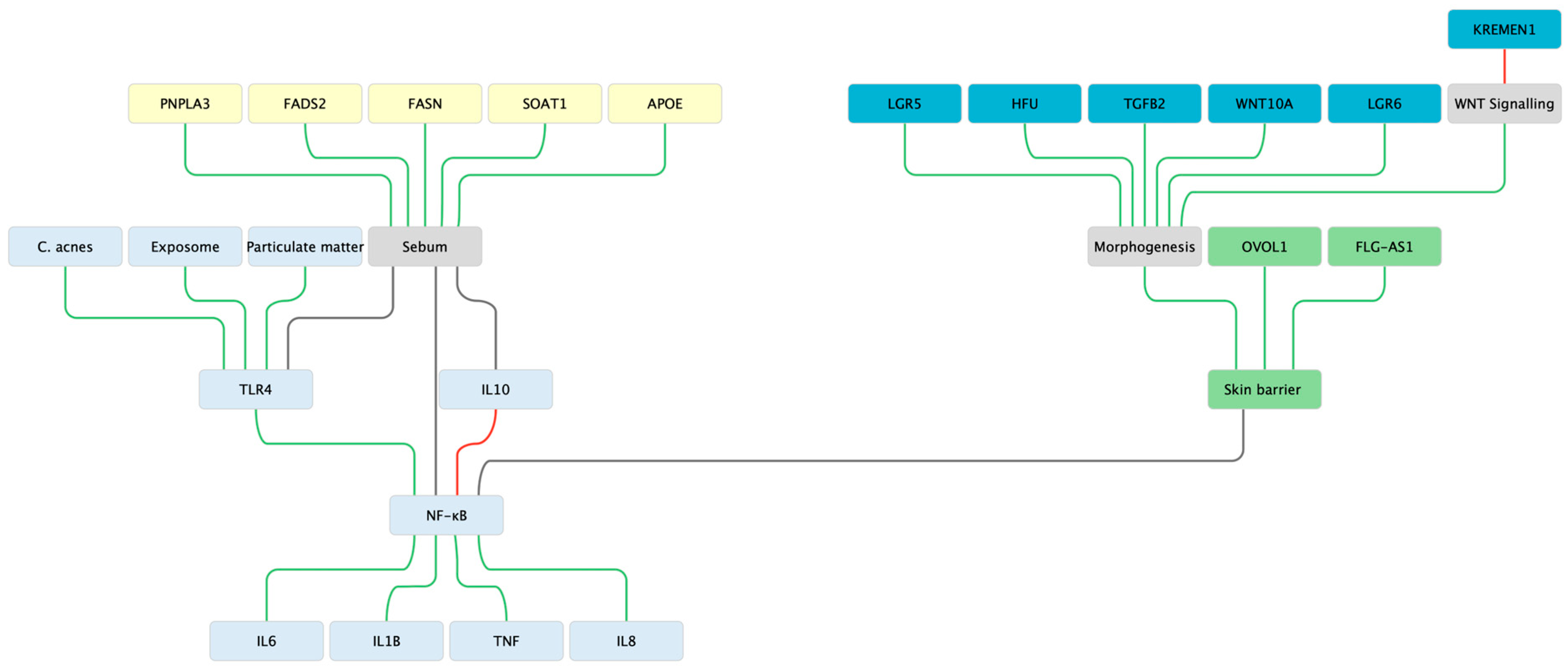
Figure 1. Network schematic of acne vulgaris integrating innate, morphogenetic, lipid, and barrier pathways. External stimuli including C. acnes and exposome components activate Toll-like receptor 4 (TLR4) in keratinocytes and sebocytes, leading to NF-κB signalling and induction of pro-inflammatory cytokines including IL-1β, IL-6, IL-8, and TNF-α, with IL-10 providing partial counter-regulation. Barrier components, represented by OVOL1, FLG-AS1, and skin barrier functionality, modulate the activation threshold of this innate cascade, highlighting how epidermal differentiation influences inflammatory tone. Sebocyte metabolism contributes an additional axis: FASN and FADS2 regulate de novo lipogenesis and sapienate synthesis, while candidate genes PNPLA3, APOE, and SOAT1 may further influence lipid trafficking and sebum composition. These lipid pathways feed back into TLR4 and NF-κB signalling, underscoring the lipid–inflammation coupling characteristic of acne lesions. At the centre of the network, a morphogenetic module comprising TGFB2, WNT10A, LGR5, LGR6, ZNRF3, and KREMEN1 integrates follicle stem-cell fate and WNT/TGF-β signalling with barrier and sebocyte biology. This central positioning reflects consistent GWAS signals and emphasises that morphogenesis connects the innate, lipid, and barrier axes into a unified pathogenic framework. Edges (lines) are coloured according to functional effect: green for stimulatory/activating interactions, red for inhibitory, and grey for modulatory associations.
Cosmetics 12 00228 g001
Figure 2. Genetic network linking androgenetic alopecia (AGA) and alopecia areata (AA) through shared biological hubs. The scheme depicts disease-specific modules (AGA, blue; AA, red) converging onto central hubs (purple). On the left, AGA is represented by androgen metabolism genes (AR, SRD5A1, SRD5A2, CYP19A1) and follicle morphogenesis regulators (WNT10A, LGR4, RSPO2, DKK2, FGF5, TWIST1, TWIST2). On the right, AA is characterised by immune-regulatory and antigen-presentation loci (HLA-DRB1, HLA-DQA1, NOTCH4, APC, LRRC32, IL15, JAK1, JAK3, STAT5, BCL2L11). Central hubs (Follicle cycling/anagen, Morphogenesis bridge, Apoptosis, Immune privilege, IFN-γ axis) represent shared biological processes where morphogenetic control, apoptotic regulation, and immune checkpoints converge. Importantly, apoptosis is shown as interconnected with follicle cycling and morphogenesis, reflecting its intrinsic role in follicular renewal. Hub-to-hub connectors clarify synergistic interactions: morphogenesis and follicle cycling are mutually activating, apoptosis modulates both morphogenesis and cycling, and the IFN-γ axis antagonises immune privilege. Edges (lines) are coloured according to functional effect: green for stimulatory/activating interactions, red for inhibitory, and grey for modulatory associations. Together, the network highlights how androgen/WNT-driven follicle cycling abnormalities in AGA and immune-mediated follicular attack in AA converge mechanistically, with follicle cycling, morphogenesis, apoptosis, and immune privilege acting as the bridging nodes.
Cosmetics 12 00228 g002
Disclaimer/Publisher’s Note: The statements, opinions and data contained in all publications are solely those of the individual author(s) and contributor(s) and not of MDPI and/or the editor(s). MDPI and/or the editor(s) disclaim responsibility for any injury to people or property resulting from any ideas, methods, instructions or products referred to in the content.