Article

Photoprotective Effect of Hydroxychloroquine on Human Keratinocytes

by
Luis Alfonso Pérez González
1,
María Antonia Martínez Pascual
2,
Elena Toledano Macías
3,
Rosa Cristina Jara Laguna
2,
Montserrat Fernández Guarino
1,
Stefano Bacci
4,
Jorge Naharro Rodriguez
1 and
María Luisa Hernández Bule
5,*
1
Dermatology Department, Ramon y Cajal University Hospital, 28034 Madrid, Spain
2
Unidad de Ensayos Clínicos de Fases Tempranas, Hospital Ramón y Cajal, 28034 Madrid, Spain
3
Photobiology and Bioelectromagnetic Laboratory, Instituto Ramón y Cajal de Investigación Sanitaria (Irycis), Hospital Ramón y Cajal, 28034 Madrid, Spain
4
Research Unit of Histology and Embriology, Department of Biology, University of Florence, 50139 Florence, Italy
5
Department of Preclinical Dentistry I, Faculty of Biomedical and Health Sciences, Universidad Europea de Madrid, 28670 Madrid, Spain
*
Author to whom correspondence should be addressed.
Cosmetics 2025, 12(5), 213; https://doi.org/10.3390/cosmetics12050213
Submission received: 21 July 2025 / Revised: 2 September 2025 / Accepted: 12 September 2025 / Published: 22 September 2025

Abstract

Hydroxychloroquine (HCQ), an antimalarial commonly used in autoimmune and dermatological conditions, may exert photoprotective effects, though its role against high-energy visible light (HEVL) remains underexplored. This study evaluated HCQ’s impact on cell viability and oxidative stress in human keratinocytes (HaCat cells) exposed to HEVL blue light. Cells were treated with HCQ (1, 2.5, or 5 µM) and irradiated with blue light doses (4.5–72 J/cm2). Assays assessed cell viability (XTT), reactive oxygen species (ROS) production, and the expression of oxidative stress-related enzymes: superoxide dismutase (SOD), glutathione peroxidase (GPX), and catalase (CAT). Results showed that low blue light doses triggered endogenous protective responses, which HCQ enhanced, potentially via SOD activation. However, higher irradiation levels caused extensive cellular damage that overwhelmed HCQ’s protective capacity. These findings suggest HCQ may confer in vitro photoprotection against sublethal HEVL exposure by modulating oxidative stress responses, although this effect diminishes beyond a certain damage threshold.

1. Introduction

Hydroxychloroquine (HCQ) is a synthetic antimalarial drug of the 4-aminochiolein family, first synthesized in 1946 from the hydroxylation of chloroquine [1]. In 1955, it was approved for the treatment of systemic lupus erythematosus (SLE) and is currently formally approved for the treatment of SLE, cutaneous lupus erythematosus, rheumatoid arthritis, primary Sjögren’s syndrome, antiphospholipid syndrome (APS), dermatomyositis/polymyositis (cutaneous forms), porphyria cutanea tarda, cutaneous sarcoidosis, and COVID-19 [2] (Table 1).
In systemic lupus erythematosus (SLE) and other inflammatory diseases such as dermatomyositis or antiphospholipid syndrome, an abnormal activation of the interferon type I (IFN-I) pathway is observed, reflected in the overexpression of interferon-stimulated genes, known as the “interferon signature”, a concept that is increasingly being focused on in the study of the physiology and treatment of lupus, currently using anifrolumab, a monoclonal antibody approved for SLE, which acts more specifically by directly blocking the interferon type I receptor (IFNAR1), preventing the signaling of all IFN-I subtypes [3]. Although less selectively, hydroxychloroquine (HCQ) helps modulate the interferon signature by inhibiting the activation of Toll-like receptors TLR7 and TLR9 [4]. This results in reduced production of IFN-α and other inflammatory cytokines such as IL-6 and TNF-α, as well as the activation of immune cells and platelets. Clinically, this has been shown to reduce the risk of vascular complications and limit the progression of mild or early forms of the disease [5].
The mechanism of action of hydroxychloroquine is still not entirely clear. It has been described as acting primarily intracellularly, as it can cross membranes, behaving like a lipophilic weak base. It preferentially accumulates in acidic organelles such as lysosomes and endosomes, where it elevates intravesicular pH [6]. This alkalinization interferes with multiple key cellular processes, such as antigen degradation, peptide presentation by MHC class II, activation of Toll-like receptors (TLR7 and TLR9), and autophagosome–lysosome fusion, thereby reducing CD4+ T and B cell activation Ferreira PMP. Through these mechanisms, HCQ exerts a selective immunomodulatory effect, decreasing the presentation of autoantigens without completely suppressing the immune response. Its anti-inflammatory action derives from the inhibition of the production of proinflammatory cytokines such as TNF-α, IL-1, and IL-6. Furthermore, at certain doses and in certain tissues, HCQ exhibits antiproliferative effects, inhibiting the expansion of activated lymphocytes, and antiplatelet effects by interfering with calcium and phospholipid-dependent signals in aggregation [7,8]. At the metabolic level, its hypoglycemic capacity has been demonstrated, possibly by reducing the lysosomal degradation of insulin and increasing insulin sensitivity, as well as a modest lipid-lowering effect, especially on LDL cholesterol [9] (Figure 1).
On the other hand, some studies suggest that HCQ could have a photoprotective effect, by stabilizing lysosomes in skin cells and reducing the inflammatory response induced by UV radiation [10,11,12]. In this regard, it has been described that antimalarials in general, and HCQ in particular, are able to reduce the concentration of porphyrins in patients with porphyria cutanea tarda and could have a beneficial effect on bone metabolism, in addition to a potential photoprotective effect, this being the least studied and worst classified effect at present [2]. Its use in dermatology has been increasing over the years, being used in a multitude of different pathologies such as porphyrias, dermatomyositis, polymorphic light eruption, solar urticaria, granuloma annulare, cutaneous sarcoidosis, various types of panniculitis, cutaneous and mucosal lichen planus, immune-mediated blistering diseases, and vasculitis and psoriatic arthritis, among other pathologies [1,13]. Although many of the dermatological pathologies in which HCQ is used have in common being diseases with photosensitivity, its photoprotective and antioxidant role against solar radiation has been little studied, especially with regard to high-energy visible light (HEVL), whose role in photoaging and cutaneous oxidative stress is becoming increasingly better known [14,15,16,17]. More recently, visible light has been proposed as an activator of immunological mechanisms involved in the pathophysiology of lupus [18,19]. The increasing importance of photoprotection against high-energy visible light is reflected in the recent development of chemical sunscreens capable of filtering HEVL and improving photoprotection against visible spectrum radiation [18,20,21].
Within the limited information available on the photoprotective and antioxidant capacities of HCQ, its potential in vitro antioxidant effect has been described [22] as well as its ability to reduce UVB radiation-induced skin inflammation [23]. These studies have shown that HCQ decreases the expression of proinflammatory genes and epidermal thickening. Furthermore, it exerts an epigenetic effect by inhibiting histone acetylation (H3K27ac), reducing the activation of inflammatory genes independently of its classical action on TLRs. This same modification of histone acetylation could be increasing DNA stabilization, thus reducing the ability of UVB to induce pyrimidine dimer formation [23]. Another study reveals that HCQ enhances UVB radiation-induced c-jun transcriptional activation in human keratinocytes [24]. This activation is mediated by the AP-1 signaling pathway, a photoprotective mechanism in the early cellular response to UV damage. Therefore, HCQ could exert part of its photoprotective effect in cutaneous lupus and other photodermatoses by enhancing these types of responses that promote survival in the face of UV damage [24]. Regarding blue light, skin hyperpigmentation is one of the effects caused by exposure to this radiation [25] and HCQ has been reported to enhance this effect of blue light in patients exposed to sunlight under ambient conditions and treated with this drug [26]. However, to our knowledge, no study has been conducted analyzing the mechanisms of action of HCQ and blue light at ambient doses. Given the lack of information in this regard, the objective of this study was to study the effect of HCQ on cell viability and the production of oxidative stress in human keratinocyte cultures treated with 440 nm blue light. Additionally, the effect of this blue light on the expression of enzymes involved in the response to oxidative stress such as catalase (CAT), glutathione peroxidase (GPX), and superoxide dismutase (SOD) was analyzed.

2. Materials and Methods

2.1. Cell Cultures

For this study, immortalized human keratinocyte cell line (HaCaT) cultures were used (HaCaT, CLS Cell Lines Service). Cells were seeded in 24-unit multiwell plates (Corning Costar TC-Treated Multiple Well Plates) with a medium composed of D-MEM rich in glucose (Biowhittaker, Lonza, Verviers, Belgium) supplemented with 10% inactivated fetal bovine serum (Gibco, Boston, MA, USA), 1% glutamine, and 1% penicillin–streptomycin (Gibco), and maintained in a 5% CO2 atmosphere at 37 °C in incubators (Thermo Fisher Scientific, Waltham, MA, USA). Cells were subcultured once a week, and the culture medium was replaced every 3 days following standard protocols.

2.2. Treatment with HEVL and HCQ

The in vitro effect of HCQ on cell viability has been described as dose-dependent, and it has been reported that the viability of human dermal fibroblasts decreases at HCQ concentrations of 10 μM or higher [27]. Prior to light treatment, the culture medium was changed to colorless D-MEM medium, and the cultures were then incubated with concentrations of 1 μM, 2.5 μM, or 5 μM HCQ. The cultures were incubated with HCQ for 30 min before light treatment. For the selection of light doses, the data described in the study by Galvez et al. [28] were taken as a reference. In this study, a measurement of the solar radiation spectrum was carried out in Malaga (a city in southern Spain), quantifying the amount of UVB, UVA, and blue light radiation throughout different times of the day. Using these reference values, the doses selected for our study were 4.5 J/cm2, 9 J/cm2, 36 J/cm2, and 72 J/cm2. These fluence values would correspond approximately to exposure to the midday sun for 10, 20, 80, and 160 min in a mid-latitude region. Keratinocyte cultures were irradiated using a custom-designed device equipped with 12 blue light lamps with a wavelength of 448 nm (Lumileds LUXEON Rebel, Royal Blue 448 nm, LXML-PR01-0500; Lumileds, Schiphol, The Netherlands). This exposure system was previously detailed [29]. Cultures were irradiated with light in a darkened room, with the beam directed from below to avoid potential scattering by the culture medium. Two controlled exposures of 20 min each were performed, separated by 24 h. Non-irradiated cells, with or without HCQ, were kept in the dark throughout the experiment and served as controls.

2.3. Cell Viability Assay

Keratinocyte cultures were seeded at a density of 4500 cells/cm2 and incubated for 3 days. Culture viability under each experimental condition was assessed using the XTT viability assay (Roche, Basel, Switzerland). These XTT assays were performed 24 h after the second light exposure, following the manufacturer’s instructions.

2.4. Reactive Oxygen Species (ROS) Production Assay

Cells were seeded at a density of 4500 cells/cm2 and incubated for 3 days. To quantify ROS, the intracellular oxidative transformation of the oxidation-sensitive probe DCFH-DA into the fluorescent dye dichlorofluorescein (DCF) was assessed. HaCat cultures were then incubated with the fluorescent probe 2′7-Dichlorodihydrofluorescein diacetate (5 μM DCFH-DA, Sigma-Aldrich, St. Louis, MO, USA) in the dark at 37 °C and 5% CO2 for 30 min. The fluorescence generated was read using a TECAN plate reader (TECAN SpectraFluor, Gödrig, Austria) at a λexc wavelength of 490 nm and λemi wavelength of 535 nm. ROS quantification was carried out immediately after the first or second exposure, thus minimizing their degradation.

2.5. Immunoblotting for the Enzymes Superoxide Dismutase (SOD), Catalase, and Glutathione Peroxidase (GPX)

HaCaT cells were seeded at a density of 6800 cells/cm2 and incubated for 4 days. The immunoblotting procedure has been described in detail elsewhere [30]. Briefly, cells were lysed in RIPA buffer (Thermo Fisher Scientific) and the protein content of the lysates was determined by BCA protein assay (Pierce; Thermo Fisher Scientific). Proteins (50 μg per lane) were separated on a 10% sodium dodecyl sulfate–polyacrylamide gel and electrophoretically transferred to a nitrocellulose membrane (Amersham, Buckinghamshire, UK). To detect SOD, GPX, and catalase, membranes were incubated overnight at 4 °C in a rabbit polyclonal anti-catalase antibody (1:1000, Invitrogen, Waltham, MA, USA), a rabbit polyclonal anti-glutathione peroxidase 2 antibody (1:1000, Abcam, Cambridge, UK), and a rabbit monoclonal anti-superoxide dismutase 1 antibody (Abcam, Ab 51254). Mouse monoclonal anti-GAPDH antibodies (1:1000, Santa Cruz Biotechnology, Dallas, TX, USA) were used as loading controls. The membranes were incubated for 1 h at room temperature with IRdye 800 CW-conjugated anti-rabbit IgG antibody (1:10,000, LI-COR Biosciences, Lincoln, NE, USA) or IRdye 680 LT-conjugated anti-mouse IgG antibody (1:15,000, LI-COR Biosciences). The ECL kit (RPN2132; GE Healthcare; Cytiva, Marlborough, MA, USA) was used to visualize the immunoreactive bands. The membranes were scanned with a LI-COR Odyssey scanner (LI-COR Biosciences). The bands obtained were evaluated densitometrically (PDI Quantity One 4.5.2 software, BioRad, Hercules, CA, USA). At least three experimental replicates were performed per protein. All values were normalized to the loading control.

2.6. Statistical Analysis

Each experiment was performed at least in triplicate. Data were normalized to a control. Statistical significance was analyzed using a one-way ANOVA with a Tukey post-test for multiple comparisons. Differences of p < 0.05 were considered statistically significant. Statistical analysis was performed using GraphPad Prism 6.01 software (GraphPad Software, San Diego, CA, USA).

3. Results

3.1. Effects on Cell Viability of HEVL and HCQ

Blue light reduced cell viability when cells were exposed twice to a dose of 9 J/cm2 or higher, whereas none of the HCQ concentrations used changed the viability of the culture compared to the control (without HCQ and in darkness). On the other hand, a concentration of 5 µM HCQ reversed the detrimental effect of 9 J/cm2, but the difference was not statistically significant. At fluences above 9 J/cm2, HCQ was unable to buffer the decrease in viability induced by light (Figure 2).

3.2. Effects on ROS Production

3.2.1. Effects on ROS Production After One Exposure to HEVL, with or Without HCQ

Fluences of 9 J/cm2 or higher significantly increased the amount of ROS produced in the cultures compared to the control, while none of the HCQ concentrations altered the cellular oxidative status. When cells were incubated with HCQ and exposed to light, ROS production was similar to that quantified in irradiated cultures incubated without HCQ (Figure 3).

3.2.2. Effects on ROS Production After Two Exposures to HEVL with or Without HCQ

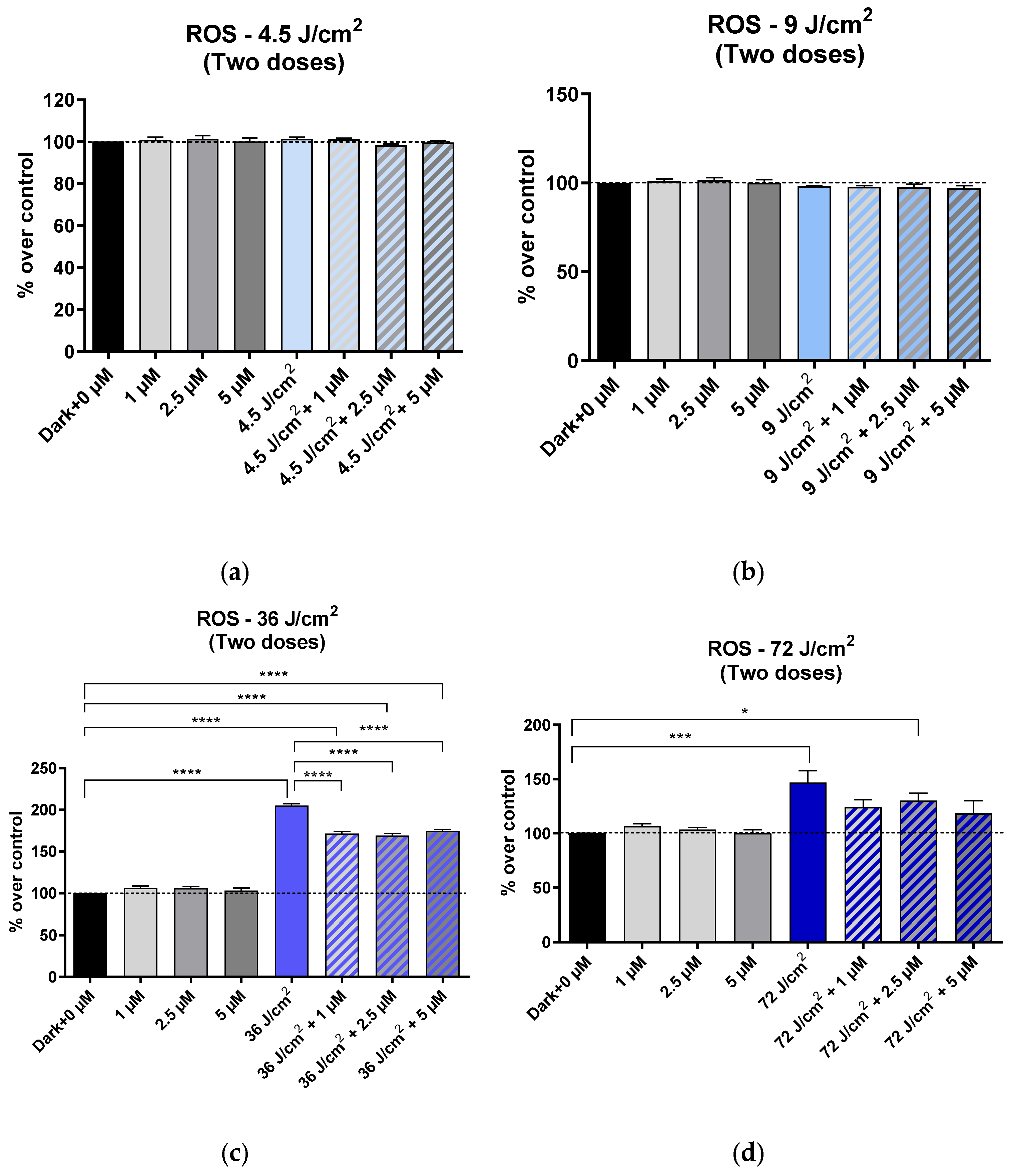
After exposing the cultures to light twice, increases in ROS relative to the control were only detected at fluences of 36 J/cm2 or 72 J/cm2. On the other hand, ROS production caused by 36 J/cm2 decreased significantly when irradiated cultures were incubated with any of the HCQ concentrations used, compared to cultures irradiated but not treated with HCQ (Figure 4).
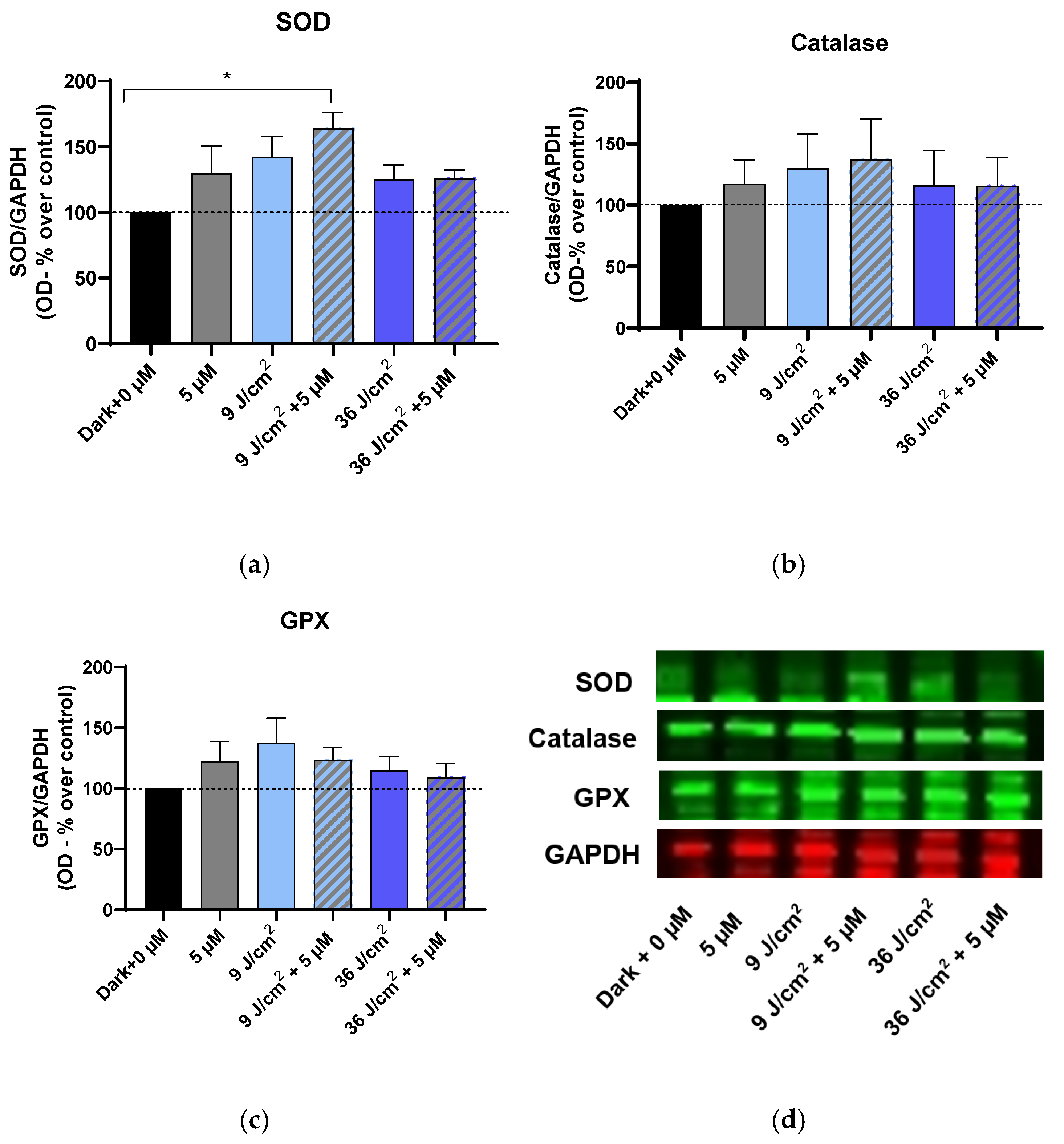
To establish the involvement of antioxidant pathways in the response of HaCat to treatment with HCQ and/or blue light, the expression of SOD, GPX, and catalase was analyzed. Blue light and HCQ increased the expression of all the enzymes analyzed compared to the control, but the differences were not statistically significant. Co-treatment of keratinocytes with light and HCQ did not significantly change the expression of catalase and GPX. In contrast, when cells were incubated with 5 µM and irradiated with 9 J/cm2, SOD expression increased significantly compared to the control (Figure 5).

4. Discussion

The harmful effects of blue light on cell viability and oxidative stress in skin cells such as fibroblasts and keratinocytes have been described and classified in several previous publications, including those by our group [29]. However, agents that can protect skin from this radiation are still very scarce, and therefore, it is a growing topic of research. Hydroxychloroquine is a drug that has been used to treat more than 20 different dermatoses, many of which have in common being photodermatoses or photoaggravated dermatoses, highlighting lupus, dermatomyositis, polymorphic light eruption, sarcoidosis, porphyria cutanea tarda, solar urticaria, and actinic prurigo [1,2]. Given this, it is conceivable that hydroxychloroquine could play a role in protecting the skin from ultraviolet radiation and possibly high-energy visible light. However, since hydroxychloroquine does not act on a specific target, the mechanism by which it exerts this effect has not yet been determined.
In the present study, the effect of blue light at levels equivalent to real-life conditions quantified by Galvez et al. throughout different times of the day [28] and the potential effect of HCQ on photoprotection were analyzed. Keratinocytes were used, as they are the cell type most exposed to ambient solar radiation. The results of the present study reflect a dose-dependent phenomenon determined by the threshold of cellular damage. Thus, an increase in ROS is observed when fluences are high and cells are exposed to a first or second blue light treatment. Thus, high doses starting at 36 J/cm2 cause an accumulation of ROS, observable after one or two exposures, which would result in a clear drop in viability. Interestingly, when keratinocytes were treated with 5 µM and exposed to this intense dose of light (36 J/cm2), HCQ was able to significantly reduce ROS levels, but this was not manifested as an improvement in cell viability. However, at lower doses (9 J/cm2), an increase in ROS was only detected after the first light treatment and, although HCQ do not seem to act on ROS production, a tendency to improve cell viability was detected in the cultures incubated with HCQ, in a dose-dependent manner. This effect could be due to the cell experiencing an adaptive response after the first exposure at lower doses, which would buffer ROS production from subsequent exposures.
Several studies have shown that exposure to blue light causes excessive generation of ROS in cultured human keratinocytes and dermal fibroblasts [31,32,33,34,35]. These ROS trigger the activation of antioxidant enzymes such as superoxide dismutase (SOD), which converts O2 into H2O2, catalase which converts H2O2 into H2O and O2, and glutathione peroxidase 2 (GPX) which reduces H2O2 or lipid peroxides to non-toxic compounds, among other enzymes such as glutathione reductase (GRD), thioredoxin, myeloperoxidase, gamma-glutamylcysteine synthetase, and heme oxygenase systems [36]. The main chromophores responsible for the overproduction of ROS induced by blue light are flavins, such as flavin adenine dinucleotide (FAD) and flavin mononucleotide (FMN), and porphyrins, such as heme. Mitochondria are one of the main intracellular locations of flavins [37]. Therefore, it is not ruled out that the effect observed in the present study is due to mitochondrial damage [38] or lipid peroxidation, which would alter cell membranes [39] and/or damage proteins and DNA [40], although such effects have not been addressed in this study. Regarding HCQ, some studies have shown that it has mild antioxidant effects, while others describe that it can potentiate oxidative stress, depending on the cell type and dose [41]. HCQ accumulates largely in specific cellular compartments, particularly lysosomes, and in fibroblasts it shows modulatory effects on cell proliferation and survival [27]. However, HCQ also induces changes in proton fluxes, which affect ROS and oxidation-reduction sensitive signal transduction pathways. These pathways include those controlled by the transcription factor nuclear factor erythroid 2, p45-related factor 2 (NRF2). Nrf2 is the most important transcription factor in the regulation of the expression of enzymatic antioxidant response genes. During increased oxidative stress, Nrf2 translocates to the nucleus, where it activates the transcription of genes for the enzymes SOD, catalase, and GPX, among others [42].
Our results show that HCQ alone does not generate ROS or modify cell viability and, therefore, does not act as a pro-oxidant or as a direct antioxidant under basal conditions. However, when cells were irradiated with low doses of light (9 J/cm2) and incubated with 5 µM HCQ, this significantly increased the expression of the SOD enzyme, although not that of catalase or GPX. SOD is the first line of defense against oxidative stress since it acts on superoxide, which is the primary ROS generated directly by blue light, and its activation is a rapid and specific response to the increase in O2 induced by irradiation. If ROS generation is not excessive, as occurs after low doses of light, H2O2 production might not exceed the threshold necessary to significantly induce GPx or catalase expression.
In sum, in our assays, cells exposed to low doses of blue light would activate protective mechanisms that HCQ could be potentiating. This could induce a transcriptional signal sufficient to significantly activate SOD but not enough to induce GPx or catalase. This SOD expression would protect the cell against subsequent exposures to blue light. At higher doses, the damage would affect multiple organelles and overwhelm HCQ’s ability to counteract it. Therefore, HCQ would only be effective against sublethal stress, and its indirect photoprotective effect would be diluted when blue light exceeded a certain toxicity threshold.
It should also be noted that there is still no evidence as to whether the effect observed in our study is due exclusively to an indirect antioxidant effect or whether a true photoprotective effect would also contribute, with HCQ being able to absorb a specific spectrum of wavelengths. In this regard, some publications unrelated to photoprotection have indicated that HCQ presents an absorption peak at 343 nm [2]. In contrast, photoaggravated skin reactions have been described, albeit in small numbers, in relation to HCQ. Therefore, we must also bear in mind that, although it is a potential photoprotective molecule, it is not exempt from being a trigger of photoallergy, as has already been described with other organic photoprotectors [21,43]. This study opens the possibility of expanding the current information on the effect of HCQ on various aspects not yet analyzed in combination with blue light. For example, it would be of great interest to analyze the anti-inflammatory potential of HCQ to ensure that the drug itself does not cause additional inflammation. Additionally, since HCQ is a photosensitizer and, at certain doses, can cause sunburn or other skin reactions, it will be essential to enhance the study to ensure that this drug does not cause a proinflammatory state.

5. Conclusions

This study demonstrates a potential in vitro photoprotective effect of hydroxychloroquine against blue light doses, given its ability to reduce cellular damage. The combination of HCQ and blue light likely generates a moderate level of oxidative stress, sufficient to significantly activate SOD expression as a primary defense against superoxide, without yet triggering a robust GPx or catalase response. Therefore, HCQ could modulate this response through indirect redox mechanisms or by enhancing the cell’s sensitivity to blue light. Since there is insufficient information to determine whether this effect is due exclusively to an indirect antioxidant effect or whether HCQ could have specific absorbance against blue light, acting as a true photoprotector, larger studies are needed to clarify the mechanism underlying this finding and determine its potential application in clinical practice.

Author Contributions

Conceptualization M.L.H.B., L.A.P.G. and M.A.M.P.; methodology, M.L.H.B., L.A.P.G., E.T.M., R.C.J.L. and M.A.M.P.; software, E.T.M.; validation, M.L.H.B.; formal analysis, M.L.H.B. and L.A.P.G.; investigation, M.L.H.B., L.A.P.G., E.T.M., M.A.M.P., R.C.J.L. and M.F.G.; resources, M.L.H.B., L.A.P.G. and M.A.M.P.; data curation M.L.H.B. and E.T.M.; writing—original draft preparation, M.L.H.B.; writing—review and editing, M.L.H.B., L.A.P.G., E.T.M., S.B., M.A.M.P. and M.F.G.; visualization, M.L.H.B., L.A.P.G., E.T.M., M.F.G., S.B., M.A.M.P., J.N.R. and R.C.J.L.; supervision, M.L.H.B.; project administration, M.L.H.B.; funding acquisition, M.L.H.B. and M.F.G. All authors have read and agreed to the published version of the manuscript.

Funding

This research received no external funding.

Institutional Review Board Statement

Not applicable.

Informed Consent Statement

Not applicable.

Data Availability Statement

The data presented in this study are available on request from the corresponding author.

Conflicts of Interest

The authors declare no conflicts of interest.

References

  1. Rodriguez-Caruncho, C.; Bielsa Marsol, I. Antipalúdicos en dermatología: Mecanismo de acción, indicaciones y efectos secundarios. Actas Dermo-Sifiliográficas 2014, 105, 243–252. [Google Scholar] [CrossRef]
  2. Sardana, K.; Sinha, S.; Sachdeva, S. Hydroxychloroquine in Dermatology and Beyond: Recent Update. Indian Dermatol. Online J. 2020, 11, 453–464. [Google Scholar] [CrossRef]
  3. Barrat, F.J.; Crow, M.K.; Ivashkiv, L.B. Interferon Target-Gene Expression and Epigenomic Signatures in Health and Disease. Nat. Immunol. 2019, 20, 1574–1583. [Google Scholar] [CrossRef] [PubMed]
  4. Nocturne, G.; Mariette, X. Interferon Signature in Systemic Autoimmune Diseases: What Does It Mean? RMD Open 2022, 8, e002687. [Google Scholar] [CrossRef] [PubMed]
  5. Willis, R.; Seif, A.; McGwin, G.; Martinez-Martinez, L.; González, E.; Dang, N.; Papalardo, E.; Liu, J.; Vilá, L.; Reveille, J.; et al. Effect of Hydroxychloroquine Treatment on Pro-Inflammatory Cytokines and Disease Activity in SLE Patients: Data from LUMINA (LXXV), a Multiethnic US Cohort. Lupus 2012, 21, 830–835. [Google Scholar] [CrossRef] [PubMed]
  6. Martinez, G.P.; Zabaleta, M.E.; Di Giulio, C.; Charris, J.E.; Mijares, M.R. The Role of Chloroquine and Hydroxychloroquine in Immune Regulation and Diseases. Curr. Pharm. Des. 2020, 26, 4467–4485. [Google Scholar] [CrossRef]
  7. Fox, R. Anti-Malarial Drugs: Possible Mechanisms of Action in Autoimmune Disease and Prospects for Drug Development. Lupus 1996, 5 (Suppl. S1), S4–S10. [Google Scholar] [CrossRef]
  8. Fox, R.I.; Kang, H.I. Mechanism of Action of Antimalarial Drugs: Inhibition of Antigen Processing and Presentation. Lupus 1993, 2 (Suppl. S1), S9–S12. [Google Scholar] [CrossRef]
  9. Solomon, D.H.; Garg, R.; Lu, B.; Todd, D.J.; Mercer, E.; Norton, T.; Massarotti, E. Effect of Hydroxychloroquine on Insulin Sensitivity and Lipid Parameters in Rheumatoid Arthritis Patients Without Diabetes Mellitus: A Randomized, Blinded Crossover Trial. Arthritis Care Res. 2014, 66, 1246–1251. [Google Scholar] [CrossRef]
  10. Siqueira-Neto, J.L.; Wicht, K.J.; Chibale, K.; Burrows, J.N.; Fidock, D.A.; Winzeler, E.A. Antimalarial Drug Discovery: Progress and Approaches. Nat. Rev. Drug Discov. 2023, 22, 807–826. [Google Scholar] [CrossRef]
  11. Fang, Q.-Q.; Wang, X.-F.; Zhao, W.-Y.; Ding, S.-L.; Shi, B.-H.; Xia, Y.; Yang, H.; Wu, L.-H.; Li, C.-Y.; Tan, W.-Q. Angiotensin-Converting Enzyme Inhibitor Reduces Scar Formation by Inhibiting Both Canonical and Noncanonical TGF-Β1 Pathways. Sci. Rep. 2018, 8, 3332. [Google Scholar] [CrossRef]
  12. Vlahopoulos, S.; Critselis, E.; Voutsas, I.; Perez, S.; Moschovi, M.; Baxevanis, C.; Chrousos, G. New Use for Old Drugs? Prospective Targets of Chloroquines in Cancer Therapy. CDT 2014, 15, 843–851. [Google Scholar] [CrossRef]
  13. Hsu, S. Comprehensive Dermatologic Drug Therapy. Arch. Dermatol. 2002, 138, 703. [Google Scholar] [CrossRef]
  14. Ezekwe, N.; Maghfour, J.; Kohli, I. Visible Light and the Skin. Photochem. Photobiol. 2022, 98, 1264–1269. [Google Scholar] [CrossRef] [PubMed]
  15. Narla, S.; Kohli, I.; Hamzavi, I.H.; Lim, H.W. Visible Light in Photodermatology. Photochem. Photobiol. Sci. 2020, 19, 99–104. [Google Scholar] [CrossRef] [PubMed]
  16. Pourang, A.; Tisack, A.; Ezekwe, N.; Torres, A.E.; Kohli, I.; Hamzavi, I.H.; Lim, H.W. Effects of Visible Light on Mechanisms of Skin Photoaging. Photodermatol. Photoimmunol. Photomed. 2022, 38, 191–196. [Google Scholar] [CrossRef]
  17. Lenane, P.; Murphy, G. Sunscreens and the Photodermatoses. J. Dermatol. Treat. 2001, 12, 53–57. [Google Scholar] [CrossRef]
  18. Neubert, E.; Bach, K.M.; Busse, J.; Bogeski, I.; Schön, M.P.; Kruss, S.; Erpenbeck, L. Blue and Long-Wave Ultraviolet Light Induce in Vitro Neutrophil Extracellular Trap (NET) Formation. Front. Immunol. 2019, 10, 2428. [Google Scholar] [CrossRef]
  19. Lood, C.; Blanco, L.P.; Purmalek, M.M.; Carmona-Rivera, C.; De Ravin, S.S.; Smith, C.K.; Malech, H.L.; Ledbetter, J.A.; Elkon, K.B.; Kaplan, M.J. Neutrophil Extracellular Traps Enriched in Oxidized Mitochondrial DNA Are Interferogenic and Contribute to Lupus-like Disease. Nat. Med. 2016, 22, 146–153. [Google Scholar] [CrossRef]
  20. Bacqueville, D.; Jacques-Jamin, C.; Dromigny, H.; Boyer, F.; Brunel, Y.; Ferret, P.J.; Redoulès, D.; Douki, T.; Bessou-Touya, S.; Duplan, H. Phenylene Bis-Diphenyltriazine (TriAsorB), a New Sunfilter Protecting the Skin against Both UVB + UVA and Blue Light Radiations. Photochem. Photobiol. Sci. 2021, 20, 1475–1486. [Google Scholar] [CrossRef]
  21. Métayer, I.; Balguerie, X.; Courville, P.; Lauret, P.; Joly, P. Photodermatosis induced by hydroxychloroquine: 4 cases. Ann. Dermatol. Venereol. 2001, 128, 729–731. [Google Scholar]
  22. Miyachi, Y.; Yoshioka, A.; Imamura, S.; Niwa, Y. Antioxidant Action of Antimalarials. Ann. Rheum. Dis. 1986, 45, 244–248. [Google Scholar] [CrossRef] [PubMed]
  23. Catalano, R.; Rocca, R.; Juli, G.; Costa, G.; Maruca, A.; Artese, A.; Caracciolo, D.; Tagliaferri, P.; Alcaro, S.; Tassone, P.; et al. A Drug Repurposing Screening Reveals a Novel Epigenetic Activity of Hydroxychloroquine. Eur. J. Med. Chem. 2019, 183, 111715. [Google Scholar] [CrossRef] [PubMed]
  24. Nguyen, T.Q.; Capra, J.D.; Sontheimer, R.D. 4-Aminoquinoline Antimalarials Enhance UV-B Induced c–Jun Transcriptional Activation. Lupus 1998, 7, 148–153. [Google Scholar] [CrossRef]
  25. Campiche, R.; Curpen, S.J.; Lutchmanen-Kolanthan, V.; Gougeon, S.; Cherel, M.; Laurent, G.; Gempeler, M.; Schuetz, R. Pigmentation Effects of Blue Light Irradiation on Skin and How to Protect against Them. Int. J. Cosmet. Sci. 2020, 42, 399–406. [Google Scholar] [CrossRef] [PubMed]
  26. Peng, J.; Yang, X.; Luo, F.; Yuan, X.; Xiong, H.; Ma, W.; Yao, X. Hydroxychloroquine-Induced Hyperpigmentation of the Skin and Bull’s-Eye Maculopathy in Rheumatic Patients: A Case Report and Literature Review. Front. Immunol. 2024, 15, 1383343. [Google Scholar] [CrossRef]
  27. Ramser, B.; Kokot, A.; Metze, D.; Weiß, N.; Luger, T.A.; Böhm, M. Hydroxychloroquine Modulates Metabolic Activity and Proliferation and Induces Autophagic Cell Death of Human Dermal Fibroblasts. J. Investig. Dermatol. 2009, 129, 2419–2426. [Google Scholar] [CrossRef]
  28. de Gálvez, E.N.; Aguilera, J.; Solis, A.; de Gálvez, M.V.; de Andrés, J.R.; Herrera-Ceballos, E.; Gago-Calderon, A. The Potential Role of UV and Blue Light from the Sun, Artificial Lighting, and Electronic Devices in Melanogenesis and Oxidative Stress. J. Photochem. Photobiol. B Biol. 2022, 228, 112405. [Google Scholar] [CrossRef]
  29. Pérez González, L.A.; Martínez-Pascual, M.A.; Toledano-Macías, E.; Jara-Laguna, R.C.; Fernández-Guarino, M.; Hernández-Bule, M.L. Effect of Combination of Blue and Red Light with Terbinafine on Cell Viability and Reactive Oxygen Species in Human Keratinocytes: Potential Implications for Cutaneous Mycosis. Int. J. Mol. Sci. 2024, 25, 12145. [Google Scholar] [CrossRef]
  30. Hernández-Bule, M.L.; Trillo, M.A.; Cid, M.A.; Leal, J.; Ubeda, A. In Vitro Exposure to 0.57-MHz Electric Currents Exerts Cytostatic Effects in HepG2 Human Hepatocarcinoma Cells. Int. J. Oncol. 2007, 30, 583–592. [Google Scholar] [CrossRef][Green Version]
  31. Suitthimeathegorn, O.; Yang, C.; Ma, Y.; Liu, W. Direct and Indirect Effects of Blue Light Exposure on Skin: A Review of Published Literature. Ski. Pharmacol. Physiol. 2022, 35, 305–318. [Google Scholar] [CrossRef] [PubMed]
  32. Magni, G.; Banchelli, M.; Cherchi, F.; Coppi, E.; Fraccalvieri, M.; Rossi, M.; Tatini, F.; Pugliese, A.M.; Rossi Degl’Innocenti, D.; Alfieri, D.; et al. Experimental Study on Blue Light Interaction with Human Keloid-Derived Fibroblasts. Biomedicines 2020, 8, 573. [Google Scholar] [CrossRef] [PubMed]
  33. Magni, G.; Cherchi, F.; Banchelli, M.; Tatini, F.; Nardini, P.; Guasti, D.; Coppi, E.; Pugliese, A.M.; Fraccalvieri, M.; Bacci, S.; et al. Effects of Short-Wavelength Blue Light on Fibroblasts, Experimental Evidence in Wound Healing and Cutaneous Fibrosis. In Proceedings of the 2nd International Electronic Conference on Biomedicines, Online, 1–15 December 2023; p. 27. [Google Scholar]
  34. Magni, G.; Cherchi, F.; Banchelli, M.; Tatini, F.; Nardini, P.; Pugliese, A.M.; Guasti, D.; Bacci, S.; Cavigli, L.; Pini, R.; et al. Experimental Evidence on Photobiomodulation Induced by Short Wavelengths Blue LED Light. In Proceedings of the Mechanisms of Photobiomodulation Therapy XVIII; Carroll, J.D., Liebert, A., Lyons, J.-A., Eds.; SPIE: San Francisco, CA, USA, 2024; p. 7. [Google Scholar]
  35. Avola, R.; Graziano, A.C.E.; Pannuzzo, G.; Bonina, F.; Cardile, V. Hydroxytyrosol from Olive Fruits Prevents Blue-light-induced Damage in Human Keratinocytes and Fibroblasts. J. Cell. Physiol. 2019, 234, 9065–9076. [Google Scholar] [CrossRef] [PubMed]
  36. Nakai, K.; Tsuruta, D. What Are Reactive Oxygen Species, Free Radicals, and Oxidative Stress in Skin Diseases? Int. J.Mol. Sci. 2021, 22, 10799. [Google Scholar] [CrossRef]
  37. Garza, Z.C.F.; Born, M.; Hilbers, P.A.J.; Van Riel, N.A.W.; Liebmann, J. Visible Blue Light Therapy: Molecular Mechanisms and Therapeutic Opportunities. CMC 2019, 25, 5564–5577. [Google Scholar] [CrossRef]
  38. McNish, H.; Mathapathi, M.S.; Figlak, K.; Damodaran, A.; Birch-Machin, M.A. The Effect of Blue Light on Mitochondria in Human Dermal Fibroblasts and the Potential Aging Implications. FASEB J. 2025, 39, e70675. [Google Scholar] [CrossRef]
  39. Habib, L.; Michael-Jubeli, R.; Abboud, M.; Lteif, R.; Tfayli, A. Impact of Blue Light on Cutaneous Barrier Structures and Properties: NPLC/HR-MSn and Raman Analyses. Analyst 2024, 149, 5693–5703. [Google Scholar] [CrossRef]
  40. Chamayou-Robert, C.; DiGiorgio, C.; Brack, O.; Doucet, O. Blue Light Induces DNA Damage in Normal Human Skin Keratinocytes. Photodermatol. Photoimmunol. Photomed. 2022, 38, 69–75. [Google Scholar] [CrossRef]
  41. Tang, S.-C.; Lu, C.-T.; Ko, J.-L.; Lin, C.-H.; Hsiao, Y.-P. Hydroxychloroquine Repairs Burn Damage through the Wnt/β-Catenin Pathway. Chem. Biol. Interact. 2023, 370, 110309. [Google Scholar] [CrossRef]
  42. Klouda, C.B.; Stone, W.L. Oxidative Stress, Proton Fluxes, and Chloroquine/Hydroxychloroquine Treatment for COVID-19. Antioxidants 2020, 9, 894. [Google Scholar] [CrossRef]
  43. Ekstein, S.F.; Hylwa, S. Sunscreens: A Review of UV Filters and Their Allergic Potential. Dermatitis 2023, 34, 176–190. [Google Scholar] [CrossRef]
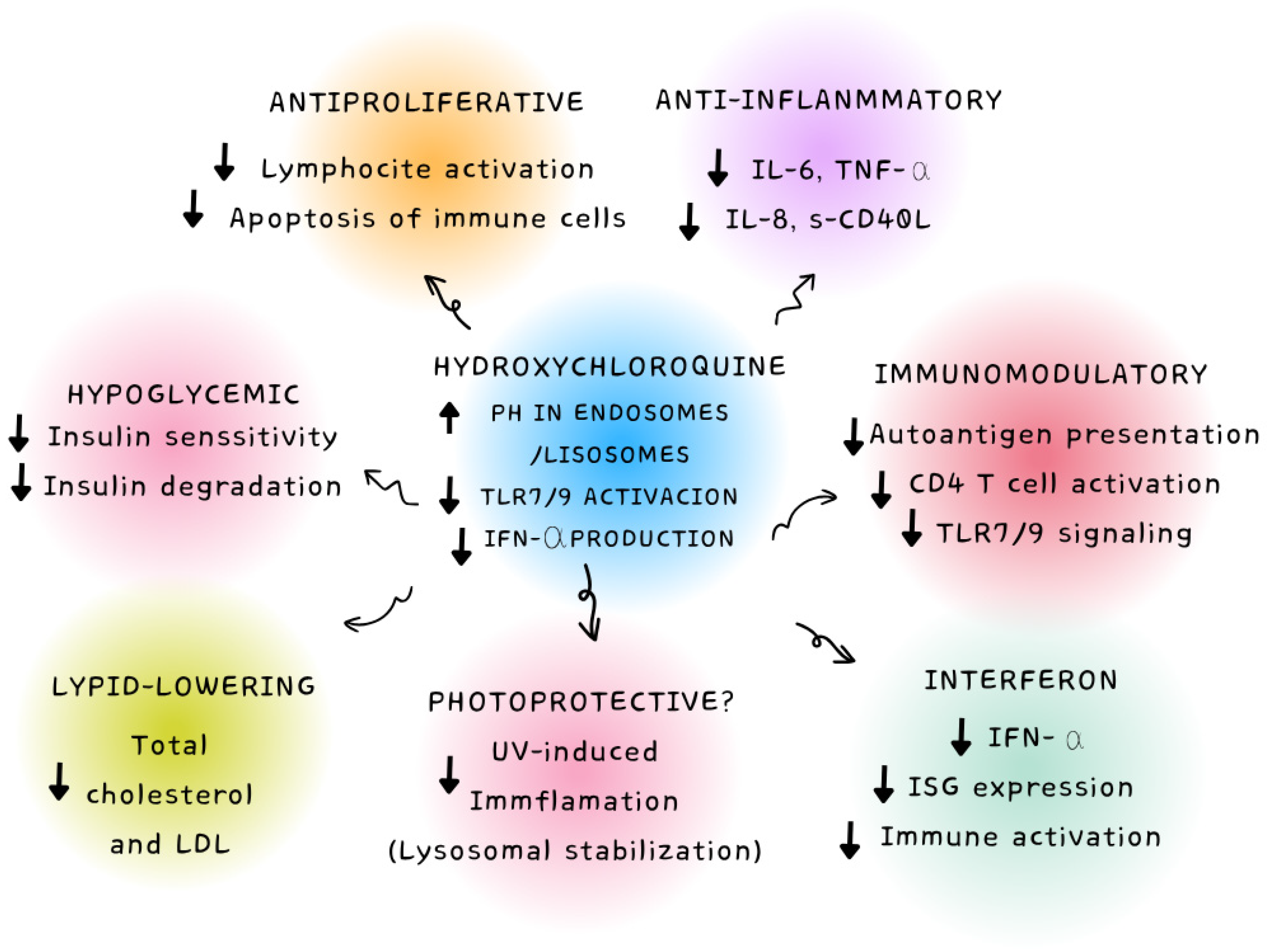
Figure 1. Visual summary of the main mechanisms of action of hydroxychloroquine at the cellular level and its main biological effects described.
Cosmetics 12 00213 g001
Figure 2. XTT viability assay in HaCaT cells treated with hydroxychloroquine (1, 2.5 or 5 µM), blue light (448 nm), or the combination of blue light + hydroxychloroquine. Cells treated twice on consecutive days. (a) Hacat irradiated with 4.5 J/cm2. (b) Hacat irradiated with 9 J/cm2. (c) Hacat irradiated with 36 J/cm2. (d) Hacat irradiated with 72 J/cm2. Mean ± SEM; 5 experimental replicates. Data normalized over the corresponding controls (dark and 0 µM of HCQ). One-way ANOVA; ns: p ≥ 0.05; *: 0.01 ≤ p < 0.05; **: 0.001 ≤ p < 0.01; ***: 0.0001 ≤ p < 0.001; ****: p < 0.0001.
Cosmetics 12 00213 g002
Figure 3. ROS production quantification assay. HaCaT cells treated with hydroxychloroquine (1, 2.5, or 5 µM), blue light (448 nm), or the combination of blue light + hydroxychloroquine. Cells treated only once (one light dose). (a) Hacat irradiated with 4.5 J/cm2. (b) Hacat irradiated with 9 J/cm2. (c) Hacat irradiated with 36 J/cm2. (d) Hacat irradiated with 72 J/cm2. Mean ± SEM; 5 experimental replicates. Data normalized over the corresponding controls (dark and 0 µM of HCQ). One-way ANOVA; ns: p ≥ 0.05; **: 0.001 ≤ p < 0.01; **** p ≤ 0.0001.
Cosmetics 12 00213 g003
Figure 4. ROS production quantification assay. HaCaT cells treated with hydroxychloroquine (1, 2.5 or 5 µM), blue light (448 nm), or the combination of blue light + hydroxychloroquine. Cells treated twice on consecutive days. (a) Hacat irradiated with 4.5 J/cm2 (b) Hacat irradiated with 9 J/cm2. (c) Hacat irradiated with 36 J/cm2. (d) Hacat irradiated with 72 J/cm2. Mean ± SEM; 5 experimental replicates. Data normalized over the corresponding controls (dark and 0 µM of HCQ). One-way ANOVA; ns: p ≥ 0.05; *: 0.01 ≤ p < 0.05; ***: 0.0001 ≤ p < 0.001; ****: p < 0.00013.3. Effect on the Expression of Enzymes Involved in Oxidative Stress SOD, GPX, and CAT.
Cosmetics 12 00213 g004
Figure 5. Superoxide dismutase (SOD), catalase, and glutathione peroxidase (GPX) expression. Cells were treated twice on consecutive days. Densitometry values for SOD (a), catalase (b), and GPX (c) expressions. Means ± SD of the protein/GAPDH ratios of at least four experimental repeats per protein. Data normalized over the corresponding controls (dark and 0 µM of HCQ). One-way ANOVA. ns: p ≥ 0.05; *: 0.01 ≤ p < 0.05 (d) Representative blots (50 µg of protein/lane). GAPDH was used as the loading control.
Cosmetics 12 00213 g005
Table 1. Clinical indications for hydroxychloroquine (HCQ) and associated clinical considerations. This table summarizes the principal approved and off-label clinical uses of hydroxychloroquine, highlighting its pharmacological rationale, therapeutic targets, and limitations in evidence-based practice.
Clinical Indication (HCQ)Pharmacological/Clinical Rationale
Systemic lupus erythematosus (SLE)Cornerstone therapy for mucocutaneous, musculoskeletal, and constitutional symptoms; reduces disease activity and flare rates.
Cutaneous lupus erythematosusEffective in discoid and subacute cutaneous variants; reduces photosensitivity and cutaneous inflammation.
Rheumatoid arthritis (RA)Conventional synthetic DMARD with immunomodulatory effects; indicated in mild-to-moderate disease often in combination therapy.
Primary Sjögren’s syndromeProvides symptomatic relief for arthralgias, fatigue, and extraglandular features; limited impact on sicca symptoms.
Antiphospholipid syndrome (APS)May confer antithrombotic and endothelial-protective effects; used adjunctively with anticoagulation.
Dermatomyositis/polymyositis (cutaneous forms)Useful for photosensitive cutaneous lesions; minimal effect on muscle involvement.
Porphyria cutanea tardaLow-dose regimens decrease hepatic porphyrin deposits and photosensitivity; alternative in patients unsuitable for phlebotomy.
Cutaneous sarcoidosisMay ameliorate granulomatous cutaneous lesions in refractory cases.
COVID-19 (investigational; not recommended)Initially explored for antiviral and immunomodulatory potential; randomized trials show no clinical benefit—currently not recommended.
Disclaimer/Publisher’s Note: The statements, opinions and data contained in all publications are solely those of the individual author(s) and contributor(s) and not of MDPI and/or the editor(s). MDPI and/or the editor(s) disclaim responsibility for any injury to people or property resulting from any ideas, methods, instructions or products referred to in the content.