Review

Advances in Extraction Technologies of Silybum marianum L. and Its Role in Protecting Against Skin Damage

1
Laboratory of Advanced Materials and Process Engineering, Faculty of Sciences, Ibn Tofaïl University, Kenitra 14000, Morocco
2
Laboratory of Natural Resources and Sustainable Development, Research Unit in Nutrition, Metabolism and Physiology, Faculty of Sciences, Ibn Tofaïl University, B.P. 133, Kenitra 14000, Morocco
3
Laboratory of Cell Biology and Molecular Genetics, Faculty of Science, Ibnou Zohr University, B.P. 8106, Agadir 80000, Morocco
4
Laboratory of Biology and Health, Team of Nutritional Sciences, Food and Healtth, Faculty of Sciences, Ibn Tofail University, B.P. 133, Kenitra 14000, Morocco
5
SIRC-Systems Engineering Laboratory (LaGeS), Hassania School of Public Works, Casablanca 20230, Morocco
*
Author to whom correspondence should be addressed.
These authors contributed equally.
Cosmetics 2025, 12(5), 211; https://doi.org/10.3390/cosmetics12050211
Submission received: 26 August 2025 / Revised: 19 September 2025 / Accepted: 19 September 2025 / Published: 21 September 2025

Abstract

Silybum marianum L., commonly known as milk thistle, is traditionally recognized for its hepatoprotective properties. This is primarily due to silymarin, a mixture of flavonolignans with strong antioxidant and anti-inflammatory activities. Although many studies have reported on its biological activities, critical syntheses that compare extraction technologies and highlight its protective roles beyond liver health remain limited. Despite the abundant literature, the protective effects of milk thistle against skin damage remain largely unexplored. To address this gap, we performed a comprehensive literature search across PubMed, Scopus, Web of Science, and Google Scholar. Our research covered publications up to February 2025 and used predefined keywords, including extraction methods for the release of silymarin, in vitro, in vivo, and clinical studies relevant to its protective effects against skin damage. The evidence indicates that silymarin exerts protective effects on the skin, including the prevention of photoaging, the management of acne, the promotion of wound healing, and the defense against UV-induced damage, through the activation of Nrf2 and the preservation of the extracellular matrix. These results highlight the promising dermatological benefits of silymarin, as well as the need for further clinical studies and the optimization of environmentally sustainable extraction techniques for large-scale production.

1. Introduction

Silybum marianum (L.) Gaertn. is also known as Marian thistle, Mary thistle, blessed milk thistle, or milk thistle [1]; in French, it is known as Chardon-Marie and in Arabic as Shouk el-nasara [2,3]. It is originally from the Mediterranean region (Figure 1). This species belongs to the Asteraceae family and is now widespread throughout the globe [4]. It is a wild plant that grows spontaneously along roadsides and on wastelands [5].
There is a legend associated with the milk thistle’s name, which tells the story of the Virgin Mary. According to the tale, as the Virgin Mary sought shelter, she took cover under a protective covering made of thorny milk thistle leaves. While breastfeeding baby Jesus, a droplet of her milk accidentally fell onto the leaves, leaving behind the distinct white veins that can still be seen on milk thistle plants today [3]. It is characterized by its glossy green leaves with distinctive milky white veins, thorny stems, and bright purple flowers [6]. Silybum marianum L. exists in two varieties: the purple-flowering type, which is more abundant, and the white-flowering type. However, it is the purple variety that has received a lot of attention in studies [3].
Traditionally, nursing mothers in Europe used it to increase milk production [7]. Its fruits, known as achenes, have historically been roasted and used as a coffee substitute. The plant’s leaves, petals, and roots were also historically regarded as vegetables in European diets. The plant’s flower heads are prepared similarly to artichokes, while the leaves can be used in salads as an alternative to spinach [8]. Milk thistle was traditionally combined with honey as a natural cough cure [9]. Additionally, the plant is sometimes cultivated for ornamental purposes in gardens, and its dried flower heads are often used in decorative arrangements [10]. For over 2000 years, the Greeks and Romans have used milk thistle to treat various liver and gallbladder conditions, including hepatitis, cirrhosis, and jaundice. It has also been used to protect the liver from toxins, such as those found in alcohol, snakebites, insect stings, and mushroom poisoning. Renowned for its healing properties, milk thistle has become a key herb in traditional medicine. Today, milk thistle remains one of the oldest and most thoroughly researched herbs for treating liver disorders [11,12,13]. This long-standing use is largely attributed to silymarin, a standardized extract from the plant consisting of a group of compounds called flavonolignans, namely silybin, isosilybin, silychristin, and silydianin. These were initially identified in the seeds [14]. The first identified and most studied compound is silybin, also known as silybinin and consisting of silybin A and silybin B. It was isolated and characterized by Pelter [15].
Research efforts are ongoing to examine the potential of milk thistle extract and its primary active compound, silymarin, in protecting the liver and other organs, such as the kidneys, lungs, and heart, from a wide range of harmful substances. Current studies are investigating how silymarin can mitigate damage to organs caused by substances such as alcohol [16,17,18], pharmaceutical drugs such as paracetamol, doxorubicin and acetaminophen [19,20,21], environmental pollutants, industrial chemicals, and heavy metals [22,23,24,25]. Researchers are investigating its mechanisms of action, including its ability to promote liver regeneration, reduce oxidative stress, and enhance detoxification processes. In addition to its protective activity, milk thistle has shown other beneficial effects such as anticancer activity. Silymarin may prevent the growth of tumor cells in various organs, including the prostate, breast, colon, ovaries, lungs, and bladder [26,27]. It exerts anti-inflammatory [28], antimicrobial [29], and antiviral activity. Additionally, it has the ability to lower cholesterol levels [30]. Recent studies have revealed that silybin exhibits notable activity against COVID-19 by inhibiting viral replication and reducing the severity of symptoms, suggesting its potential as a therapeutic agent in combating the ongoing pandemic [31]. In recent years, the cosmetic and dermatological industries have experienced increasing demand for products that are safe, effective and environmentally sustainable. This has driven research towards bioactive ingredients that can improve skin health, prevent and manage skin conditions, and support overall skin appearance and resilience. Consequently, there is increasing interest in innovative compounds that combine therapeutic potential with safety and sustainability for modern skincare applications.
This review aims to provide a comprehensive summary of different extraction methods used to extract silymarin from milk thistle, particularly by comparing alternative techniques with conventional methods. Additionally, this review aims to update our understanding of silymarin’s role in protecting the skin against aging and dermatological disorders, such as acne, impaired wound healing, and skin cancer, through its antioxidant, anti-inflammatory, and regenerative effects.

2. Methodology

This article is a narrative review based on a structured search of the scientific literature. Major databases, including PubMed, Scopus, Web of Science, and Google Scholar, were searched up to February 2025, with no publication date restrictions. A combination of relevant keywords was used in the search strategy, including “milk thistle”, “silymarin”, “extraction methods”, “skin protection”, “in vivo”, “in vitro”, and “clinical studies”. Studies were selected based on their relevance to the scope of the review. Only peer-reviewed articles were included as they are considered reliable and unbiased, while conference abstracts and theses were excluded.

3. Flavonolignans from Milk Thistle

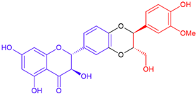
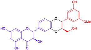
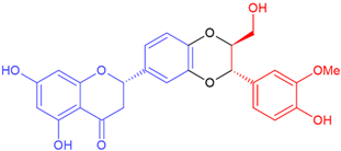
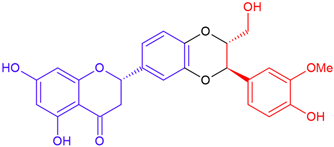
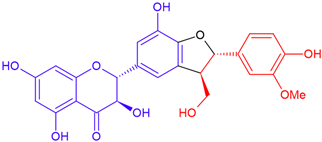
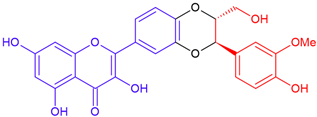
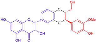
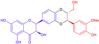
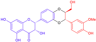
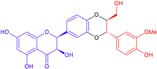
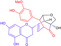
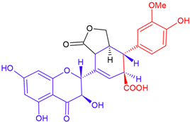
Flavonolignans are a chemical class belonging to the group of natural phenols known as non-conventional lignans, composed of a part of flavonoid and a part of lignan. Milk thistle is the richest known source of this particular group of compounds [32]. These chemicals are distinctive to the Asteraceae, Berberidaceae, Chenopodiaceae, Flacourtiaceae, Fabaceae, Poaceae, and Scrophulariaceae families of plants [29,33]. A mixture of flavonolignans called silymarin is obtained from seeds after fat is removed. This mixture is composed of approximately 30% silybin A and B, with silybin B typically being slightly more abundant. It also contains around 5% isosilybin A and B, 7–10% silychristin A, and 10% silydianin. The mixture also includes a smaller proportion of taxifolin (2–5%) and less than 3% 2,3-dehydroflavonolignans [34].
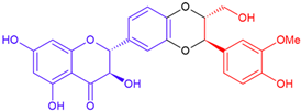
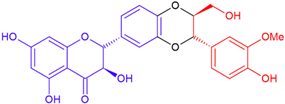
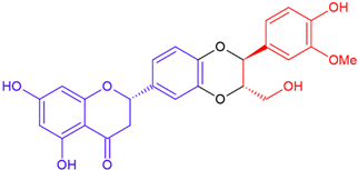
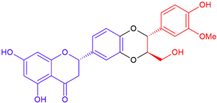
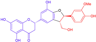
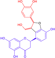
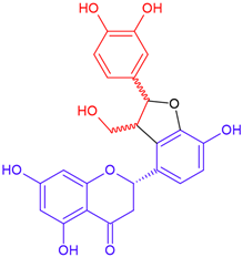
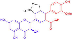
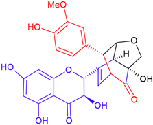
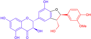
Silymarin from milk thistle consists of a flavonoid, taxifolin, which is fused with coniferyl alcohol through an oxeran ring (Figure 2). Except for silybin and isosilybin, the other flavonolignans found in milk thistle are made up of taxifolin fused with coniferyl alcohol via various structural arrangements [35]. Another type of flavonolignans exists: non-taxifolin-derived flavonolignans, which are present in the white-flowering type [35]. Flavonolignans can have different structures, with the flavonoid part fused to a coniferyl alcohol unit via various linkages, including a dioxane ring, a cyclic ether, a simple ether side chain, a lactone, a carbon-carbon bond, and B-ring fission [36]. Table 1 presents the flavonolignans isolated from milk thistle.

4. Extraction of Bioactive Compounds from Milk Thistle

It has always been difficult to extract bioactive components from plant material completely and effectively. Currently, there is no single extraction technique that can effectively extract bioactive chemicals from a variety of sources. The chemical composition of the compounds, the method of extraction, the solvent used, and the particle size all affect how bioactive chemicals are extracted from plants [38].
European Pharmacopoeia outlines a two-step process for extracting silymarin from milk thistle seeds, which contain a high level of lipids. In the first step, the seeds are defatted for 6 h using hexane to remove the lipids. Silymarin is then extracted in the second step for 5 h using methanol with a Soxhlet apparatus [39]. Oil is a by-product of silymarin production [40]. According to Wallace et al. [41], defatting increases the silymarin content twofold, making lipid removal a crucial extraction step. Omar et al. [42] have also noted that the highest concentration of active ingredients is obtained by extracting defatted seeds with methanol.
Conventional methods are limited in that they are time-consuming and require large quantities of harmful solvents such as hexane, ethanol, methanol, ethyl acetate, among others. Furthermore, the Soxhlet method involves two steps for extracting silymarin, which may result in silymarin loss during the defatting stage.
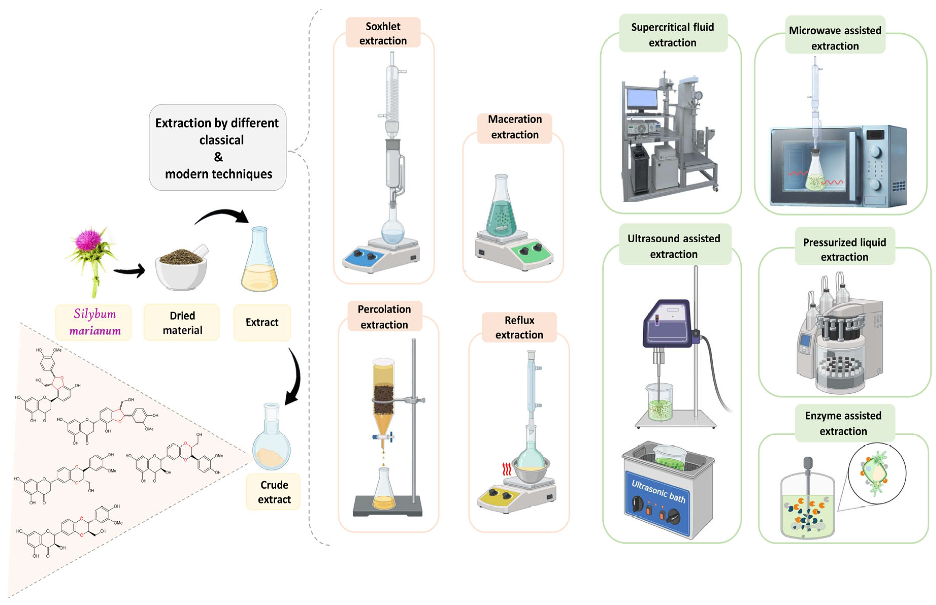
Numerous studies have investigated more efficient, environmentally friendly extraction techniques to enhance silymarin production. Researchers have compared conventional extraction methods with alternative techniques to assess their efficacy (Figure 3). Alternative methods such as Microwave-Assisted Extraction (MAE), Pressurized Liquid Extraction (PLE), Ultrasound-Assisted Extraction (UAE), Supercritical Fluid Extraction (SFE), Enzyme-Assisted Extraction (EAE), and Subcritical Water Extraction (SWE) have been evaluated. These comparisons aim to identify extraction techniques that maximize silymarin yield while minimizing time, solvent use, energy demand, and environmental impact. These findings contribute to the development of sustainable silymarin production practices, paving the way for greater efficiency and a smaller ecological footprint in the industry.
Microwave Assisted Extraction (MAE).
MAE is one of the most advanced techniques and a rapid extraction method that uses microwave radiation to speed up the extraction process. It offers faster extraction, efficient recovery, and minimal solvent use, while protecting thermolabile compounds during the process. Extraction can be completed in just a few minutes [43]. Saleh et al. [44] compared conventional methods such as Soxhlet extraction and heat reflux extraction with MAE using 80% methanol as the extraction solvent, to evaluate their effectiveness in isolating silymarin. The results showed that MAE not only reduced the extraction time but also improved the extraction yield. At 400 W, the silymarin content increased as the extraction time was extended from 5 to 30 min. However, at 800 W, the silymarin content increased at 15 min but decreased at 30 min. The effectiveness of MAE is influenced by various factors, including extraction time, power consumption, solvent, and temperature. Another study compared conventional methods with MAE using ethanol and methanol, exploring parameters such as extraction time. The findings showed that MAE of the methanolic extract for only 1 min resulted in the highest yield (11.2%) and a content of 1813.3 mg/g, followed by Soxhlet extraction with a yield of 1656.5 mg/g after 6 h. These results suggest that MAE can extract higher amounts of phenolic compounds in significantly less time [45].
Pressurized Liquid Extraction (PLE)
PLE is another alternative method employed for extracting silymarin from milk thistle. It is alternatively recognized as Accelerated Solvent Extraction (ASE) or Subcritical Solvent Extraction (SSE). This method shortens extraction time and reduces solvent use, making it a more environmentally friendly alternative to conventional techniques [46]. It involves using organic solvents at elevated pressures and temperatures that exceed their typical boiling points. The temperature is typically maintained within the range of 40–200 °C and the pressure is usually set between 500 and 3000 psi. The extraction process lasts for a relatively short duration of 5–15 min. The high temperature speeds up the diffusivity of solvents, thereby improving the kinetics of extraction, while the elevated pressure serves to retain the solvent in its liquid form at higher temperatures [43]. There is often a question regarding whether alternative extraction methods can eliminate the need for defatting. Wianowska and Wisniewski [47] conducted a study comparing the European Pharmacopoeia-recommended method, Soxhlet extraction, and PLE, both with and without defatting. The study showed that removing lipids through PLE did not significantly affect the yield of silymarin. However, extracts obtained from defatted fruit were found to be clearer, making chromatographic analysis easier. PLE yielded a higher amount of silymarin in a shorter time and with lower solvent consumption than the two-step Soxhlet extraction. The optimal PLE conditions involved acetone as the solvent at a temperature of 125 °C for 10 min, without the need for a preliminary defatting step. The PLE approach enables the efficient isolation of silymarin mixtures without the need for defatting.
Subcritical Water Extraction (SWE)
When 100% water is used as a solvent in PLE, the method is often referred to as Subcritical Water Extraction. It is also known by various other names, including Superheated Water Extraction, Pressurized Low Polarity Water Extraction, or Pressurized Hot Water Extraction. This variant of PLE leverages the unique properties of water under high-pressure and high-temperature conditions to facilitate the extraction process [48]. The process involves using liquid water as an extractant at temperatures higher than water’s ambient boiling point (100 °C/273 K, 0.1 MPa) but not exceeding the critical point of water (374 °C/647 K, 22.1 MPa) [49]. SWE offers a significant advantage in that it reduces the need for organic solvents by using water as a substitute. Furthermore, water is abundant and harmless, and its reuse or disposal causes minimal environmental impact. The selectivity and extraction efficiency of SWE are primarily affected by temperature, pressure, extraction time, flow rate, and the use of modifiers or additives; temperature being the main factor. A high temperature changes the properties of water, bringing its polarity closer to that of non-polar substances. This helps non-polar compounds to dissolve in water, making their extraction easier [50].
Duan et al. [51] investigated the use of SWE, employing water at temperatures above its boiling point (ranging from 100 to 140 °C). Their results showed that, while higher temperatures accelerated extraction and reached maximum concentrations faster, they did not significantly increase total silymarin compound yield. This was likely due to temperature-induced degradation. The time required to achieve maximum yields of the compounds decreased from 200 min at 100 °C to 55 min at 140 °C. Thus, while temperature positively influenced the extraction rate, it did not significantly increase total yields. To minimize the degradation of bioactive compounds caused by the high temperature of the water, Bunnell et al. [52] designed and validated the performance of a countercurrent pressurized hot water reactor. The aim was to evaluate the yield of silymarin and the extent of product degradation in comparison to two other methods: ethanol-Soxhlet extraction and batch pressurized hot water extraction. The ethanol-Soxhlet method using 0.59 mm seed meal yielded the highest levels of flavonolignans, including silychritin, silydianin, silybin A, silybin B, isosilybin A, and isosilybin B. However, the yield of silymarin obtained from 1.48 mm seed meal using the Batch Parr reactor and the countercurrent reactor was higher than that obtained using the ethanol-Soxhlet with 1.48 mm seed meal. When normalized to silybin, the countercurrent reactor provided the highest proportions of SC, SD, and total silymarin. It yielded the highest proportions of silychristin, silydianin, and total silymarin per silybin B, followed by the batch Parr reactor and the ethanol-Soxhlet method. The countercurrent reactor exhibited exceptional performance in terms of flavonolignan yield, surpassing the yields obtained from the batch Parr reactor and the ethanol-Soxhlet method using 1.48 mm seed meal. Its ability to optimize extraction conditions and facilitate efficient solute-solvent contact makes it a promising choice for maximizing flavonolignan extraction from milk thistle seeds.
Ultrasound-Assisted Extraction (UAE)
UAE is another environmentally friendly technique employed for extracting silymarin. It offers clear advantages over traditional methods, including reduced solvent use and faster bioactive compound extraction. The breakdown of cell walls caused by ultrasound enables bioactive compounds in the plant matrix to be extracted more quickly. This enhanced mass transfer improves the efficiency of the extraction process. Ultrasound equipment is less expensive and easier to use than other innovative extraction methods such as MAE [46].
Saleh et al. [53] evaluated UAE in comparison to maceration. They applied indirect sonication (40 kHz) using a water bath and direct sonication (20 kHz) using a probe for different time intervals (15, 30, and 60 min). The study demonstrated that UAE was more effective than maceration. Notably, extraction efficiency was higher when the 20 kHz probe system was used. However, the authors emphasized the need for further research to evaluate the long-term effectiveness of the probe system, especially after 60 min. In contrast, Nowak et al. [54] investigated the impact of extraction methods and solvents on the antioxidant properties of various milk thistle parts, finding that Soxhlet extraction was generally more effective than UAE, resulting in higher antioxidant activity. However, they observed that extending the UAE duration from 15 to 60 min increased polyphenol content, regardless of the solvent used. In another study investigating the influence of extraction methods on silymarin content, four techniques were employed: UAE, maceration, percolation and extraction in a water bath. This study revealed that extracting in a water bath for 30 min produced the highest silymarin yield. However, prolonging the extraction time to 60 min resulted in a decrease in silymarin content [55]. Drouet et al. [56] focused on optimizing UAE using aqEtOH and compared it to maceration. They reported that UAE resulted in a significant increase in silymarin content, yielding nearly six times more than conventional maceration. The optimal UAE conditions identified in their study were an ethanol concentration of 54.5% (v/v), an ultrasound frequency of 36.6 kHz, an extraction time of 60 min and a temperature of 45 °C, yielding 20.28 ± 0.41 mg/g DW of silymarin, compared to 3.40 ± 0.14 mg/g DW obtained by maceration.
Overall, these studies emphasize the complexity of extraction techniques and the influence of various factors, such as extraction time, solvent composition, and temperature, on the yield and content of silymarin. It is important to consider these factors when selecting an extraction method for optimal results.
Supercritical Fluid Extraction (SFE)
SFE is an alternative method of extracting silymarin and other bioactive constituents to conventional techniques. It employs organic solvents at pressures and temperatures above their typical boiling points, resulting in a supercritical fluid state. Supercritical CO2 is the most frequently used supercritical fluid, although other SCFs are also employed, including ethane, butane, pentane, nitrous oxide, ammonia, trifluoromethane and water. Supercritical CO2 is a safe, environmentally friendly, non-flammable, non-toxic solvent with a low critical temperature (Tc = 31.1 °C). It can easily be removed and may reduce the risk of degradation processes due to a lack of light and air [43,46]. Çelik and Gürü [57] investigated the extraction of oil and silybin from milk thistle using supercritical carbon dioxide (CO2) without a co-solvent. They studied the effects of temperature (40–80 °C), pressure (160–220 bar), CO2 flow rate (3, 4, and 5 mL/min), and particle size (0.3125, 0.925, and 1.2 mm) on the extraction process. The results showed that increasing the temperature beyond 80 °C reduced the levels of oil and silybin. Decreasing the particle size resulted in higher amounts of oil, total silybin, silybin A, and silybin B, due to the increased contact area between the CO2 and the milk thistle seeds. Regarding pressure, silybin B concentrations decreased significantly above 180 bar, while silybin A concentrations increased within the 180–220 bar range. The overall amounts of silybin and oil content were unaffected by pressures above 180 bar. The authors noted that the supercritical extraction process becomes more energy-intensive at higher pressure ranges, which may not be economically feasible. Therefore, the optimal pressure chosen was 180 bar. The optimal conditions recommended were a temperature of 40 °C, a pressure of 180 bar, a CO2 flow rate of 4 mL/min, and a particle size of 0.3025 mm. Under these conditions, the amounts obtained were 327 mg/g of oil, 2.29 mg/g of Silybin A and 1.29 mg/g of Silybin B.
A recent study by Milovanovic et al. [58] investigated the factors affecting oil extraction from milk thistle, as well as the impact of temperature and pressure on the concentrations of phenolic compounds, flavonoids, and antioxidant activity. The study tested the pressure of 450 bar. The results showed that, at constant pressure, the IC50 value (indicating radical scavenging activity) increased with temperature. The extract produced at 300 bar and 60 °C exhibited the lowest IC50 value (12.7 mg/mL), indicating the highest radical scavenging activity. Total polyphenolic content decreased as temperature increased from 40 °C to 80 °C at pressures of 200 and 450 bar, likely due to the heat sensitivity of polyphenols. Similarly, the total flavonoid content decreased as pressure increased from 200 bar to 300 bar and 450 bar while maintaining the same temperature. An increase in temperature from 40 °C to 80 °C at constant pressure resulted in a modest increase in total flavonoid content. The maximum yield (31.5%) and the highest total phenolic (9.2 mg GAE/g) and flavonoid content (123.8 μg QE/g) were achieved at 450 bar and 80 °C. However, the authors acknowledged that the high-pressure requirement could make the process costly. Thus, a pressure of 300 bar and a temperature of 60 °C may be a more suitable alternative for the industrial extraction of high-value extracts from milk thistle [58].
Enzyme Assisted Extraction (EAE)
The cell wall of plants is composed of various polysaccharides, including hemicellulose, cellulose, and pectin. These polysaccharides can act as barriers, hindering the extraction of target compounds using ordinary solvent extraction methods. However, enzymes can be used to improve the yield of natural extracts from plant matrices by breaking down or modifying these polysaccharides [59]. EAE represents a novel extraction method involving the use of enzymes such as cellulases, pectinases, and hemicellulases. These enzymes play a vital role in breaking down the cell wall components and disrupting the structural integrity of plant cells. This disruption allows for the efficient release of intracellular components during the extraction process [60]. To maximize the effectiveness of enzymes in extraction processes, a comprehensive understanding of their catalytic properties, mechanisms of action, and ideal operating conditions is essential. To achieve optimal outcomes, it is crucial to identify the enzyme or blend of enzymes that best matches the specific plant material under consideration [61]. Various operational factors are considered during EAE studies, including reaction temperature, extraction time, system pH, enzyme concentration and substrate particle size. These parameters are carefully considered and optimized to ensure an efficient and effective extraction process. [60].
EAE has several advantages over traditional methods. These include lower energy consumption, faster extraction rates, improved extraction yields, and simpler recovery processes involving reduced solvent use [43]. When it comes to silymarin extraction, using enzymes has been found to result in a higher silybin content than traditional extraction methods. Liu et al. [62] examined the extraction of silybin from the defatted seeds using EAE, comparing it to traditional reflux extraction using ethanol as the solvent. The study reported that the yield of silybin obtained through EAE three times in 1 h was 24.81 ± 1.93 mg/g, whereas the traditional reflux over 3 h three times yielded 10.42 ± 1.65%. This indicates a significant 138% increase in silybin yield using EAE compared to traditional extraction methods. To optimize the EAE conditions for silybin extraction, the authors employed the Box–Behnken experimental design. The optimal conditions were an extraction temperature of 40 °C, a pH of 4.5 for the enzyme solution, and a seed particle size of 7003 µm.
Key comparisons and conclusions of the extraction methods are summarized in Table 2.

5. Skin-Protective Activity of Silymarin

The skin, as the body’s primary barrier against environmental aggressors, is continuously exposed to physical, chemical, and biological insults that contribute to various dermal disorders. These include premature aging, inflammatory conditions such as acne, impaired wound healing, and carcinogenesis. Ultraviolet (UV) radiation remains one of the major extrinsic factors driving oxidative stress, photoaging, and skin malignancies [63,64]. Additionally, endogenous factors such as hormonal imbalances, impaired antioxidant defenses, and metabolic dysfunctions exacerbate dermal damage, contributing to collagen degradation, transepidermal water loss (TEWL), inflammation, and reduced regenerative capacity [63,65].
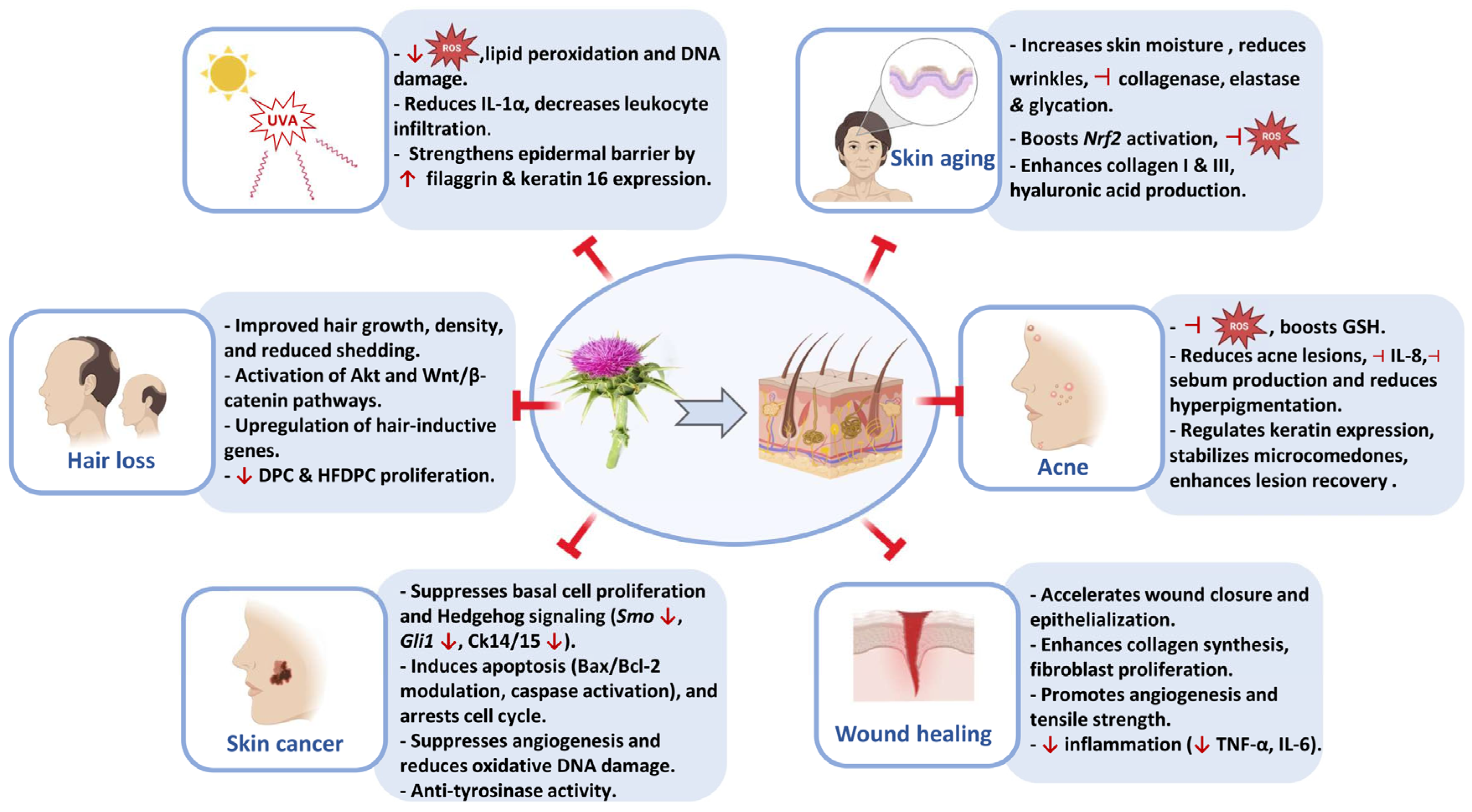
Among various phytochemicals evaluated for skin protection, Silybum marianum has emerged as a potent bioactive compound with broad dermoprotective properties. Its efficacy has been investigated across a range of experimental models and clinical trials targeting skin aging, UV-induced damage, acne, wound healing, hair loss, and skin cancer (Figure 4) [66,67,68,69,70,71,72].
A detailed overview of the main findings supporting the skin-protective activity of silymarin is summarized in Table 3.

5.1. Skin Aging

The aging of skin is governed by numerous intrinsic and extrinsic factors, contributing to a gradual loss of its structural and functional characteristics. Intrinsic aging, often referred to as chronological aging is a natural process primarily determined by genetic and physiological factors [73,74]. It is characterized by a gradual decline in collagen and elastin production, leading to decreased skin elasticity and the formation of fine lines and wrinkles [75]. Additionally, cellular senescence increases, while the skin’s ability to repair itself diminishes, resulting in progressive thinning of the epidermis and dermis. Among the primary factors involved in intrinsic aging is oxidative stress, triggered by the excessive presence of reactive oxygen species (ROS) that damage various cell components, including proteins, lipids, and DNA. Over time, these molecular changes lead to the visible signs of aging, such as loss of skin firmness, reduced hydration, and an overall decline in skin function [76].
Silymarin has shown promising anti-aging effects. Rasul and Akhtar [67] reported that a topical water-in-oil emulsion containing 4% Silybum marianum seed extract improved hydration and reduced transepidermal water loss (TEWL). This led to a significant wrinkle reduction in a clinical study. Shin et al. [77] demonstrated that silibinin inhibits protein glycation and increases fibrillin-1 expression in skin explants. In a recent animal study by He and Fan [78], it was found that silymarin treatment in D-galactose-induced aged mice enhanced collagen and hyaluronic acid (HA) synthesis and up-regulated Nrf2 signaling, which is a key regulator of the antioxidant defense. Furthermore, Vostálová et al. [79] confirmed its ability to suppress collagenase and elastase activity in vitro, thereby preventing the degradation of extracellular matrix components.

5.2. UVA-Induced Skin Damage

Among the various forms of ultraviolet (UV) radiation, UVA (320–400 nm) is of particular concern due to its ability to penetrate deeply into the dermis. Once there, it exerts damaging effects on connective tissues, blood vessels, and dermal cells [80]. In contrast to UVB, which predominantly causes direct DNA damage, UVA exposure leads to the generation of large amounts of ROS, contributing to oxidative stress, lipid peroxidation, and indirect DNA damage, such as the formation of 8-oxo-7,8-dihydroguanine (8-oxoG) [81,82]. The oxidative stress induced by UVA activates key transcription factors, notably NF-κB and AP-1, which subsequently drive the expression of pro-inflammatory cytokines, including IL-6, IL-1β, and TNF-α, as well as matrix metalloproteinases (particularly MMP-1 and MMP-3). These enzymes degrade structural components of the extracellular matrix, such as collagen and elastin, leading to hallmark signs of photoaging: wrinkle formation, skin sagging, and loss of dermal elasticity [83,84]. In addition, UVA exposure can impair mitochondrial function and trigger apoptosis via intrinsic pathways. Sustained oxidative stress also activates the p53 signaling pathway, which governs DNA repair and cell death mechanisms. However, chronic UVA exposure may result in the accumulation of p53 mutations, potentially facilitating the development of skin cancers, especially basal and squamous cell carcinomas [85,86]. Although melanocytes play a protective role through melanin production, they are not exempt from UVA-induced damage. UVA can alter melanocyte signaling pathways, contributing to hyperpigmentation and oxidative stress responses linked to melanoma. These effects are mediated, in part, by cAMP and p53 regulatory interactions [87,88].
Silymarin’s photoprotective capacity has been extensively studied. In keratinocyte models, Svobodová et al. [68] demonstrated that pretreatment with silymarin markedly reduced UVA-induced intracellular ROS accumulation, prevented glutathione (GSH) depletion, and limited apoptotic events, including caspase-3 activation and DNA fragmentation. In vivo, Katiyar et al. [89] showed that topical application of silymarin significantly attenuated UVA-induced leukocyte infiltration, particularly by inhibiting the recruitment and activation of CD11b+ inflammatory cells. More recent studies have highlighted silymarin’s role in strengthening the epidermal barrier. Boira et al. [90] reported that topical silymarin upregulated the expression of key barrier proteins such as filaggrin and keratin 16, which are essential for maintaining corneocyte structure and skin integrity. Interestingly, Fidrus et al. [91] highlighted a dose-dependent dual response to silymarin: at lower concentrations, it exhibited clear antioxidant and photoprotective effects; however, at higher doses, it paradoxically increased ROS production and phototoxicity. This biphasic effect underscores the necessity for precise formulation and controlled delivery to ensure therapeutic efficacy while minimizing adverse reactions.

5.3. Acne

Acne vulgaris ranks among the most prevalent inflammatory skin disorders, affecting up to 85% of adolescents and a significant proportion of adults, with an increasing incidence in women beyond their twenties [92]. The development of acne involves several contributing factors, such as follicular hyperkeratinization, increased sebum production, colonization by Cutibacterium acnes (formerly Propionibacterium acnes), and a robust inflammatory response mediated by cytokines such as IL-1β, IL-6, and IL-8 [93]. Oxidative stress is also a key contributor, as lipid peroxidation of sebum and depletion of cutaneous antioxidants can exacerbate inflammation and comedogenesis [94]. Conventional acne treatments, including topical retinoids, benzoyl peroxide, antibiotics, and hormonal therapies, are often limited by skin irritation, antibiotic resistance, or systemic side effects, particularly with prolonged use [95]. As a result, natural compounds with anti-inflammatory, antioxidant, and sebum-regulating properties are increasingly being investigated as complementary or alternative therapeutic options.
Silybum marianum has demonstrated promising efficacy in acne management through multiple mechanisms. Its potent antioxidant activity reduces oxidative stress and restores glutathione levels, while its anti-inflammatory properties inhibit cytokines such as IL-8 [69]. Clinical trials have shown that both oral and topical formulations of silymarin significantly improve acne severity, reduce sebum production, and diminish post-inflammatory hyperpigmentation [96,97,98]. In addition, a 12-month international real-life clinical cohort study reported that long-term topical application of Silybum marianum fruit extract significantly reduced acne lesion counts, improved seborrhea, and restored skin homeostasis, with excellent tolerance [99].

5.4. Hair Loss

Hair loss, or alopecia, is a widespread dermatological concern that affects both men and women and can significantly impair psychological well-being and quality of life [100]. Among the various types of alopecia, androgenetic alopecia is the most common, characterized by a gradual miniaturization of hair follicles due to androgen sensitivity and genetic predisposition [101]. The pathogenesis of hair loss is multifactorial, involving hormonal dysregulation, oxidative stress, chronic inflammation, and dysfunction of dermal papilla cells (DPCs), which are essential for hair follicle development and cycling [102]. Current treatments for androgenetic alopecia include topical minoxidil and oral finasteride. However, both drugs have limitations: minoxidil often requires lifelong use and has limited efficacy in some patients, while finasteride carries the risk of adverse effects such as decreased libido and mood disturbances [103]. Consequently, there is growing interest in botanical compounds that support hair follicle regeneration and reduce inflammatory and oxidative stress without systemic toxicity.
Several in vitro studies have shown that silymarin and its major flavonolignan, silibinin, promote the proliferation of human dermal papilla cells (DPCs), enhance their antioxidant defense, and reduce the expression of inflammatory mediators such as COX-2 and iNOS [104]. Notably, silibinin activates the Akt and Wnt/β-catenin signaling pathways -two key regulators of hair growth- leading to increased expression of hair-inductive genes, including ALPL, VCAN, FGF7, and BMP2 [70]. Clinical evidence also supports its efficacy. In a human trial, a topical serum containing Silybum marianum extract reduced hair shedding and significantly improved hair density and volume over the course of treatment [105]. Additionally, apigenin, a flavonoid also present in Silybum marianum flower extracts, has been proven to raise HFDPC proliferation, enhance VEGF secretion, and reduce markers of cellular senescence, supporting its role in promoting angiogenesis and follicle vitality [106].

5.5. Wound Healing

Wound healing is a complex and precisely controlled biological process that occurs in several overlapping phases: hemostasis, inflammation, proliferation, and tissue remodeling. Each phase relies on the coordinated activity of numerous cellular and molecular mechanisms, including cell migration, the release of cytokines and growth factors, extracellular matrix production, and the formation of new blood vessels (angiogenesis), all working in concert to restore tissue integrity [107,108]. While conventional treatments for wound healing (such as antiseptics, antibiotics, and surgical procedures) remain standard practice, they often fall short in contexts involving severe oxidative stress or underlying systemic conditions. As a result, increasing attention is being directed toward natural compounds with pro-regenerative, antioxidant, and anti-inflammatory properties, which show promise as therapeutic adjuvants to enhance tissue repair and improve clinical outcomes [109].
In vitro studies on human skin fibroblasts show that silymarin enhances antioxidant defense, suppresses inflammatory responses (notably COX-2 expression), and protects cells from oxidative stress-induced apoptosis [110]. Animal studies corroborate these findings: topical or systemic administration of silymarin and silibinin significantly accelerated wound contraction, increased fibroblast recruitment, enhanced collagen and glycosaminoglycan synthesis, and promoted angiogenesis and re-epithelialization [111,112,113]. Notably, silymarin also modulates key inflammatory and tissue remodeling markers. For instance, it downregulates TNF-α and upregulates markers of epithelial–mesenchymal, supporting both anti-inflammatory and pro-regenerative activity [113]. Silymarin has demonstrated significant wound healing benefits, including complete epithelialization in patients with second-degree burns [114], as well as reduced inflammation and pain severity in perineal incisions among postpartum women [115].

5.6. Skin Cancer

Skin cancer is the most prevalent form of malignancy worldwide. It includes non-melanoma skin cancers (NMSCs), primarily basal cell carcinoma (BCC) and squamous cell carcinoma (SCC), as well as malignant melanoma. Chronic exposure to UV radiation is the principal etiological factor, inducing direct DNA damage (e.g., the formation of cyclobutane pyrimidine dimers), oxidative stress, immunosuppression, and activation of oncogenic signaling pathways such as MAPK, PI3K/Akt, and β-catenin [116,117]. While early-stage tumors are typically treated with surgical excision, advanced cases often require systemic therapies (such as chemotherapy or immunotherapy) which can be costly and cause significant side effects [118]. As a result, polyphenolic compounds with chemopreventive potential are increasingly explored as complementary or topical alternatives.
Among them, silymarin has shown significant anticancer effects in various skin cancer models. In vivo, topical silibinin significantly inhibited the initiation and progression of UVB-induced basal cell carcinoma in Ptch+/− mice, reducing both the number and size of tumors. This effect was associated with decreased basal cell proliferation (Ki-67), downregulation of Hedgehog signaling mediators (Smo, Gli1), and reduced expression of cytokeratins 14 and 15 [72]. In melanoma models, silymarin also reduced tumor growth and weight in mice implanted with A375 melanoma xenografts. It promoted apoptosis by upregulating caspase-3, inhibited angiogenesis through downregulation of VEGF and CD31, and suppressed cellular proliferation by decreasing PCNA expression [119]. Silymarin also exhibits strong antioxidant and anti-inflammatory properties that contribute to its chemopreventive effects. Nanoformulations such as silymarin-loaded lipid carriers or β-cyclodextrin nanosponges have further improved their bioavailability and tumor-targeting capabilities. These formulations enhanced apoptosis, inhibited tyrosinase activity, and significantly reduced oxidative stress and pro-inflammatory cytokines in experimental melanoma models [120,121]. Additionally, silymarin has demonstrated protective effects on DNA integrity in normal cells, reducing 8-OHdG levels and chromosomal instability, suggesting a dual role in both chemoprevention and therapeutic intervention [122].
Table 3. Summary of studies on the skin-protective effects of Milk Thistle extract (Silybum marianum) and its compounds.
Skin Disorder/ConditionModelActive CompoundConcentration/DoseBiological EffectReference
Skin agingIn vitro cell-free assaySilybum marianum seed extractNot disclosed
  • Strong inhibition of collagenase and elastase, antioxidant activity, inhibition of advanced glycation end products (AGEs).
[56]
Human clinical studySilybum marianum seed extract4% topical (W/O emulsion)
  • Increased skin moisture, reduced transepidermal water loss (TEWL), improved skin smoothness, reduced wrinkles.
[67]
Human skin explantsSilibinin (from Silybum marianum flower extract)1% topical application
  • Inhibits glycation, reduces N2-(carboxymethyl) lysine (CML) expression, stimulates fibrillin-1 expression.
[77]
D-galactose-induced aging miceSilybum marianum extract50, 100, 200 mg/kg,
2 mg/cm2 (topical)
  • Increased skin hydration, HYP & HA levels; upregulated collagen I & III, downregulated MMP-1 & MMP-3, activated Nrf2 pathway.
[78]
In vitro enzyme assaysSilymarin


Dehydrosilybin


Silybin
0.05–100 mg/L


4.1 mg/L (anti-elastase), 11.2 mg/L (anti-collagenase)

59.1 mg/L (anti-elastase), 25.2 mg/L (anti-collagenase)
  • Inhibits collagenase and elastase activity, protects against UV-induced skin aging.
  • Strongest anti-collagenase and anti-elastase activity, high UVA protection factor.
  • Inhibits collagenase and elastase activity, moderate UVB protection.
[79]
Human clinical studySilybum marianum seed oil1% in cream
  • Reduced wrinkles, improved elasticity, increased dermal density, and improved skin tone.
[123]
Human skin explantsSilybum marianum extract0.8% topical application
  • Increased collagen III and hyaluronic acid production, reduced wrinkles (21% forehead, 17% circumference).
[124]
UVA-induced skin damageHuman keratinocytes (HaCaT cell line)Silymarin 0.7–34 mg/L
  • Reduced oxidative stress, GSH depletion, ROS production, lipid peroxidation, DNA damage, and caspase-3 activation.
[68]
C3H/HeN miceSilymarin1 mg/cm2 (topical)
  • Reduced ROS production and leukocyte infiltration by targeting CD11b+ cells, decreasing oxidative stress in UV-exposed skin.
[89]
Human Reconstructed Epidermis (RHE)Silymarin1% topical
  • Reduced ROS and IL-1α levels, upregulated Nrf2 and AHR pathways and improved epidermal barrier function (increased filaggrin and keratin 16 expression).
[90]
Human keratinocytes (HaCaT cell line) Silymarin10–250 µg/mL
  • Low doses (≤100 µg/mL): Antioxidant effect, reducing ROS by up to 40% after UVA exposure.
  • High dose (250 µg/mL): Dual effect—ROS reduction accompanied by increased phototoxicity.
[91]
HaCaT keratinocytesSilibinin75 µM
  • Increased ROS production and enhanced apoptosis.
  • Upregulated ER stress (CHOP, GRP78).
[125]
Primary human dermal fibroblastsSilymarin and Silybin 3.013–36.15 mg/L
  • Reduced UVA-induced ROS generation, prevented GSH depletion, decreased caspase-3 activity, reduced DNA single-strand breaks (SSB), and lowered MMP-1 levels.
[126]
AcneHuman clinical trialSilymarin210 mg/day orally
  • Reduced inflammatory lesions, decreased IL-8, increased GSH, and inhibited oxidative stress.
[69]
Human clinical trialSilymarin140 mg oral tablet
  • Antioxidant and anti-inflammatory effects, reduced acne severity, potential therapeutic option in combination with doxycycline.
[96]
Human clinical studySilymarin 0.5% topical
(in antioxidant serum)
  • Reduced acne severity, decreased sebum production, and reduced melanin pigmentation.
[97]
Human clinical trialSilymarin1.4% topical
  • Significant reduction in acne severity.
  • Improved post-inflammatory hyperpigmentation.
[98]
Human clinical studySilybum marianum fruit extract (SMFE, patented)Not disclosed
  • Significant reduction in total acne lesion counts (−59.6% at 12 months)
  • Decrease in inflammatory and non-inflammatory lesions
  • Improvement in skin homeostasis and microcomedone index.
[99]
Human observational studySilybum marianum fruit extract7% topical
  • Reduced microcomedones and improved acne lesion stability and regulation of infundibular keratin expression.
[127]
Human clinical studySilybum marianum fruit extract (patented as ComedoclastinTM)Not disclosed
  • Reduced both inflammatory and noninflammatory acne lesions, with stronger effects on larger follicles.
[128]
Hair loss3D spheroid-cultured human dermal papilla cellsSilibinin10 µM
  • Activation of Akt and Wnt/β-catenin signaling pathways, increased expression of hair-inductive genes (ALPL, VCAN, FGF7, BMP2), enhanced viability and size of dermal papilla spheroids.
[70]
Human follicle dermal papilla cells (DPCs) in vitroSilymarin50 & 100 µM
  • Increased DPC proliferation (BrdU assay), reduced COX-2 and iNOS levels, enhanced total antioxidant capacity (TAC) and decreased reactive oxygen species.
[104]
Human clinical trial Silybum marianum extractPart of a serum (exact % not disclosed)
  • Reduced hair shedding and improved hair density/volume.
[105]
Human dermal papilla cells (HFDPC) in vitro


Human clinical trial
Apigenin (from Silybum marianum flower extract)
2% apigenin (10–100 µg/mL)


0.001% apigenin (0.05% extract in shampoo formulation)
  • Increased HFDPC proliferation, enhanced VEGF secretion, reduced cellular senescence (SA-β-gal) and suppressed IL-6 and ROS.
  • Increased hair density and improved hair growth.
[106]
Wound healingWistar rats (experimental)Silibinin6 & 12 mg/mL topically
  • Reduced inflammatory cells (lymphocytes, macrophages), increased fibrocytes, collagen, and GAGs (critical for wound strength) and enhanced tensile strength and wound maturity.
[71]
Human skin fibroblasts in vitroSilymarin4.5–36 µg/mL
  • Increased cellular antioxidant capacity, suppressed LPS-induced COX-2 expression, and protected fibroblasts from H2O2 damage.
[110]
Swiss albino miceSilibinin0.2% hydrogel topically
  • Reduced inflammatory cells, increased fibroblasts and collagen synthesis.
  • Enhanced wound contraction and tensile strength and promoted angiogenesis and tissue remodeling.
[111]
Wistar rats (abdominal excision)Silymarin ointment2% (in eucerin base)
  • Accelerated wound closure (↓ wound area), reduced clinical signs of inflammation (redness, swelling).
  • Enhanced angiogenesis (↑ blood vessels) and iNOS expression.
  • Elevated estradiol levels, promoting re-epithelialization via growth factor activation (e.g., HB-EGF, VEGF).
[112]
Wistar ratsSilymarin3% topical
  • Enhanced epithelialization and collagenization.
  • Reduced inflammation (↓ TNF-α) and regulated EMT markers (↑ E-cadherin, ↓ N-cadherin/Vimentin).
[113]
Human patients (clinical trial)Silymarin140 mg/day orally
  • Accelerated and complete wound healing (epithelialization).
[114]
Human clinical trial (primiparous women)Silybum marianum seeds ointment3% topical (in eucerin base)
  • Accelerated wound healing (↓ REEDA scores), reduced inflammation (↓ redness, edema) and reduced pain severity (↓ VAS scores).
[115]
Wistar ratsSilymarin nanoemulsion1% (in loaded chitosan gel)
  • Enhanced wound closure, reduced TNF-α and IL-6 levels and accelerated re-epithelialization and collagen deposition.
[129]
Skin cancerPtch+/− mice, UVB-induced BCC in vivoSilibinin9 mg/200 µL
topically
  • Reduced BCC lesion number and area (45–94%).
  • Inhibited basal cell proliferation (↓ Ki67).
  • Downregulated Ck14, Ck15, Smo and Gli1 expression.
  • Decreased incidence of dysplasia, fibrosarcoma, and squamous cell carcinoma.
[72]
Human melanoma cells (A375, Hs294t)


Athymic nude mice (A375 xenografts)
Silymarin10–80 µg/mL


500 mg/kg orally
  • Reduced cell viability, induced G0/G1 (A375) & G2-M (Hs294t) cell cycle arrest and increased apoptosis via Bax/Bcl-2 modulation and caspase activation.
  • Decreased tumor volume and weight, reduced proliferation (↓ PCNA), increased apoptosis (↑ caspase-3) and inhibited angiogenesis (↓ MMP-2/9, VEGF, CD31).
[119]
B16F10 melanoma cellsSilymarin-loaded in β-cyclodextrin nanosponges10–200 µg/mL
  • Increased apoptosis in melanoma cells, reduced inflammation (97% protection), and exhibited anti-tyrosinase activity (87.86% inhibition).
  • Strong antioxidant.
[120]
B16 murine melanoma cells in vitro

Albino mice in vivo
Silymarin-loaded
NLC gel

Silymarin-NLC gel
50, 100, 200 μg/mL
(applied in gel)

1 mg/cm2
  • Enhanced cytotoxicity against B16 melanoma cells, reduced oxidative stress markers (↑ SOD, CAT; ↓ MDA).
  • Reduced pro-inflammatory cytokines (IL-1α and TNF-α).
  • Decreased tumor volume.
[121]
Balb/c mice in vivo


Human lymphocytes in vitro
Silybum marianum
leaf extract
100 mg/kg orally


10–1000 µg/mL
  • Delayed tumor onset, reduced tumor incidence and frequency.
  • Suppressed papilloma formation.
  • Reduced 8-OHdG (DNA oxidative damage) and decreased sister chromatid exchange (chromosomal protection).
[122]
Human melanoma (A2058) and epidermal carcinoma (A431)Silybum marianum
callus extract
15–125 µg/mL
  • Suppressed IL-6 in epidermal carcinoma cells (A431).
  • Strongest radical-scavenging capacity.
[130]
B16F10 melanoma
cell line
Silymarin Inclusion Complex-Based Gel 10–500 µg/mL
  • Showed concentration-dependent cytotoxicity against B16F10 melanoma cells.
  • Inhibited protein denaturation and reduced inflammation linked to skin cancer progression.
[131]
Arrows indicate change compared to control: ↑ = significant increase, ↓ = significant decrease.

6. Limitations

Despite the promising evidence available, several limitations remain. Most clinical trials on Silybum marianum are small-scale, short in duration, and heterogeneous in terms of formulations and dosages. In addition, the low solubility and limited dermal bioavailability of silymarin continue to restrict its clinical translation.
A further challenge is the paradoxical dose-dependent behavior observed in some studies. At low concentrations, silymarin may act as an antioxidant, while at higher doses it can exert pro-oxidant or even phototoxic effects. This highlights the importance of pharmacokinetic studies and the development of safe and effective therapeutic ranges for both topical and systemic applications. An additional concern lies in the design of clinical studies. Interindividual variability related to sex, age, skin type, and the presence of comorbidities is often overlooked, despite these factors having a significant impact on efficacy and safety in dermatological contexts. Addressing these variables will require more rigorously controlled and stratified clinical trials. Although modern extraction techniques offer clear advantages in terms of speed and efficiency, they still face limitations, such as the possible degradation of bioactive compounds and the need for further optimization and standardization.

7. Future Perspectives

Future research on Silybum marianum and its bioactive compounds should prioritize the rigorous standardization of silymarin extracts to ensure reproducibility across studies and formulations. Large-scale, randomized, and well-designed clinical trials are essential to validate their efficacy, particularly in cancer therapy and chronic dermatological conditions.
Silymarin nanoformulations represent a promising approach to enhance the cutaneous efficacy of its active compounds. Although in vitro and in vivo studies have demonstrated good tolerability, the deep penetration of nanoparticles still requires long-term toxicological and immunological evaluations to ensure safety in chronic applications. In parallel, exploring synergistic strategies combining silymarin with other natural compounds may broaden its therapeutic spectrum and enhance clinical outcomes.
At the technological level, combining various extraction methods, such as PLE with UAE, or coupling PLE with SFE, or UAE with MAE, and similar approaches, can create a synergistic effect that enhances extraction efficiency and maximizes the yield of silymarin and its key bioactive compounds. These hybrid techniques optimize the breakdown of plant material, improve solvent penetration, and accelerate the release of active compounds, resulting in higher-quality and more concentrated extracts. This approach not only increases the overall yield but also reduces extraction time and solvent consumption, making the process more sustainable and cost-effective.

8. Conclusions

This review aimed to examine advances in the extraction technologies of Silybum marianum and to synthesize current knowledge on the protective role of silymarin in skin health. While silymarin is well-studied for hepatic disorders, its dermatological potential remains comparatively underexplored. This work addressed this gap by providing an integrated overview of both technological innovations and biomedical evidence.
Key findings from the literature highlight that modern extraction methods, including MAE, UAE, SFE, PLE, EAE, and SWE, offer superior efficiency and sustainability compared with conventional techniques, while preserving the biochemical integrity of flavonolignans. Optimized extraction strategies facilitate eco-friendly and economically viable industrial production, supporting the development of high-quality silymarin formulations. Evidence from in vitro, in vivo, and clinical studies consistently demonstrates silymarin’s capacity to protect the skin against aging, UV-induced damage, acne, impaired wound healing, hair loss, and carcinogenesis.
By bridging technological and biomedical insights, this review positions silymarin as a promising natural compound at the interface of pharmacology, dermatology, and industrial biotechnology. Its relevance lies in both advancing theoretical understanding of antioxidant and photoprotective mechanisms and enabling practical applications in dermatological product development. These findings have two major implications: they will guide the rational development of standardized, targeted dermatological formulations, and they advance theoretical understanding by revealing how natural antioxidants modulate key signaling pathways such as Nrf2, NF-κB, Wnt/β-catenin, and Hedgehog in skin biology.
Despite encouraging progress, several challenges remain, including optimal dosing strategies, long-term safety, and large-scale clinical validation. Addressing these issues will be essential for fully realizing the therapeutic and cosmetic potential of silymarin and underscores the importance of ongoing research and technological optimization.

Author Contributions

O.I.: Writing—original draft, Methodology, Conceptualization, Investigation. M.J. (Mariam Jalal): Writing—original draft, Methodology, Conceptualization, Investigation. I.E.M. Writing—original draft, Methodology. M.J. (Mourad Jbene): Writing—review & editing, Visualization, Software. Y.T.: Writing—review & editing, Visualization, Validation, Supervision. A.H.: Writing—review & editing, Visualization, Validation, Supervision. All authors have read and agreed to the published version of the manuscript.

Funding

This research received no external funding.

Institutional Review Board Statement

Not applicable.

Informed Consent Statement

Not applicable.

Data Availability Statement

Not applicable.

Conflicts of Interest

The authors declare no conflicts of interest.

References

  1. Abouzid, S.; Ahmed, O.M. Silymarin Flavonolignans: Structure–Activity Relationship and Biosynthesis. Stud. Nat. Prod. Chem. 2013, 40, 469–484. [Google Scholar] [CrossRef]
  2. Chambers, C.S.; Holečková, V.; Petrásková, L.; Biedermann, D.; Valentová, K.; Buchta, M.; Křen, V. The Silymarin Compositio and Why Does It Matter??? Food Res. Int. 2017, 100, 339–353. [Google Scholar] [CrossRef] [PubMed]
  3. Batanouny, K.; Hammouda, F.M.; Ismail, S.I.; Abdel-Azim, N.S.; Shams, K.A. A Guide to Medicinal Plants in North Africa; IUCN Centre for Mediterranean Cooperation: Malaga, Spain, 2005. [Google Scholar]
  4. Karkanis, A.; Bilalis, D.; Efthimiadou, A. Cultivation of Milk Thistle (Silybum marianum L. Gaertn.), a Medicinal Weed. Ind. Crops Prod. 2011, 34, 825–830. [Google Scholar] [CrossRef]
  5. Montemurro, P.; Fracchiolla, M.; Lonigro, A. Effects of Some Environmental Factors on Seed Germination and Spreading Potentials of Silybum marianum Gaertner. Ital. J. Agron. 2007, 2, 315–320. [Google Scholar] [CrossRef]
  6. Eita, A.A.B. Milk Thistle (Silybum marianum (L.) Gaertn.): An Overview about Its Pharmacology and Medicinal Uses with an Emphasis on Oral Diseases. J. Oral Biosci. 2022, 64, 71–76. [Google Scholar] [CrossRef]
  7. Kaur, A.K.; Wahi, A.K.; Kumar, B.; Bhandari, A.; Prasad, N. Milk Thistle (Silybum marianum): A Review. Int. J. Pharm. Res. Dev. 2011, 3, 1–10. [Google Scholar]
  8. Pepping, J. Milk Thistle: Silybum marianum. Am. J. Heal. Pharm. 1999, 56, 1195. [Google Scholar] [CrossRef]
  9. Giordano, M.; Luongo, G.; Davinelli, S.; Ladhari, A.; Nappo, G.R.; Giordano, M. Silybum marianum: Not Just Silymarin and Flavonolignans. Rec. Nat. Prod. 2021, 15, 338–350. [Google Scholar] [CrossRef]
  10. Hlangothia, D.; Abdel Rahman, F.; Nguyen, T. Distribution of Silymarin in the Fruit of Silybum marianum L. Pharm. Anal. Acta 2016, 7, 1000511. [Google Scholar] [CrossRef]
  11. Karimi, G.; Vahabzadeh, M.; Lari, P.; Rashedinia, M.; Moshiri, M. Silymarin, a Promising Pharmacological Agent for Treatment of Diseases. Iran. J. Basic Med. Sci. 2011, 14, 308–317. [Google Scholar]
  12. Abenavoli, L.; Capasso, R.; Milic, N.; Capasso, F. Milk Thistle in Liver Diseases: Past, Present, Future. Phytother. Res. 2011, 24, 1423–1432. [Google Scholar] [CrossRef]
  13. Loguercio, C.; Festi, D. Silybin and the Liver: From Basic Research to Clinical Practice. World J. Gastroenterol. 2011, 17, 2288–2301. [Google Scholar] [CrossRef]
  14. Biedermann, D.; Vavříková, E.; Cvak, L.; Křen, V. Chemistry of Silybin. Nat. Prod. Rep. 2014, 31, 1138–1157. [Google Scholar] [CrossRef]
  15. Ung, L.; Le, Q.-U.; Lay, H.-L.; Wu, M.-C.; Kumar Joshi, R.; Ming-Chang Wu, C. Phytoconstituents and Pharmacological Activities of Silybum marianum (Milk Thistle): A Critical Review. Am. J. Essent. Oils Nat. Prod. 2018, 6, 41–47. [Google Scholar]
  16. Das, S.K.; Mukherjee, S. Biochemical and Immunological Basis of Silymarin Effect, a Milk Thistle (Silybum marianum) against Ethanol-Induced Oxidative Damage. Toxicol. Mech. Methods 2012, 22, 409–413. [Google Scholar] [CrossRef] [PubMed]
  17. Talbi, A.; Khelili, K.; Remita, F.; Abdennour, C. The Benefit of Silybum marianum in Ethanol-Induced Reprotoxicity of Male Wistar Rat. Braz. J. Pharm. Sci. 2022, 58, 1–10. [Google Scholar] [CrossRef]
  18. Song, Z.; Deaciuc, I.; Song, M.; Lee, D.Y.W.; Liu, Y.; Ji, X.; McClain, C. Silymarin Protects against Acute Ethanol-Induced Hepatotoxicity in Mice. Alcohol. Clin. Exp. Res. 2006, 30, 407–413. [Google Scholar] [CrossRef]
  19. Kim, Y.C.; Na, J.D.; Kwon, D.Y.; Park, J.H. Silymarin Prevents Acetaminophen-Induced Hepatotoxicity via up-Regulation of the Glutathione Conjugation Capacity in Mice. J. Funct. Foods 2018, 49, 235–240. [Google Scholar] [CrossRef]
  20. Okiljević, B.; Martić, N.; Govedarica, S.; Andrejić Višnjić, B.; Bosanac, M.; Baljak, J.; Pavlić, B.; Milanović, I.; Rašković, A. Cardioprotective and Hepatoprotective Potential of Silymarin in Paracetamol-Induced Oxidative Stress. Pharmaceutics 2024, 16, 520. [Google Scholar] [CrossRef]
  21. Rašković, A.; Stilinović, N.; Kolarović, J.; Vasović, V.; Vukmirović, S.; Mikov, M. The Protective Effects of Silymarin against Doxorubicin-Induced Cardiotoxicity and Hepatotoxicity in Rats. Molecules 2011, 16, 8601–8613. [Google Scholar] [CrossRef] [PubMed]
  22. Thi, D.; Lien, P.; Thi, C.; Hoang, K.; Hanh, N.T. Hepatoprotective Effect of Silymarin on Chronic Hepatotoxicity in Mice Induced by Carbon Tetrachloride. J. Pharmacogn. Phytochem. 2016, 5, 262–266. [Google Scholar]
  23. Chtourou, Y.; Garoui, E.M.; Boudawara, T.; Zeghal, N. Therapeutic Efficacy of Silymarin from Milk Thistle in Reducing Manganese-Induced Hepatic Damage and Apoptosis in Rats. Hum. Exp. Toxicol. 2013, 32, 70–81. [Google Scholar] [CrossRef]
  24. Farjad, E.; Momeni, H.R. Silymarin Ameliorates Oxidative Stress and Enhances Antioxidant Defense System Capacity in Cadmium-Treated Mice. Cell J. 2018, 20, 422–426. [Google Scholar] [CrossRef] [PubMed]
  25. Oda, S.S.; El-Ashmawy, I.M. Protective Effect of Silymarin on Mercury-Induced Acute Nephro-Hepatotoxicity in Rats. Glob. Vet. 2012, 9, 376–383. [Google Scholar] [CrossRef]
  26. Emadi, S.A.; Rahbardar, M.G.; Mehri, S.; Hosseinzadeh, H. A Review of Therapeutic Potentials of Milk Thistle (Silybum marianum L.) and Its Main Constituent, Silymarin, on Cancer, and Their Related Patents. Iran. J. Basic Med. Sci. 2022, 25, 1166–1176. [Google Scholar] [CrossRef]
  27. Koltai, T.; Fliegel, L. Role of Silymarin in Cancer Treatment: Facts, Hypotheses, and Questions. J. Evid. Based Integr. Med. 2022, 27, 1–38. [Google Scholar] [CrossRef] [PubMed]
  28. Juma’a, K.M.; Ahmed, Z.A.; Numan, I.T.; Hussain, S.A.R. Dose-Dependent Anti-Inflammatory Effect of Silymarin in Experimental Animal Model of Chronic Inflammation. Afr. J. Pharm. Pharmacol. 2009, 3, 242–247. [Google Scholar]
  29. Lee, D.G.; Kim, H.K.; Park, Y.; Park, S.C.; Woo, E.R.; Jeong, H.G.; Hahm, K.S. Gram-Positive Bacteria Specific Properties of Silybin Derived from Silybum marianum. Arch. Pharm. Res. 2003, 26, 597–600. [Google Scholar] [CrossRef]
  30. Sobolová, L.; Škottová, N.; Večeřa, R.; Urbánek, K. Effect of Silymarin and Its Polyphenolic Fraction on Cholesterol Absorption in Rats. Pharmacol. Res. 2006, 53, 104–112. [Google Scholar] [CrossRef]
  31. Nautiyal, B.; Kumar, A.; Malik, J.K. SARS-CoV-2: Silibinin Prospects in Antiviral Drug Development. EAS J. Anaesthesiol. Crit. Care 2021, 3, 55–59. [Google Scholar] [CrossRef]
  32. Begum, S.A.; Sahai, M.; Ray, A.B. Non-Conventional Lignans: Coumarinolignans, Flavonolignans, and Stilbenolignans. Fortschr. Chem. Org. Naturst. Prog. Chem. Org. Nat. Prod. 2010, 93, 1–70. [Google Scholar]
  33. Nadeem, M.; Khan, I.T.; Khan, F.; Shah, M.A.; Niaz, K. Lignans and Flavonolignans. Recent Adv. Nat. Prod. Anal. 2020, 98–116. [Google Scholar]
  34. Křen, V.; Valentová, K. Silybin and Its Congeners: From Traditional Medicine to Molecular Effects. Nat. Prod. Rep. 2022, 39, 1264–1281. [Google Scholar] [CrossRef]
  35. Abouzid, S. Silymarin, Natural Flavonolignans from Milk Thistle. In Phytochemicals—A Global Perspective of Their Role in Nutrition and Health; InTech: Rijeka, Croatia, 2012; Volume 11, pp. 255–272. [Google Scholar]
  36. Song, K.; Li, M.; Yang, Y.; Zhang, Z.; Zhu, Q.; Liu, J.; Wang, A. Natural Flavonolignans as Potential Therapeutic Agents against Common Diseases. J. Pharm. Pharmacol. 2022, 74, 337–350. [Google Scholar] [CrossRef]
  37. Hammad, W.; Sweidan, N.; Zarqa, M.A. A New Flavonolignan from Milk Thistle (Silybum marianum). J. Asian Nat. Prod. Res. 2024, 26, 739–746. [Google Scholar] [CrossRef]
  38. Zia, S.; Khan, M.R.; Shabbir, M.A.; Aslam, A.; Kashif, M.; Khan, I.; Nadeem, M.; Ahmed, A.; Din, A.; Aadil, R.M. An Inclusive Overview of Advanced Thermal and Nonthermal Extraction Techniques for Bioactive Compounds in Food and Food-Related Matrices An Inclusive Overview of Advanced Thermal and Nonthermal. Food Rev. Int. 2020, 38, 1166–1196. [Google Scholar] [CrossRef]
  39. AbouZid, S.F.; Chen, S.N.; Pauli, G.F. Silymarin Content in Silybum marianum Populations Growing in Egypt. Ind. Crops Prod. 2016, 83, 729–737. [Google Scholar] [CrossRef]
  40. Hadolin, M.; Škerget, M.; Knez, Ž.; Bauman, D. High Pressure Extraction of Vitamin E-Rich Oil from Silybum marianum. Food Chem. 2001, 74, 355–364. [Google Scholar] [CrossRef]
  41. Wallace, S.N.; Carrier, D.J.; Clausen, E.C. Extraction of Nutraceuticals from Milk Thistle: Part II. Extraction with Organic Solvents. Appl. Biochem. Biotechnol. 2003, 108, 891–903. [Google Scholar] [CrossRef]
  42. Omar, A.A.; Hadad, G.M.; Badr, J.M. First Detailed Quantification of Silymarin Components in the Leaves of Silybum marianum Cultivated in Egypt during Different Growth Stages. Acta Chromatogr. 2012, 24, 463–474. [Google Scholar] [CrossRef]
  43. Ajila, C.M.; Brar, S.K.; Verma, M.; Tyagi, R.D.; Godbout, S.; Valéro, J.R. Extraction and Analysis of Polyphenols: Recent Trends. Crit. Rev. Biotechnol. 2011, 31, 227–249. [Google Scholar] [CrossRef]
  44. Saleh, I.A.; Vinatoru, M.; Mason, T.J.; Abdel-Azim, N.S.; Shams, K.A.; Aboutabl, E.; Hammouda, F.M. Extraction of Silymarin from Milk Thistle (Silybum marianum) Seeds—A Comparison of Conventional and Microwave-Assisted Extraction Methods. J. Microw. Power Electromagn. Energy 2017, 51, 124–133. [Google Scholar] [CrossRef]
  45. Jahan, N.; Khalil-ur-Rahman; Basra, S.M.A.; Sajid, S.; Afzal, I. Seed Enhancement of Silybum marianum and Optimization of Silymarin Extraction. Int. J. Agric. Biol. 2016, 18, 464–470. [Google Scholar] [CrossRef]
  46. Mandal, S.C.; Mandal, V.; Das, A.K. Classification of Extraction Methods. In Essentials of Botanical Extraction; Academic Press: Amsterdam, The Netherlands, 2015; pp. 83–136. [Google Scholar] [CrossRef]
  47. Wianowska, D.; Wisniewski, M. Simplified Procedure of Silymarin Extraction from Silybum marianum L. Gaertner. J. Chromatogr. Sci. 2015, 53, 366–372. [Google Scholar] [CrossRef] [PubMed]
  48. Wijngaard, H.; Hossain, M.B.; Rai, D.K.; Brunton, N. Techniques to Extract Bioactive Compounds from Food By-Products of Plant Origin. Food Res. Int. 2012, 46, 505–513. [Google Scholar] [CrossRef]
  49. Plaza, M.; Turner, C. Pressurized Hot Water Extraction of Bioactives. TrAC Trends Anal. Chem. 2015, 71, 39–54. [Google Scholar] [CrossRef]
  50. Teo, C.C.; Tan, S.N.; Yong, J.W.H.; Hew, C.S.; Ong, E.S. Pressurized Hot Water Extraction (PHWE). J. Chromatogr. A 2010, 1217, 2484–2494. [Google Scholar] [CrossRef] [PubMed]
  51. Duan, L.; Carrier, D.J.; Clausen, E.C. Silymarin Extraction from Milk Thistle Using Hot Water. Appl. Biochem. Biotechnol. Part A Enzyme Eng. Biotechnol. 2004, 114, 559–568. [Google Scholar] [CrossRef]
  52. Bunnell, K.A.; Wallace, S.N.; Clausen, E.C.; Penney, W.R.; Carrier, D.J. Comparison of Silymarin Extraction from Silybum marianum Using a Soxhlet Apparatus, Batch Parr, and Countercurrent Pressurized Hot Water Reactors. Trans. ASABE 2010, 53, 1935–1940. [Google Scholar] [CrossRef]
  53. Saleh, I.; Vinatoru, M.; Mason, T.; Abdel Azim, N.; Aboutabl, E.A.; Hammouda, F. Ultrasonic Assisted Extraction and Conventional Extraction of Silymarin from Silybum marianum Seeds: A Comparison. Res. J. Pharm. Biol. Chem. Sci. 2015, 6, 709–717. [Google Scholar]
  54. Nowak, A.; Florkowska, K.; Zielonka-Brzezicka, J.; Duchnik, W.; Muzykiewicz, A.; Klimowicz, A. The Effects of Extraction Techniques on the Antioxidant Potential of Extracts of Different Parts of Milk Thistle (Silybum marianum L.). Acta Sci. Pol. Technol. Aliment. 2021, 20, 37–46. [Google Scholar] [CrossRef]
  55. Đorđević, S.; Janković, T.; Mihailović, M. The Influence of the Extraction Method on the Content of Silymarin in Silybi Mariani Fructus. Lek. Sirovine 2018, 38, 5–8. [Google Scholar] [CrossRef]
  56. Drouet, S.; Leclerc, E.A.; Garros, L.; Tungmunnithum, D.; Kabra, A.; Abbasi, B.H.; Lainé, É.; Hano, C. A Green Ultrasound-Assisted Extraction Optimization of the Natural Antioxidant and Anti-Aging Flavonolignans from Milk Thistle Silybum marianum (L.) Gaertn. Fruits for Cosmetic Applications. Antioxidants 2019, 8, 304. [Google Scholar] [CrossRef]
  57. Çelik, H.T.; Gürü, M. Extraction of Oil and Silybin Compounds from Milk Thistle Seeds Using Supercritical Carbon Dioxide. J. Supercrit. Fluids 2015, 100, 105–109. [Google Scholar] [CrossRef]
  58. Milovanovic, S.; Lukic, I.; Kamiński, P.; Dębczak, A.; Klimkowska, K.; Tyśkiewicz, K.; Konkol, M. Green Manufacturing of High-Value Extracts from Milk Thistle Seeds: Parameters That Affect the Supercritical CO2 Extraction Process. J. CO2 Util. 2022, 63, 102134. [Google Scholar] [CrossRef]
  59. Nadar, S.S.; Rao, P.; Rathod, V.K. Enzyme Assisted Extraction of Biomolecules as an Approach to Novel Extraction Technology: A Review. Food Res. Int. 2018, 108, 309–330. [Google Scholar] [CrossRef] [PubMed]
  60. Streimikyte, P.; Viskelis, P.; Viskelis, J. Enzymes-Assisted Extraction of Plants for Sustainable and Functional Applications. Int. J. Mol. Sci. 2022, 23, 2359. [Google Scholar] [CrossRef]
  61. Puri, M.; Sharma, D.; Barrow, C.J. Enzyme-Assisted Extraction of Bioactives from Plants. Trends Biotechnol. 2012, 30, 37–44. [Google Scholar] [CrossRef]
  62. Liu, H.; Du, X.; Yuan, Q.; Zhu, L. Optimisation of Enzyme Assisted Extraction of Silybin from the Seeds of Silybum marianum by Box-Behnken Experimental Design. Phytochem. Anal. 2009, 20, 475–483. [Google Scholar] [CrossRef] [PubMed]
  63. Quan, T. Molecular Insights of Human Skin Epidermal and Dermal Aging. J. Dermatol. Sci. 2023, 112, 48–53. [Google Scholar] [CrossRef]
  64. Kammeyer, A.; Luiten, R.M. Oxidation Events and Skin Aging. Ageing Res. Rev. 2015, 21, 16–29. [Google Scholar] [CrossRef] [PubMed]
  65. Sahawneh, P. Factors Influencing Skin Health from Within. J. Integr. Health 2024, 3, 156–163. [Google Scholar] [CrossRef]
  66. Bocheva, G.; Slominski, R.M.; Slominski, A.T. Neuroendocrine Aspects of Skin Aging. Int. J. Mol. Sci. 2019, 20, 2798. [Google Scholar] [CrossRef]
  67. Rasul, A.; Akhtar, N. Anti-Aging Potential of a Cream Containing Milk Thistle Extract: Formulation and In Vivo Evaluation. Afr. J. Biotechnol. 2012, 11, 1509–1515. [Google Scholar] [CrossRef]
  68. Svobodová, A.; Zdarilová, A.; Malisková, J.; Mikulková, H.; Walterová, D.; Vostalová, J. Attenuation of UVA-Induced Damage to Human Keratinocytes by Silymarin. J. Dermatol. Sci. 2007, 46, 21–30. [Google Scholar] [CrossRef]
  69. Sahib, A.; Al-Anbari, H.; Abdullah, F. Effects of Oral Antioxidants on Lesion Counts Associated with Oxidative Stress and Inflammation in Patients with Papulopustular Acne. J. Clin. Exp. Dermatol. Res. 2012, 3, 163. [Google Scholar] [CrossRef]
  70. Cheon, H.I.; Bae, S.; Ahn, K.J. Flavonoid Silibinin Increases Hair-Inductive Property Via Akt and Wnt/β-Catenin Signaling Activation in 3-Dimensional-Spheroid Cultured Human Dermal Papilla Cells. J. Microbiol. Biotechnol. 2019, 29, 321–329. [Google Scholar] [CrossRef]
  71. Oryan, A.; Tabatabaei Naeini, A.; Moshiri, A.; Mohammadalipour, A.; Tabandeh, M.R. Modulation of Cutaneous Wound Healing by Silymarin in Rats. J. Wound Care 2012, 21, 457–464. [Google Scholar] [CrossRef]
  72. Paudel, S.; Raina, K.; Tiku, V.R.; Maurya, A.; Orlicky, D.J.; You, Z.; Agarwal, R. Chemopreventive Efficacy of Silibinin against Basal Cell Carcinoma Growth and Progression in UVB-Irradiated Ptch+/– Mice. Carcinogenesis 2022, 43, 557–570. [Google Scholar] [CrossRef]
  73. George, J.; Sneed, K.; Pathak, Y. The Skin Aging Process and Anti-Aging Strategies. BJSTR 2022, 42, 33377–33386. [Google Scholar] [CrossRef]
  74. Kang, H.Y.; Lee, J.W.; Papaccio, F.; Bellei, B.; Picardo, M. Alterations of the Pigmentation System in the Aging Process. Pigment Cell Melanoma Res. 2021, 34, 800–813. [Google Scholar] [CrossRef]
  75. Bonté, F.; Girard, D.; Archambault, J.-C.; Desmoulière, A. Skin Changes During Ageing. Subcell. Biochem. 2019, 91, 249–280. [Google Scholar] [CrossRef]
  76. Rinnerthaler, M.; Bischof, J.; Streubel, M.K.; Trost, A.; Richter, K. Oxidative Stress in Aging Human Skin. Biomolecules 2015, 5, 545. [Google Scholar] [CrossRef]
  77. Shin, S.; Lee, J.-A.; Kim, M.; Kum, H.; Jung, E.; Park, D. Anti-Glycation Activities of Phenolic Constituents from Silybum marianum (Milk Thistle) Flower in Vitro and on Human Explants. Molecules 2015, 20, 3549–3564. [Google Scholar] [CrossRef]
  78. He, M.; Fan, L. Evaluation of Anti-Aging Effect of Percutaneous Application of Silybum marianum Extract. China Surfactant Deterg. Cosmet. 2024, 54, 981–987. [Google Scholar] [CrossRef]
  79. Vostálová, J.; Tinková, E.; Biedermann, D.; Kosina, P.; Ulrichová, J.; Rajnochová Svobodová, A. Skin Protective Activity of Silymarin and Its Flavonolignans. Molecules 2019, 24, 1022. [Google Scholar] [CrossRef]
  80. Tyrrell, R.M.; Sage, E. The Biology of UVA Radiation. In CRC Handbook of Organic Photochemistry and Photobiology, 3rd ed.; CRC Press: Boca Raton, FL, USA, 2019; pp. 1395–1434. [Google Scholar]
  81. Lee, H.; Hong, Y.; Kim, M. Structural and Functional Changes and Possible Molecular Mechanisms in Aged Skin. Int. J. Mol. Sci. 2021, 22, 12489. [Google Scholar] [CrossRef]
  82. Liu, C.; Wei, J.; Wang, X.; Zhao, Q.; Lv, J.; Tan, Z.; Xin, Y.; Jiang, X. Radiation-Induced Skin Reactions: Oxidative Damage Mechanism and Antioxidant Protection. Front. Cell Dev. Biol. 2024, 12, 1480571. [Google Scholar] [CrossRef] [PubMed]
  83. Ciążyńska, M.; Olejniczak-Staruch, I.; Sobolewska-Sztychny, D.; Narbutt, J.; Skibińska, M.; Lesiak, A. Ultraviolet Radiation and Chronic Inflammation-Molecules and Mechanisms Involved in Skin Carcinogenesis: A Narrative Review. Life 2021, 11, 326. [Google Scholar] [CrossRef]
  84. Dunaway, S.; Odin, R.; Zhou, L.; Ji, L.; Zhang, Y.; Kadekaro, A.L. Natural Antioxidants: Multiple Mechanisms to Protect Skin From Solar Radiation. Front. Pharmacol. 2018, 9, 392. [Google Scholar] [CrossRef] [PubMed]
  85. Kim, Y.; He, Y.-Y. Ultraviolet Radiation-Induced Non-Melanoma Skin Cancer: Regulation of DNA Damage Repair and Inflammation. Genes Dis. 2014, 1, 188–198. [Google Scholar] [CrossRef]
  86. Garmyn, M.; Yarosh, D.B. The Molecular and Genetic Effects of Ultraviolet Radiation Exposure on Skin Cells. In Photodermatology; CRC Press: Boca Raton, FL, USA, 2007; pp. 41–54. [Google Scholar]
  87. Premi, S.; Wallisch, S.; Mano, C.M.; Weiner, A.B.; Bacchiocchi, A.; Wakamatsu, K.; Bechara, E.J.H.; Halaban, R.; Douki, T.; Brash, D.E. Chemiexcitation of Melanin Derivatives Induces DNA Photoproducts Long after UV Exposure. Science 2015, 347, 842. [Google Scholar] [CrossRef]
  88. Cui, R.; Widlund, H.R.; Feige, E.; Lin, J.Y.; Wilensky, D.L.; Igras, V.E.; D’Orazio, J.; Fung, C.Y.; Schanbacher, C.F.; Granter, S.R.; et al. Central Role of P53 in the Suntan Response and Pathologic Hyperpigmentation. Cell 2007, 128, 853–864. [Google Scholar] [CrossRef] [PubMed]
  89. Katiyar, S.K.; Meleth, S.; Sharma, S.D. Silymarin, a Flavonoid from Milk Thistle (Silybum marianum L.), Inhibits UV-Induced Oxidative Stress through Targeting Infiltrating CD11b+ Cells in Mouse Skin. Photochem. Photobiol. 2008, 84, 266–271. [Google Scholar] [CrossRef]
  90. Boira, C.; Chapuis, E.; Scandolera, A.; Reynaud, R. Silymarin Alleviates Oxidative Stress and Inflammation Induced by UV and Air Pollution in Human Epidermis and Activates β-Endorphin Release through Cannabinoid Receptor Type 2. Cosmetics 2024, 11, 30. [Google Scholar] [CrossRef]
  91. Fidrus, E.; Ujhelyi, Z.; Fehér, P.; Hegedűs, C.; Janka, E.A.; Paragh, G.; Vasas, G.; Bácskay, I.; Remenyik, É. Silymarin: Friend or Foe of UV Exposed Keratinocytes? Molecules 2019, 24, 1652. [Google Scholar] [CrossRef]
  92. Tan, J.K.L.; Bhate, K. A Global Perspective on the Epidemiology of Acne. Br. J. Dermatol. 2015, 172 (Suppl. 1), 3–12. [Google Scholar] [CrossRef]
  93. Kurokawa, I.; Danby, F.W.; Ju, Q.; Wang, X.; Xiang, L.F.; Xia, L.; Chen, W.; Nagy, I.; Picardo, M.; Suh, D.H.; et al. New Developments in Our Understanding of Acne Pathogenesis and Treatment. Exp. Dermatol. 2009, 18, 821–832. [Google Scholar] [CrossRef] [PubMed]
  94. Arican, O.; Kurutas, E.B.; Sasmaz, S. Oxidative Stress in Patients With Acne Vulgaris. Mediat. Inflamm. 2005, 2005, 380–384. [Google Scholar] [CrossRef] [PubMed]
  95. Zaenglein, A.L.; Pathy, A.L.; Schlosser, B.J.; Alikhan, A.; Baldwin, H.E.; Berson, D.S.; Bhushan, R. Guidelines of Care for the Management of Acne Vulgaris. J. Am. Acad. Dermatol. 2016, 74, 945–973. [Google Scholar] [CrossRef]
  96. Shie Morteza, M.; Hayati, Z.; Namazi, N.; Abdollahimajd, F. Efficacy and Safety of Oral Silymarin in Comparison with Oral Doxycycline and Their Combination Therapy in the Treatment of Acne Vulgaris. Dermatol. Ther. 2019, 32, e13095. [Google Scholar] [CrossRef]
  97. Kim, J.; Lee, Y.N.; Lee, J.; Lee, S.G.; Kim, H.; Choi, Y.S.; Draelos, Z.D.; Kim, J. Efficacy and Safety of Silymarin Containing Antioxidant Serum as an Adjuvant Treatment of Mild-to-Moderate Acne Vulgaris: A Prospective, Open-Label Pilot Study. J. Cosmet. Dermatol. 2023, 22, 561–568. [Google Scholar] [CrossRef]
  98. Atallah, D.A.-A.; Badran, A.Y.; Makhlouf, A.G.; Mekkawy, M.M. Topical Silymarin Cream as a Novel Therapy Versus Salicylic Acid Peels in Acne Vulgaris: A Split-Face Clinical Trial. J. Cutan. Med. Surg. 2024, 28, 22–28. [Google Scholar] [CrossRef]
  99. Saurat, J.-H.; Reygagne, P.; Josse, G.; Hamidou, Z.; Bianovici, S.; Ramel, F.; Durbise, E.; Lovati, C.; Bellani, E.; Bystrzanowska, D.; et al. Long-Term Use of Silybum marianum Fruit Extract Contributes to Homeostasis in Acne-Prone Skin-A 12-Month Follow-Up International “Real Life” Cohort Study. J. Pers. Med. 2022, 13, 96. [Google Scholar] [CrossRef]
  100. Cash, T.F. The Psychological Effects of Androgenetic Alopecia in Men. J. Am. Acad. Dermatol. 1992, 26, 926–931. [Google Scholar] [CrossRef] [PubMed]
  101. Trüeb, R.M. Molecular Mechanisms of Androgenetic Alopecia. Exp. Gerontol. 2002, 37, 981–990. [Google Scholar] [CrossRef] [PubMed]
  102. Liang, A.; Fang, Y.; Ye, L.; Meng, J.; Wang, X.; Chen, J.; Xu, X. Signaling Pathways in Hair Aging. Front. Cell Dev. Biol. 2023, 11, 1278278. [Google Scholar] [CrossRef]
  103. Blumeyer, A.; Tosti, A.; Messenger, A.; Reygagne, P.; Del Marmol, V.; Spuls, P.I.; Blume-Peytavi, U. Evidence-Based (S3) Guideline for the Treatment of Androgenetic Alopecia in Women and in Men. J. Dtsch. Dermatol. Ges. 2011, 9, S1–S57. [Google Scholar] [CrossRef]
  104. Ashtiani, H.R.A.; Dadgar, A.; Akaberi, M. Improvement of Cell Proliferation and Antioxidant Activity of Silymarin in Hair Follicles Dermal Papillae Isolated from the Human Scalp: Comparison with Vitamin C Effects. Int. J. Trichology 2020, 12, 168–175. [Google Scholar] [CrossRef] [PubMed]
  105. Turlier, V.; Froliger, M.; Ribet, V.; Mengeaud, V.; Reygagne, P. A Well-Tolerated Hair Serum Containing New Natural Active Ingredients Reduced Hair Loss and Improved Quality of Life in Women with Chronic Telogen Effluvium: A 16-Week Controlled Study. J. Cosmet. Dermatol. 2024, 23 (Suppl. 5), 12–21. [Google Scholar] [CrossRef]
  106. You, J.; Woo, J.; Roh, K.-B.; Jeon, K.; Jang, Y.; Choi, S.-A.; Ryu, D.; Cho, E.; Park, D.; Lee, J.; et al. Evaluation of Efficacy of Silybum marianum Flower Extract on the Mitigating Hair Loss in Vitro and in Vivo. J. Cosmet. Dermatol. 2024, 23, 529–542. [Google Scholar] [CrossRef]
  107. Gurtner, G.C.; Werner, S.; Barrandon, Y.; Longaker, M.T. Wound Repair and Regeneration. Nature 2008, 453, 314–321. [Google Scholar] [CrossRef]
  108. Guo, S.; DiPietro, L.A. Factors Affecting Wound Healing. J. Dent. Res. 2010, 89, 219–229. [Google Scholar] [CrossRef] [PubMed]
  109. Sorg, H.; Tilkorn, D.J.; Hager, S.; Hauser, J.; Mirastschijski, U. Skin Wound Healing: An Update on the Current Knowledge and Concepts. Eur. Surg. Res. 2017, 58, 81–94. [Google Scholar] [CrossRef]
  110. Sharifi, R.; Pasalar, P.; Kamalinejad, M.; Dehpour, A.R.; Tavangar, S.M.; Paknejad, M.; Mehrabani Natanzi, M.; Nourbakhsh, M.; Ahmadi Ashtiani, H.R.; Akbari, M.; et al. The Effect of Silymarin (Silybum marianum) on Human Skin Fibroblasts in an in Vitro Wound Healing Model. Pharm. Biol. 2013, 51, 298–303. [Google Scholar] [CrossRef] [PubMed]
  111. Samanta, R.; Pattnaik, A.K.; Pradhan, K.K.; Mehta, B.K.; Pattanayak, S.P.; Banerjee, S. Wound Healing Activity of Silibinin in Mice. Pharmacogn. Res. 2016, 8, 298–302. [Google Scholar] [CrossRef] [PubMed]
  112. Tabari, S.A.; Carpi, S.; Polini, B.; Nieri, P.; Esfahani, M.L.; Moghadamnia, A.A.; Ghorbani, H.; Ranaei, M.; Kazemi, S. Topical Application of Silymarin Enhances Cutaneous Wound Healing in Rats. S. Afr. J. Bot. 2019, 124, 494–498. [Google Scholar] [CrossRef]
  113. Aslan, E.; Aladağ, T.; Demirel, H.H.; Pektaş, M.B. Silymarin Promotes Wound Healing through Regulating Epithelial-Mesenchymal Transition in Rat Model: Histopathological and Immunohistochemical Evidences. Health Sci. Q. 2024, 4, 195–205. [Google Scholar] [CrossRef]
  114. Mahmoodi-Nesheli, M.; Alizadeh, S.; Solhi, H.; Mohseni, J.; Mahmoodi-Nesheli, M. Adjuvant Effect of Oral Silymarin on Patients’ Wound Healing Process Caused by Thermal Injuries. Casp. J. Intern. Med. 2018, 9, 341–346. [Google Scholar] [CrossRef]
  115. Toomari, E.; Hajian, S.; Mojab, F.; Omidkhah, T.; Nasiri, M. Evaluation the Effect of Silybum marianum Ointment on Episiotomy Wound Healing and Pain Intensity in Primiparous Women: A Randomized Triple Blind Clinical Trial. BMC Complement. Med. Ther. 2021, 21, 253. [Google Scholar] [CrossRef]
  116. Narayanan, D.L.; Saladi, R.N.; Fox, J.L. Ultraviolet Radiation and Skin Cancer. Int. J. Dermatol. 2010, 49, 978–986. [Google Scholar] [CrossRef]
  117. Wang, S.Q.; Setlow, R.; Berwick, M.; Polsky, D.; Marghoob, A.A.; Kopf, A.W.; Bart, R.S. Ultraviolet A and Melanoma: A Review. J. Am. Acad. Dermatol. 2001, 44, 837–846. [Google Scholar] [CrossRef] [PubMed]
  118. Luke, J.J.; Flaherty, K.T.; Ribas, A.; Long, G.V. Targeted Agents and Immunotherapies: Optimizing Outcomes in Melanoma. Nat. Rev. Clin. Oncol. 2017, 14, 463–482. [Google Scholar] [CrossRef] [PubMed]
  119. Vaid, M.; Singh, T.; Prasad, R.; Katiyar, S.K. Silymarin Inhibits Melanoma Cell Growth Both in Vitro and in Vivo by Targeting Cell Cycle Regulators, Angiogenic Biomarkers and Induction of Apoptosis. Mol. Carcinog. 2015, 54, 1328–1339. [Google Scholar] [CrossRef] [PubMed]
  120. Dalal, P.; Rao, R. β-Cyclodextrin Nanosponges for Enhanced Anti-Melanoma Potential of Silymarin with Functions of Anti-Oxidant, Anti-Inflammatory and Anti-Tyrosinase. Results Chem. 2023, 6, 101006. [Google Scholar] [CrossRef]
  121. Iqbal, B.; Ali, J.; Ganguli, M.; Mishra, S.; Baboota, S. Silymarin-Loaded Nanostructured Lipid Carrier Gel for the Treatment of Skin Cancer. Nanomedicine 2019, 14, 1077–1093. [Google Scholar] [CrossRef]
  122. Alzoubi, K.H.; Khabour, O.F.; Alkofahi, A.S.; Mhaidat, N.M.; Abu-Siniyeh, A.A. Anticancer and Antimutagenic Activity of Silybum marianum L. and Eucalyptus camaldulensis Dehnh. against Skin Cancer Induced by DMBA: In Vitro and In Vivo Models. Pak. J. Pharm. Sci. 2021, 34, 987–993. [Google Scholar] [CrossRef]
  123. Hahn, H.J.; Jung, H.J.; Schrammek-Drusios, M.C.; Lee, S.N.; Kim, J.-H.; Kwon, S.B.; An, I.-S.; An, S.; Ahn, K.J. Instrumental Evaluation of Anti-Aging Effects of Cosmetic Formulations Containing Palmitoyl Peptides, Silybum marianum Seed Oil, Vitamin E and Other Functional Ingredients on Aged Human Skin. Exp. Ther. Med. 2016, 12, 1171. [Google Scholar] [CrossRef]
  124. Boira, C.; Chapuis, E.; Lapierre, L.; Tiguemounine, J.; Scandolera, A.; Reynaud, R. Silybum Marianum Extract: A Highly Effective Natural Alternative to Retinoids to Prevent Skin Aging Without Side Effects. J. Cosmet. Dermatol. 2025, 24, e16613. [Google Scholar] [CrossRef]
  125. Narayanapillai, S.; Agarwal, C.; Tilley, C.; Agarwal, R. Silibinin Is a Potent Sensitizer of UVA Radiation-Induced Oxidative Stress and Apoptosis in Human Keratinocyte HaCaT Cells. Photochem. Photobiol. 2012, 88, 1135–1140. [Google Scholar] [CrossRef]
  126. Rajnochová Svobodová, A.; Gabrielová, E.; Michaelides, L.; Kosina, P.; Ryšavá, A.; Ulrichová, J.; Zálešák, B.; Vostálová, J. UVA-Photoprotective Potential of Silymarin and Silybin. Arch. Dermatol. Res. 2018, 310, 413–424. [Google Scholar] [CrossRef] [PubMed]
  127. Fontao, F.; von Engelbrechten, M.; Seilaz, C.; Sorg, O.; Saurat, J.H. Microcomedones in Non-Lesional Acne Prone Skin New Orientations on Comedogenesis and Its Prevention. J. Eur. Acad. Dermatol. Venereol. 2020, 34, 357–364. [Google Scholar] [CrossRef] [PubMed]
  128. Crocco, E.I.; Alves, R.O.; Carvalho, G.S.M.; Silva, R.R.A.; Silva, R.S.B.; Calbucci, K.B.C.V.; Coutinho, A.L.F.; Andrade, C.S.; Braga, J.C.T. Reflectance Confocal Microscopy and Clinical Evaluation of a Product Containing Silybum marianum Fruit Extract in Monotherapy for Acne Vulgaris Treatment: A Prospective Study. JEADV Clin. Pract. 2024, 3, 1085–1094. [Google Scholar] [CrossRef]
  129. Albarqi, H.A.; Ahmad, M.Z.; Albekairi, T.H.; Alqhamdi, E.M.; Albawardi, S.S.; Ahmad, J.; Alshahrani, R.S.; Algahtani, M.S.; Alqahtani, A.A.; Alasiri, A.; et al. Investigating the Wound Healing Activity of Silymarin Nanoemulsion Loaded in Chitosan Gel. Sci. Adv. Mater. 2024, 16, 357–366. [Google Scholar] [CrossRef]
  130. Wingren, A.G.; Faik, R.Z.; Holefors, A.; Filecovic, E.; Gustafsson, A. In Vitro Effects of Undifferentiated Callus Extracts from Plantago major L., Rhodiola rosea L. and Silybum marianum L. in Normal and Malignant Human Skin Cells. Heliyon 2023, 9, e16480. [Google Scholar] [CrossRef]
  131. Mahdi, W.A.; Imam, S.S.; Alotaibi, A.; Alhallaf, S.; Alzhrani, R.F.; Alshehri, S. Formulation and Evaluation of a Silymarin Inclusion Complex-Based Gel for Skin Cancer. ACS Omega 2025, 10, 3006–3017. [Google Scholar] [CrossRef] [PubMed]
Figure 1. Milk thistle (Silybum marianum L.) (Photo: O. Iraqi).
Cosmetics 12 00211 g001
Figure 2. Chemical structure of flavonolignans: Silybin as taxifolin fused with coniferyl alcohol by oxeran ring.
Cosmetics 12 00211 g002
Figure 3. An overview of classical and modern techniques for extracting bioactive compounds from Milk Thistle (Silybum marianum) (Created with BioRender.com).
Cosmetics 12 00211 g003
Figure 4. Mechanisms of skin-protective effects of Milk Thistle (Silybum marianum) (Created with BioRender.com).
Cosmetics 12 00211 g004
Table 1. Flavonolignans isolated from milk thistle [31,35,37,38].
Compounds/
Flavonoid Part
VarietyLinkageStructureMolecular Weight
(g/mol)
Silybin A/taxifolinPurple floweringDioxane ringCosmetics 12 00211 i001482.4
Silybin B/taxifolinPurple floweringDioxane ringCosmetics 12 00211 i002482.4
Isosilybin A/taxifolinPurple floweringDioxane ringCosmetics 12 00211 i003482.4
Isosilybin B/taxifolinPurple floweringDioxane ringCosmetics 12 00211 i004482.4
Isosilybin C/taxifolinPurple floweringDioxane ringCosmetics 12 00211 i005482.4
Isosilybin D/taxifolinPurple floweringDioxane ringCosmetics 12 00211 i006482.4
Isosilandrin A/eriodictyolWhite floweringDioxane ringCosmetics 12 00211 i007466.4
Isosilandrin B/eriodictyolWhite floweringDioxane ringCosmetics 12 00211 i008466.4
Silandrin A/eriodictyolWhite floweringDioxane ringCosmetics 12 00211 i009466.4
Silandrin B/eriodictyolWhite floweringDioxane ringCosmetics 12 00211 i010466.4
Silyhermin/eriodictyolWhite floweringcyclic etherCosmetics 12 00211 i011466.4
Neosilyhermin A/eriodictyolWhite floweringCyclic ether Cosmetics 12 00211 i012466.4
Neosilyhermin B/eriodictyolWhite floweringCyclic etherCosmetics 12 00211 i013466.4
Silyamandin/taxifolinWhite floweringB-ring fissionCosmetics 12 00211 i014498.4
Silydianin/taxifolinPurple floweringB-ring fissionCosmetics 12 00211 i015482.4
Silychristin A/taxifolinPurple floweringCyclic etherCosmetics 12 00211 i016482.4
Silychristin B/taxifolinPurple floweringCyclic etherCosmetics 12 00211 i017482.4
2,3-Dehydrosilybin A/quercetinPurple floweringDioxane ringCosmetics 12 00211 i018480.42
2,3-Dehydrosilybin B/quercetinPurple floweringDioxane ringCosmetics 12 00211 i019480.42
Neusilychristin/taxifolinWhite floweringCyclic etherCosmetics 12 00211 i020482.4
2,3-cis Silybin A/taxifolinPurple floweringDioxane ringCosmetics 12 00211 i021482.4
2,3-cis Silybin A/taxifolinPurple floweringDioxane ringCosmetics 12 00211 i022482.4
2,3-cis Silybin B/taxifolinPurple floweringDioxane ringCosmetics 12 00211 i023482.4
2,3-cis Silybin B/taxifolinPurple floweringDioxane ringCosmetics 12 00211 i024482.4
Silymonin/eriodictyolWhite floweringB-ring fissionCosmetics 12 00211 i025482.4
Sonyamandin/taxifolinPurple floweringB-ring fissionCosmetics 12 00211 i026498.4
Table 2. Summary of the most effective and optimized method for silymarin extraction.
Extraction MethodsSolventCompounds/ExtractsBest Method IdentifiedKey FindingsReferences
MAE, Soxhlet, Heat refluxMethanol 80%Silybin A, silybin B, taxifolin, silychristin, isosilybin A isosilybin B, silydianinMAE
400 W, 30 min: 254.2 mg/10 g seeds.
800 W, 15 min: 263.1 mg/10 g seeds.
[44]
Reflux, Soxhlet, Maceration, MAEMethanol, EthanolSilymarin mixtureMAE followed by Soxhlet
MAE at 1000 W for 1 min produced a yield of 1813.3 mg/g of silymarin.
Soxhlet extraction for 6 h yielded 1656.5 mg/g.
[45]
Maceration, Ultrasound direct and indirect sonicationMethanol 80%Silybin A, silybin B, taxifolin, silychristin, isosilybin A, isosilybin B, silydianinUltrasound direct sonication
20 Hz prob system for 60 min, gave 61.1 mg/10 g seeds. Outperformed indirect sonication and maceration
[53]
Soxhlet, Shaking, UAEEthanol (96%, 70%), methanol, acetone and petroleum etherSeeds, leaves and flowers extractsSoxhlet extraction
Five full cycles of Soxhlet extraction showed the highest antioxidant activity for ethanol (96% 70%), methanol and acetone.
[54]
Percolation, Maceration, UAE, Extraction on a water bathEthanol 60%Taxifolin, silychristin, silydianin, silybin A, silybin B, isosilybinWater bath
A 30-min water bath extraction produced the highest silymarin content (2.33%), whereas increasing the duration to 60 min resulted in a decrease in yield.
[55]
UAE, MacerationEthanol(v/v)%Taxifolin, silybin A, silybin B, isosilybin A, isosilybin B, silychristin, silydianinUAE
54.5% ethanol, 36.6 kHz frequency, 60 min at 45 °C, and a liquid-to-solid ratio of 25:1 mL/g; silymarin content (20.28 mg/g DW), ~6-fold higher than maceration (3.40 mg/g)
[56]
PLE, SoxhletMethanol, Acetone, Ethyl acetateSilybin A, silybin B, silychristin, isosilybin A isosilybin B, silydianinPLE
At 125 °C for 10 min with acetone on non-defatted fruits; highest silymarin yield: silychristin 3.3, silydianin 6.9, silybin A 3.3, silybin B 5.1, isosilybin A 2.6, isosilybin B 1.5 mg/g; no defatting required
[47]
Supercritical CO2_Oil, silybin A and silybin B_
Under optimized conditions at 40 °C, 200 bar, a CO2 flow rate of 4 mL/min, 0.3025 mm; yields were 327 mg/g oil, 2.29 mg/g silybin A, and 1.92 mg/g silybin B.
[57]
Reflux, EAEEthanolSilybinEAE using cellulase
Optimal at 40 °C, pH 4.5, 7003 µm; yield 24.6 mg/g (138% higher than reflux)
[62]
SWEHot waterTaxifolin, silybin A,
silybin B, silychristin
_
Raising temperature from 100 °C to 140 °C shortened extraction time (200 → 55 min) but did not increase yield.
[51]
Soxhlet, Batch PHWE, Countercurrent PHWEHot water, ethanolSilybin A, silybin B, silychristin, silydianin, isosilybin A, isosilybinBSoxhlet: 0.59 mm seed meal; PHWE & Countercurrent PHWE: 1.48 mm seed meal
Soxhlet with 0.59 mm seed meal gave the highest silymarin (18.3 mg/g).
Batch and Countercurrent PHWE (1.48 mm) produced more silymarin than Soxhlet with 1.48 mm seeds, but less than Soxhlet with 0.59 mm seeds.
Countercurrent PHWE (1.48 mm) gave the highest silymarin per mg silybin B.
[52]
Disclaimer/Publisher’s Note: The statements, opinions and data contained in all publications are solely those of the individual author(s) and contributor(s) and not of MDPI and/or the editor(s). MDPI and/or the editor(s) disclaim responsibility for any injury to people or property resulting from any ideas, methods, instructions or products referred to in the content.