Review

Laser-Assisted Exosome Delivery (LAED) with Fractional CO2 Laser: A Pilot Two-Case Report and Narrative Review

1
Department of Dermatology, University of Modena and Reggio Emilia, 41124 Modena, Italy
2
Dermatology Unit, Department of Medicine (DIMED), University of Padua, 35121 Padua, Italy
3
Department of Clinical Sciences, Sapienza University of Rome, 00185 Rome, Italy
4
Unit of Dermatology, Department of Integrated Medical and General Activity, University of Verona, 37100 Verona, Italy
5
Department of Health Sciences, Magna Graecia University, 88100 Catanzaro, Italy
*
Author to whom correspondence should be addressed.
Cosmetics 2025, 12(5), 199; https://doi.org/10.3390/cosmetics12050199
Submission received: 10 July 2025 / Revised: 31 August 2025 / Accepted: 10 September 2025 / Published: 12 September 2025
(This article belongs to the Special Issue Feature Papers in Cosmetics in 2025)

Abstract

Laser-assisted exosome delivery (LAED) combines ablative fractional lasers with immediate topical application of exosomes. Here, we introduce the LAED concept and report two uncontrolled feasibility observations: a 62-year-old man with atrophic acne scars and a 68-year-old woman with diffuse dyschromia underwent fractional CO2 laser treatment followed by topical exosomes. Both cases showed early, encouraging signals of clinical improvement and shorter downtime, with good tolerability. An exploratory day-7 patient self-evaluation using a 5-point Likert scale for speed/comfort of recovery yielded 5/5 in both cases. Given the two-case, non-comparative design, causality cannot be inferred, and efficacy remains hypothesis-generating. These preliminary findings motivate controlled trials with standardized objective and patient-reported measures and longer follow-up to determine whether LAED truly enhances the cosmetic benefits of fractional laser treatment.

1. Introduction

Fractional ablative lasers are an established standard in esthetic and regenerative dermatology for the treatment of scars and skin rejuvenation [1,2]. Their ability to induce controlled dermal regeneration has expanded the therapeutic options for scars and skin aging, improving clinical efficacy and safety compared to traditional methods [3,4]. Among these technologies, the fractional CO2 laser stands out for its versatility and effectiveness in a wide range of indications beyond rejuvenation, including the treatment of atrophic acne scars, hypertrophic scars, rhinophyma, striae distensae, surgical scars and photoaging damage [5,6,7,8,9,10]. Its ability to create microthermal ablation zones surrounded by healthy tissue promotes rapid re-epithelialization and prolonged dermal remodeling through the stimulation of neocollagenesis and elastin production [11,12,13]. However, the outcomes are not always optimal, and limitations persist, such as prolonged healing times and the risk of adverse effects (erythema, post-inflammatory dyschromia) [14,15,16,17]. At the same time, new ‘cell-free’ therapies based on exosomes, small extracellular vesicles (40–160 nm), have emerged in recent years. Exosomes mediate intercellular communication by transporting proteins, nucleic acids and other bioactive factors [18,19]. Numerous studies have demonstrated their safety and efficacy in the medical field, stimulating a growing interest in dermatological regenerative applications [20,21]. Recent evidence suggests the potential of exosomes to promote skin rejuvenation, wound healing and the maintenance of healthy skin [22,23,24].
In this context, the integration of laser technologies with exosome-based therapies constitutes an innovative frontier. Several preclinical studies and initial clinical reports indicate that exosomes, whether applied topically or via minimally invasive methods, can amplify the regenerative outcomes of different dermatological procedures. Their content of growth factors, cytokines, and microRNAs is believed to enhance collagen production, regulate inflammation, and support more effective tissue remodeling [25,26,27,28].
Exosomes have been investigated not only in association with laser treatments but also combined with microneedling or as stand-alone therapy, underscoring their versatility as agents for esthetic skin regeneration [29,30,31,32].
Numerous recent publications have highlighted how exosomes can help overcome some of the limitations of laser treatments by modulating inflammation, promoting neocollagenesis and accelerating the restoration of the epidermal barrier. This combined action could be decisive in optimizing healing pathways and reducing the incidence of esthetic complications, especially in patients with skin at risk of hyperpigmentation or in protocols involving large skin areas [33,34,35,36]. The added value of exosomes lies in their ability to influence the dermal microenvironment at different levels: stimulation of fibroblasts and keratinocytes, modulation of melanocyte activity and control of local immune responses [37,38].
Furthermore, exosomes are also the subject of growing interest in other dermatological fields.
In the treatment of androgenetic alopecia, for example, clinical and preclinical studies have documented an increase in hair density and quality thanks to the use of mesenchymal cell-derived exosomes, suggesting a role in stimulating the dermal papilla and extending the anagen phase [39,40].
Similarly, in melasma, exosomes have been associated with a modulating effect on chronic inflammation and melanocyte activity, opening prospects for combined protocols with minimally invasive physical techniques [41,42].
These observations reinforce the rationale for exploring LAED as an integrated approach that combines physical stimulation and biological support to enhance skin regeneration. Fractional CO2 laser, thanks to its ability to induce controlled microdamage and its established efficacy profile in the treatment of scars, striae distensae, rhynophima, and photoaging, represents the ideal platform for the application of synergistic strategies with exosomes [5,6,7,8,9,10]. In this context, the goal is to maximize clinical benefits while minimizing recovery times and side effects, with a view to increasingly personalized regenerative medicine.
In this article, we aim to provide a comprehensive review of the scientific rationale supporting the combination of fractional laser-induced microchannels and exosomes, highlighting the biological basis for this therapeutic synergy.
We will also provide a detailed overview of the main preclinical and clinical evidence demonstrating the possible role of exosomes in enhancing skin repair and remodeling processes. A specific section will be dedicated to the presentation of two original clinical cases illustrating the application of Laser-Assisted Exosome Delivery (LAED) for the treatment of acne scars and photoaging-induced dyschromic pigmentation, with standardized photographic documentation.
Lastly, we will address possible protocol optimizations and future research directions to support large-scale controlled trials and promote the safe and effective integration of this technique into clinical practice.

2. Laser-Assisted Drug Delivery (LADD)

Laser-Assisted Drug Delivery (LADD) represents a technological advancement in the transcutaneous administration of therapeutic agents. This technique was developed to overcome the limitations associated with the low skin bioavailability of conventional topical drugs [43]. LADD employs the controlled ablative action of lasers to increase the permeability of the skin barrier, particularly the stratum corneum, thereby enhancing the penetration and diffusion of active molecules into deeper skin layers [44].
The most used lasers for this purpose are CO2 and Er:YAG (Erbium-doped Yttrium Aluminum Garnet) in both conventional ablative and fractional modes. Their ability to generate vertical microchannels in a precise and reproducible manner makes them particularly suitable for LADD [45].
Preclinical and clinical studies have demonstrated that fractional ablative lasers significantly increase tissue drug concentration. A single pass of fractional CO2 laser, for example, can create microchannels approximately 300 µm in diameter and 1.8 mm deep, allowing radial diffusion up to 1.5 mm from the channel margins [46].
Among its main dermatological applications, LADD enhances the penetration of corticosteroids for the treatment of hypertrophic and keloid scars [47,48,49].
It is also used for delivering photosensitizing agents in photodynamic therapy, as well as depigmenting and biostimulating agents for esthetic and regenerative purposes [50,51,52]. Recent studies have highlighted its potential in increasing the bioavailability of topical chemotherapeutics, such as 5-fluorouracil, for the treatment of precancerous lesions and superficial skin carcinomas [53]. This results in significantly improved intracutaneous absorption compared to conventional topical application.
LADD has also shown promise in local anesthesia. The use of fractional lasers to enhance the penetration of topical anesthetics, such as articaine with epinephrine, has enabled effective anesthesia within 10–15 min, with precise control of microchannel depth and density [54]. The success of these applications is linked to the ability of laser-created microchannels to bypass skin barriers and facilitate the transport of large or hydrophilic molecules.
There is growing interest in comparing LADD with other transcutaneous delivery techniques, such as microneedling and radiofrequency [55,56]. While these methods share the goal of overcoming the stratum corneum, LADD offers superior control over microchannel depth, density, thermal effect, and lesion geometry. This precision allows tailoring to the specific active agent and clinical indication, making LADD particularly suitable for temperature-sensitive molecules and for delicate or difficult-to-treat areas [57].
Despite significant progress, LADD still presents areas for development. Further work is needed to standardize protocols, define optimal parameters for different clinical indications, and select the most appropriate agents for laser-assisted delivery.
In this context, the integration of exosomes offers particularly innovative possibilities. Their nanometric size and rich bioactive content make exosomes ideal candidates for delivery via laser-induced microchannels. Combining physical laser stimulation with biological support from exosomes could define a new frontier in regenerative medicine and support the development of more effective and safer protocols.
Finally, one of the main limitations of LADD is the difficulty in accurately quantifying the amount of active ingredients absorbed into the skin. Absorption can also vary depending on skin type and anatomical location [58,59].
These challenges underscore the need for further experimental and clinical studies to optimize delivery strategies and ensure predictable, reproducible outcomes in daily practice.

3. Exosomes in Dermatology

Exosomes are small extracellular vesicles (40–160 nm in diameter) produced by almost all cell types and considered to be fundamental mediators of intercellular communication. By transporting proteins, lipids, mRNA, microRNA and other metabolites inside them, exosomes transfer biochemical signals between different cells, modulating their behavior. Due to their nanometric size, biocompatibility and ability to cross biological membranes, they show considerable therapeutic potential [60,61].
Exosomes have attracted increasing interest in dermatology due to their multifunctional role in skin homeostasis and disease modulation. These extracellular vesicles are naturally involved in maintaining tissue integrity, immune regulation, and cellular communication within the cutaneous microenvironment [62,63,64].
Recent preclinical and clinical studies have investigated the application of exosomes in a wide range of dermatological conditions, including chronic wounds, inflammatory and autoimmune skin diseases (such as psoriasis and atopic dermatitis), and superficial skin cancers. In these contexts, exosomes have demonstrated the ability to modulate angiogenesis, fibroblast activation and extracellular matrix remodeling, supporting faster and more balanced tissue repair. They can downregulate pro-inflammatory cytokines (e.g., TNF-α, IL-6), inhibit pathogenic T-cell activation, and restore the balance between effector and regulatory pathways [65,66,67].
Exosomes derived from mesenchymal stem cells (MSCs) have emerged as promising regenerative agents capable of reproducing many of the beneficial effects of stem cells without associated risks. They show strong anti-inflammatory, antioxidant and pro-regenerative properties. In animal models of wounding, MSC-derived exosomes accelerated healing and reduced scar formation [68].
Moreover, exosomes from bovine colostrum, rich in growth factors, have been shown to protect skin cells from UV (ultraviolet) damage and reduce melanin production in UVB-irradiated melanocytes, contributing to the prevention of post-inflammatory hyperpigmentation [69].
Several recent studies have further documented the potential of exosomes as standalone treatments or in combination with other minimally invasive procedures.
For example, Proietti et al. reported encouraging results with the combination of microneedling and exosomes derived from Rosa damascena stem cells for melasma. This approach demonstrated significant reductions in pigmentation indices and improved skin homogeneity [41].
Similarly, Ersan et al. showed that the intradermal injection of foreskin-derived mesenchymal stromal cell exosomes significantly increased hair density in patients with androgenetic alopecia, with sustained patient satisfaction and no adverse effects [39].
These findings reinforce the versatility of exosomes as therapeutic agents not only in laser-assisted protocols but also as monotherapy or combined with microneedling.
In summary, thanks to their ability to regenerate tissue, modulate inflammation and regulate skin pigmentation, exosomes represent a new therapeutic frontier in esthetic and regenerative dermatology.

4. Integrated Methods and Clinical Evidence

In this pilot study, we combined state-of-the-art fractional CO2 laser resurfacing with rigorously characterized bovine colostrum exosomes to explore the feasibility of laser-assisted exosome delivery (LAED) in two prototypical esthetic indications (Table 1). Exosome sera (Exolight C for collagen stimulation and Exolight M for depigmentation; DEKA M.E.L.A., Florence, Italy) were manufactured in an ISO 13485-compliant and CE-marked facility [70]. Vesicles were purified by sequential low-speed centrifugation, tangential flow filtration, and ultracentrifugation (120,000× g, 2 h). Nanoparticle tracking analysis (NanoSight NS300, Malvern Panalytical Ltd., Malvern, UK) showed a mean diameter of 110 ± 18 nm with 3.2 × 10¹¹ particles/mL−1; >90% of the particles expressed CD63/CD81 and were calnexin-negative by bead-based flow cytometry. Endotoxin content was <0.05 EU mL−1 by LAL assay, and sterility was confirmed after 14-day aerobic/anaerobic culture. Aliquots were stored at −80 °C and thawed once immediately before use. This study was approved by the Local Ethics Committee of the Calabria Region (protocol code 374, 17 December 2019).
Both patients were treated with the SmartXide Tetra PRO fractional CO2 platform and the DOT PRO scanner (DEKA M.E.L.A.). For atrophic acne scars (62-year-old man), standard resurfacing parameters were 14 W, 800 µs dwell time, and 550 µm spacing in Smart-Pulse mode; two sessions were performed four weeks apart. For diffuse photoaging-related discoloration (68-year-old woman), a single CoolPeel pass (5 W, 500 µm spacing, High-Pulse mode) provided gentle fractional ablation. Immediately after laser irradiation, the skin was cleansed with sterile saline, then 2 mL of the relevant exosomal serum was massaged into the treated area for two minutes and left under hydrocolloid occlusion for 20 min. Residual serum was not removed to maximize transepidermal absorption.
Post-procedure care was consistent: Vaseline twice daily for 48 h, followed by 5% panthenol cream until re-epithelialization and SPF 50+ sunscreen from day 3 onwards. Analgesia was limited to 1 g of paracetamol as needed.
Objective endpoints are being prospectively acquired. Acne scars are classified using the Échelle d’Évaluation Clinique des Cicatrices d’Acné (ECCA). Standardized photographs were obtained with the VISIA® (Canfield Scientific, Parsippany, NJ, USA) complexion analysis system. In cases of dyschromia, UV images were acquired to derive a UV pigmentation score, reported in Results and Table 1. High-frequency ultrasound (DermaScan C, 20 MHz, Cortex Technology, Aalborg, Denmark) and optical coherence tomography (VivoSight DX, Michelson Diagnostics Ltd., Maidstone, UK) were used to quantify epidermal thickness, collagen density, and vascularity at baseline and at weeks 4, 12, and 24.
In addition to these objective endpoints, we administered a 5-point Likert scale for speed/comfort of recovery on day 7 after the procedure in both cases (reference points: 1 = very slow and uncomfortable; 2 = slow and slightly uncomfortable; 3 = moderate; 4 = rapid and somewhat comfortable; 5 = unexpectedly rapid and comfortable). This tool is not validated and is used solely for feasibility/tolerability purposes; validated FACE-Q modules were programmed as above. Future studies will standardize PROM selection, timing (including day 7 acquisition), minimal clinically relevant difference, and analysis.
In summary, for acne scars, post-laser erythema and edema resolved in 5–6 days, faster than the 8–12 days typically reported for comparable CO2 settings, and VISIA® photography at week 8 documented significant smoothing of box-shaped and wavy depressions without post-inflammatory hyperpigmentation. For dyschromia, downtime was limited to two days of mild erythema and microexfoliation; by week 4, analysis in UV mode indicated an approximately 30% reduction in UV pigmentation score with a visibly brighter and more even texture. No adverse events occurred in any of the patients.

4.1. Case 1—Atrophic Acne Scars

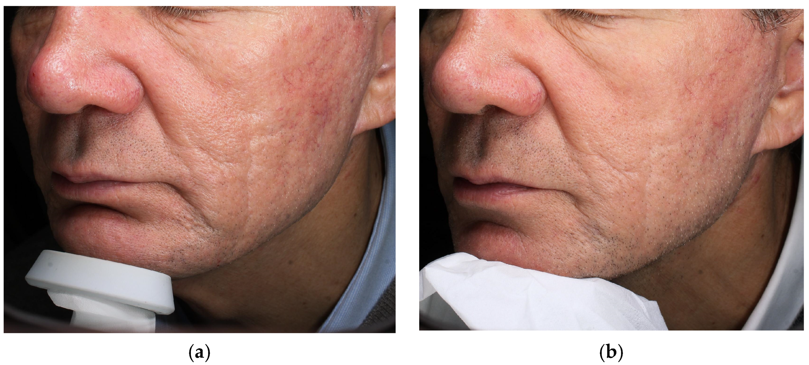
A 62-year-old male patient presented with multiple atrophic acne scars on his face, mainly located on the cheeks (Figure 1 and Figure 2). A LAED protocol was proposed to take advantage of the greater efficacy of fractional ablative CO2 combined with the regenerative effect of exosomes. The treatment consisted of two sessions, four weeks apart. In each session, after application of topical anesthetic cream, an ablative fractional CO2 laser treatment (SmartXide Tetra PRO, DEKA M.E.L.A Srl, Florence, Italy), equipped with a dedicated scanner system (DOT PRO scanner, DEKA M.E.L.A, Florence, Italy) was performed on the entire face. The parameters used were as follows: power 14 W, dwell time 800 µs, spacing 550 µm, Smart Pulse mode. Immediately after the laser treatment, the area was cleansed with saline solution and then covered with Exolight C.
At day 7, the patient rated the speed and comfort of recovery with a score of 5/5 on a 5-point Likert scale. Clinically post-laser erythema and edema resolved within 5–6 days (compared to 8–12 days typical for fractional CO2 at those settings), with minimal desquamation.
After the second session, at 8 weeks, clinical evaluation showed a marked reduction in the depth and visibility of acne scars, with improvement in the surrounding skin texture (Figure 1 and Figure 2). Pre- and post-treatment comparison photos confirmed a clear smoothing of the scars. The patient tolerated the procedures well, with no infections or post-inflammatory hyperpigmentation (PIH). Patient-reported recovery was favorable; even if Future studies should incorporate validated PROMs, e.g., FACE-Q, and blinded grading. In summary, the addition of exosomes may have contributed to improved clinical outcomes and appeared to shorten healing times.

4.2. Case 2—Diffuse Hyperpigmentation of the Face

A 68-year-old female patient presented with diffuse hyperchromic spots on her face (senile lentigines and photoaging-induced dyschromic pigmentation), associated with uneven skin texture and fine roughness (Figure 3).
A single treatment was chosen with ablative fractional CO2 laser (SmartXide Tetra PRO, DEKA M.E.L.A Srl, Florence, Italy) and topical exosomes with depigmenting activity (Exolight M).
In particular, the CoolPeel mode (DEKA M.E.L.A Srl, Florence, Italy) of the CO2 laser was used to perform light resurfacing focused on the hyperpigmented areas, while minimizing downtime (power 5 W, spacing 500 µm, High pulse mode). Immediately after the laser treatment, a layer of Exolight M was applied to the face.
The patient experienced only mild erythema and fine exfoliation in the following two days, allowing her to resume normal social activities with no erythema, as early as the third day after treatment (downtime significantly lower than her previous experiences with fractional lasers).
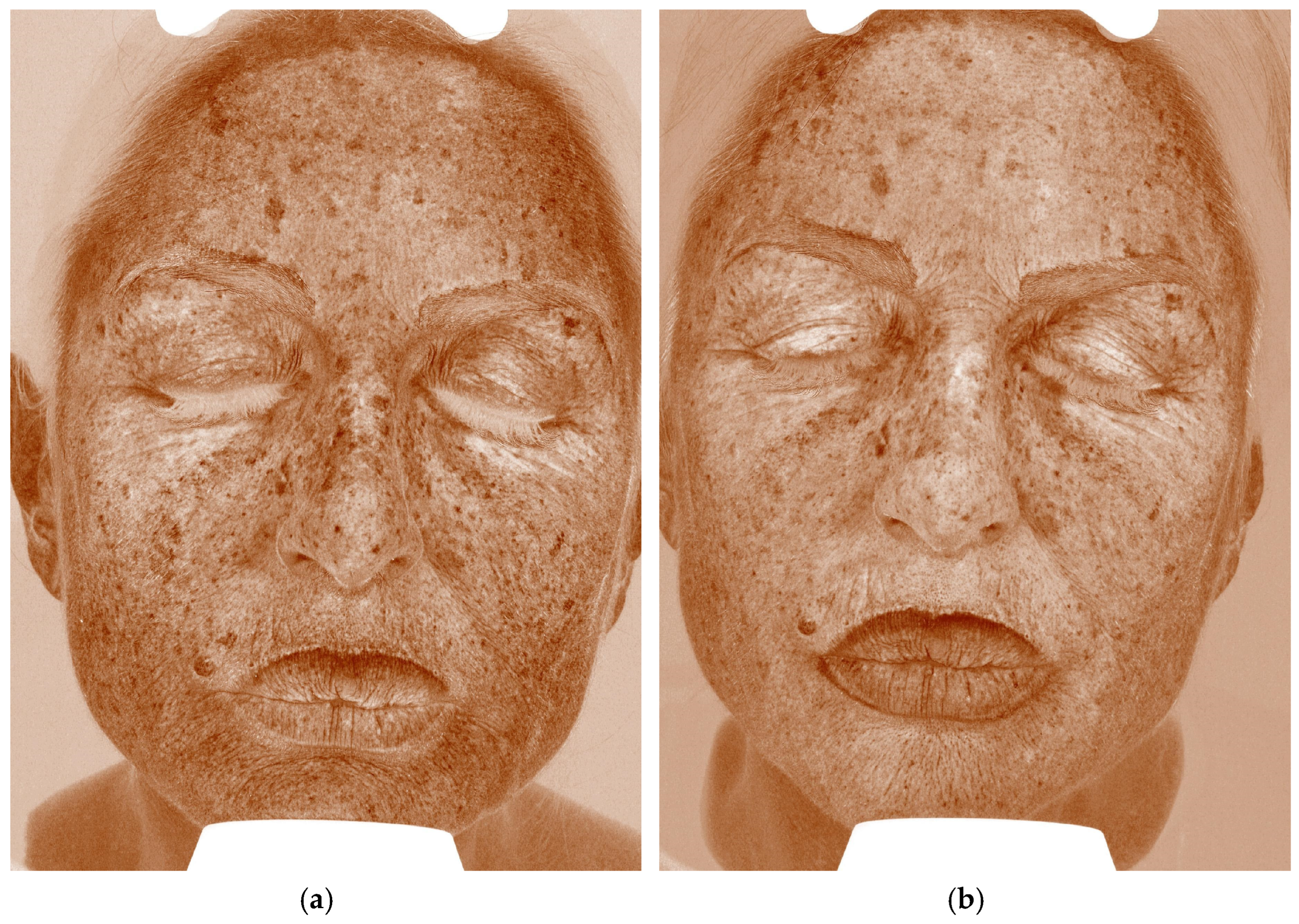
Four weeks after the session, clinical and instrumental examination showed an overall reduction in hyperpigmentation of approximately 30% compared to the baseline (Figure 3). Digital analysis using UV photography confirmed the decrease in pigment index in the treated areas (Figure 4).
The patient reported a subjective improvement in skin brightness and evenness, consistent with the attending physician’s clinical assessment and confirmed by comparative photographic documentation. At day 7, the patient rated the speed and comfort of recovery as 5/5 on the 5-point Likert scale. While these observations are encouraging, they are based on non-standardized assessments. Future studies should integrate validated PROMs, such as the FACE-Q, along with objective assessment tools to provide reproducible and comparable evidence. No complications were observed in this case.

5. Laser-Assisted Exosome Delivery (LAED)

Laser-Assisted Exosome Delivery (LAED) is a new therapeutic approach that delivers exosomes through laser-generated microchannels in the stratum corneum.
This method is based on the same principles as Laser-Assisted Drug Delivery (LADD), previously discussed, exploiting the formation of microablative zones (MAZs) in the stratum corneum to facilitate the penetration of active molecules into deep skin tissues. The use of fractional CO2 lasers allows the creation of epidermal and dermal microchannels through which exosomes can be more effectively assimilated by fibroblasts, keratinocytes and melanocytes. Thanks to their bioactive properties, exosomes contribute to modulating the post-procedural inflammatory response, stimulate regenerative processes and improve the overall safety profile, reducing recovery times.
The two clinical cases presented in this study offer preliminary clinical observations consistent with potential benefits of LAED. In both patients, topical application of exosomes immediately after fractional CO2 treatment appeared to result in faster and more marked improvement than typically expected based on laser use alone; however, these signals are hypothesis-generating and non-confirmatory.
Recent studies have further explored the integration of exosomes with laser technologies.
A significant contribution to the clinical validation of LAED comes from the randomized, double-blind, split-face study conducted by Kwon et al. on 25 patients with atrophic acne scars. Patients were treated with three sessions of fractional CO2 laser on the entire face, immediately followed by the application of a gel containing adipose stem cell-derived exosomes (ASCE) or a placebo gel on each half. Efficacy was assessed using the ECCA (Échelle d’Évaluation Clinique des Cicatrices d’Acné) score and standardized imaging assessment at 12 weeks. The side treated with exosomes showed a significantly greater reduction in total ECCA score than the control side (32.5% vs. 19.9%; p < 0.01), with superior results also for the icepick, boxcar and rolling acne scar subtypes. In addition, patients reported a shorter subjective recovery time (mean 4.1 days vs. 4.3; p = 0.03) and less post-treatment erythema. These data confirm the clinical efficacy of the combination of fractional CO2 and exosomes in improving not only the quality of dermal regeneration but also the tolerability of the treatment [71].
Further interesting evidence is provided by the preclinical study conducted by Fusco et al., which evaluated the efficacy of bovine-derived Exolight exosomes as adjuvants to fractional CO2 laser treatment. This study included both in vitro analyses and in vivo preclinical models, documenting anti-melanogenic, pro-collagenic and anti-inflammatory activity. In particular, combined use with CO2 laser showed a significant reduction in post-treatment healing time, associated with faster crust detachment and less residual vascularization in the first few days after the procedure, as evidenced by high-resolution OCT imaging. The 3D and cross-sectional images obtained with Optical Coherence Tomography (OCT) have documented a faster resolution of erythema and early normalization of the vascular network, confirming the anti-inflammatory effect of exosomes in the context of post-laser inflammation [72]. These results highlight how Laser-Assisted Exosome Delivery (LAED) can not only enhance clinical efficacy but also reduce healing time, improving patient quality of life. Faster recovery allows for a quicker return to daily and social activities—an increasingly relevant factor in a society where visual appearance, especially on social media, plays a major role. This is particularly significant among younger individuals, who use platforms like Instagram and TikTok as primary sources of skin health content [73].
Lueangarun et al. described a clinical case of pilary repigmentation in a patient with androgenetic alopecia with circumscribed poliosis. The subject was treated with fractional 1064 nm Nd:YAG (Neodymium-doped Yttrium Aluminum Garnet) picosecond laser combined with topical application of exosomes. The protocol led to a clear improvement in both hair density and pigmentation of the depigmented hair tuft, with excellent tolerability and no significant side effects. These data suggest a dual effect of exosomes in the trichological field, acting both on follicular regeneration and on the modulation of the epithelial pigmentary unit, thanks to paracrine communication mediated by miRNAs contained in the exosomes themselves [40].
Finally, Mirzadeh et al. presented a series of cases in which patients with androgenetic alopecia, unresponsive to conventional treatments (platelet-rich plasma, minoxidil, mesotherapy), were treated with a combination of Low-Level Laser Therapy (LLLT) and autologous exosomes obtained from adipose MSCs. The clinical results, observed after two sessions, documented an improvement in hair density and thickness, with a synergistic effect attributed to the interaction between laser stimulation and the regenerative potential of exosomes. The effect was also attributed to the modulation of intracellular pathways such as Transforming Growth Factor-beta (TGF-β), Nuclear Factor kappa-B (NF-κB) and/or Hypoxia-Inducible Factor-1 (HIF-1) and to the increase in mitochondrial ATP production induced by laser [74].
These initial observations suggest the strong biological rationale and clinical potential of LAED and open broader application scenarios. The synergistic combination of laser and exosomes could be used in various dermatological indications. For example, in the treatment of striae distensae, the addition of pro-collagenic exosomes could improve the quality of dermal regeneration.
Another potential application is the management of skin discoloration using fractional picosecond lasers combined with depigmenting exosomes.
Furthermore, it remains an area of active investigation whether exosomes could be effectively combined with other laser sources or light-based devices, such as dye lasers or intense pulsed light systems, to expand therapeutic possibilities [75].
LAED also raises open questions. The regulation of exosomes, currently situated in a regulatory gray zone between cosmetic products, biological agents and medical devices, will require clearer legal definitions depending on their origin, processing, and intended use. Critical aspects such as standardization of production, sterilization and storage must be strictly controlled [76].

6. Limitations

We acknowledge that this work represents an exploratory proof-of-concept rather than a definitive clinical study, and several factors limit the robustness of our conclusions.
The evidence comes from only two uncontrolled cases, which, while useful for illustrating feasibility and providing preliminary clinical documentation, do not allow us to quantify the true effect size or separate the potential contribution of exosomes from the natural course of wound healing.
Furthermore, outcomes were assessed primarily through clinical photographs, physician judgment, and patient satisfaction. While these approaches reflect daily practice, they lack the rigor of blinded assessment and standardized outcome measures. Future studies should therefore include validated.
Patient-reported questionnaires, such as the FACE-Q, along with objective quantifications such as the ECCA score, Mexameter indices, or advanced imaging methods (OCT, high-frequency ultrasound) to ensure reproducibility and comparability. Another limitation is the short follow-up, limited to 4–8 weeks, which is sufficient to document early re-epithelialization but not long-term pigment stability, collagen remodeling, or delayed adverse events. Further observation will be needed to establish both durability and safety. Although quality control parameters for exosome preparations have been reported, important aspects such as batch-to-batch consistency, post-thaw stability, and dose–response relationships have not been studied, limiting reproducibility between centers. Finally, both patients were older adults with lighter skin types, and the results cannot be generalized to other age groups, skin types, or comorbid conditions.
Overall, these limitations indicate that this report should be interpreted only as a preliminary feasibility study, primarily documenting the tolerability of combining fractional CO2 laser with topical exosomes. The hypothesis that LAED can improve clinical outcomes or reduce recovery times remains a hypothesis and cannot be confirmed based on this work. Moreover, the literature is currently limited; additional well-designed studies are needed. Explicitly acknowledging these limitations, our intention is to stimulate and guide the design of larger, controlled clinical trials with standardized outcome measures and longer follow-ups, which will be essential to establish whether LAED can evolve into a reliable, evidence-based therapeutic strategy in regenerative dermatology.

7. Future Perspectives and Conclusions

Laser-assisted exosome delivery (LAED) is a promising emerging approach in esthetic and regenerative dermatology, but current evidence remains preliminary. The two cases presented suggest that the combination of fractional CO2 laser and topical exosomes is technically feasible and appears well-tolerated, with encouraging early signs of clinical improvement. However, these observations cannot establish efficacy and should be considered only as hypothesis-generating.
Before LAED can be considered a reliable and reproducible therapeutic strategy, several challenges must be addressed. Standardized protocols are needed to clarify the optimal parameters for each indication, including laser settings, exosome source and concentration, number and frequency of sessions, timing and method of application, and most importantly, the tools used to assess outcomes. Future studies should employ validated patient-reported questionnaires and objective quantitative measures to ensure reproducibility and comparability across centers. Long-term safety also requires systematic investigation to rule out late-onset adverse events and confirm the durability of results. Equally important is the standardization of exosome manufacturing, sterilization, storage, and quality control procedures, along with a clearer regulatory framework defining their classification and use in clinical practice. Advances in these areas will enable the design of well-controlled clinical trials on larger populations, capable of quantifying the true contribution of exosomes beyond conventional laser treatment and testing the underlying biological rationale with statistical validity.
Only through such a rigorous and collaborative scientific approach will it be possible to determine whether LAED can evolve from a proof-of-concept proposal to an evidence-based therapeutic option, supporting its safe and effective integration into modern dermatology. Until such evidence is available, LAED, while promising, should still be considered experimental.

Author Contributions

Conceptualization, validation, writing—review and editing: A.C., F.C. and S.P.N.; methodology, investigation, data curation: A.C., F.C., G.C. and S.P.N.; writing—original draft preparation: A.C. and F.C.; visualization, supervision: S.P.N., G.C., C.L., E.Z., R.M., A.D., L.G. and L.B. All authors have read and agreed to the published version of the manuscript.

Funding

This research received no external funding.

Institutional Review Board Statement

The study was conducted in accordance with the Declaration of Helsinki and approved by the Local Ethical Committee of Calabria Region (protocol code 374; 17 December 2019).

Informed Consent Statement

Informed consent was obtained from all subjects involved in the study.

Data Availability Statement

The data supporting the findings of this study are available within the article.

Conflicts of Interest

The authors declare no conflicts of interest.

Abbreviations

ASCEAdipose Stem Cell–derived Exosomes
ATPAdenosine Triphosphate
CO2Carbon Dioxide
ECCAÉchelle d’Évaluation Clinique des Cicatrices d’Acné
Er:YAGErbium-doped Yttrium Aluminum Garnet
EUEndotoxin Unit(s)
FACE-QFacial Aesthetic Clinical Evaluation Questionnaire
HIF-1Hypoxia-Inducible Factor-1
ISOInternational Organization for Standardization
LADDLaser-Assisted Drug Delivery
LAEDLaser-Assisted Exosome Delivery
LALLimulus Amebocyte Lysate (assay)
LLLTLow-Level Laser Therapy
MAZ(s)Microablative Zone(s)
MSC(s)Mesenchymal Stem Cell(s)
Nd:YAGNeodymium-doped Yttrium Aluminum Garnet
NF-κBNuclear Factor kappa-B
OCTOptical Coherence Tomography
PIHPost-Inflammatory Hyperpigmentation
PRPPlatelet-Rich Plasma
PRO(s)Patient-Reported Outcome(s)
PROM(s)Patient-Reported Outcome Measure(s)
SPFSun Protection Factor
TNF-αTumor Necrosis Factor-alpha
UVUltraviolet
UVBUltraviolet B
VISIA®VISIA® Complexion Analysis System

References

  1. Goel, A.; Krupashankar, D.; Aurangabadkar, S.; Nischal, K.; Omprakash, H.; Mysore, V. Fractional lasers in dermatology—Current status and recommendations. Indian J. Dermatol. Venereol. Leprol. 2011, 77, 369. [Google Scholar] [CrossRef]
  2. Cohen, S.R.; Goodacre, A.; Lim, S.; Johnston, J.; Henssler, C.; Jeffers, B.; Saad, A.; Leong, T. Clinical outcomes and complications associated with fractional lasers: A review of 730 patients. Aesthetic Plast. Surg. 2017, 41, 171–178. [Google Scholar] [CrossRef]
  3. Jih, M.H.; Kimyai-Asadi, A. Fractional photothermolysis: A review and update. Semin. Cutan. Med. Surg. 2008, 27, 63–71. [Google Scholar] [CrossRef]
  4. Bodendorf, M.O.; Grunewald, S.; Wetzig, T.; Simon, J.C.; Paasch, U. Fractional laser skin therapy. JDDG J. Ger. Soc. Dermatol. 2009, 7, 301–308. [Google Scholar] [CrossRef]
  5. Yang, Y.J.; Lee, G.Y. Treatment of striae distensae with nonablative fractional laser versus ablative CO2 fractional laser: A randomized controlled trial. Ann. Dermatol. 2011, 23, 481–489. [Google Scholar] [CrossRef] [PubMed]
  6. Clementi, A.; Cannarozzo, G.; Guarino, L.; Zappia, E.; Cassalia, F.; Alma, A.; Sannino, M.; Longo, C.; Nisticò, S.P. Sequential fractional CO2 and 1540/1570 nm lasers: A narrative review of preclinical and clinical evidence. J. Clin. Med. 2025, 14, 3867. [Google Scholar] [CrossRef]
  7. Campolmi, P.; Cannarozzo, G.; Bennardo, L.; Clementi, A.; Sannino, M.; Nisticò, S.P. Fractional micro-ablative CO2 laser as therapy in penile lichen sclerosus. J. Lasers Med. Sci. 2021, 12, e61. [Google Scholar] [CrossRef]
  8. Ong, M.W.S.; Bashir, S.J. Fractional laser resurfacing for acne scars: A review. Br. J. Dermatol. 2012, 166, 1160–1169. [Google Scholar] [CrossRef] [PubMed]
  9. Borges, J.; Manela-Azulay, M.; Cuzzi, T. Photoaging and the clinical utility of fractional laser. Clin. Cosmet. Investig. Dermatol. 2016, 9, 107–114. [Google Scholar] [CrossRef] [PubMed]
  10. Amato, S.; Nisticò, S.P.; Clementi, A.; Stabile, G.; Cassalia, F.; Dattola, A.; Rizzuto, G.; Cannarozzo, G. Multispectral imaging and OCT-guided precision treatment of rhinophyma with CO2 and dye lasers: A comprehensive diagnostic and therapeutic approach. Cosmetics 2024, 11, 221. [Google Scholar] [CrossRef]
  11. Kołodziejczyk, K.; Hop, I.; Branewska, J.; Ostrowska, B.; Matysek, M.; Olszanicka, A.; Imioło, J.; Maciąg, A.; Niemiec, R.; Galas, A. CO2 ablative fractional laser—Mechanism of action and assessment of safety, effectiveness and possible side effects: Literature review. J. Educ. Health Sport 2023, 21, 72–77. [Google Scholar] [CrossRef]
  12. Nisticò, S.P.; Bennardo, L.; Zingoni, T.; Pieri, L.; Fusco, I.; Rossi, F.; Magni, G.; Cannarozzo, G. Synergistic sequential emission of fractional 10,600 and 1540 nm lasers for skin resurfacing: An ex vivo histological evaluation. Medicina 2022, 58, 1308. [Google Scholar] [CrossRef]
  13. Berlin, A.L.; Hussain, M.; Phelps, R.; Goldberg, D.J. A prospective study of fractional scanned nonsequential carbon dioxide laser resurfacing: Clinical and histopathologic evaluation. Dermatol. Surg. 2009, 35, 222–228. [Google Scholar] [CrossRef] [PubMed]
  14. Fife, D.J.; Fitzpatrick, R.E.; Zachary, C.B. Complications of fractional CO2 laser resurfacing: Four cases. Lasers Surg. Med. 2009, 41, 179–184. [Google Scholar] [CrossRef]
  15. Kaur, J.; Sharma, S.; Kaur, T.; Bassi, R. Complications of fractional ablative carbon dioxide laser in various aesthetic procedures: A retrospective study. Int. J. Res. Dermatol. 2019, 5, 664–667. [Google Scholar] [CrossRef]
  16. Ramsdell, W.M. Fractional CO2 laser resurfacing complications. Semin. Plast. Surg. 2012, 26, 137–140. [Google Scholar] [CrossRef] [PubMed]
  17. Lodi, G.; Cassalia, F.; Sannino, M.; Cannarozzo, G.; Baroni, A.; Amato, S.; Zappia, E.; Pellacani, G.; Nisticò, S.P. Blue light therapy in dermatological practice: A review. Cosmetics 2025, 12, 30. [Google Scholar] [CrossRef]
  18. Kalluri, R.; LeBleu, V.S. The biology, function, and biomedical applications of exosomes. Science 2020, 367, eaau6977. [Google Scholar] [CrossRef]
  19. Gurunathan, S.; Kang, M.H.; Jeyaraj, M.; Qasim, M.; Kim, J.H. Review of the isolation, characterization, biological function, and multifarious therapeutic approaches of exosomes. Cells 2019, 8, 307. [Google Scholar] [CrossRef]
  20. Yousefian, F.; Espinoza, L.; Yadlapati, S.; Lorenc, Z.P.; Gold, M. A comprehensive review of the medical and cosmetic applications of exosomes in dermatology. J. Cosmet. Dermatol. 2024, 23, 1224–1228. [Google Scholar] [CrossRef]
  21. Xiong, M.; Zhang, Q.; Hu, W.; Zhao, C.; Lv, W.; Yi, Y.; Wang, Y.; Tang, H.; Wu, M.; Wu, Y. The novel mechanisms and applications of exosomes in dermatology and cutaneous medical aesthetics. Pharmacol. Res. 2021, 166, 105490. [Google Scholar] [CrossRef]
  22. Basu, J.; Ludlow, J.W. Exosomes for repair, regeneration and rejuvenation. Expert. Opin. Biol. Ther. 2016, 16, 489–506. [Google Scholar] [CrossRef]
  23. Kim, Y.J.; Yoo, S.-M.; Park, H.H.; Lim, H.J.; Kim, Y.L.; Lee, S.; Seo, K.W.; Kang, K.S. Exosomes derived from human umbilical cord blood mesenchymal stem cells stimulate rejuvenation of human skin. Biochem. Biophys. Res. Commun. 2017, 493, 1102–1108. [Google Scholar] [CrossRef]
  24. Qiu, H.; Liu, S.; Wu, K.; Zhao, R.; Cao, L.; Wang, H. Prospective application of exosomes derived from adipose-derived stem cells in skin wound healing: A review. J. Cosmet. Dermatol. 2020, 19, 574–581. [Google Scholar] [CrossRef]
  25. Novis, T.; Gomes Menezes, A.H.; Vilaça Lima, L.C.; Lötvall, J.; Borges do Nascimento, I.J.; Maeda Takiya, C. The therapeutic, diagnostic, and prognostic values of extracellular vesicles (exosomes) in dermatology: A systematic review. JAAD Rev. 2024, 1, 135–174. [Google Scholar] [CrossRef]
  26. Semsarzadeh, N.; Andrasik, W.; Khetarpal, S. Stem cells and exosomes in aesthetic medicine. Adv. Cosmet. Surg. 2021, 4, 59–70. [Google Scholar] [CrossRef]
  27. Pinto, H.; Sánchez-Vizcaíno Mengual, E. Exosomes in the real world of medical aesthetics: A review. Aesthetic Plast. Surg. 2024, 48, 2513–2527. [Google Scholar] [CrossRef] [PubMed]
  28. Thakur, A.; Shah, D.; Rai, D.; Parra, D.C.; Pathikonda, S.; Kurilova, S.; Cili, A. Therapeutic values of exosomes in cosmetics, skin care, tissue regeneration, and dermatological diseases. Cosmetics 2023, 10, 65. [Google Scholar] [CrossRef]
  29. Park, G.H.; Kwon, H.H.; Seok, J.; Yang, S.H.; Lee, J.; Park, B.C.; Shin, E.; Park, K.Y. Combined treatment with human adipose tissue stem cell-derived exosome solution and microneedling for facial skin aging: 12-week prospective, randomized, split-face study. J. Cosmet. Dermatol. 2023, 22, 3418–3426. [Google Scholar] [CrossRef]
  30. Zhang, B.; Gong, J.; He, L.; Khan, A.; Xiong, T.; Shen, H.; Li, Z. Exosomes-based advancements for application in medical aesthetics. Front. Bioeng. Biotechnol. 2022, 10, 1083640. [Google Scholar] [CrossRef] [PubMed]
  31. Ku, Y.C.; Omer Sulaiman, H.; Anderson, S.R.; Abtahi, A.R. The potential role of exosomes in aesthetic plastic surgery: A review of current literature. Plast. Reconstr. Surg. Glob. Open 2023, 11, e5051. [Google Scholar] [CrossRef]
  32. Yang, G.H.; Lee, Y.B.; Kang, D.; Choi, E.; Nam, Y.; Lee, K.H.; You, H.J.; Kang, H.J.; An, S.H.; Jeon, H. Overcome the barriers of the skin: Exosome therapy. Biomater. Res. 2021, 25, 22. [Google Scholar] [CrossRef]
  33. Zhou, C.; Zhang, B.; Yang, Y.; Jiang, Q.; Li, T.; Gong, J.; Tang, H.; Zhang, Q. Stem cell-derived exosomes: Emerging therapeutic opportunities for wound healing. Stem Cell Res. Ther. 2023, 14, 107. [Google Scholar] [CrossRef]
  34. Li, M.; Wang, T.; Tian, H.; Wei, G.; Zhao, L.; Shi, Y. Macrophage-derived exosomes accelerate wound healing via anti-inflammation in diabetic rats. Artif. Cells Nanomed. Biotechnol. 2019, 47, 3793–3803. [Google Scholar] [CrossRef] [PubMed]
  35. Bian, D.; Wu, Y.; Song, G.; Azizi, R.; Zamani, A. Mesenchymal stromal cells and their exosomes in skin wound healing: A comprehensive review. Stem Cell Res. Ther. 2022, 13, 14. [Google Scholar] [CrossRef] [PubMed]
  36. Rani, S.; Ritter, T. The exosome—A naturally secreted nanoparticle—And its application to wound healing. Adv. Mater. 2016, 28, 5542–5552. [Google Scholar] [CrossRef] [PubMed]
  37. Lei, L.; Zhou, S.; Zeng, L.; Gu, Q.; Xue, H.; Wang, F.; Feng, J.; Cui, S.; Shi, L. Exosome-based therapeutics in dermatology. Biomater. Res. 2025, 29, 0148. [Google Scholar] [CrossRef]
  38. Javeed, N.; Mukhopadhyay, D. Exosomes and their role in the micro/macro-environment: A comprehensive review. J. Biomed. Res. 2017, 31, 386–394. [Google Scholar] [CrossRef]
  39. Ersan, M.; Ozer, E.; Akin, O.; Tasli, P.N.; Sahin, F. Effectiveness of exosome treatment in androgenetic alopecia: Outcomes of a prospective study. Aesthetic Plast. Surg. 2024, 48, 4262–4271. [Google Scholar] [CrossRef]
  40. Lueangarun, S.; Cho, B.S.; Tempark, T. Hair repigmentation of poliosis circumscripta in androgenetic alopecia treated with exosomes and fractional picosecond laser. J. Cosmet. Dermatol. 2024, 23, 2307–2311. [Google Scholar] [CrossRef]
  41. Proietti, I.; Battilotti, C.; Svara, F.; Innocenzi, C.; Spagnoli, A.; Potenza, C. Microneedling plus exosomes for melasma: Efficacy and tolerability. Appl. Sci. 2024, 14, 7252. [Google Scholar] [CrossRef]
  42. Wang, T.; Gao, H.; Wang, D.; Zhang, C.; Hu, K.; Zhang, H.; Lin, J.; Chen, X. Stem cell-derived exosomes in melasma treatment and percutaneous penetration. Lasers Surg. Med. 2023, 55, 178–189. [Google Scholar] [CrossRef]
  43. Wenande, E.; Anderson, R.R.; Haedersdal, M. Fundamentals of fractional laser-assisted drug delivery: Methodology and data interpretation. Adv. Drug Deliv. Rev. 2020, 153, 169–184. [Google Scholar] [CrossRef]
  44. Sklar, L.R.; Burnett, C.T.; Waibel, J.S.; Moy, R.L.; Ozog, D.M. Laser-assisted drug delivery: A review of an evolving technology. Lasers Surg. Med. 2014, 46, 249–262. [Google Scholar] [CrossRef]
  45. Omi, T.; Numano, K. The role of the CO2 laser and fractional CO2 laser in dermatology. Laser Ther. 2014, 23, 49–60. [Google Scholar] [CrossRef] [PubMed]
  46. Hædersdal, M.; Sakamoto, F.H.; Farinelli, W.A.; Doukas, A.G.; Tam, J.; Anderson, R.R. Fractional CO2 laser-assisted drug delivery. Lasers Surg. Med. 2010, 42, 113–122. [Google Scholar] [CrossRef] [PubMed]
  47. Machado, B.H.B.; Zhang, J.; Frame, J.; Najlah, M. Laser-assisted delivery of growth factors and vitamin C for scars: Randomised double-blind early clinical trial. Aesthetic Plast. Surg. 2021, 45, 2363–2374. [Google Scholar] [CrossRef] [PubMed]
  48. Waibel, J.S.; Wulkan, A.J.; Rudnick, A.; Daoud, A. Hypertrophic scars: Laser-assisted corticosteroid vs 5-fluorouracil delivery. Dermatol. Surg. 2019, 45, 423–430. [Google Scholar] [CrossRef]
  49. Truong, K.; Prasidha, I.; Wain, T. Laser-assisted drug delivery for keloid/hypertrophic scars: Systematic review of RCTs. Lasers Med. Sci. 2022, 37, 47–59. [Google Scholar] [CrossRef]
  50. Steeb, T.; Schlager, J.G.; Kohl, C.; Ruzicka, T.; Heppt, M.V.; Berking, C. Laser-assisted photodynamic therapy for actinic keratosis: Systematic review and meta-analysis. J. Am. Acad. Dermatol. 2019, 80, 947–956. [Google Scholar] [CrossRef]
  51. Haykal, D.; Cartier, H.; Goldberg, D.; Gold, M. Advancements in laser technologies for skin rejuvenation: Efficacy and safety. J. Cosmet. Dermatol. 2024, 23, 3078–3089. [Google Scholar] [CrossRef]
  52. Khedr, M.M.; Elhefnawy, A.A.; Mahmoud, W.H.; Mostafa, I.M.E.; Hantash, S.A. Laser-assisted delivery of tranexamic acid for facial post-burn hyperpigmentation. Eur. J. Plast. Surg. 2023, 46, 1349–1356. [Google Scholar] [CrossRef]
  53. Wenande, E.; Olesen, U.H.; Nielsen, M.M.B.; Janfelt, C.; Hansen, S.H.; Anderson, R.R.; Haedersdal, M. Fractional laser-assisted topical delivery enhances cutaneous 5-fluorouracil uptake. Expert Opin. Drug Deliv. 2017, 14, 307–317. [Google Scholar] [CrossRef]
  54. Meesters, A.A.; Nieboer, M.J.; Kezic, S.; de Rie, M.A.; Wolkerstorfer, A. Parameters in fractional laser-assisted delivery of topical anesthetics: Role of laser type/settings. Lasers Surg. Med. 2018, 50, 813–818. [Google Scholar] [CrossRef] [PubMed]
  55. Cameli, N.; Abril, E.; Mariano, M.; Berardesca, E. Monopolar radiofrequency and transdermal drug delivery in melasma. Dermatol. Surg. 2014, 40, 748–755. [Google Scholar] [CrossRef] [PubMed]
  56. Nagarkar, R.; Singh, M.; Nguyen, H.X.; Jonnalagadda, S. Advances in microneedle technology for transdermal drug delivery. J. Drug Deliv. Sci. Technol. 2020, 59, 101923. [Google Scholar] [CrossRef]
  57. Muskat, A.; Kost, Y.; Balazic, E.; Cohen, J.L.; Kobets, K. Laser-assisted drug delivery in scars, rhytids, and melasma: Literature review. Aesthet. Surg. J. 2023, 43, NP181–NP198. [Google Scholar] [CrossRef]
  58. Ibrahim, O.; Wenande, E.; Hogan, S.; Arndt, K.A.; Haedersdal, M.; Dover, J.S. Challenges to laser-assisted drug delivery: From theory to clinical practice. Lasers Surg. Med. 2018, 50, 20–27. [Google Scholar] [CrossRef]
  59. Searle, T.; Ali, F.R.; Al-Niaimi, F. Lessons learned from the first decade of laser-assisted drug delivery. Dermatol. Ther. Heidelb. 2021, 11, 93–104. [Google Scholar] [CrossRef]
  60. Théry, C.; Zitvogel, L.; Amigorena, S. Exosomes: Composition, biogenesis and function. Nat. Rev. Immunol. 2002, 2, 569–579. [Google Scholar] [CrossRef]
  61. Pegtel, D.M.; Gould, S.J. Exosomes. Annu. Rev. Biochem. 2019, 88, 487–514. [Google Scholar] [CrossRef] [PubMed]
  62. Shen, X.; Song, S.; Chen, N.; Liao, J.; Zeng, L. Stem cell-derived exosomes: A supernova in cosmetic dermatology. J. Cosmet. Dermatol. 2021, 20, 3812–3817. [Google Scholar] [CrossRef]
  63. Dal’Forno-Dini, T.; Birck, M.S.; Rocha, M.; Bagatin, E. Exploring the reality of exosomes in dermatology. An. Bras. Dermatol. 2025, 100, 121–130. [Google Scholar] [CrossRef]
  64. McBride, J.D.; Rodriguez-Menocal, L.; Badiavas, E.V. Extracellular vesicles as biomarkers and therapeutics in dermatology: A focus on exosomes. J. Invest. Dermatol. 2017, 137, 1622–1629. [Google Scholar] [CrossRef]
  65. Bin Dayel, S.; Hussein, R.S. Exosomes in dermatology: Emerging roles in skin health and disease. Pharmaceutics 2025, 17, 600. [Google Scholar] [CrossRef]
  66. Kose, O.; Botsali, A.; Caliskan, E. Role of exosomes in skin diseases. J. Cosmet. Dermatol. 2022, 21, 3219–3225. [Google Scholar] [CrossRef] [PubMed]
  67. Nahm, W.J.; Nikas, C.; Goldust, M.; Horneck, N.; Cervantes, J.A.; Burshtein, J.; Tsoukas, M. Exosomes in dermatology: Current applications, clinical evidence, and future directions. Int. J. Dermatol. 2025; Online ahead of print. [Google Scholar] [CrossRef]
  68. Quiñones-Vico, M.I.; Sanabria-de la Torre, R.; Sánchez-Díaz, M.; Sierra-Sánchez, Á.; Montero-Vílchez, T.; Fernández-González, A.; Arias-Santiago, S. Exosomes derived from mesenchymal stromal cells in dermatology. Front. Cell Dev. Biol. 2021, 9, 647012. [Google Scholar] [CrossRef]
  69. Adriano, B.; Cotto, N.M.; Chauhan, N.; Jaggi, M.; Chauhan, S.C.; Yallapu, M.M. Milk exosomes: Nature’s abundant nanoplatform for theranostics. Bioact. Mater. 2021, 6, 2479–2490. [Google Scholar] [CrossRef]
  70. DEKA M.E.L.A. Exolight C & M; DEKA M.E.L.A.: Florence, Italy, 2025; Available online: https://www.dekalaser.com (accessed on 9 September 2025).
  71. Kwon, H.H.; Yang, S.H.; Lee, J.; Park, B.C.; Park, K.Y.; Jung, J.Y.; Bae, Y.; Park, G.H. Adipose-derived exosomes plus fractional CO2 laser for acne scars: A 12-week double-blind split-face RCT. Acta Derm. Venereol. 2020, 100, adv00147. [Google Scholar] [CrossRef] [PubMed]
  72. Fusco, I.; Zingoni, T.; Madeddu, F.; Colombo, C.; Amato, S.; Greco, M.E.; Rizzuto, G.; Zappia, E.; Bonan, P.; Nisticò, S. Preclinical results on Exolight exosomes as adjuvant to CO2 laser for skin aging management. JOJ Dermatol. Cosmet. 2025, 6, 555694. [Google Scholar] [CrossRef]
  73. Cassalia, F.; Ciolfi, C.; Scolaro, F.; Danese, A.; Lunardon, A.; Caroppo, F.; Belloni Fortina, A. Public use of social media on skin health: Effects on awareness and preventive behaviours. Acta Derm. Venereol. 2023, 103, adv15341. [Google Scholar] [CrossRef]
  74. Mirzadeh, S.; Hashesmi, T.; Jafari, A.M.; Hamidnia, L.; Ansari, A.; Ayazi, M.; Harandi, N.T.; Mehran, Y.Z. Low-level laser therapy plus autologous exosomes in hair growth: Case series. Sch. J. Med. Case Rep. 2024, 12, 1250–1256. [Google Scholar] [CrossRef]
  75. Clementi, A.; Cannarozzo, G.; Amato, S.; Zappia, E.; Bennardo, L.; Michelini, S.; Morini, C.; Sannino, M.; Longo, C.; Nisticò, S.P. Dye laser applications in cosmetic dermatology: Efficacy and safety for vascular lesions and scars. Cosmetics 2024, 11, 227. [Google Scholar] [CrossRef]
  76. Wang, C.K.; Tsai, T.H.; Lee, C.H. Regulation of exosomes as biologic medicines: Development and manufacturing challenges. Clin. Transl. Sci. 2024, 17, e13904. [Google Scholar] [CrossRef]
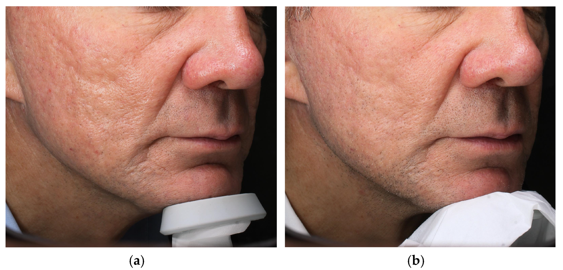
Figure 1. (a) Right side of the face before treatment and (b) four weeks after the last LAED session.
Cosmetics 12 00199 g001
Figure 2. (a) Left side of the face before treatment and (b) four weeks after the last LAED session.
Cosmetics 12 00199 g002
Figure 3. (a) Frontal clinical image before treatment and (b) four weeks after the LAED session.
Cosmetics 12 00199 g003
Figure 4. (a) Pre-treatment UV imaging acquired via the VISIA® system and (b) four weeks after the LAED session.
Cosmetics 12 00199 g004
Table 1. Cases description.
CaseAge/SexIndicationLaser—Mode and ParametersExosome SerumDowntime *Clinical
Improvement
Patient Self-Evaluation (Day 7) Adverse Events
162 MAtrophic acne scarsFractional CO2, Smart-Pulse 14 W · dwell 800 µs · spacing 550 µm (2 sessions, 4 weeks apart)Exolight C (pro-collagenic)5–6 days (erythema + edema)Visible scar depth/texture less marked (clinician estimate)5/5None
268 FPhoto-aging dyschromia Fractional CO2, CoolPeel 5 W · spacing 500 µm · High-Pulse (single session)Exolight M (depigmenting)2 days (mild erythema/micro-peel)UV pigment score less ~30% at 4 weeks 5/5None
* Time to social-downtime resolution (no concealing make-up). Objective imaging (VISIA® UV or standard, Canfield Scientific, Parsippany, NJ, USA) at 4–8 weeks. Exploratory 5-point Likert scale for speed/comfort of recovery (1 = very slow/uncomfortable; 5 = unexpectedly fast/comfortable); non-validated, used only for feasibility/tolerability.
Disclaimer/Publisher’s Note: The statements, opinions and data contained in all publications are solely those of the individual author(s) and contributor(s) and not of MDPI and/or the editor(s). MDPI and/or the editor(s) disclaim responsibility for any injury to people or property resulting from any ideas, methods, instructions or products referred to in the content.