Review

Cydonia oblonga: A Comprehensive Overview of Applications in Dermatology and Cosmetics

1
Pharmacy Institution “Zdravlje Lek”, Prvomajska 100, 11000 Belgrade, Serbia
2
Department of Pharmacy, Faculty of Medical Sciences, University of Kragujevac, 34000 Kragujevac, Serbia
3
Center for Molecular Medicine and Stem Cell Research, Faculty of Medical Sciences, University of Kragujevac, 34000 Kragujevac, Serbia
4
Center of Excellence for Redox Balance Research in Cardiovascular and Metabolic Disorders, 34000 Kragujevac, Serbia
5
Department of Dentistry, Faculty of Medical Sciences, University of Kragujevac, 34000 Kragujevac, Serbia
*
Authors to whom correspondence should be addressed.
Cosmetics 2025, 12(5), 187; https://doi.org/10.3390/cosmetics12050187
Submission received: 30 June 2025 / Revised: 28 July 2025 / Accepted: 26 August 2025 / Published: 28 August 2025
(This article belongs to the Section Cosmetic Dermatology)

Abstract

This review aims to provide a comprehensive overview of the botany, phytochemical composition, and dermatological effects of Cydonia oblonga (CO), with a particular focus on its therapeutic mechanisms across various skin conditions. Among the different parts of the plant, the fruit and peel are especially rich in bioactive compounds, primarily polyphenols such as phenolic acids, anthocyanins, and flavonoids, which are known for their potent antioxidant activity. These constituents contribute significantly to the fruit and peel’s health-promoting properties. To date, multiple extracts derived from various CO parts have been studied in both in vitro and in vivo models. Reported dermatological effects include antioxidant, antimicrobial, anti-inflammatory, anti-allergic, UV-protective, moisturizing, and anti-aging effects, as well as beneficial outcomes in conditions such as wound healing, erythema, and hyperpigmentation. As a result, formulations containing CO-derived compounds have been developed for use in both diseased and healthy skin care. However, only a limited number of these effects have been validated in human clinical studies. Given the promising results from preclinical research, future directions should prioritize in vivo investigations in human subjects to determine optimal concentrations and delivery systems for targeting specific skin disorders.

1. Introduction

Plants are recognized as a rich source of secondary metabolites (SMs), which are extensively studied for their potential applications in the production of food additives, functional foods, and pharmaceutical agents. The SMs as bioactive compounds are known to exert a wide range of pharmacological effects, such as antibacterial, antiviral, antitumor, antioxidant, anticonvulsant, analgesic, anti-inflammatory, and antidepressant activities. These various effects of SMs significantly contribute to human health and disease prevention [1]. Plants represent not only a fundamental dietary component for humans and animals, but also a source of relatively safe therapeutic agents. Historically, phytomedicines have been integral to traditional medical systems such as Traditional Chinese Medicine, Unani, and Ayurveda. The therapeutic potential of medicinal plants is increasingly supported by a growing body of scientific evidence. As a result of emerging challenges in healthcare worldwide, the interest in the isolation and characterization of bioactive phytochemicals from medicinal plants has increased. Therefore, reliance on phytomedicines for the prevention and treatment of various disorders has reached high levels in contemporary medicine [2]. The extensive data about various medicinal applications of Cydonia oblonga Mill. (CO), along with its rich phytochemical composition, forms the subject of this comprehensive review aimed at elucidating its bioactive constituents and dermatological effects, which are not the primary focus of existing pharmacological reviews (Figure 1). While several reviews have addressed the phytochemical and ethnomedicinal aspects of CO, this review uniquely focuses on its dermatological relevance. By emphasizing topical and cosmetic applications—particularly antioxidant, anti-inflammatory, antimicrobial, UV-protective, and anti-aging properties—this paper aims to synthesize evidence from both traditional uses and contemporary biomedical research. In doing so, it highlights the translational potential of CO-based formulations and identifies current gaps in clinical validation and formulation standardization.

2. Taxonomy, Botany, and Bioactive Compounds of Cydonia oblonga

2.1. Taxonomy

CO Mill., commonly known as quince, is a member of the plant kingdom (Plantae), which comprises all multicellular photosynthetic organisms. It is further classified under the subkingdom Tracheophyta, denoting its status as a vascular plant. Within the superdivision Spermatophyta, it is identified as a seed-producing plant. The species belongs to the division Magnoliophyta, which includes all angiosperms or flowering plants, and is placed in the class Magnoliopsida, characterizing it as a dicotyledonous plant. It is further categorized under the subclass Rosidae, and assigned to the order Rosales. At the family level, CO is classified within Rosaceae and a subfamily of Amigdaloideae. The genus Cydonia Mill. encompasses this species, with CO Mill. being the only one and widely cultivated species [3].

2.2. Botany

The CO, commonly known as quince, aiva, bier, or marmelo, is widely recognized for its diverse applications in medicinal, nutritional, and ornamental contexts. The genus name Cydonia is etymologically linked to “Kydonia”, an ancient region located on the northwestern coast of Crete, Greece, where the species has been cultivated since ancient times [4,5]. The CO is an ancient fruit-bearing tree, both wild and cultivated, with origins tracing back to Asia Minor. Historical records indicate that it was domesticated around 200 BCE by early civilizations such as the Greeks and Babylonians. Nowadays, the species is primarily distributed across the western Mediterranean region and parts of China. In Italy, the CO is cultivated throughout all regions and is also found in a sub-spontaneous state at elevations of up to approximately 1500 m above sea level. The species typically flowers in the spring, with blooming occurring between April and May. The fruit is either subglobose or pyriform (pear-shaped), exhibiting a bright yellow color, astringent flavor, and a distinctive aroma. Two main morphological varieties are recognized: one bearing apple-shaped fruits and the other bearing pear-shaped fruits. These varieties also differ in organoleptic properties, with the apple-shaped type characterized by firmer pulp and a more pronounced astringency compared to the softer and milder-tasting pear-shaped variety [1,6].
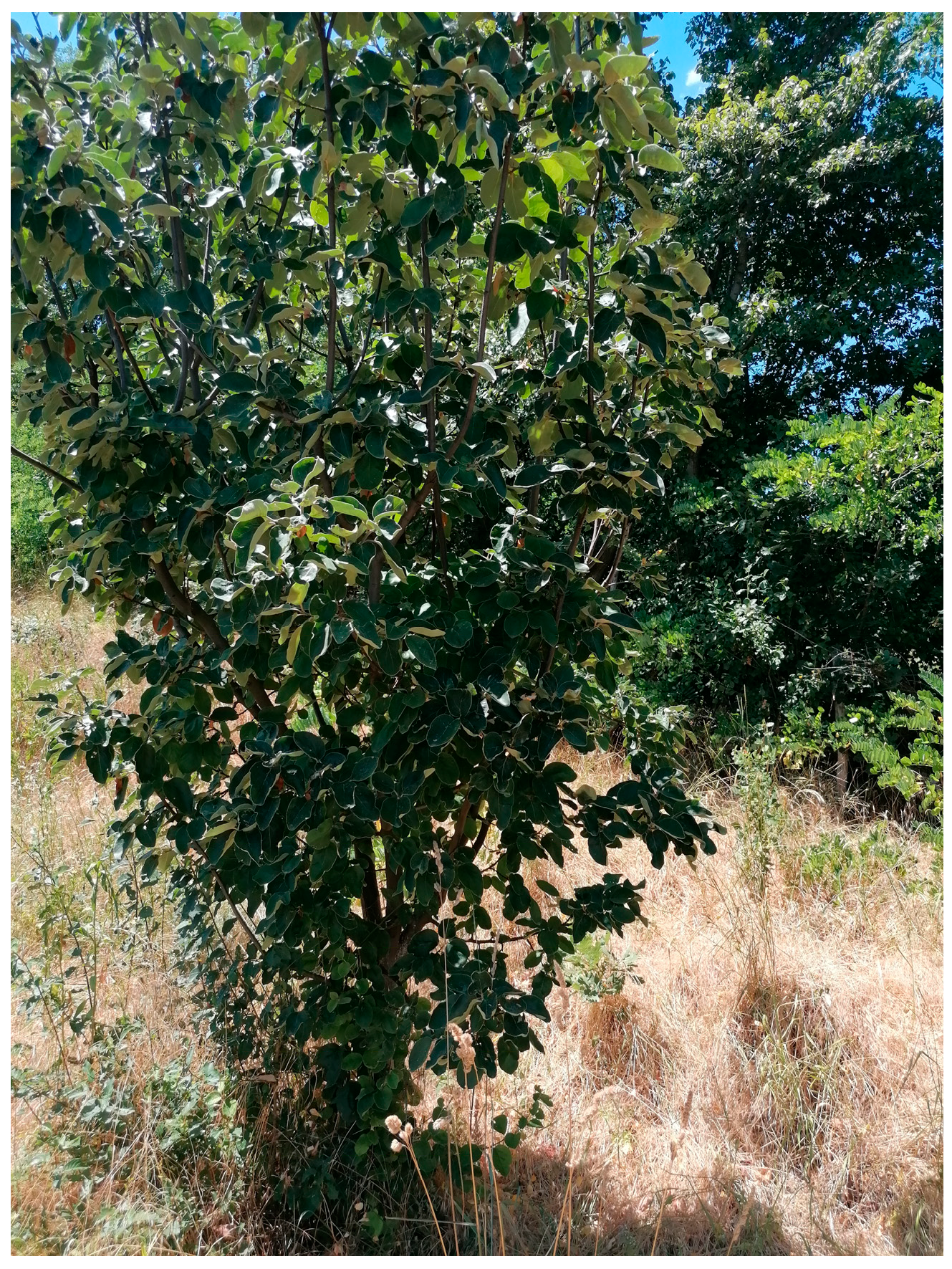
The CO grows as a small tree or large shrub, reaching heights of up to 8 m. It exhibits a dense, spreading, and often pendulous canopy structure (Figure 2). The young shoots are initially purplish-red in color, later maturing to a purplish-brown hue. Morphologically, the shoots are cylindrical in shape and are densely tomentose during early developmental stages, gradually becoming glabrous with maturity [7]. The vegetative and floral buds of the CO are purplish-brown and covered with dense tomentose hairs. Stipules are caduceus, ovate in shape, and abscise early during development. The petioles measure approximately 0.8 to 1.5 cm in length and are similarly tomentose. Leaf blades are ovate to oblong, ranging from 5 to 10 cm in length and 3 to 5 cm in width. The CO possesses prominent venation located on the lower surface, while the upper surface is glabrous or sparsely pubescent during early growth. The shape of the leaf base is typically rounded or subcordate, with entire margins, and the apex is either acute or emarginate [7]. The flowers of the CO are large, solitary, and borne terminally on short shoots. The diameter of the CO flowers is approximately 4–5 cm, while the length of the petals is about 1,8 cm. They are typically white or pale pink in color, contributing to the ornamental appeal of the species. The floral initiation in the CO occurs shortly before anthesis, indicating a rapid transition from bud development to flower opening [8,9]. The fruits are typically spherical to oblong in shape, measuring approximately 8–12 cm in diameter and exhibiting an average weight ranging from 100 to 250 g. During early stages of development, the epidermis displays a brown to light green coloration, which gradually transitions to a bright yellow hue upon full ripening [10]. The fruit of the CO is aromatic, with firm flesh that contains numerous grit cells which are large, irregular, and similar in structure to those found in Pyrus species. The shape of the fruit can vary, typically being pyriform, globose, or maliform, with some genotypes exhibiting a ribbed contour [7].

2.3. Active Compounds

The CO is rich in vitamins and minerals, as well as secondary metabolites such as phenolic compounds and essential oils.
The CO fruit and peel are rich sources of various minerals, including potassium, sodium, calcium, and phosphorus [11]. Furthermore, the fruit and peel are rich in various antioxidants, including phenolic compounds such as caffeoylquinic acids, flavonoids such as rutin, and other components like ascorbic, citric, malic, D-(–)-quinic, fumaric, and L-shikimic acids [12]. The monosaccharide profile revealed the presence of rhamnose, mannose, D-glucose, L-arabinose, fructose, sucrose, maltose, and galactose. Sugar alcohols identified in the CO fruit include D-sorbitol, D-mannitol, and D-galactitol. The most abundant phenolics in the CO peel are rutin and chlorogenic acid [2].
The most abundant flavonoid present in the CO leaves is rutin. Other flavonoids found in leaf extract include quercetin-3-O-galactoside, quercetin-3-O-rutinoside kaempferol-3-O-glycoside, kaempferol-3-O-rutinoside, kaempferol-3-O-glucoside, and 4-O-caffeoylquinic acid [13]. The phenolic acid content of the fruit and leaf extract of the CO is characterized by the presence of 5-O-caffeoylquinic acid, 3,5-O-dicaffeoylquinic acid, 3-O-caffeoylquinic acid, 4-O-caffeoylquinic acid, quercitin-3-O-rutinoside, kaempferol-3-O-rutinoside, quercitin-3-O-galactoside, kaempferol-3-O-glycoside, and kaempferol-3-O-glucoside [14]. During the flowering and fruiting season, the presence of 40 different essential oils, aldehydes, fatty acids, monoterpenes, and norisoprenoids was revealed [15].
Major phytochemical constituents in the CO seeds are tannins, glycosides, and flavones, such as isoschaftoside, caffeoylquinic acids, and 5-O-caffeoylquinic acid. Phenolics isolated from the CO seed extract include 3-, 4-, and 5-O-caffeoyl quinic acids, 3,5-dicaffeoyl quinic acid, apigenin derivatives, leucenin-2, 6-C-pentosyl-8-C-glucosyl chrysoeriol, 6-C-glucosyl-8-C-pentosyl chrysoeriol, and stellarin-2. Furthermore, the seed extract is rich in various organic acids and a wide range of amino acids [16]. The significant amount of 6,8-di-C-glucosyl luteolin (lucenin2), 6,8-di-C-glucosyl apigenin (vicenin-2), 6,8-di-C-glucosylchrysoeriol (stellarin-2), 6-C-arabinosyl-8-C-glucosylapigenin (isoschaftoside), 6-C-glucosyl-8-C-arabinosyl apigenin (schaftoside), 6-C- pentosyl-8-C-glucosyl chrysoeriol, and 6-C-glucosyl-8-C-pentosyl was also revealed while investigating the complex phytochemical composition of the CO seeds [17]. According to the study by Ghopur, along with the three known phytopharmaceutical constituents (ursolic acid, tormentic acid, and β-daucosterol), the presence of a new chromone compound (5,7-dihydroxy-2-n-pentacosanylchromen-4-one) was identified [18].
Recent phytochemical investigations of the CO ethanol extract with the use of different spectroscopic methods and X-ray crystallography led to the identification of three new dibenzofurans, which were determined to be 6-hydroxy-2,3,4,7-tetramethoxydibenzofuran, 1,6-dihydroxy-2,3,4,7-tetramethoxydibenzofuran, and 2,3,4,7-tetramethoxydibenzofuran-6,9-quinone [19].
Various parts of the CO, including its seeds, buds, bark, leaves, and fruits, which are rich in phytochemical compounds, have been traditionally employed for their medicinal properties. The plant is utilized both internally and externally in ethnomedicine. Different parts of the CO are commonly used in the management of respiratory conditions such as rhinitis, dryness of the throat, dry cough, bronchitis, tuberculosis, and excessive sneezing. It has also been used for systemic conditions including fever and gastrointestinal disorders such as dysentery, gastric acidity, duodenal ulcers, vomiting, and constipation. Additionally, it is used to treat urinary tract conditions, including dysuria and hemoptysis, and is believed to have a cooling effect, making it beneficial in cases of burning sensations such as burning of the tongue. Externally, preparations derived from the CO are traditionally applied for the treatment of ulcers, burns, and scalds [20].

3. Dermatological Effects of Cydonia oblonga

CO contains a wide spectrum of bioactive phytochemicals distributed throughout various parts of the plant. Upon oral administration, these compounds exhibit diverse pharmacological effects that contribute to the management of multiple health conditions. Clinical evidence supports the therapeutic potential of CO and its constituents in the treatment of different gastrointestinal disorders and urinary and respiratory tract conditions [21]. Nevertheless, further clinical investigations are required to comprehensively elucidate the molecular pathways through which CO exerts therapeutic effects against these conditions.
Beyond systemic applications, CO has also shown considerable promise in dermatology. Different plant parts have been traditionally and scientifically applied to both diseased and healthy skin. Experimental and clinical studies have reported favorable outcomes in treating various dermatological conditions, indicating the potential of CO-derived compounds for skin health. This review compiles and analyzes existing research on the dermatological properties of CO, highlighting the underlying mechanisms of action and therapeutic outcomes associated with different plant-derived preparations.

3.1. Antioxidant and UV-Protective Effects

Polyphenols derived from plant sources are known to exhibit multiple antioxidant mechanisms within the human body. These compounds can function as reducing agents, hydrogen donors, free radical scavengers, and singlet oxygen quenchers, thereby contributing to cellular protection and minimizing oxidative damage. Consequently, there has been growing scientific interest in the isolation and characterization of antioxidants from plant materials, particularly fruits, leaves, and seeds, with the aim of evaluating their potential in modulating the progression of oxidative stress-related disorders [22].
The antioxidant activity of the CO fruit was determined with the use of 2,2-diphenyl-1-picrylhydrazyl (DPPH) and 2,2′-azobis(2-amidinopropane) dihydrochloride (AAPH)-induced oxidative hemolysis of human erythrocytes. The pulp, peel, and seed methanolic extract of the CO exhibited certain antioxidant activity. This study revealed the superior effect in antioxidant activity of the pulp and peel extract in comparison with the seed extract [22].
The CO methanolic leaf extract also exhibited antioxidant activity which was comparable to the antioxidant activity of the Camellia sinensis leaf extract, which is known for its high antioxidant effects. Although the DPPH scavenging activity of the CO leaf extract was lower in comparison to the Camellia sinensis leaf extract, the protection of the erythrocyte membrane from hemolysis was similar in both analyzed extracts and the reducing power of the CO leaf extract was significantly higher in comparison to the Camellia sinensis leaf extract. The findings of this study demonstrate that the methanolic extract of the CO leaf exhibits potent antioxidant activity, comparable to that of the Camellia sinensis leaf extract, which is known as a potent antioxidant. The extract showed significant free radical scavenging capacity, particularly against DPPH and peroxyl radicals, and provided protective effects against oxidative damage in erythrocytes. The results suggest that the CO leaf extract represents a promising natural source of antioxidants, with potential applications for the prevention and management of oxidative stress-related diseases [23].
The ferric reducing antioxidant power (FRAP) and total phenolic content (TPC) were analyzed and compared between the CO-intact and peeled fruit samples. The antioxidant capacity was found to be significantly higher in the intact fruits compared to the peeled fruit extracts. This investigation suggests that the CO fruit extract may be regarded as a valuable source of antioxidants and polyphenolic compounds. Furthermore, the fruit peel represents a very significant part of the plant with strong antioxidant activity [24].
The CO fruit was fractionated into pulp, peel, and seed components, from which methanolic extracts were prepared and evaluated for their antioxidant properties through the DPPH method, as well as the superoxide anion radical and molybdenum reducing capacity assessment. Both pulp and peel extracts exhibited comparable DPPH radical scavenging activities, with EC50 values of 0.6 mg/mL and 0.8 mg/mL, respectively. In contrast, the seed extract demonstrated considerably lower antioxidant activity, with an EC50 of 12.2 mg/mL. Moreover, under AAPH-induced oxidative stress, the pulp and peel extracts significantly protected erythrocyte membranes from hemolysis in a concentration- and time-dependent manner [25].
The antioxidant effects of various extracts of the CO are summarized in Table 1.
Multiple studies have demonstrated the potent antioxidant activity of CO extracts derived from the seeds, leaves, and whole fruit, as well as the pulp and peel. This high antioxidant capacity positions CO as a promising ingredient for inclusion in dermatological and cosmetic formulations, particularly those aimed at skincare. The significant antioxidant activity of the CO has been confirmed in many studies. Regarding the solvent used for the extraction of various parts of the CO, the methanolic extract exhibited the highest level of antioxidant activity, while the ethanolic extract demonstrated the lowest level of antioxidant activity [26].
Prolonged exposure of the skin to ultraviolet (UV) radiation induces significant immunological alterations that contribute to photoaging, a process that closely resembles intrinsic, chronological aging. More than three decades ago, it was demonstrated that UV radiation exposure leads to an immunosuppressive state, characterized by the reduced capacity to attenuate contact hypersensitivity responses in the skin [27].
Evaluation of the protective effects of the CO methanol leaf extract against UV-A-induced histopathological alterations was performed in selected tissues of the fish specimen Clarias gariepinus. In the group exposed to UV-A radiation and treated with 20 mL of quince leaf extract, a notable improvement in skin cell structure was observed compared to the groups exposed to UV-A radiation or to UV-A radiation and 10 mL of quince extract, which exhibited mild enlargement of alarm cells and rupture of epithelial cells, indicating less protective efficacy [28].
The CO pulp and peel extract exhibited the second-highest UV-A photoprotective activity in comparison with Fortunella margarita, Diospyros kaki Thumb, and Annona cherimola L. pulp and peel extract. The calculated Sun Protective Factor (SPF) of the CO peel extract was higher than the calculated SPF of the CO pulp extract. These results suggest that the potent application of the CO peel extract in anti-aging and skin-protecting cosmetic products [29]. The UV-protective effects of various extracts of the CO are summarized in Table 1.
The CO leaf extract, as well as the pulp and peel extracts, could be incorporated into sun protective formulations due to their UV-protective activity. A range of strategies employing antioxidants and anti-inflammatory agents, such as the CO extracts, has been developed to augment the skin’s natural defenses against UV radiation. In this context, plant-derived compounds have been extensively investigated for their potential to mitigate UV-induced skin aging, pathological conditions, and carcinogenesis [30].
Table 1. Antioxidant and UV-protective effects of Cydonia oblonga.
Cydonia oblonga FormulationEffectModelMaterialDosage/
Concentration
MechanismStudy
Peel, pulp, and seed extractAntioxidant
activity
DPPH and AAPH methodsHuman erythrocytes25 µLFree radical scavenging and increasing the red blood cell resistance to oxidative stressMagalhães et al. [22]
Leaf extractAntioxidant
activity
DPPH, AAPH, and Folin–Ciocalteu methodHuman erythrocyte suspensions and hemolysis assay25 µLInterruption of the free radical
chain reaction and erythrocyte membrane damage, inhibitory effects of hemolysis
Costa et al. [23]
Fruit extractAntioxidant
activity
FRAP methodSupernatants of the whole and peeled fruitsNo informationFree radical scavengingPapp et al. [24]
Peel, pulp, and seed extractAntioxidant
activity
DPPH method, superoxide anion radical, and molybdenum reducing capacity assessmentHuman hepatoblastoma cell line (HepG2), lung epithelial cell line (A549), and
cervical carcinoma cell line (HeLa)
5–500 mg/mLFree radical scavenging,
oxidative stress reduction and
apoptosis induction
Pacifico et al. [25]
Leaf extractUV-protective activityin vivo animal studyfish specimen Clarias gariepinus20 mLAntioxidant activity and improvement in the pathological alterations recorded in red blood cells, skin, and liver.Sayed et al. [28]
Peel and pulp extractUV-protective activityin vitro determination of the SPFminimal erythema dose (MED) on protected skin1%, 2.5%, 5% Photoprotective activity of the present carotenoids and flavonoidsLasota et al. [29]

3.2. Anti-Inflammatory and Anti-Allergic Activity

Inflammation represents a physiological response of the host immune system to tissue injury and/or pathogenic stimuli. While this process is essential for initiating tissue repair and eliminating harmful agents, persistent or chronic inflammation may result in progressive tissue damage and, if unregulated, can ultimately lead to organ dysfunction or failure [31].
The anti-inflammatory effects of the CO ethanol leaf extract were evaluated in an albino rat model with the use of the induced ear edema by arachidonic acid and the induced paw edema by carrageenan. The CO leaf extract at a concentration of 100 mg/kg significantly reduced the arachidonic acid-induced ear edema (by 52% in comparison with the control group). Furthermore, the extract has also shown a significant inhibitory effect on carrageenan-induced hind paw edema in a dose-dependent manner. The biochemical analysis demonstrated that the CO leaf extract led to a reduction in lipid peroxidation (LPO), nitric oxide (NO), interleukin-6 (IL-6), and tumor necrosis factor-alpha (TNF-α) levels, while concurrently increasing the level of reduced glutathione (GSH). Moreover, the elevated activities of the antioxidant enzymes catalase (CAT) and glutathione S-transferase (GST) were modulated and normalized in the experimental group. These results suggest that the CO leaf extract exhibits significant anti-inflammatory activity, indicating its potential as a promising natural therapeutic agent for the management of inflammatory disorders [32].
The CO peel extract has also demonstrated anti-inflammatory activity in the lipopolysaccharide (LPS)-treated human myelomonocytic cell line THP-1. The CO peel extract significantly attenuated the LPS-induced secretion of TNF-α and IL-8 in a dose-dependent manner. With a dose of the extract of 20 µg/mL, the maximum inhibition of 52% of TNF-α production and the inhibition of 50% of pro-inflammatory chemokines have been achieved. Moreover, the CO peel extract at a dose of 20 µg/mL increased the level of IL-10, which leads to a reduction in the TNF-α level, which can be beneficial in the treatment of several inflammatory diseases. The observed increase in IL-10 secretion by activated macrophages further supports the potential of this extract as an alternative or complementary therapeutic strategy for the treatment and/or prevention of inflammatory diseases [33].
The anti-inflammatory effects of various extracts of the CO are summarized in Table 2.
The anti-inflammatory effects of the CO leaf and peel extracts are mediated through multiple mechanisms, including enhancing the levels of GSH, suppression of the production of pro-inflammatory mediators (NO, IL-6, TNF-α), suppression of TNF-α and IL-8 production, increased production of IL-10, and inhibition of LPS-mediated activation of NF-κB. These bioactivities suggest their potential therapeutic utility in the management and symptomatic relief of inflammatory dermatological conditions.
If inadequately controlled, allergic responses can lead to a range of clinical conditions such as dermatitis, allergic rhinitis, anaphylactic shock, and asthma. Conventional pharmacological treatments for allergic disorders include antihistamines, immunosuppressants, and corticosteroids. These treatment options are often associated with significant adverse effects, such as hypertension, diabetes mellitus, osteoporosis, and growth retardation. Consequently, there is a growing interest in identifying plant-derived compounds and dietary materials with anti-allergic properties that offer therapeutic benefits without the undesirable side effects of conventional drugs [21].
The CO aqueous fruit extract reduced the elevation of IL-13 and TNF-α expression levels in Ig E antigen-stimulated mast cell-like RBL-2H3 cell line. This extract has also shown a significant inhibitory effect on leukotriene C4 and prostaglandin D2 production, as well as the attenuation of the induction of intracellular cyclooxygenase COX-2 in mouse bone marrow-derived mast cells. These findings suggest that the CO fruit extract exerts inhibitory effects on a broad spectrum of late-phase immune responses mediated by mast cells [34].
Further investigation of the anti-allergic effect of the CO fruit extract led to a study which included the combination of the extract with Citrus limon juice, where this product with marketing authorization (Gencydo®) exhibited significant inhibitory activity in Ig E-stimulated basophilic and mast cells in terms of the inhibition of degranulation and histamine, cytokine, and chemokine release, as well as the inhibition of chemokine production from lung epithelial cells. The results of this investigation indicate the potential of topical treatment with Gencydo® in the treatment of various skin allergy disorders [35].
The anti-allergic effects of various extracts of the CO are summarized in Table 2.
In vitro studies demonstrated that the allergic response of IgE-stimulated cell lines was significantly attenuated by treatment with the CO fruit extract, either alone or in combination with Citrus limon juice. The observed synergistic anti-allergic effects of this combination highlight its potential for inclusion in topical formulations aimed at managing skin conditions associated with allergic responses.

3.3. Other Dermocosmetic Effects

The antimicrobial activity of the CO pulp and peel aqueous acetone extracts was evaluated against various microbial strains. Among the tested samples, the CO peel extract demonstrated the highest antibacterial efficacy, with minimum inhibitory concentrations (MIC) and minimum bactericidal concentrations (MBC) ranging from 102 to 5 × 10³ µg polyphenol/mL. The observed antimicrobial effects are likely attributed to a synergistic interaction between chlorogenic acid and other bioactive constituents present in the extract [36].
The efficacy of the antimicrobial effects is determined by various factors, including the solvent used for extraction. The CO fruit and seed extracts were prepared using ethanol, acetone, and water as solvents to evaluate their antimicrobial activity against the selected bacterial strains, including Escherichia coli, Klebsiella pneumoniae, Staphylococcus aureus, and Enterobacter aerogenes. Among the tested extracts, the ethanolic seed extract exhibited the highest inhibitory effect on bacterial growth. In contrast, aqueous extracts demonstrated the lowest antimicrobial efficacy [37].
Furthermore, the antibacterial activity of the CO seed extract was evaluated against Staphylococcus aureus, Staphylococcus epidermidis, Klebsiella pneumoniae, Escherichia coli, and Moraxella spp. using the agar well diffusion method. The extract demonstrated notable antibacterial properties, with the highest antibacterial activity against Staphylococcus aureus. In contrast, Escherichia coli and Moraxella spp. were the most resistant to the extract. These findings suggest that the CO seed extract possesses greater antibacterial efficacy against Gram-positive bacteria compared to Gram-negative bacteria [38].
Moreover, the synergistic antifungal effects of the CO leaf extracts and silver nanoparticles against Aspergillus niger were evaluated in vitro. Ethanolic and acetonic CO leaf extracts were prepared alongside silver nanoparticles. The antifungal activity of the individual extracts and silver nanoparticles against A. niger were assessed using the agar dilution method. The minimum inhibitory concentration (MIC) and minimum fungicidal concentration (MFC) were determined via the broth macrodilution technique. The CO leaf extracts and silver nanoparticles demonstrated antifungal activity against A. niger. The ethanolic extract exhibited greater antifungal efficacy compared to the acetonic extract. Furthermore, a synergistic antifungal effect was observed when the ethanolic extract was combined with silver nanoparticles, resulting in enhanced inhibition of A. niger growth. The ethanolic CO leaf extract in combination with silver nanoparticles exhibits a synergistic antifungal effect against A. niger and may serve as a promising alternative or adjunct in the control of fungal infections caused by this pathogen [39].
The antimicrobial effects of various extracts of the CO are summarized in Table 3.
The antimicrobial activity exhibited by the CO seeds, leaves, and whole fruit, as well as the pulp and peel, has demonstrated efficacy against a variety of bacterial and fungal strains. These findings highlight the potential of the CO-derived compounds for therapeutic application in conditions which represent the consequence of microbial infections. Recent investigations suggested the antimicrobial activity of the CO extracts could be enhanced with the incorporation into silver nanoparticles [40].
Wound healing is characterized by the critical involvement of fibroblasts, which facilitate fibrin clot degradation and contribute to wound contraction through their active role in extracellular matrix (ECM) remodeling and collagen synthesis [41].
The effect of the CO seed mucilage dissolved in distilled water on wound healing was determined through the analysis of its influence on the proliferation of human skin fibroblasts. The study assessed various concentrations of the CO seed mucilage, ranging from 50 to 400 µg/mL, applied to a human skin fibroblast cell line over time intervals of 12, 24, 48, and 72 h. Using the microculture tetrazolium assay (MTT), the results demonstrated that the CO seed mucilage significantly enhanced fibroblast proliferation, notably even at the lowest concentration of 50 µg/mL after 48 h of exposure [42].
Furthermore, the wound-healing efficiency of the CO seed mucilage, formulated as 5%, 10%, and 20% creams in Eucerin base, on the modulation of the growth factors involved in wound healing in the Iranian rabbit animal model was evaluated. The results demonstrated statistically significant differences in wound contraction between the CO seed mucilage cream concentrations of the 10% and 20% treatment groups compared to the control group on most evaluation days. Rabbits receiving the 20% CO seed mucilage cream exhibited the most pronounced healing outcomes, including complete wound closure within 13 days, elevated hydroxyproline content, increased tissue tensile strength, and higher concentrations of key growth factors in wound exudate. Regarding these findings, the CO seed mucilage exhibits great potential in the formulation of natural topical preparations that would be used for wound-healing purposes [43].
Further investigation led to the formulation of creams with the CO seed mucilage and the evaluation of the therapeutic effects on dermal toxicity induced by T-2 toxin. The creams were prepared with the use of Eucerin base containing 5%, 10%, and 15% (w/w) CO seed mucilage. The wound-healing effect was defined by the reduction in wound margins, erythema, and blister formation. The observed healing durations were 9–14 days, depending on the treatment group. The CO seed mucilage preparation at a concentration of 15% (w/w) exhibits superior healing effects on T-2 toxin-induced dermal toxicity compared to both the untreated control and the Eucerin base cream without the CO mucilage [44].
The wound-healing effects of various extracts of the CO are summarized in Table 3. The wound-healing properties of the CO seed mucilage have been well-documented. Recent studies have identified nanofibers composed of polyvinyl alcohol (PVA) and CO seed mucilage as a promising candidate for advanced wound treatment applications [45].
Pigmentation disorders are among the most prevalent cosmetic concerns and have contributed to the rising demand for skin-lightening products. Many of the active ingredients used to treat hyperpigmentation are derived from natural sources and primarily act by inhibiting tyrosinase, the copper-containing oxidoreductase that plays a key role in the early stages of melanin biosynthesis. In the search for new depigmenting agents, the mushroom tyrosinase inhibition assay is widely used due to its simplicity, affordability, and high throughput. However, it is important to note that significant structural and functional differences exist between fungal and mammalian tyrosinase, which may affect the biological relevance of screening results obtained using the mushroom enzyme [46].
The tyrosinase inhibitory potential of the CO hydroglycolic peel and pulp extracts was evaluated in commercially available mushroom tyrosinase and murine tyrosinase obtained from melanoma cell lysates. The CO pulp extract demonstrated moderate tyrosinase inhibitory activity, reducing mushroom tyrosinase activity by 38–57% and murine tyrosinase activity by 25–28%. A comparable inhibitory effect was observed for the CO peel extract, which inhibited mushroom tyrosinase by 41–49% and murine tyrosinase by 26–27%. The observed inhibitory activity against both enzyme types is likely attributable to the synergistic interaction of flavonoids and phenolic acids naturally present in the extracts. The CO pulp and peel extracts exhibited the most significant tyrosinase inhibitory activity in comparison with Fortunella margarita (kumquat), Diospyros kaki Thumb. (Japanese persimmon), and Annona cherimola L. (cherimoya), which suggests great potential of the use of the CO extracts in skin-lightening preparations [29].
Further investigation of the CO ethanolic peel extract revealed significant anti-tyrosinase activity with the IC50 value of 7.84 ± 0.03 mg/mL. This study highlighted the influence of the drying technology of the extracts on their anti-tyrosinase activity. The low-temperature and vacuum conditions present in freeze-dried ethanolic CO peel extract effectively inhibited both thermal and oxidative degradation of anti-tyrosinase bioactive compounds, thereby preserving the anti-tyrosinase activity to a greater extent. In contrast, hot air drying was characterized by elevated temperatures and exposure to atmospheric oxygen, which significantly diminished the concentration and biological activity of these compounds and therefore their anti-tyrosinase activity [47].
The purified protein from the CO petroleum ether seed extract also exhibited anti-tyrosinase activity in an in vitro study in which two analyzed amino acid sequences of two purified peptides resulted in IC50 values of 1.15 mg/mL and 8.69 mg/mL which were higher in comparison to kojic acid (IC50 value of 0.06 mg/mL) and lower in comparison to citric acid (IC50 value of 9.77 mg/mL). These findings indicate that the CO seed protein exhibits moderate anti-tyrosinase activity, which is less potent than that of kojic acid but more effective than the activity demonstrated by citric acid, indicating its potential in skin-lightening products as a new, effective, and safe alternative to conventional treatment options with various side effects such as tretinoin and arbutin [48].
The skin-lightening effects of various extracts of the CO are summarized in Table 3.
Conventional hyperpigmentation therapy can cause various adverse effects, such as local irritation like erythema, xerosis, discrete exfoliation, and burning sensation [49]. Accordingly, the CO pulp and peel extracts, along with their purified seed protein, represent effective and potentially safer alternatives for the management of skin pigmentation disorders, owing to their pronounced anti-tyrosinase activity. Moreover, the demonstrated ability of various CO plant-derived components to reduce skin erythema suggests their therapeutic relevance in conditions associated with these inflammatory skin responses.
A moisturizer’s function is characterized by a reduction in transepidermal water loss (TEWL) through the formation of a semi-occlusive layer over the stratum corneum. This barrier enhances the hydration and pliability of the stratum corneum. Cutaneous hydration is influenced by multiple intrinsic and extrinsic factors, including environmental conditions, nutritional status, and the natural aging process. With advancing age, the skin’s ability to retain moisture diminishes, necessitating the use of topical moisturizers to support and maintain skin barrier function and overall dermal health [49].
The CO fruit extract incorporated into an emulgel at a concentration of 4% exhibited significant cosmetic effects in healthy volunteers. The formulation demonstrated effective moisturizing properties by reducing the TEWL from the facial skin, resulting in a significant increase in both skin hydration and elasticity. The measured moisture level of the skin in healthy volunteers reached 56.49%, which was significantly higher in comparison to the moisture level of the control group (10.76%) after 12 weeks of the study. Owing to its substantial moisturizing potential, the formulation may serve as a promising candidate for cosmetic applications aimed at improving facial skin resilience [50,51].
The moisturizing effects of various extracts of the CO are summarized in Table 3.
TEWL represents the most widely employed quantitative parameter for evaluating the functional integrity of the skin barrier, both in healthy individuals and in patients with dermatological conditions characterized by impaired barrier function, such as atopic dermatitis. TEWL quantifies the passive diffusion of water vapor through a defined area of the stratum corneum to the skin surface over a specified time interval [52]. Given its pronounced efficacy in reducing TEWL, the CO fruit extract demonstrates potential as a bioactive component in cosmetic formulations aimed at enhancing skin hydration. Improved moisture retention contributes to the overall appearance of the skin, promoting a healthier and more youthful facial complexion.
Skin erythema can arise from a variety of etiological factors. One of the most prevalent causes is prolonged exposure to UV radiation. In addition to photodamage, erythema is frequently observed as a clinical manifestation in several dermatological conditions, including acne, psoriasis, melasma, and post-inflammatory hyperpigmentation. It may also occur secondary to systemic conditions such as fever, or as a response to exposure to specific bands of electromagnetic radiation [53].
The formulation of an emulgel which contains 4% of the CO fruit extract demonstrated a reduction in erythema in healthy volunteers. Variations in erythema levels were observed for both the test and control formulations over the course of the study. The active formulation induced a progressive and consistent reduction in erythema values among participants, with a decrease of 10.35% by 12 weeks. In contrast, the control formulation exhibited a more modest and less consistent effect, with an erythema reduction of 2.87% after 12 weeks of the study. These results suggest that the potential of the CO fruit extract formulations for skin erythema treatment [51].
The effects on skin erythema of various extracts of the CO are summarized in Table 3.
Table 3. Other dermocosmetic effects of Cydonia oblonga.
Cydonia oblonga FormulationEffectModelMaterialDosage/
Concentration
MechanismStudy
Peel and pulp
extract
Antimicrobial and antioxidant activityAgar diffusion method,
determination of MIC and MBC
Staphylococcus aureus, Escherichia coli, Pseudomonas aeruginosa, Salmonella species, Candida
albicans, Aspergillus niger strains
100 µLBactericide and
bacteriostatic activity
Fattouch et al. [36]
Fruit and seed extractAntibacterial activityMuller-Hinton agar media method, determination of MIC and MBCE. coli, Klebsiella pneumoniae, S. aureus, and Enterobacter aerogenes strains80 µLInhibition of the
bacterial growth
Alizadeh et al. [37]
Seed extractAntibacterial activityAgar diffusion methodS. aureus, S. epidermidis, Klebsiella pneumoniae, E. coli, and Moraxella spp. strains125–500 mg/mLPrevention of the
development
of microorganisms by precipitating microbial protein
Al-khazraji et al. [38]
Leaf extractAntifungal activityAgar diffusion method, Broth macrodilution assay, determination of MIC and MFCAspergillus niger strains25–800 mg/mLInhibition of the fungal growthHamed et al. [39]
Seed mucilageWound-healing activityin vitro study in the fibroblast cell linehuman skin fibroblasts50–400 µg/mLstimulation of fibroblast proliferationGhafourian et al. [42]
Seed mucilage incorporated into a Eucerin-based creamWound-healing activityin vivo study in the animal modelmale Iranian rabbits5–20%elevation of hydroxyproline content, increase in tissue tensile strength, and concentrations of key growth factorsPari et al. [43]
Seed mucilage incorporated into a Eucerin-based creamWound-healing activityin vivo study in the animal modelNew
Zealand
rabbits
5–15%activation of
growth
factors, increase in
collagen
production, neutralizing
dermal
toxicity
Hemmati et al. [44]
Peel and pulp
extract
Tyrosinase inhibition in vitro study in enzyme assaysmushroom tyrosinase and murine tyrosinase1–5%synergistic inhibition of tyrosinase activity by flavonoids and phenolic acidsLasota et al. [29]
Peel extractTyrosinase inhibitionin vitro study in enzyme assaystyrosinase solution10 mg/mLinhibition of tyrosinase activity by phenolic compoundsWang et al. [47]
Purified seed
protein
Tyrosinase inhibitionin vitro study in enzyme assaysmushroom tyrosinase100 μLinhibition of tyrosinase activity by arginine residueDeng et al. [48]
Fruit extract incorporated into an emulgelMoisturizing effect, effects against skin erythemain vivo study in healthy volunteersHuman skin4%Control of the TEWLKhiljee et al. [51]
The reviewed literature indicates that no adverse health effects or safety concerns have been documented following the administration of CO at established therapeutic dosages. Nonetheless, caution is warranted when using preparations derived from whole seeds, such as quince mucilage, due to the presence of cyanogenic glycosides. Upon ingestion, these compounds, although typically present in low concentrations, may undergo enzymatic hydrolysis in the gastrointestinal tract, leading to the release of hydrogen cyanide, a potentially toxic volatile compound [10]. Despite this, the available evidence supports the overall safety of CO-derived plant parts for topical application, particularly when formulations exclude unprocessed seed material.
Despite the growing body of in vitro and in vivo evidence, several limitations persist across the current literature. A major drawback lies in the heterogeneity of extraction protocols, solvent systems, and plant part selection, which impairs reproducibility and comparability between studies. Furthermore, many studies lack thorough phytochemical standardization, making it difficult to attribute observed effects to specific compounds. Although a few formulations have been tested in human volunteers, most data remain preclinical. The scarcity of randomized, controlled clinical trials is a significant barrier to validating dermatological efficacy in real-world applications.

4. Conclusions and Future Perspective

This review underscores the dermatological relevance of CO, with particular focus on its polyphenol-rich peel and seed mucilage as bioactive sources for topical application. Although existing evidence is primarily preclinical, reported antioxidant, anti-inflammatory, antimicrobial, and photoprotective effects support its candidacy as a multifunctional botanical ingredient in skin care. However, its integration into dermatological practice remains hindered by a lack of standardized extraction protocols, limited clinical validation, and insufficient data on formulation efficacy and safety.
Moving forward, the dermatological value of CO should be assessed through comparative studies with established natural actives such as green tea, arbutin, and licorice, which already hold defined roles in evidence-based skin care. Furthermore, the sustainability of quince by-products, particularly fruit peel, aligns with current trends in clean-label and upcycled cosmetics, adding to its practical appeal. While still in the early stages of development, COoffers a promising but as yet unproven addition to the repertoire of plant-derived dermatological agents. Unlocking its full potential will require mechanistic studies, formulation optimization, and well-designed clinical trials to bridge the gap between experimental promise and clinical applicability.

Author Contributions

Conceptualization, M.T. and M.A. (Marijana Andjic); writing—original draft preparation, A.A., J.D., M.G. and M.A. (Miljan Adamovic); writing—review and editing, M.T. and M.A. (Marijana Andjic); visualization, M.T.; supervision, M.T. and M.A. (Miljan Adamovic). All authors have read and agreed to the published version of the manuscript.

Funding

This research was funded by the Pharmacy Institution “Zdravlje Lek”, Prvomajska 100, 11000 Belgrade, Serbia.

Institutional Review Board Statement

Not applicable.

Informed Consent Statement

Not applicable.

Data Availability Statement

No new data were created or analyzed in this study. Data sharing is not applicable to this article.

Acknowledgments

The research was supported by the Ministry of Science, Technological Development, and Innovation, Republic of Serbia through Grant Agreements with the University of Kragujevac—Faculty of Medical Sciences No. 451-03-137/2025-03/200111 and the Faculty of Medical Sciences, University of Kragujevac (Junior Project 15/25).

Conflicts of Interest

The authors declare no conflicts of interest.

Abbreviations

The following abbreviations are used in this manuscript:
COCydonia oblonga
SMsecondary metabolites
DPPH2,2-diphenyl-1-picrylhydrazyl
AAPH2,2′-azobis(2-amidinopropane) dihydrochloride
FRAP ferric reducing antioxidant power
TPC total phenolic content
UVultraviolet
SPF Sun Protective Factor
LPO lipid peroxidation
NO nitric oxide
IL interleukin
TNF-α tumor necrosis factor-alpha
GSH reduced glutathione
CAT catalase
CST glutathione S-transferase
LPSlipopolysaccharide
MICminimum inhibitory concentrations
MBC minimum bactericidal concentrations
TEWLtransepidermal water loss

References

  1. Gubitosa, F.; Fraternale, D.; De Bellis, R.; Gorassini, A.; Benayada, L.; Chiarantini, L.; Albertini, M.C.; Potenza, L. Cydonia oblonga Mill. Pulp Callus Inhibits Oxidative Stress and Inflammation in Injured Cells. Antioxidants 2023, 12, 1076. [Google Scholar] [CrossRef] [PubMed]
  2. Ashraf, M.U.; Muhammad, G.; Hussain, M.A.; Bukhari, S.N. Cydonia oblonga M., A Medicinal Plant Rich in Phytonutrients for Pharmaceuticals. Front. Pharmacol. 2016, 7, 163. [Google Scholar] [CrossRef]
  3. Schoch, C.L.; Ciufo, S.; Domrachev, M.; Hotton, C.L.; Kannan, S.; Khovanskaya, R.; Leipe, D.; Mcveigh, R.; O’nEill, K.; Robbertse, B.; et al. NCBI Taxonomy: A comprehensive update on curation, resources and tools. Database 2020, 2020, baaa062. [Google Scholar] [CrossRef] [PubMed Central]
  4. Lee, H.S.; Jung, J.I.; Hwang, J.S.; Hwang, M.O.; Kim, E.J. Cydonia oblonga Miller fruit extract exerts an anti-obesity effect in 3T3-L1 adipocytes by activating the AMPK signaling pathway. Nutr. Res. Pract. 2023, 17, 1043–1055. [Google Scholar] [CrossRef] [PubMed]
  5. Abed, S.N.; Bibi, S.; Jan, M.; Talha, M.; Islam, N.U.; Zahoor, M.; Al-Joufi, F.A. Phytochemical Composition, Antibacterial, Antioxidant and Antidiabetic Potentials of Cydonia oblonga Bark. Molecules 2022, 27, 6360. [Google Scholar] [CrossRef]
  6. Kostecka-Gugała, A. Quinces (Cydonia oblonga, Chaenomeles sp., and Pseudocydonia sinensis) as Medicinal Fruits of the Rosaceae Family: Current State of Knowledge on Properties and Use. Antioxidants 2024, 13, 71. [Google Scholar] [CrossRef]
  7. Bell, R.L.; Leitao, J. Cydonia. In Wild Crop Relatives: Genomic and Breeding Resources; Springer: Berlin/Heidelberg, Germany, 2010; pp. 1–16. [Google Scholar] [CrossRef]
  8. Webster, A.D. Cydonia oblonga quince. In Encyclopedia of Fruit and Nuts; Janick, J., Paull, R.E., Eds.; CABI: Wallingford, UK, 2008; pp. 634–642. [Google Scholar]
  9. Sabir, S.; Qureshi, R.; Arshad, M.; Amjad, M.S.; Fatima, S.; Masood, M.; Saboon; Chaudhari, S.K. Pharmacognostic and clinical aspects of Cydonia oblonga: A review. Asian Pac. J. Trop. Dis. 2015, 5, 850–855. [Google Scholar] [CrossRef]
  10. Al-Snafi, A.E. The medical importance of Cydonia oblonga—A review. IOSR J. Pharm. 2016, 6, 87–99. [Google Scholar]
  11. Rop, O.; Balík, J.; ŘEZNÍČEK, V.; Juríková, T.; Škardová, P.; Salaš, P.; SOCHOR, J.; MLČEK, J.; Kramářová, D. Chemical characteristics of fruits of some selected quince (Cydonia oblonga Miller) cultivars. Cazech J. Food Sci. 2011, 29, 65–73. [Google Scholar] [CrossRef]
  12. Silva, B.M.; Andrade, P.B.; Mendes, G.C.; Seabra, R.M.; Ferreira, M.A. Phenolic profile of quince fruit (Cydonia oblonga Miller) (Pulp and Peel). J. Agric. Food Chem. 2002, 50, 4615–4618. [Google Scholar] [CrossRef]
  13. Benzarti, S.; Hamdi, H.; Lahmayer, I.; Toumi, W.; Kerkeni, A.; Belkadhi, K.; Sebei, H. Total phenolic compounds and antioxidant potential of quince (Cydonia oblonga Miller) leaf methanol extract. Int. J. Inov. Appl. Stud. 2015, 13, 518–526. [Google Scholar]
  14. Oliveira, A.P.; Pereira, J.A.; Andrade, P.B.; Valentao, P.; Seabra, R.M.; Silva, B.M. Phenolic profile of Cydonia oblonga Miller leaves. J. Agric. Food Chem. 2007, 55, 7926–7930. [Google Scholar] [CrossRef]
  15. Erdogan, T.; Gonenç, T.; Hortoglu, Z.S.; Demirci, B.; Baser, K.H.C.; Kıvçak, B. Chemical composition of the essential oil of quince (Cydonia oblonga Miller) leaves. Med. Aromat. Plants 2012, 1, 134. [Google Scholar] [CrossRef]
  16. Silva, B.M.; Andrade, P.B.; Ferreres, F.; Seabra, M.R.; Oliveira, M.B.; Margarida, A.F. Composition of Quince (Cydonia oblonga Miller) seeds: Phenolics, organic acids and free amino acids. Nat. Prod. Res. 2005, 19, 275–281. [Google Scholar] [CrossRef]
  17. Ferreres, F.; Silva, B.M.; Andrade, P.B.; Seabra, R.M.; Ferreira, M.A. Approach to the study of C-glycosyl flavones by ion trap HPLC-PADESI/MS/MS: Application to seeds of Quince (Cydonia oblonga). Phytochem. Anal. 2003, 14, 352–359. [Google Scholar] [CrossRef]
  18. Ghopur, H.; Usmanova, S.K.; Ayupbek, A.; Aisa, H.A. A new chromone from seeds of Cydonia oblonga. Chem. Nat. Compd. 2012, 48, 562–564. [Google Scholar] [CrossRef]
  19. Wei, J.J.; Wang, W.Q.; Song, W.B.; Xuan, L.J. Three new dibenzofurans from Cydonia oblonga Mill. Nat. Prod. Res. 2020, 34, 1146–1151. [Google Scholar] [CrossRef] [PubMed]
  20. Fazeenah, A.H.A.; Quamri, M.A. Behidana (Cydonia oblonga Miller)—A review. World J. Pharm. Res. 2016, 5, 79–94. [Google Scholar]
  21. Cvetkovska, K.; Bauer, B. Ethnopharmacological and toxicological review of Cydonia oblonga M. Maced. Pharm. Bull. 2018, 64, 3–16. [Google Scholar] [CrossRef]
  22. Magalhães, A.S.; Silva, B.M.; Pereira, J.A.; Andrade, P.B.; Valentão, P.; Carvalho, M. Protective effect of quince (Cydonia oblonga Miller) fruit against oxidative hemolysis of human erythrocytes. Food Chem. Toxicol. 2009, 47, 1372–1377. [Google Scholar] [CrossRef] [PubMed]
  23. Costa, R.M.; Magalhães, A.S.; Pereira, J.A.; Andrade, P.B.; Valentão, P.; Carvalho, M.; Silva, B.M. Evaluation of free radical-scavenging and antihemolytic activities of quince (Cydonia oblonga) leaf: A comparative study with green tea (Camellia sinensis). Food Chem. Toxicol. 2009, 47, 860–865. [Google Scholar] [CrossRef]
  24. Papp, N.; Szabó, T.; Szabó, Z.; Nyéki, J.; Stefanovits-Bányai, É.; Hegedûs, A. Antioxidant capacity and total polyphenolic content in quince (Cydonia oblonga Mill.) fruit. J. Int. J. Hortic. Sci. 2013, 19, 33–35. [Google Scholar] [CrossRef]
  25. Pacifico, S.; Gallicchio, M.; Fiorentino, A.; Fischer, A.; Meyer, U.; Stintzing, F.C. Antioxidant properties and cytotoxic effects on human cancer cell lines of aqueous fermented and lipophilic quince (Cydonia oblonga Mill.) preparations. Food Chem. Toxicol. 2012, 50, 4130–4135. [Google Scholar] [CrossRef]
  26. Yilmaz, D.C.; Seyhan, S.A. Antioxidant potential of Cydonia oblonga Miller leaves. Istanb. J. Pharm. 2017, 47, 9–11. [Google Scholar] [CrossRef]
  27. Salminen, A.; Kaarniranta, K.; Kauppinen, A. Photoaging: UV radiation-induced inflammation and immunosuppression accelerate the aging process in the skin. Inflamm. Res. 2022, 71, 817–831. [Google Scholar] [CrossRef] [PubMed]
  28. Sayed, A.H.; Abdel-Tawab, H.S.; Abdel Hakeem, S.S.; Mekkawy, I.A. The protective role of quince leaf extract against the adverse impacts of ultraviolet—A radiation on some tissues of Clarias gariepinus (Burchell, 1822). J. Photochem. Photobiol. B. 2013, 119, 9–14. [Google Scholar] [CrossRef] [PubMed]
  29. Lasota, M.; Lechwar, P.; Kukula-Koch, W.; Czop, M.; Czech, K.; Gawel-Beben, K. Pulp or Peel? Comparative Analysis of the Phytochemical Content and Selected Cosmetic-Related Properties of Annona cherimola L., Diospyros kaki Thumb., Cydonia oblonga Mill. and Fortunella margarita Swingle Pulp and Peel Extracts. Molecules 2024, 29, 1133. [Google Scholar] [CrossRef]
  30. Boo, Y.C. Emerging Strategies to Protect the Skin from Ultraviolet Rays Using Plant-Derived Materials. Antioxidants 2020, 9, 637. [Google Scholar] [CrossRef]
  31. Mosser, D.M.; Edwards, J.P. Exploring the full spectrum of macrophage activation. Nat. Rev. Immunol. 2008, 8, 958–969. [Google Scholar] [CrossRef]
  32. Ahmed, M.M.; Bastawy, S. Evaluation of antiinflammatory properties and possible mechanism of action of Egyptian quince (Cydonia oblonga) leaf. Egypt. J. Biochem. Mol. Biol. 2014, 32, 190–205. [Google Scholar]
  33. Essafi-Benkhadir, K.; Refai, A.; Riahi, I.; Fattouch, S.; Karoui, H.; Essafi, M. Quince (Cydonia oblonga Miller) peel polyphenols modulate LPS-induced inflammation in human THP-1-derived macrophages through NF-κB, p38MAPK and Akt inhibition. Biochem. Biophy. Res. Commun. 2012, 418, 180–185. [Google Scholar] [CrossRef] [PubMed]
  34. Kawahara, T.; Iizuka, T. Inhibitory effect of hot-water extract of quince (Cydonia oblonga) on immunoglobulin E-dependent late-phase immune reactions of mast cells. Cytotechnology 2011, 63, 143–152. [Google Scholar] [CrossRef]
  35. Gründemann, C.; Papagiannopoulos, M.; Lamy, E.; Mersch-Sundermann, V.; Huber, R. Immunomodulatory properties of a lemon-quince preparation (Gencydo®) as an indicator of anti-allergic potency. Phytomedicine 2011, 18, 760–768. [Google Scholar] [CrossRef]
  36. Fattouch, S.; Caboni, P.; Coroneo, V.; Tuberoso, C.I.G.; Angioni, A.; Dessi, S.; Marzouki, N.; Cabras, P. Antimicrobial Activity of Tunisian Quince (Cydonia oblonga Miller) Pulp and Peel Polyphenolic Extracts. J. Agric. Food Chem. 2007, 55, 963–969. [Google Scholar] [CrossRef]
  37. Alizadeh, H.; Rahnema, M.; Semnani, S.; Hajizadeh, N. Detection of Compounds and Antibacterial Effect of Quince (Cydonia oblonga Miller) Extracts in vitro and in vivo. J. Biol. Act. Prod. Nat. 2013, 3, 303–309. [Google Scholar] [CrossRef]
  38. Al-khazraji, S.K. Phytochemical screening and antibacterial activity of the crude extract of Cydonia oblonga seeds. Glob. Adv. Res. J. Microbiol. 2013, 2, 137–140. [Google Scholar]
  39. Hamed, A.; Mehdi, R.; Shahrzad, N.S.; Ajalli, M. Synergistic antifungal effects of quince leaf’s extracts and silver nanoparticles on Aspergillus niger. J. Appl. Biol. Sci. 2014, 8, 10–13. [Google Scholar]
  40. Baran, M.F. Synthesis, characterization and investigation of antimicrobial activity of silver nanoparticles from Cydonia oblonga leaf. Appl. Ecol. Environ. Res. 2019, 17, 2583–2592. [Google Scholar] [CrossRef]
  41. Stortelers, C.; Kerkhoven, R.; Moolenaar, W. Multiple actions of LPA on fibroblasts revealed by transcriptional profiling. BMC Genom. 2008, 9, 387. [Google Scholar] [CrossRef]
  42. Ghafourian, M.; Tamri, P.; Hemmati, A. Enhancement of human skin fibroblasts proliferation as a result of treating with quince seed mucilage. Jundishapur J. Nat. Pharm. Prod. 2015, 10, e18820. [Google Scholar] [CrossRef] [PubMed]
  43. Pari, T.; Aliasghar, H.; Mehri Ghafourian, B. Wound healing properties of quince seed mucilage:In vivoevaluation in rabbit full-thickness wound model. Int. J. Surg. 2014, 12, 843–847. [Google Scholar] [CrossRef]
  44. Hemmati, A.A.; Kalantari, H.; Jalali, A.; Rezai, S.; Zadeh, H. Healing effect of quince seed mucilage on T-2 toxin-induced dermal toxicity in rabbit. Exp. Toxicol. Pathol. 2012, 64, 181–186. [Google Scholar] [CrossRef]
  45. Jafari, M.; Baniasadi, H.; Rezvanpour, A.; Lotfi, M. Fabrication and characterisation of a wound dressing composed of polyvinyl alcohol and quince seed mucilage. J. Wound Care 2021, 30, XIIIi–XIIIx. [Google Scholar] [CrossRef]
  46. Pillaiyar, T.; Manickam, M.; Namasivayam, V. Skin Whitening Agents: Medicinal Chemistry Perspective of Tyrosinase Inhibitors. J. Enzym. Inhib. Med. Chem. 2017, 32, 403–425. [Google Scholar] [CrossRef]
  47. Wang, P.; Lv, W.; Wang, H. Effects of freeze-hot air drying on physicochemical properties and anti-tyrosinase activity of quince peels. Food Chem. 2025, 463, 141507. [Google Scholar] [CrossRef]
  48. Deng, Y.; Huang, L.; Zhang, C.; Xie, P.; Cheng, J.; Wang, X.; Liu, L. Skin-care functions of peptides prepared from Chinese quince seed protein: Sequences analysis, tyrosinase inhibition and molecular docking study. Ind. Crops Prod. 2020, 148, 112331. [Google Scholar] [CrossRef]
  49. Raminelli, A.C.P.; Romero, V.; Semreen, M.H.; Leonardi, G.R. Nanotechnological Advances for Cutaneous Release of Tretinoin: An Approach to Minimize Side Effects and Improve Therapeutic Efficacy. Curr. Med. Chem. 2018, 25, 3703–3718. [Google Scholar] [CrossRef]
  50. Purnamawati, S.; Indrastuti, N.; Danarti, R.; Saefudin, T. The Role of Moisturizers in Addressing Various Kinds of Dermatitis: A Review. Clin. Med. Res. 2017, 15, 75–87. [Google Scholar] [CrossRef] [PubMed]
  51. Khiljee, T.; Akhtar, N.; Khiljee, S.; Khiljee, B.; Rasheed, H.M.; Ansari, S.A.; Alkahtani, H.M.; Ansari, I.A. Gauging Quince Phytonutrients and Its 4% Emulgel Effect on Amplifying Facial Skin Moisturizing Potential. Gels 2023, 9, 934. [Google Scholar] [CrossRef]
  52. Alexander, H.; Brown, S.; Danby, S.; Flohr, C. Research Techniques Made Simple: Transepidermal Water Loss Measurement as a Research Tool. J. Investig. Dermatol. 2018, 138, 2295–2300.e1. [Google Scholar] [CrossRef] [PubMed]
  53. Abdlaty, R.; Hayward, J.; Farrell, T.; Fang, Q. Skin erythema and pigmentation: A review of optical assessment techniques. Photodiagnosis Photodyn. Ther. 2021, 33, 102127. [Google Scholar] [CrossRef] [PubMed]
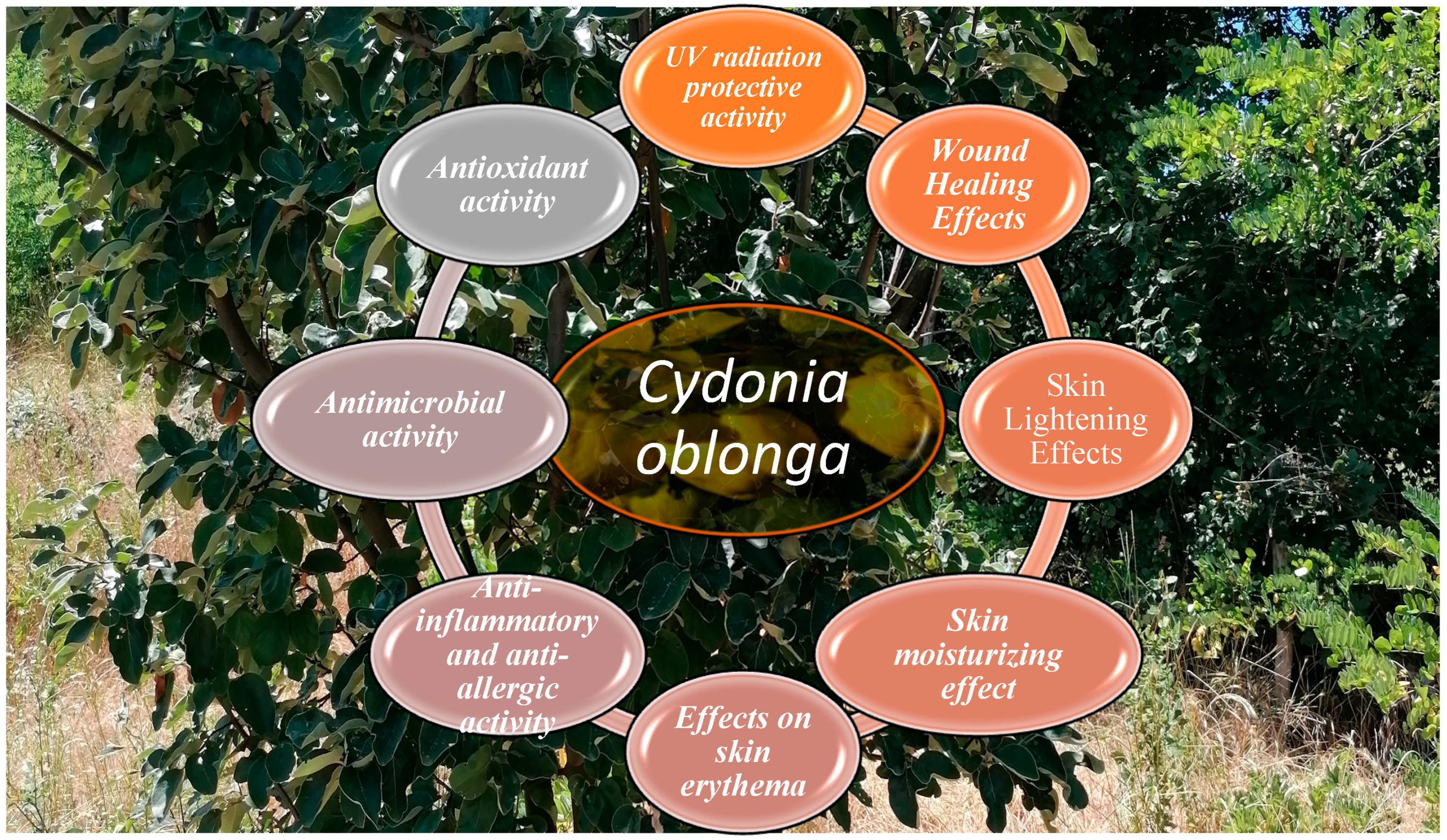
Figure 1. Skin protection induced by Cydonia oblonga.
Cosmetics 12 00187 g001
Figure 2. The Cydonia oblonga tree.
Cosmetics 12 00187 g002
Table 2. Anti-inflammatory and anti-allergic activity of Cydonia oblonga.
Cydonia oblonga FormulationEffectModelMaterialDosage/
Concentration
MechanismStudy
Leaf extractAnti-inflammatory activityArachidonic acid-induced ear edema, carrageenan-induced hind paw edemaAlbino rats25 mg/kg; 50 mg/kg; 100 mg/kgAntioxidative and free radical scavenging activities, enhancing the levels of GSH; suppression of the production of pro-inflammatory mediators (NO, IL-6, TNF-α) Ahmed et al. [32]
Peel extractAnti-inflammatory activityLPS-induced inflammationHuman
myelomonocytic cell line THP-1
20 µg/mLSuppression of the TNF-α and IL-8 production, increased production of the IL-10, inhibition of the LPS-mediated activation of NF-κB, pγ8MAPK, and AktEssafi-Benkhadir et al. [33]
Fruit extractAnti-allergic activity in vitro study in the
Ig E antigen-stimulated cell cultures
mast cell-like RBL-2H3 cell line, mouse bone marrow-derived mast cells50 μg/mL,
500 μg/mL
Inhibition of the IL-13 TNF-α expression level in Ig E antigen; inhibition of the leukotriene C4 and prostaglandin D2 production; reduction in the COX-2 expressionKawahara et al. [34]
Fruit extract in combination with Citrus limon juiceAnti-allergic activityin vitro study in the
Ig E-stimulated basophilic cell line,
β-Hexosaminidase and histamine release assay
RBL-2H3 (rat basophilic cells), human mast cells HMC-1, lung epithelial BEAS-2B cells0.2 mg/mL, 0.4 mg/mL, 0.8 mg/mLinhibition of the degranulation and histamine, cytokine, and chemokine release of basophilic cells and mast
cells, modulation of the chemokine production from lung epithelial
cells
Gründemann et al. [35]
Disclaimer/Publisher’s Note: The statements, opinions and data contained in all publications are solely those of the individual author(s) and contributor(s) and not of MDPI and/or the editor(s). MDPI and/or the editor(s) disclaim responsibility for any injury to people or property resulting from any ideas, methods, instructions or products referred to in the content.