Review

Enzymes DNA Repair in Skin Photoprotection: Strategies Counteracting Skin Cancer Development and Photoaging Strategies

by
Ewelina Musielak
and
Violetta Krajka-Kuźniak
*
Department of Pharmaceutical Biochemistry, Poznan University of Medical Sciences, Rokietnicka 3, 60-806 Poznan, Poland
*
Author to whom correspondence should be addressed.
Cosmetics 2025, 12(4), 172; https://doi.org/10.3390/cosmetics12040172
Submission received: 21 May 2025 / Revised: 11 July 2025 / Accepted: 25 July 2025 / Published: 12 August 2025
(This article belongs to the Special Issue Feature Papers in Cosmetics in 2025)

Abstract

Ultraviolet radiation (UVR) is a major contributor to skin aging and carcinogenesis, primarily through the induction of DNA damage. While conventional sunscreens provide passive protection by blocking UVR, active photoprotection using DNA repair enzymes offers a strategy to reverse UV-induced DNA lesions at the molecular level. Enzymes such as photolyase, T4 endonuclease V, and 8-oxoguanine glycosylase address distinct types of DNA damage through light-dependent and -independent mechanisms, complementing the skin’s endogenous repair systems. Advances in nanocarrier technologies and encapsulation methods have improved the stability and delivery of these enzymes in topical formulations. Emerging evidence from clinical studies indicates their potential in reducing actinic keratoses, pigmentation disorders, and photoaging signs, although challenges in regulatory approval, long-term efficacy validation, and formulation optimization remain. This review provides a comprehensive synthesis of the mechanistic, clinical, and formulation aspects of enzyme-based photoprotection, outlines regulatory and ethical considerations, and highlights future directions, including CRISPR-based repair and personalized photoprotection strategies, establishing enzyme-assisted sunscreens as a next-generation approach to comprehensive skin care.

1. Introduction to DNA in the Skin

The skin serves as a primary barrier against environmental insults and pathogenic threats. It is composed of three main layers: the epidermis (outermost), the dermis (rich in blood vessels, lymphatic vessels, nerve endings, fibroblasts, macrophages, and intracellular matrix), and the subcutaneous tissue (consisting of fat cells (lipocytes)). Despite its durability, the skin is exposed to various external factors, such as ultraviolet radiation (UVR), pollution, and medications [1]. One of the most significant threats to skin cells is sunlight’s ultraviolet (UV) radiation. Long-term exposure to the sun induces photoaging, a process in which the skin changes the thickness of the epidermis, significantly increasing pigment heterogeneity, degradation of dermal collagen, and mutagenesis of keratinocytes and melanocytes (Figure 1) [2]. Constant exposure to UV light can lead to DNA damage in skin cells, especially in the epidermis, where actively dividing keratinocytes are particularly vulnerable. Skin DNA is continually exposed to various damaging factors from the environment. Ultraviolet radiation is one of the most significant sources, particularly UV-A and UV-B [3]. Recent systematic reviews have highlighted the importance of understanding UV-induced DNA damage and its implications for photoaging and photocarcinogenesis [4]. These forms of radiation are the primary environmental agents responsible for inducing DNA lesions in skin cells [5,6]. Among environmental factors, UV-B radiation is especially harmful due to its direct interaction with DNA [6]. In addition to UV radiation, reactive oxygen species (ROS) play a crucial role in DNA damage. ROS are chemically reactive molecules generated both by UV exposure and environmental pollution. They contribute to oxidative DNA damage, disrupting base pairing and compromising genomic integrity [7]. Another contributing factor is exposure to chemical pollutants and toxins, which may be present in the atmosphere or even in cosmetic and skincare products. These substances can interact with DNA directly or increase the skin’s sensitivity to UV-induced damage. While thermal and mechanical stress are less directly damaging to DNA, they can initiate inflammatory responses that elevate oxidative stress within the skin, indirectly affecting DNA stability [8].
Compared to cosmetics, biological products may offer better specificity. Of particular note are enzymes that have been studied for topical use to treat the harmful effects of sunlight and other external factors. Biological products, particularly enzymes, have attracted attention due to their greater specificity in treating UV—induced skin damage [9]. Topical enzymes can be divided into three main categories: antioxidants, proteases, and DNA repair enzymes. This review will discuss DNA repair enzymes in dermatological applications and the main challenges and perspectives related to delivering these enzymes to the skin.

2. Types of DNA Damage in the Skin

As the body’s outermost barrier, the skin is frequently exposed to environmental factors that can damage cellular DNA [10]. Among these, ultraviolet (UV) radiation is the most prominent cause of direct and indirect DNA lesions in skin cells. This exposure results in various types of DNA damage that, if unrepaired, can contribute to photoaging, inflammation, and carcinogenesis [11]. One of the most characteristic and harmful outcomes of UV exposure is the formation of pyrimidine dimers. These lesions occur when UV-B radiation causes abnormal covalent bonds to form between adjacent pyrimidine bases, most commonly thymine or cytosine. The two main types of such dimers are cyclobutane pyrimidine dimers (CPDs) and 6—4 photoproducts [12]. CPDs result from the direct linkage of adjacent pyrimidines, while 6—4 photoproducts involve a bond between the sixth carbon of one pyrimidine and the fourth carbon of the neighboring one. These structural distortions impede proper base pairing and interfere with transcription and DNA replication. If not efficiently repaired, they can result in mutations—especially cytosine-to-thymine (C → T) transitions—frequently found in UV-induced skin cancers [13].
In addition to direct photolesions, UV-A radiation penetrates deeper into the skin and promotes the formation of reactive oxygen species (ROS) [14]. These chemically reactive molecules can cause oxidative damage to DNA, including modifications to bases and breaks in the sugar-phosphate backbone. One of the most common oxidative lesions is 8-oxo-7,8-dihydroguanine (8-oxoG), which tends to mispair with adenine during replication, leading to G → T transversions [15]. ROS can also induce single-strand breaks and create abasic sites—regions in DNA where the base has been removed due to depurination or depyrimidination. Although these types of damage do not distort the DNA helix as significantly as pyrimidine dimers, they are still highly mutagenic and can be cytotoxic if not repaired promptly (Figure 2) [1,14].
Environmental toxins, pollutants, and residues from cigarette smoke may also contribute to DNA damage by promoting the formation of DNA-protein crosslinks. These covalent attachments between DNA and associated skin proteins can severely disrupt DNA processing during replication or transcription and are particularly detrimental during cell division, increasing the risk of genomic instability [16].
Although less frequently associated with UV exposure, double-strand breaks (DSBs) can occur in skin cells as a secondary consequence of replication stress or the collapse of replication forks due to unresolved DNA lesions [17]. DSBs are among the most dangerous forms of DNA damage because they involve the breakage of both DNA strands and, if improperly repaired, can lead to chromosomal rearrangements, deletions, or cell death [16]. Furthermore, chronic UV exposure can lead to replication stress, especially in aging skin, where DNA lesions accumulate over time. Persistent damage can cause replication forks to stall or collapse, triggering DNA damage response pathways and reducing the efficiency of genome duplication. Telomeres, which cap and protect the ends of chromosomes, are particularly vulnerable to oxidative damage [18,19]. With repeated exposure, they undergo shortening and dysfunction, contributing to cellular senescence and the visible signs of aging, such as wrinkles and pigmentation changes. Altogether, the variety and severity of DNA damage in skin cells underscore the importance of efficient DNA repair mechanisms in preserving skin health and preventing carcinogenesis [17].
These DNA alterations, if left unrepaired, can lead to various clinical manifestations associated with UV exposure. The most common UV-induced skin conditions, along with key characteristics, prevention strategies, and symptomatic treatments, are summarized in Table 1 [20,21].

3. Active Photoprotection with DNA Repair Enzymes

Sunscreens are now essential in the fight against photoaging, as they block UV-R. Studies have shown that regular use of sunscreens can prevent the development of skin cancer. Sunscreens containing DNA repair enzymes as well as antioxidants provide “active photoprotection” [27]. According to a report published by Grand View Research (2023) [28], the global market for enzymes used in dermatology was valued at $60.48 billion. Enzymes in personal care and cosmetics are among the fastest-growing segments in the cosmetics and dermatology industry [29].
DNA repair enzymes are being studied for their potential as an active strategy against the effects of UVR radiation. Sunscreens absorb, scatter, or reflect UVR radiation, reducing DNA photodamage. However, active photoprotection, on the other hand, can reverse UVR-induced DNA damage. UV-B radiation causes direct DNA lesions such as cyclobutane pyrimidine dimers (CPDs), while UV-A generates oxidative stress leading to base modifications (e.g., 8-oxoG). Photolyase repairs CPDs using blue light activation. T4 endonuclease V recognizes and excises pyrimidine dimers, initiating base excision repair. 8-oxoguanine glycosylase removes oxidative guanine lesions to maintain genomic integrity (Figure 3) [30].

3.1. Photolyases

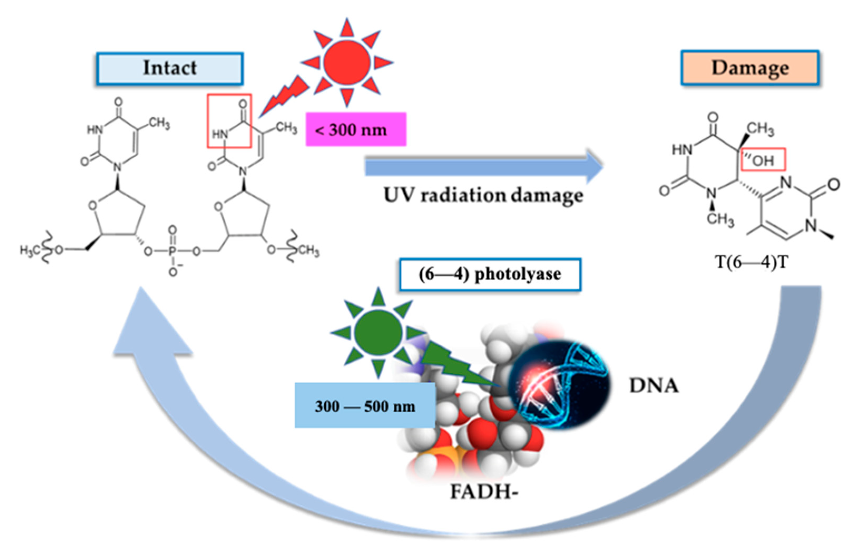
Photolyases are specialized DNA repair enzymes capable of directly reversing ultraviolet (UV)-induced DNA lesions through a process known as photoreactivation [31]. These enzymes belong to the broader flavoprotein family and require visible light—particularly within the blue to near-UV spectrum—for their activity. Photolyases can be observed in fish, amphibians, birds, and a few marsupials. However, in higher plants and animals, DNA repair has been lost during evolution. Therefore, their function is limited to growth regulation and functioning as blue light photoreceptors. In skin health, photolyases represent a promising mechanism for mitigating the harmful effects of UV exposure, including mutagenesis, inflammation, and photoaging (Figure 4) [32].
Although absent in humans, photolyases are naturally expressed in various organisms and can be delivered topically. Recent advances in dermatological science have enabled their incorporation into topical formulations such as liposomal sprays and creams [33]. These products aim to support the skin’s endogenous DNA repair mechanisms, particularly in individuals with high cumulative sun exposure or impaired repair capacity. Photolyases exert their protective effects by recognizing and repairing specific UV-induced DNA lesions, primarily cyclobutane pyrimidine dimers (CPDs) and, in the case of a subclass known as 6—4 photolyases, (6—4) photoproducts [34]. These lesions distort the DNA double helix, disrupt base pairing, and interfere with vital cellular processes such as transcription and replication. In skin cells, such disruptions contribute to sunburn via apoptosis and increase the risk of skin cancer by introducing mutagenic errors during cell division. Upon locating and binding to a DNA lesion, photolyase becomes activated when exposed to light in the 300–500 nm range—typically within the blue portion of the visible spectrum [35]. Two essential cofactors capture the absorbed light energy: reduced flavin adenine dinucleotide (FADH), which initiates electron transfer to cleave abnormal covalent bonds in the lesion, and an antenna chromophore, which acts as a light-harvesting molecule. Common antenna chromophores include methenyltetrahydrofolate (MTHF) or 8-hydroxy-5-deazaflavin, depending on the organism of origin. These cofactors work synergistically to ensure rapid and efficient repair of UV-induced DNA damage [36,37].
This light-dependent repair mechanism is rapid, accurate, and mutation-free, offering a significant advantage over excision-based repair systems [33]. Unlike other enzymes used in dermatological applications—such as T4 endonuclease V or 8-oxoguanine glycosylase, which operate through light-independent (dark) mechanisms—photolyases require visible light as an energy source, not as a target of protection. This allows for immediate correction of DNA damage in the skin, especially after sun exposure, when CPD accumulation in epidermal keratinocytes is highest [35]. Most organisms, including humans, lack mechanisms to mitigate the effects of long-term exposure to UV radiation, which causes DNA damage that can ultimately lead to the development of skin diseases.
In topical formulations, photolyase cofactors are stabilized or encapsulated (e.g., in liposomes) to preserve their activity upon application. Liposomal delivery systems enhance enzyme stability and facilitate effective skin penetration, ensuring that photolyases can reach target cells in the epidermis and repair DNA damage within keratinocytes [38].

3.2. T4-bacteriophage Endonuclease V (T4 Endonuclease V)

T4 Endonuclease V (T4N5) is a 16.5 kDa polypeptide isolated in 1975 from Escherichia coli infected with T4 bacteriophage by Tanaka et al. [39] It plays a critical role in repairing UV-induced DNA lesions, particularly cyclobutane pyrimidine dimers (CPDs), which, if left unrepaired, can give rise to mutagenic events associated with actinic keratosis and nonmelanoma skin cancers (NMSCs). By enhancing nucleotide excision repair pathways, T4N5 accelerates the correction of UV-induced DNA damage by up to fourfold and contributes to protecting UV-compromised cells.
Mechanistically, T4N5 recognizes UV-induced CPDs and initiates repair through a dual-action process involving pyrimidine dimer–DNA glycosylase activity and apurinic/apyrimidinic endonuclease activity. The enzyme binds to DNA in a salt-dependent manner and uses facilitated diffusion to locate damage sites, initiating targeted repair with high efficiency. This enzyme can enhance natural DNA repair processes by up to about fourfold. In addition, T4 endonuclease V promotes skin regeneration and repair while preventing the breakdown of extracellular matrix components, thus helping to combat photoaging. T4N5 treatment can reduce MMP-1 induction in human skin cells similarly to photolyase treatment, resulting in reduced collagen degradation.
Furthermore, a single T4N5 polypeptide can successfully replace the human multienzyme complex to initiate excision repair, effectively repairing CPD [3]. In the 1980s, researchers demonstrated that T4N5 could be encapsulated in liposomes, significantly improving the enzyme’s stability, facilitating percutaneous absorption, and ensuring efficient cellular delivery. In vitro experiments have confirmed that liposomal formulations of T4N5 enhance DNA repair capacity and modulate UV-induced responses, such as the suppression of MMP-1 expression, thereby reducing collagen degradation and helping to maintain the extracellular matrix [40]. Clinically, topical application of T4N5 liposomes has been shown to reduce the incidence of actinic keratosis (AK) and basal cell carcinoma (BCC), with long-term studies indicating sustained efficacy even after treatment cessation. Furthermore, preclinical studies in murine models support its use as an additive in broad-spectrum sunscreens, reducing UV-induced damage, including the formation of sunburn cells [41].

3.3. 8-Oxoguanine Glycosylase

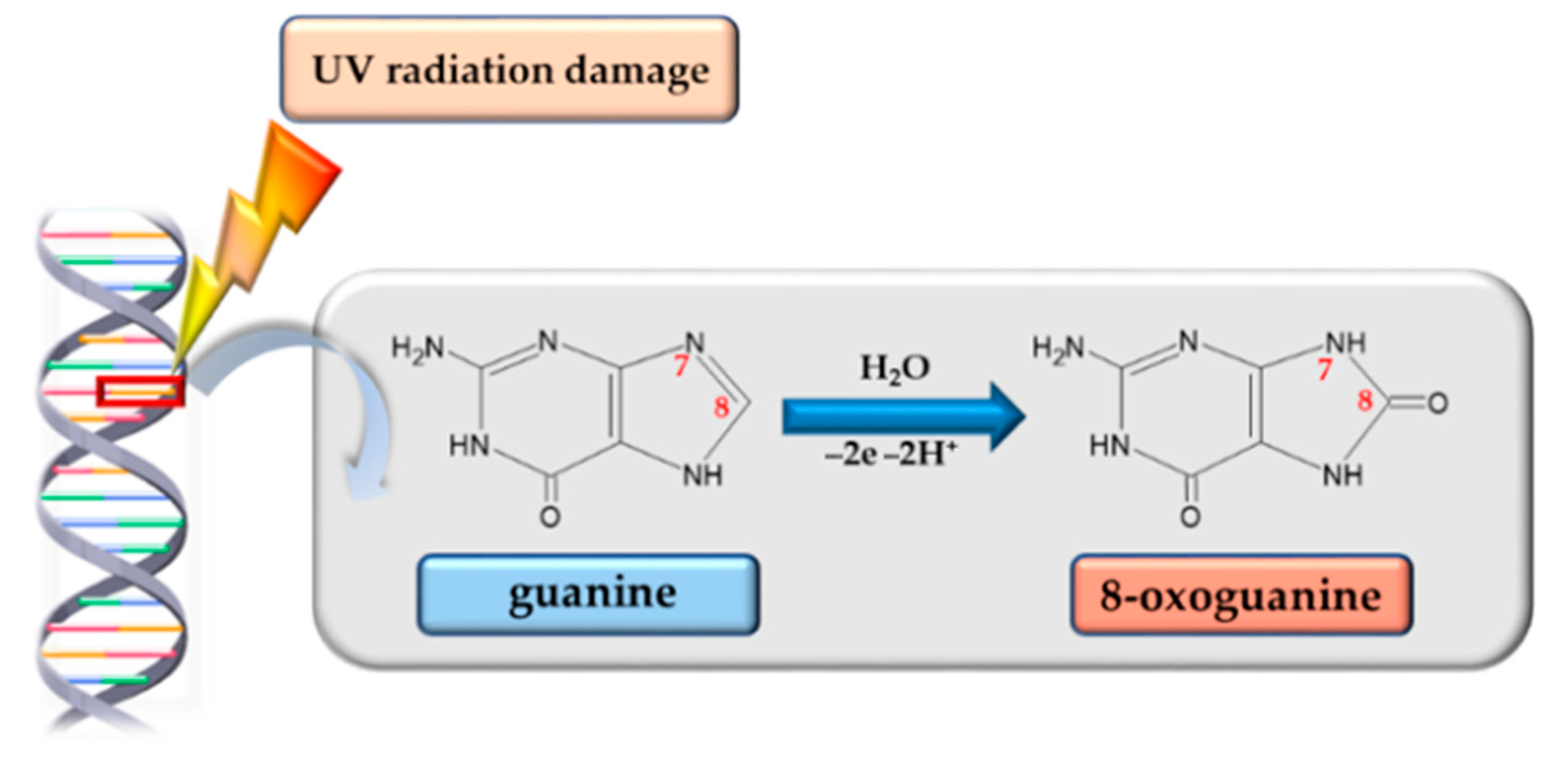
Various environmental stimuli can trigger the formation of reactive oxygen species (ROS), either directly or through secondary signaling pathways. At physiological levels, ROS act as important intracellular messengers; however, their excessive accumulation disrupts redox balance, causing oxidative stress and promoting the aberrant oxidation of essential biomolecules. Among DNA bases, guanine is particularly prone to oxidative damage due to its low redox potential. This leads to the formation of 7,8-dihydro-8-oxoguanine (8-oxoG), the most prevalent oxidative lesion, typically occurring at a frequency of 2–3 lesions per 106 guanine bases [42]. Guanine oxidation can result from direct ROS interaction or via charge transfer mechanisms, especially in 5′-GG-3′ sequences, which are especially susceptible [43].
If unrepaired, 8-oxoG can mispair with adenine during replication, leading to G:C → T:A transversions—a mutagenic event associated with cancer development. To prevent such outcomes, cells utilize the evolutionarily conserved guanine oxidation (GO) repair system. Initially described in prokaryotes, this system comprises three enzymes—MutT, MutM, and MutY—whose human homologs are NUDT1 (also known as MTH1), 8-oxoguanine DNA glycosylase (OGG1), and MUTYH. Together, they eliminate oxidized guanine nucleotides and restore base fidelity in DNA (Figure 5) [44].
Experimental studies highlight the clinical relevance of this system: topical delivery of OGG1, for example, does not alter UV-B-induced tumor multiplicity but significantly reduces tumor volume and suppresses malignant progression [45].

3.4. Base and Nucleotide Excision Repair Mechanisms in UV-Induced Damage

In the context of UV-induced DNA damage, two primary repair pathways operate in skin cells: Base Excision Repair (BER) and Nucleotide Excision Repair (NER). BER is responsible for repairing small, non-helix-distorting base lesions caused by oxidative stress, such as 8-oxoguanine, and is facilitated by enzymes like 8-oxoguanine glycosylase (OGG1). This process involves recognition, excision of the damaged base, and restoration of the correct nucleotide, preserving genomic stability [46]. NER, in contrast, addresses bulky, helix-distorting lesions such as cyclobutane pyrimidine dimers and 6—4 photoproducts resulting from UV exposure. It involves damage recognition, removal of a short single-stranded DNA segment containing the lesion, and resynthesis using the undamaged strand as a template. The integration of DNA repair enzymes into dermocosmetic formulations aligns with these cellular repair processes, enhancing the skin’s natural ability to correct UV-induced damage and reduce the risk of photoaging and photocarcinogenesis. The complementary roles of BER and NER in addressing UV-induced DNA lesions and maintaining genomic stability have been comprehensively discussed [4]. Photoprotection encompasses a broad range of behavioral, physical, and chemical measures. These aim to prevent or limit the acute and chronic harmful effects of ultraviolet radiation (UVR) on human skin. The most effective photostrategy is undoubtedly avoiding sun exposure, but this is extremely difficult and often impractical [47]. Broad-spectrum (BSP) sunscreens typically consist of mineral and organic compounds (filters) capable of scattering and neutralizing radiation of various wavelengths [48]. Due to ease of application, ongoing advances in pharmacology, and increasing awareness, their use has become widespread. Based on their mechanism of action, sunscreens can be divided into “passive” and “active” sunscreens (Figure 6).
Passive photoprotection, the most commonly used approach, involves the topical application of ultraviolet (UVR) filters. However, these filters are ineffective in reversing radiation-induced skin damage. To address this gap, research is being conducted using DNA repair enzymes [47,48].

3.5. Synergistic Use of DNA Repair Enzymes and Antioxidants

Recent studies have highlighted the potential of combining DNA repair enzymes with topical antioxidants to enhance the skin’s defense mechanisms against UV-induced damage. While DNA repair enzymes such as photolyase or OGG1 directly address molecular lesions, antioxidants like vitamin C, E, and polyphenols prevent the formation of ROS that cause secondary DNA damage. This dual strategy may help protect cellular structures and preserve extracellular matrix integrity more effectively than either component alone [3,49]. In vitro and ex vivo studies have demonstrated that co-formulations can reduce markers of inflammation (e.g., IL-6, TNF-α), lower levels of oxidative lesions like 8-oxoG, and prevent collagen breakdown. Additionally, antioxidants may stabilize enzymes within cosmetic formulations and enhance their shelf life by reducing oxidative degradation [50]. However, clinical trials evaluating the synergy between antioxidants and repair enzymes are scarce. Future research should validate whether such combinations yield additive or synergistic effects on clinical endpoints like wrinkle reduction, pigment uniformity, or epidermal thickness in photoaged skin. This approach reflects a shift from unifunctional sunscreens to multi-active photoprotective formulations aligned with modern cosmeceutical trends.

4. Clinical and Cosmetic Relevance

Advancements in understanding cutaneous biology have facilitated the development of various anti-aging strategies and pharmacological agents to promote skin regeneration and repair. According to current expert consensus, an ideal sunscreen should offer broad-spectrum protection against both UV-B and UV-A radiation, demonstrate reactive oxygen species (ROS)-neutralizing capacity, exhibit high photostability, ensure a favorable safety profile, and ideally include DNA repair-enhancing enzymes. Emerging evidence suggests that incorporating such enzymes into conventional sunscreen formulations provides superior protection against UV-induced molecular damage compared to traditional sunscreens alone. However, integrating DNA repair enzymes into topical formulations presents formulation challenges—particularly regarding enzyme stability and desirable cosmetic attributes. Due to their macromolecular and hydrophilic nature, enzymes can negatively impact the texture, viscosity, and absorption of creams or lotions. Without proper encapsulation—such as through liposomal or nanocarrier systems—these formulations may feel sticky and greasy or leave visible residues on the skin, potentially reducing user acceptance and long-term compliance. Therefore, from both cosmetic and clinical perspectives, it is essential to balance biochemical efficacy with user-friendly sensory properties during product development [51]. The most important examples of DNA repair enzymes are presented in Table 2.
Recent research has focused on developing sunscreen formulations with enhanced photoprotective properties. One example is Eryfotona® AK-NMSC (Isdin SA, Provencals, Barcelona, Spain), a CPD photolyase cream that has demonstrated clinical and histological efficacy in managing actinic keratosis (AK). Light-induced and ROS-mediated DNA damage significantly contributes to the pathogenesis of AK and is closely linked to molecular mechanisms underlying photoaging. In response, novel formulations have been designed to incorporate DNA repair enzymes alongside traditional UV filters [9].
Ateia® (Kwizda Pharma, Vienna, Austria) is one such product, combining standard photofilters with Nopasome®—a proprietary complex that includes liposome-encapsulated CPD photolyase (Photosome®), T4 endonuclease V (Ultrasome®), and prickly pear cactus extract. Another example is Ladival® med (STADA Arzneimittel, Bad Vilbel, Germany), a commercially available sunscreen containing photolyase, offered in SPF 15 and 20 variants. These formulations illustrate a growing trend toward multifunctional sunscreens integrating clinical dermatology with cosmetic innovation. Table 3 presents selected commercial sunscreens available in European and U.S. markets that contain DNA repair enzymes and are supported by scientific literature regarding their efficacy [55]. The products listed serve illustrative and educational purposes only and do not imply endorsement. Given the dynamic nature of the cosmetics and dermatology industries, new products and updated formulations may emerge. The examples reflect current trends in integrating DNA repair enzymes into evidence-based photoprotection strategies.
It should be noted that the listed products represent only a selection of commercially available formulations. Due to the dynamic nature of the cosmetics and dermatology market, new products may emerge, and formulations may change over time. The examples provided are based on published studies and illustrate how DNA repair enzymes are currently being integrated into clinical and cosmetic photoprotection strategies.
Recent studies further suggest that combining DNA repair enzymes with topical antioxidants may yield synergistic effects in protecting against UV-induced skin damage, including photocarcinogenesis and photoaging. However, clinical evidence remains limited, particularly regarding anti-aging outcomes in humans. Only one clinical trial has demonstrated a measurable benefit of enzyme-based sunscreens on skin aging parameters [60]. Unlike prior overviews, Table 3 includes only products evaluated in peer-reviewed clinical studies, helping readers distinguish between marketed claims and evidence-based efficacy.

5. Ethical and Regulatory Barriers in the Use of DNA Repair Enzymes in Cosmetics

In the EU, enzyme-based skincare products are primarily regulated under the Cosmetics Regulation (No 1223/2009 of the European Parliament and of the Council of 30 November 2009 on EC cosmetic products. OJ L 342, 22.12.2009, p. 59–209) [61], requiring safety assessments, labeling compliance, and notification through the CPNP, while some enzyme-containing products may be classified as medical devices depending on claims. In the U.S, the FDA distinguishes between cosmetics and drugs based on intended use, and products making therapeutic claims may require OTC monograph compliance or New Drug Applications. In Asia, regulatory approaches vary, with Japan’s quasi-drug category and Korea’s functional cosmetics regulations, requiring efficacy data for enzyme claims [62,63].
It is essential to ensure transparency in enzyme efficacy and safety claims, avoiding overstatement of anti-aging or cancer prevention benefits without robust clinical data. Ethical marketing should inform consumers about the limitations of enzyme-based products and avoid implying they replace medical treatment. Enzymes used in topical formulations may pose allergenicity or irritation risks; thus, rigorous testing, including patch testing and long-term tolerability studies, is essential. Encapsulation techniques may mitigate immunogenicity while improving stability [64].
Sourcing enzymes sustainably requires consideration of production methods, energy consumption, and waste management. Using recombinant enzyme production in controlled systems reduces environmental impacts. Adopting ESG principles in the supply chain aligns enzyme-based photoprotection with sustainability goals, meeting consumer demand for environmentally responsible skincare products [62].

6. Barriers to the Use of DNA Repair Enzymes in Sunscreens

A primary limitation of using DNA repair enzymes in sunscreens is their limited skin penetration. Although liposomal encapsulation has been employed to enhance delivery, current evidence does not conclusively support its preventive effects on human photoaging. Most clinical studies have focused on endpoints such as lesion count or carcinogenesis, with relatively few addressing validated aging markers like skin elasticity, hydration, or wrinkle depth. Moreover, the lack of long-term, head-to-head clinical trials comparing enzyme-containing sunscreens to conventional formulations significantly limits conclusions regarding their anti-aging efficacy. Short follow-up durations and insufficient use of standardized dermatological scoring systems or imaging tools (e.g., cutometry, TEWL, high-resolution wrinkle grading) further constrain interpretability. Future studies should incorporate larger cohorts and focus specifically on quantifiable markers of skin aging to establish the true benefit of enzyme-based formulations in this domain [65].
Another concern is potential allergenicity. As exogenous proteins, DNA repair enzymes such as photolyase or T4 endonuclease V may provoke cutaneous hypersensitivity in susceptible individuals. Although the risk appears low, isolated reports have noted mild skin irritation, erythema, or contact sensitivity, particularly in those with a history of atopic or allergic contact dermatitis. To ensure safe use—especially for long-term or daily application on sensitive skin—such formulations should undergo rigorous allergenicity and tolerability testing before market approval [19,40].

7. Future Directions and Emerging Technologies

As the field of photoprotection evolves beyond conventional sunscreens, future research is increasingly focused on novel DNA repair strategies and delivery technologies that address current limitations while opening new therapeutic and cosmetic possibilities. One promising area involves the exploration of CRISPR-based or engineered DNA repair systems. Although still at an early experimental stage, modified CRISPR/Cas platforms may offer highly specific recognition and correction of UV-induced DNA lesions without introducing double-strand breaks. Synthetic DNA glycosylases or recombinase-based technologies could further expand the toolbox for targeted repair, especially in individuals with compromised repair capacity or high susceptibility to photo-induced skin disorders [66,67].
Simultaneously, nanotechnology is expected to be critical in overcoming key formulation and delivery barriers associated with enzyme-based skin products. Advanced nanocarriers—including liposomes, polymeric nanoparticles, dendrimers, and exosome-mimetic vesicles—are being developed to enhance the skin penetration, stability, and bioavailability of macromolecular repair enzymes [68,69]. These systems can also facilitate co-delivery of complementary agents such as antioxidants or peptides, increasing therapeutic synergy while maintaining desirable cosmetic properties [70].
Another emerging concept is the development of “smart” or responsive formulations. These systems are designed to activate under specific environmental stimuli, such as UV radiation or oxidative stress, allowing for targeted release of DNA repair enzymes when and where they are most needed. This approach holds the potential to reduce enzyme degradation, minimize unwanted exposure, and increase formulation efficiency [71,72].
Finally, integrating DNA repair actives into personalized skincare regimens represents an exciting future direction. Advances in genomics and molecular diagnostics may soon allow for individualized risk profiling based on a person’s DNA repair capacity or skin phototype. This could enable the creation of customized sunscreens or cosmeceuticals tailored to a user’s genetic predisposition to photoaging, pigmentation disorders, or skin cancers [73,74]. Together, these innovations suggest a shift toward more biologically intelligent, evidence-driven, and patient-centered approaches in dermatological photoprotection.

8. Perspectives of Personalization

As photoprotection research develops, there is growing interest in personalizing skin protection strategies using DNA repair enzymes. It has been shown that skin phototype (according to the Fitzpatrick scale) affects susceptibility to UV damage and the effectiveness of endogenous repair mechanisms, which may determine the selection of repair enzymes in dermocosmetic products. In people with lighter phototypes, who are more susceptible to the formation of CPD, preparations with photolyase and T4 endonuclease V may be indicated, while in people with darker phototypes, where oxidative processes dominate, products containing 8-oxoguanine glycosylase (OGG1) in synergy with antioxidants may potentially bring greater benefits [75].
In addition, the progressive implementation of genetic profiling in dermatology allows for the identification of polymorphisms in genes encoding DNA repair enzymes, e.g., OGG1 Ser326Cys, which may affect the effectiveness of oxidative damage repair and predisposition to photoaging or skin cancer. In the future, this may enable the selection of products containing specific repair enzymes depending on the patient’s genetic profile, supporting precise photoprotection [76,77].
At the same time, the trend of personalized dermocosmetics is developing, which adjust the composition to the user’s skin parameters, such as the level of hydration, the degree of UV damage, or lipid profile. Integration of DNA repair enzymes within such personalized products may be the next step in the evolution of dermocosmetics from passive protection to active skin regeneration, responding to individual needs related to age, lifestyle, and environmental exposure [78].

9. Conclusions

In recent years, significant progress has been made in understanding the molecular mechanisms of photoaging and the role of DNA damage in skin degeneration. While traditional sunscreens offer passive protection by blocking or reflecting ultraviolet radiation (UVR), they are ineffective once DNA damage has occurred. Incorporating DNA repair enzymes into topical formulations represents a promising strategy beyond prevention, offering active photoprotection by restoring DNA integrity. Enzymes such as photolyase, T4 endonuclease V, and 8-oxoguanine glycosylase have demonstrated the ability to reduce UV-induced genotoxicity and may help mitigate the risk of skin cancer and premature skin aging. Integrating DNA repair enzymes into sunscreens and dermatological products provides a novel and scientifically supported approach to protecting skin health from increasing UV exposure. To assess the translational feasibility and broader dermatological relevance of topical DNA repair enzymes, a strategic SWOT (Strengths, Weaknesses, Opportunities, Threats) analysis can be used. This tool helps contextualize the emerging role of these agents in the clinical and consumer photoprotection landscape (Table 4).
Future research should aim to optimize delivery systems, validate long-term clinical benefits, and further explore synergistic effects with antioxidants and other bioactive compounds. As the demand for multifunctional cosmeceuticals grows, DNA repair enzyme-based products may become essential to next-generation photoprotection strategies.

Author Contributions

E.M., writing—original draft preparation, writing—review and editing; V.K.-K., conceptualization, funding acquisition, writing—original draft preparation, writing—review and editing. All authors have read and agreed to the published version of the manuscript.

Funding

This research received no external funding.

Institutional Review Board Statement

Not applicable.

Informed Consent Statement

Not applicable.

Data Availability Statement

Data will be available if requested.

Conflicts of Interest

The authors declare no conflicts of interest.

References

  1. Nascimento, N.S.; Torres-Obreque, K.M.; Oliveira, C.A.; Rabelo, J.; Baby, A.R.; Long, P.F.; Young, A.R.; Rangel-Yagui, C.d.O. Enzymes for dermatological use. Exp. Dermatol. 2024, 33, 15008. [Google Scholar] [CrossRef]
  2. Passeron, T.; Krutmann, J.; Andersen, M.; Katta, R.; Zouboulis, C. Clinical and biological impact of the exposome on the skin. J. Eur. Acad. Dermatol. Venereol. 2020, 34, 4–25. [Google Scholar] [CrossRef]
  3. Garcia-Mouronte, E.; Pérez-González, L.A.; Naharro-Rodriguez, J.; Fernández Guarino, M. Understanding Active Photoprotection: DNA-Repair Enzymes and Antioxidants. Life 2024, 14, 822. [Google Scholar] [CrossRef]
  4. Luze, H.; Nischwitz, S.P.; Zalaudek, I.; Müllegger, R.; Kamolz, L.P. DNA repair enzymes in sunscreens and their impact onphotoageing-A systematic review. Photodermatol. Photoimmunol. Photomed. 2020, 36, 424–432. [Google Scholar] [CrossRef]
  5. Qian, M.; Kalbina, I.; Rosenqvist, E.; Jansen, M.A.K.; Strid, A. Supplementary UV-A and UV-B radiation differentially regulate morphology in Ocimum basilicum. Photochem. Photobiol. Sci. 2023, 22, 2219–2230. [Google Scholar] [CrossRef]
  6. Pfeifer, G.P. Mechanisms of UV-induced mutations and skin cancer. Genome Instab. Dis. 2020, 1, 99–113. [Google Scholar] [CrossRef]
  7. Hong, Y.; Boiti, A.; Vallone, D.; Foulkes, N.S. Reactive Oxygen Species Signaling and Oxidative Stress: Transcriptional Regulation and Evolution. Antioxidants 2024, 13, 312. [Google Scholar] [CrossRef] [PubMed]
  8. Tang, H.; Yang, T.; Yu, D.; Xiong, H.; Zhang, S. Current insights and future perspectives of ultraviolet radiation (UV) exposure: Friends and foes to the skin and beyond the skin. Environ. Int. 2024, 185, 160–4120. [Google Scholar] [CrossRef] [PubMed]
  9. Lee, J.W.; Ratnakumar, K.; Hung, K.F.; Rokunohe, D.; Kawasumi, M. Deciphering UV-induced DNA Damage Responses to Prevent and Treat Skin Cancer. Photochem. Photobiol. 2020, 96, 478–499. [Google Scholar] [CrossRef]
  10. Lv, X.; Han, Z.; Huo, H.; Liu, X.; Zhang, J.; Chi, J.; Han, B.; Jiang, Z. Anti-inflammatory and antioxidant succinyl-chitosan oligosaccharide protects human epidermal cell and mouse skin against ultraviolet B-induced photodamage. Carbohydr. Polym. 2025, 351, 123102. [Google Scholar] [CrossRef] [PubMed]
  11. Jin, S.G.; Padron, F.; Gerd, P.; Pfeifer, G.P. UVA Radiation, DNA Damage, and Melanoma. ACS Omega 2022, 7, 32936–32948. [Google Scholar] [CrossRef] [PubMed]
  12. Kciuk, M.; Marciniak, B.; Mojzych, M.; Kontek, R. Focus on UV-Induced DNA Damage and Repair—Disease Relevance and Protective Strategies. Int. J. Mol. Sci. 2020, 21, 7264. [Google Scholar] [CrossRef] [PubMed]
  13. Hung, K.F.; Sidorova, J.M.; Nghiem, P.; Kawasumi, M. The 6-4 photoproduct is the trigger of UV-induced replication blockage and ATR activation. Proc. Natl. Acad. Sci. USA 2020, 117, 12806–12816. [Google Scholar] [CrossRef] [PubMed]
  14. Nakai, K.; Tsuruta, D. What Are Reactive Oxygen Species, Free Radicals, and Oxidative Stress in Skin Diseases? Int. J. Mol. Sci. 2021, 22, 10799. [Google Scholar] [CrossRef]
  15. Sies, H.; Jones, D.P. Reactive oxygen species (ROS) as pleiotropic physiological signalling agents. Nat. Rev. Mol. Cell Biol. 2020, 21, 363–383. [Google Scholar] [CrossRef]
  16. Herr, L.M.; Schaffer, E.D.; Fuchs, K.F.; Datta, A.; Brosh, R.M. Replication stress as a driver of cellular senescence and aging. Commun. Biol. 2024, 7, 616. [Google Scholar] [CrossRef]
  17. Provasek, V.E.; Mitra, J.; Malojirao, V.H.; Hegde, M.L. DNA Double-Strand Breaks as Pathogenic Lesions in Neurological Disorders. Int. J. Mol. Sci. 2022, 23, 4653. [Google Scholar] [CrossRef]
  18. Rai, S.; Rai, G.; Kumar, A. Eco-evolutionary impact of ultraviolet radiation (UVR) exposure on microorganisms, with a specialfocus on our skin microbiome. Microbiol. Res. 2022, 260, 127044. [Google Scholar] [CrossRef]
  19. Milićević, S.M.; Mijatović, Z.; Stanojević, G.; Radovanović, M.M.; Popović, V. Health risks of extended exposure to low-level UV radiation—An analysis of ground-based and satellite-derived data. Sci. Total Environ. 2022, 831, 154899. [Google Scholar] [CrossRef]
  20. Available online: https://www.cdc.gov/radiation-health/features/uv-radiation.html (accessed on 18 May 2025).
  21. Thawabteh, A.M.; Jibreen, A.; Karaman, D.; Thawabteh, A.; Karaman, R. Skin Pigmentation Types, Causes and Treatment-A Review. Molecules 2023, 28, 4839. [Google Scholar] [CrossRef]
  22. Shin, S.H.; Lee, Y.H.; Rho, N.K.; Park, K.Y. Skin aging from mechanisms to interventions: Focusing on dermal aging. Front. Physiol. 2023, 14, 1195272. [Google Scholar] [CrossRef]
  23. He, X.; Wan, F.; Su, W.; Xie, W. Research Progress on Skin Aging and Active Ingredients. Molecules 2023, 28, 5556. [Google Scholar] [CrossRef]
  24. Milanowska, M.; Grudzińska, A.; Jarosz, D.; Tsitko, H.; Dudzińska, P. Skin cancer’s prevention in the light of current medical knowledge. J. Educ. Health Sport 2023, 23, 35–39. [Google Scholar] [CrossRef]
  25. Subhadarshani, S.; Athar, M.; Elmets, C.A. Photocarcinogenesis. Curr. Derm. Rep. 2020, 9, 189–199. [Google Scholar] [CrossRef]
  26. Kaltchenko, M.V.; Chien, A.L. Photoaging: Current Concepts on Molecular Mechanisms, Prevention, and Treatment. Am. J. Clin. Dermatol. 2025, 26, 321–344. [Google Scholar] [CrossRef]
  27. Douki, T.; Caillat, S.; Bacqueville, D.; Géniès, C.; Huyghe, C.; Duplan, H.; Le Digabel, J.; Lauze, C.; Filiol, J.; Marinescu, R.; et al. Nuclear and Urinary Measurements Show the Efficacy of Sun-Protection Factor 50+ Sunscreen against DNA Photoproducts upon Real-Life Recreational Exposure. JID Innov. 2023, 3, 100227. [Google Scholar] [CrossRef]
  28. Grand View Research. Enzymes Market Size, Share & Trends Analysis Report, 2030. 2020. Available online: https://www.grandviewresearch.com/industry-analysis/specialty-enzymes-market (accessed on 9 May 2025).
  29. D’Augustin, O.; Huet, S.; Campalans, A.; Radicella, J.P. Lost in the Crowd: How Does Human 8-Oxoguanine DNA Glycosylase 1 (OGG1) Find 8-Oxoguanine in the Genome? Int. J. Mol. Sci. 2020, 21, 8360. [Google Scholar] [CrossRef]
  30. Nitulescu, G.; Lupuliasa, D.; Adam-Dima, I.; Nitulescu, G.M. Ultraviolet Filters for Cosmetic Applications. Cosmetics 2023, 10, 101. [Google Scholar] [CrossRef]
  31. Hosokawa, Y.; Maestre-Reyna, M. Light-driven DNA repair at atomic and picosecond resolution revealed via time-resolved serial femtosecond crystallography. Clin. Transl. Med. 2024, 14, 1566. [Google Scholar] [CrossRef]
  32. Ramírez-Gamboa, D.; Díaz-Zamorano, A.L.; Meléndez-Sánchez, E.R.; Reyes-Pardo, H.; Villaseñor-Zepeda, K.R.; López-Arellanes, M.E.; Sosa-Hernández, J.E.; Coronado-Apodaca, K.G.; Gámez-Méndez, A.; Afewerki, S.; et al. Photolyase Production and Current Applications: A Review. Molecules 2022, 27, 5998. [Google Scholar] [CrossRef]
  33. Haney, A.M.; Sanfilippo, J.E.; Garczarek, L.; Partensky, F.; Kehoe, D.M. Multiple photolyases protect the marine cyanobacterium Synechococcus from ultraviolet radiation. Mbio 2022, 13, 1511–1522. [Google Scholar] [CrossRef]
  34. Wang, Z.; Li, Z.; Lei, Y.; Liu, Y.; Feng, Y.; Chen, D.; Ma, S.; Xiao, Z.; Hu, M.; Deng, J.; et al. Recombinant Photolyase-Thymine Alleviated UVB-Induced Photodamage in Mice by Repairing CPD Photoproducts and Ameliorating Oxidative Stress. Antioxidants 2022, 11, 2312. [Google Scholar] [CrossRef]
  35. Wang, P.-H.; Hosokawa, Y.; Soares, J.C.; Emmerich, H.-J.; Fuchs, V.; Caramello, N.; Engilberge, S.; Bologna, A.; Rosner, C.J.; Nakamura, M.; et al. Redox-State-Dependent Structural Changes within a Prokaryotic 6–4 Photolyase. J. Am. Chem. Soc. 2025, 147, 16084–16098. [Google Scholar] [CrossRef]
  36. Ramírez, N.; Serey, M.; Illanes, A.; Piumetti, M.; Ottone, C. Immobilization strategies of photolyases: Challenges and perspectives for DNA repairing application. J. Photochem. Photobiol B Biol. 2021, 215, 112113. [Google Scholar] [CrossRef] [PubMed]
  37. Granger, C.; Trullàs, C.; Bauza, G.; Garre, A. 17895 Anti-photoaging effect of a novel tinted facial sunscreen with high sun protection, peptide complex, and encapsulated photolyase after 1 month of use. J. Am. Acad. Dermatol. 2020, 83, AB202. [Google Scholar] [CrossRef]
  38. Patel, V.A.; Arron, S.T.; Berman, B.; Chapman, M.S.; Jambusaria-Pahlajani, A.; Martin, G.; Rossi, A.M.; Schlesinger, T.; Zeitouni, N.C.; Bhatia, N. Expert consensus-based recommendations on the use of photodynamic therapy in actinic keratosis patients. JAAD Int. 2025, 20, 62–73. [Google Scholar] [CrossRef]
  39. Gupta, A.; Singh, A.P.; Singh, V.K.; Singh, P.R.; Jaiswal, J.; Kumari, N.; Upadhye, V.; Singh, S.C.; Sinha, R.P. Natural Sun-Screening Compounds and DNA-Repair Enzymes: Photoprotection and Photoaging. Catalysts 2023, 13, 745. [Google Scholar] [CrossRef]
  40. Ivanova, I.; Svilenska, T.; Maisch, T.; Karrer, S.; Niebel, D.; Berneburg, M.; Kurz, B. The role of UV-induced cutaneous matrix metalloproteinases and mi-RNAs in the pathogenesis of lupus erythematosus. J. Trans. Autoim. 2025, 10, 100265. [Google Scholar] [CrossRef]
  41. De Rosa, M.; Johnson, S.A.; Opresko, P.L. Roles for the 8-Oxoguanine DNA Repair System in Protecting Telomeres From Oxidative Stress. Front. Cell Dev. Biol. 2021, 9, 758402. [Google Scholar] [CrossRef]
  42. De Rosa, M.; Barnes, R.P.; Detwiler, A.C.; Nyalapatla, P.R.; Wipf, P.; Opresko, P.L. OGG1 and MUTYH repair activities promote telomeric 8-oxoguanine induced senescence in human fibroblasts. Nat. Commun. 2025, 16, 893. [Google Scholar] [CrossRef]
  43. Wang, J.; Li, C.; Han, J.; Xue, Y.; Zheng, X.; Wang, R.; Radak, Z.; Nakabeppu, Y.; Boldogh, I.; Ba, X. Reassessing the roles of oxidative DNA base lesion 8-oxoGua and repair enzyme OGG1 in tumorigenesis. J. Biomed. Sci. 2025, 32, 1. [Google Scholar] [CrossRef] [PubMed]
  44. Caldecott, K.W. Mammalian DNA Base Excision Repair: Dancing in the Moonlight. DNA Repair 2020, 93, 102921. [Google Scholar] [CrossRef] [PubMed]
  45. Marciniak, E.; Osuch, B.; Młotkowska, P.; Kowalczyk, P.; Roszkowicz-Ostrowska, K.; Misztal, T. Gene Expression and Activity of Selected Antioxidant and DNA Repair Enzymes in the Prefrontal Cortex of Sheep as Affected by Kynurenic Acid. Int. J. Mol. Sci. 2025, 26, 2381. [Google Scholar] [CrossRef] [PubMed]
  46. Abril, S.P.; Rincón-Díaz, N.; Puyana, M.; Castellanos, L.; Ramos, F.A. Photoprotective activity from Colombian Caribbean brown algae using HPLC-DAD metabolic profiling by MCR-ALS data analysis. Sci. Rep. 2025, 15, 16204. [Google Scholar] [CrossRef]
  47. Patwa, H.; Babcock, N.; Kurian, P. Quantum-enhanced photoprotection in neuroprotein architectures emerges from collective light-matter interactions. Front. Phys. 2024, 12, 1387271. [Google Scholar] [CrossRef]
  48. Erber, L.; Groehler, A.S.; Cyuzuzo, C.I.; Baker-Wainwright, J.; Maskey, R.S.; Li, L.; Machida, Y.J.; Tretyakova, N. SPRTN metalloprotease participates in repair of ROS-mediated DNA-protein crosslinks. Sci. Rep. 2024, 14, 30919. [Google Scholar] [CrossRef]
  49. Neacșu, S.M.; Mititelu, M.; Ozon, E.A.; Musuc, A.M.; Iuga, I.D.M.; Manolescu, B.N.; Petrescu, S.; Cusu, J.P.; Rusu, A.; Surdu, V.-A.; et al. Comprehensive Analysis of Novel Synergistic Antioxidant Formulations: Insights into Pharmacotechnical, Physical, Chemical, and Antioxidant Properties. Pharmaceuticals 2024, 17, 690. [Google Scholar] [CrossRef]
  50. Wang, Y.; Deng, X.; Zhou, M. DNA damage mediated by UV radiation and relative repair mechanisms in mammals. Genome Instab. Dis. 2022, 3, 331–337. [Google Scholar] [CrossRef]
  51. Minoretti, P.; Emanuele, E.; García Martín, Á.; Liaño Riera, M.; Gómez Serrano, M.; Santiago Sáez, A. Exploring the Protective Efficacy of Topical Products for Actinic Keratosis Against Ultraviolet-Induced DNA and Protein Damage: An Experimental, Double-Blind Irradiation Study. Cureus 2023, 15, 44065. [Google Scholar] [CrossRef]
  52. Cao, X.; Luyao Yan, L.; Yang, C.; Wang, L.; Zhang, M.; Zhong, D. Dynamics of DNA Repair by Class-II Photolyases via a Unified Electron-Transfer Bifurcating Mechanism. J. Am. Chem. Soc. 2025, 147, 11291–11300. [Google Scholar] [CrossRef]
  53. Kong, X.Y.; Vik, E.S.; Nawaz, M.S.; Berges, N.; Dahl, T.B.; Vågbø, C.; Suganthan, R.; Segers, F.; Holm, S.; Quiles-Jiménez, A.; et al. Deletion of Endonuclease V suppresses chemically induced hepatocellular carcinoma. Nucleic Acids Res. 2020, 48, 4463–4479. [Google Scholar] [CrossRef]
  54. Pan, L.; Hao, W.; Xue, Y.; Wang, K.; Zheng, X.; Luo, J.; Ba, X.; Xiang, Y.; Qin, X.; Bergwik, J.; et al. 8-Oxoguanine targeted by 8-oxoguanine DNA glycosylase 1 (OGG1) is central to fibrogenic gene activation upon lung injury. Nucleic Acids Res. 2023, 51, 1087–1102. [Google Scholar] [CrossRef]
  55. Chaudhary, M.; Khan, A.; Gupta, M. Skin Ageing: Pathophysiology and Current Market Treatment Approaches. Curr. Aging Sci. 2020, 13, 22–30. [Google Scholar] [CrossRef]
  56. Wolf, P.; Müllegger, R.R.; Soyer, H.P.; Hofer, A.; Smolle, J.; Horn, M.; Cerroni, L.; Hofmann-Wellenhof, R.; Kerl, H.; Maier, H.; et al. Topical Treatment with Liposomes Containing T4 Endonuclease V Protects Human Skin In Vivo from Ultraviolet-Induced Upregulation of Interleukin-10 and Tumor Necrosis Factor-α. J. Investig. Dermatol. 2000, 114, 149–156. [Google Scholar] [CrossRef]
  57. Puviani, M.; Barcella, A.; Milani, M. Efficacy of a photolyase-based device in the treatment of cancerization field in patients with actinic keratosis and non-melanoma skin cancer. G. Ital. Dermatol. Venereol. Organo Uff. Soc. Ital. Dermatol. Sifilogr. 2013, 148, 693–698. [Google Scholar]
  58. Stege, H.; Roza, L.; Vink, A.A.; Grewe, M.; Ruzicka, T.; Krutmann, J. Enzyme plus light therapy to repair DNA damage in ultraviolet-B-irradiated human skin. Proc. Natl. Acad. Sci. USA 2000, 97, 1790–1795. [Google Scholar] [CrossRef]
  59. Krutmann, J.; Hansen, P.M. Algenenzym Photolyase verbessert Schutz vor UVB-Schäden. Pharm Ztg. 2004, 149, 50–53. [Google Scholar]
  60. Wu, R.; Li, W.; Yang, P.; Shen, N.; Yang, A.; Liu, X.; Ju, Y.; Lei, L.; Fang, B. DNA hydrogels and their derivatives in biomedical engineering applications. J. Nanobiotechnol. 2024, 22, 518. [Google Scholar] [CrossRef] [PubMed]
  61. Available online: https://eur-lex.europa.eu/eli/reg/2009/1223/oj (accessed on 18 May 2025).
  62. Alnuqaydan, A.M. The dark side of beauty: An in-depth analysis of the health hazards and toxicological impact of synthetic cosmetics and personal care products. Front. Public Health 2024, 12, 1439027. [Google Scholar] [CrossRef]
  63. Liang, J.; Liu, W.; Zhang, T.; Guo, D.; Gong, J.; Yang, Z. Utilization of natural products in diverse pathogeneses of diseases associated with single or double DNA strand damage repair. Chin. Med. 2025, 20, 46. [Google Scholar] [CrossRef] [PubMed]
  64. Huang, R.; Zhou, P.K. DNA damage repair: Historical perspectives, mechanistic pathways and clinical translation for targeted cancer therapy. Signal Transduct. Target. Ther. 2021, 6, 254. [Google Scholar] [CrossRef]
  65. Chavda, V.P.; Acharya, D.; Hala, V.; Daware, S.; Vora, L.K. Sunscreens: A comprehensive review with the application of nanotechnology. J. Drug Deliv. Sci. Technol. 2023, 86, 104720. [Google Scholar] [CrossRef]
  66. Stahl-Rommel, S.; Li, D.; Sung, M.; Li, R.; Vijayakumar, A.; Atabay, K.D.; Bushkin, G.G.; Castro, C.L.; Foley, K.D.; Copeland, D.S.; et al. A CRISPR-based assay for the study of eukaryotic DNA repair onboard the International Space Station. PLoS ONE 2021, 16, 0253403. [Google Scholar] [CrossRef]
  67. Zeballos, C.M.; Gaj, T. Next-Generation CRISPR Technologies and Their Applications in Gene and Cell Therapy. Trends Biotechnol. 2021, 39, 692–705. [Google Scholar] [CrossRef]
  68. Nakamura, Y.; Mochida, A.; Chono, S. Nanocarrier technologies in dermatology: Recent advances and future perspectives. Adv. Drug Deliv. Rev. 2022, 187, 114383. [Google Scholar]
  69. Villarreal-Gómez, L.J.; Origel-Lucio, S.; Hernández-Hernández, D.A.; Pérez-González, G.L. Use of Exosomes for Cosmetics Applications. Cosmetics 2025, 12, 9. [Google Scholar] [CrossRef]
  70. Haque, T.; Crowther, J.M.; Lane, M.E. Polymeric nanoparticles in topical drug delivery: A review of recent advancements. J. Control Release 2021, 336, 171–187. [Google Scholar]
  71. Tran, T.T.; Wright, D.W.; Hoa, N.T. Stimuli-responsive materials for controlled release of bioactives in skin. J. Mater. Chem. B 2020, 8, 10021–10034. [Google Scholar]
  72. Kang, H.; Park, H.J.; Kang, J.; Hwang, Y.; Lee, Y.; Park, S.-U.; Kim, J.; Lee, K.H.; Jang, S. Advances in DNA damage detection: Current progress, challenges, and future directions. TrAC Trends Anal. Chem. 2025, 189, 118246. [Google Scholar] [CrossRef]
  73. Chen, J.; Potlapalli, R.; Quan, H.; Chen, L.; Xie, Y.; Pouriyeh, S.; Sakib, N.; Liu, L.; Xie, Y. Exploring DNA Damage and Repair Mechanisms: A Review with Computational Insights. BioTech 2024, 13, 3. [Google Scholar] [CrossRef]
  74. Slominski, R.M.; Kim, T.-K.; Janjetovic, Z.; Brożyna, A.A.; Podgorska, E.; Dixon, K.M.; Mason, R.S.; Tuckey, R.C.; Sharma, R.; Crossman, D.K.; et al. Malignant Melanoma: An Overview, New Perspectives, and Vitamin D Signaling. Cancers 2024, 16, 2262. [Google Scholar] [CrossRef]
  75. Gracia-Cazaña, T.; Aguilera, J.; Navarro-Bielsa, A.; González, S.; Lim, H.W.; Gilaberte, Y. New trends on personalized sunscreens. Photodermatol. Photoimmunol. Photomed. 2024, 40, 12967. [Google Scholar] [CrossRef]
  76. Sullivan, M.; Gonzalez Obezo, C.; Lipsky, Z.; Panchal, A.; Jensen, J. Frontiers in Topical Photoprotection. Cosmetics 2025, 12, 96. [Google Scholar] [CrossRef]
  77. Eg, R.; Tønnesen, Ö.D.; Tennfjord, M.K. A scoping review of personalized use experiences on social media: The interplay between algorithms and human factors. Comput. Hum. Behav. Rep. 2023, 9, 100253. [Google Scholar] [CrossRef]
  78. Peng, S.; Long, M.; Chen, Q.; Yin, Z.; Zeng, C.; Zhang, W.; Wen, Q.; Zhang, X.; Ke, W.; Wu, Y. Perspectives on cancer therapy—Synthetic lethal precision medicine strategies, molecular mechanisms, therapeutic targets and current technical challenges. Cell Death Discov. 2025, 11, 179. [Google Scholar] [CrossRef]
Figure 1. Clinical symptoms of photoaging.
Cosmetics 12 00172 g001
Figure 2. Schematic diagram illustrating the differential effects of cyclobutane pyrimidine dimers (CPDs) and 6—4 photoproducts on the etiology and progression of skin carcinomas and malignant melanoma. Distinct pathways for developing carcinoma (left) and melanoma (right) are presented, highlighting their association with two photoreactivatable DNA lesions induced by solar UV-A and UV-B radiation in the skin.
Cosmetics 12 00172 g002
Figure 3. Schematic representation of the role of DNA repair enzymes in protecting human skin from UV-induced damage.
Cosmetics 12 00172 g003
Figure 4. Potential mechanism of action of photolyase.
Cosmetics 12 00172 g004
Figure 5. Potential mechanism of action of 8-oxoguanine glycosylase.
Cosmetics 12 00172 g005
Figure 6. Comparison of passive (sunscreen-based) versus active (DNA repair enzyme-based) photoprotection approaches in skin care.
Cosmetics 12 00172 g006
Table 1. Examples of skin diseases caused by UV radiation.
Skin DamageCharacteristicPreventionReduction of
Symptoms
Ref.
Hyperpigmentation
Darkening of certain skin areas due to overproduction and accumulation of melanin pigment, resulting in uneven skin tone.
  • Daily use of sunscreen with SPF ≥ 30
  • Avoid prolonged sun exposure
  • Treat hormonal imbalances
  • Topical treatments
  • Chemical peeling
  • Laser therapy
[22]
Skin aging Premature aging (photoaging), which includes thin, dry, translucent skin, fine lines, loss of subcutaneous fat, reduced sweating, and sunken features.
  • Daily use of sunscreen
  • Avoid prolonged sun exposure
  • Healthy lifestyle
  • Proper skincare
  • Topical treatments
  • Collagen peptides
  • Vitamin C
  • Laser therapy
  • Ultrasound tightening
[23,24]
PhotocarcinogenesisUV-induced processes lead to skin cancer via DNA damage and mutations caused by direct or indirect UV radiation exposure.
  • Daily use of sunscreen with SPF ≥ 30
  • Limit UV exposure
  • General sun protection
  • Sunscreens block UV radiation
  • Antioxidants (vitamin C, E)
  • Immunomodulatory treatments
[25,26]
Table 2. Enzymes for dermatological use—potential uses and limitations.
Type EnzymeMechanism of ActionClinical ApplicationsFormulation BarriersRef.
PhotolyaseRepairs CPDs via blue-light photoreactivationPost-UV skin recovery; prevention of photoaging; adjunct in actinic keratosis managementLimited stability without encapsulation; requires light activation[52]
T4 endonucleaseInitiates NER by recognizing and excising CPDsReduces UV-induced lesion counts; supports high-risk patient skin protectionAllergenicity potential; encapsulation needed for delivery[53]
8-Oxoguanine glycosylaseRemoves oxidative lesions (8-oxoG) via BERRemoves oxidative DNA mutations; supports anti-photaging interventionsLarge molecular size limits penetration; stability challenges[54]
Table 3. Representative examples of commercially available sunscreens containing DNA repair enzymes in Europe and the USA.
Product NameCompanySPFKey Bioactive
Components
Type of DNA
Repair
Enzyme(s)
Mechanism/Claimed BenefitClinical Study TypeOutcome
Assessed
Ref.
Eryfotona® AK-NMSCISDIN,
Barcelona, Spain
100+DNA
Repairsomes®,
vitamin E,
panthenol
PhytolyaseEnhances CPD repair; supports actinic keratosis regressionRandomized clinical trial (RCT)AK lesion reduction[56]
Heliocare 360◦AK FluidCantabria Labs, Madrid, Spain100+Fernblock®+, Genorepair® Complex, sulforaphanePhytolyase, T4 Endonuclease V, OGG1Reduces UV damage; protects DNA; antioxidant synergyNot availableAK lesion reduction; protective treatment for AK and NMSC[57]
Ateia®Kwizda Pharma, Vienna,
Austria
25–50+Nopasome®, cactus extract, jojoba oil, vitamin EPhytolyase, T4 Endonuclease VTargets CPDs and 6—4PPs; supports extracellular matrix integrityRandomized clinical trial (RCT)AK lesion reduction; sun allergy prevention[58]
Ladival® medTADA, Bad Vilbel,
Germany
15–20Vitis vinifera seed
extract, titanium dioxide
PhytolyaseBroad-spectrum UV filter with enzyme-assisted DNA repairNot availableProtection against sunburn and sun-induced skin aging[59]
Neova® DNA Damage ControlPharma
Cosmetics, New York, NY, USA
40–44Photolysome, cooper peptides, antioxidantsPhytolyase, T4 Endonuclease VReduces DNA mutation burden; supports collagen preservationOpen-label human studyReduced CPD levels[56]
Priori
Tetra®
PRIORI Skincare, Richmond, VA, USA50Titanium dioxide, melanin, carnostine, mustard seed extractsPhytolyase, T4 Endonuclease V, OGG1Combines ROS neutralization with DNA repairNot availableReduced pigmentation wrinkles[57]
Table 4. Appendix: SWOT summary of enzymatic photoprotection in dermatology.
AspectDetails
StrengthsDirects DNA repair; complements UV filters; clinically supported for lesion reduction: potential anti-photoaging benefits.
WeaknessesLimited skin penetration; stability challenges; potential allergenicity; high production costs.
OpportunitiesIntegration into multifunctional cosmeceuticals; combination with antioxidants; use in high-risk and personalized skincare.
ThreatsRegulatory challenges; consumer cost sensitivity; lack of standardized testing models; market saturation with unproven claims.
Disclaimer/Publisher’s Note: The statements, opinions and data contained in all publications are solely those of the individual author(s) and contributor(s) and not of MDPI and/or the editor(s). MDPI and/or the editor(s) disclaim responsibility for any injury to people or property resulting from any ideas, methods, instructions or products referred to in the content.