Article

Skin Anti-Aging Properties of the Glycopeptide- and Glycoprotein-Enriched Fraction from a Cosmetic Variation of the Longevity Medicine, Gongjin-Dan

1
R&D Center, LG Household & Health Care, Seoul 07795, Republic of Korea
2
R&D Center, DURAE Corporation, Anyang-si 14048, Republic of Korea
*
Author to whom correspondence should be addressed.
Cosmetics 2025, 12(3), 91; https://doi.org/10.3390/cosmetics12030091
Submission received: 7 March 2025 / Revised: 14 April 2025 / Accepted: 17 April 2025 / Published: 1 May 2025
(This article belongs to the Section Cosmetic Dermatology)

Abstract

:
This study deals with the extraction of active compounds for a formula (Angelica gigas, Cornus officinalis, Ganoderma lucidum, Thymus vulgaris, and Asparagus cochinchinensis) and the evaluation of its skin anti-aging properties. This formulation was inspired by the oriental medicine Gongjin-dan (Angelica gigas, Cornus officinalis, deer antler, and musk), which has been used as a restorative drug for longevity. Enzyme-based extraction and chemical purification were used to obtain a mixed fraction (GEF) enriched in glycopeptides and glycoproteins from the five herbal materials. The chemical characteristics of GEF, including the carbohydrate groups attached to the peptides and proteins, the total carbohydrate and protein contents, and the composition of monosaccharides and amino acids were determined. The chemical characteristics that were significantly different from those of the extract, generally prepared in the same ratio, were the abundance of glycopeptides and glycoproteins and the high proportions of conditionally essential amino acids (51.0%) and acidic/basic amino acids (67.7%). These are necessary components for strengthening the skin layers against aging. The in vitro skin anti-aging properties of GEF on human fibroblasts (HS68), keratinocytes (HaCaT), and adipose-derived mesenchymal stem cells (ADMSCs) were evaluated. It was found that MMP-1 gene expression was inhibited (18–28%) and fibrillin-1 protein (23–37%) was restored contrary to the effect of UV irradiation. COL1A1 and COL4A1 gene expression (25–35%), HAS2 gene expression (22–213%), and adipogenesis (15%) were facilitated. These results demonstrate the potential of GEF as a raw material for skin anti-aging and reinforce the scientific evidence supporting a traditional medicine with a long history.

1. Introduction

Skin aging is characterized by wrinkles, fine lines, loss of elasticity, sagging, pigmentation, and dry skin, and this is due to internal (chronological, genetic, and hormonal factors) and extrinsic (lifestyle, ultraviolet (UV) rays, and accumulation of reactive oxygen species (ROS)) factors [1]. Statistically, the most significant visible sign that makes people look older than their actual age is a lack of skin plumpness through features such as wrinkles and sagging [2]. This indicates that in order to prevent skin aging and lower perceived age, the elements making up the skin layers should be produced in sufficient volumes, while their breakdown by various factors should be reduced.
Fibrillin-1 is a major protein in the elastic fiber network at the dermal–epidermal junction (DEJ), providing elasticity to the dermal layer of the skin [3]. The fibrillin-rich microfibrillar network situated at the upper dermis undergoes extensive remodeling by photoaging, leading to wrinkle formation and loss of elasticity [4]. Extracellular matrix proteins such as collagen and elastin, which support skin elasticity, are regulated for tissue remodeling and are degraded by a group of endopeptidases known as matrix metalloproteinases (MMPs) in the dermis [5]. The expression of MMPs is a characteristic of photoaging and inflammaging and, therefore, studies have been conducted to find materials that suppress this expression during aging [6].
Wrinkles are also caused by atrophy of the dermis and subcutaneous (SC) fat layer, with the SC fat layer adjacent to the dermis becoming thinner with aging [7]. To address this issue, surgical trials such as skin grafts and reconstructive surgery have been conducted using mesenchymal stem cells from adipose tissue (ADMSCs) [8], and case studies have found that plant extracts inducing adipocyte differentiation and adipogenesis improve wrinkles [9]. Another important element, hyaluronic acid (HA), which holds approximately 1000 times its weight in water, is vital for hydration balance and is mainly found in the extracellular matrix; it is more abundant in the dermis than the epidermis. HA maintains the homeostasis of the skin, regulates biological functions, including wound healing and tissue regeneration, and is also correlated with tissue elasticity. Due to its non-immunogenic and non-toxic characteristics, HA can be replenished by dermal fillers, topical application, and HA-inducing substances [10,11]. In sum, suppressing the degradation of fibrillin proteins and the activation of MMPs (signs of photoaging) and facilitating the synthesis of important elements such as collagen fibers, SC fat, and HA are potential anti-aging strategies.
Glycopeptides and glycoproteins are peptides and proteins in which oligosaccharide chains are covalently bonded to amino acid side chains, respectively. In plants, most proteins of the extracellular and endomembrane compartments are glycosylated forms, and the glycosylation of proteins has a strong influence on their biological activities [12]. Although they are difficult to separate, purify, and identify, glycopeptides and glycoproteins are a dominant category among the field of biopharmaceuticals because of their strong effects. In the cosmetic industry, the development of materials for anti-aging is centered on plant-derived glycoproteins [13,14,15].
Gongjin-dan is a medicine that has been used for hundreds of years, mainly in Korea and China, as a tonic or restorative drug for ill patients to restore energy and promote longevity. As well as being used as a traditional Chinese medicine (TCM), its use has also been recorded in the Donguibogam, a Korean medical classic documented by UNESCO’s Memory of the World Programme [16]. It is currently one of the most popular herbal medicines in Korea (ranked 3rd out of 134 in 2021) and has been scientifically shown to improve chronic fatigue, ameliorate oxidative damage via the Sirtuin 1 pathway, and modulate the immune system [17,18]. In line with the movement towards developing cosmetic materials that do not use animal-derived raw materials, we modified Gongjin-dan, originally consisting of four ingredients (the root of Angelica gigas, the fruit of Cornus officinalis, the antlers of Cervus nippon or Cervus elaphus, and the preputial gland of Moschus moschiferus), to contain five ingredients possessing anti-aging potential (the root of Angelica gigas, the fruit of Cornus officinalis, the fruiting body of Ganoderma lucidum, the aerial part of Thymus vulgaris, and the root of Asparagus cochinchinensis) [19,20,21,22]. The extract of the improved formula exhibited skin anti-aging properties in preliminary experiments.
The method of preparing the original medicine does not feature boiling or extraction but, instead, the raw materials are ground into a powder and rolled into pills. This suggests that in addition to the alcohol-based extracts and low-molecular-weight secondary metabolites mainly used in the modern cosmetic industry, there may be important anti-aging constituents in the structural components of their plant cells or high-molecular compounds. Therefore, the first aim of this study was to obtain a glycopeptide- and glycoprotein-enriched fraction from cell compartments using proteases and a chemical purification process. The second aim was to investigate this fraction’s anti-aging properties on human foreskin fibroblasts, human epidermal keratinocytes, and human adipose-derived mesenchymal stem cells as a raw material for the cosmetic industry.

2. Materials and Methods

2.1. Raw Materials

Five raw materials, the roots of Angelica gigas Nakai and Asparagus cochinchinensis (Lour.) Merr., the fruit of Cornus officinalis Sieb. Et Zucc., the fruiting body of Ganoderma lucidum (Leyss. ex Fr.) Karst., and the aerial part of Thymus vulgaris L., were purchased from local suppliers in Korea. The raw materials were carefully washed, removing dirt and residue on the surface. After drying at 40 °C, these materials were evenly cut into small pieces with a stainless-steel knife. Particles ranging 500–600 μm (30–35 mesh) were obtained with sieves after pulverizing the raw materials with a grinder. The processed materials were stored at −20 °C until use to prevent loss or degradation of their metabolites.

2.2. Chemicals

The reagents and standards used for fractionation and chemical analysis were Protamex and Alcalase as commercial proteases (Novozymes, Bagsværd, Denmark); ammonium sulfate, bovine serum albumin (BSA), copper sulfate, seven monosaccharides (arabinose, fructose, fucose, galactose, glucose, rhamnose, and xylose), Folin–Ciocalteu phenol reagent, hydrochloric acid, potassium sodium tartrate, sodium carbonate, sodium hydroxide, sodium phosphate, and trifluoroacetic acid (Sigma–Aldrich, St. Louis, MO, USA); HPLC-grade acetonitrile, ethanol, methanol, and water (J.T. Baker, Radnor, PA, USA); and an amino acid mixture (alanine, arginine, aspartic acid, citrulline, GABA, glutamic acid, glycine, histidine, isoleucine, leucine, lysine, phenylalanine, proline, serine, threonine, tyrosine, and valine), borate buffer, O-phthalaldehyde (OPA), and 9-fluorenylmethyl chloroformate (FMOC) reagent (Agilent Technologies, Santa Clara, CA, USA). The materials used in cell culture and in vitro experiments are as follows: Dulbecco’s modified Eagle medium (DMEM) and fetal bovine serum (FBS) (Gibco, Waltham, MA, USA); Dulbecco’s phosphate-buffered saline (DPBS) and phosphate-buffered saline (PBS) (SolBio, Suwon, Korea); and 4′,6-Diamidino-2-phenylindole dihydrochloride (DAPI), formalin, isopropanol, paraformaldehyde, and Triton X-100 (Sigma–Aldrich, St. Louis, MO, USA).

2.3. Preparation of the Mixed Extract (ME)

The dried raw materials (25 g each) were mixed with 500 g of purified water and extracted by stirring at 50 °C for 24 h. Each extract was cooled, filtered through a 0.45 μm membrane filter, and concentrated to a powder using a freeze-dryer (FD 8508, IlShinBioBase, Seoul, Korea). Powders of Angelica gigas, Cornus officinalis, Ganoderma lucidum, Thymus vulgaris, and Asparagus cochinchinensis were mixed in weight ratios of 7.5, 7.5, 2.5, 1, and 7.5, respectively. The mixed powder was dissolved in purified water by stirring, with a total concentration of 0.5%. The ratio and total concentration were determined by considering the color and odor of the solution and the amount of precipitate, used as a control group for comparing chemical compositions.

2.4. Preparation of Glycopeptide- and Glycoprotein-Enriched Fraction (GEF)

The glycopeptide- and glycoprotein-enriched fraction was collected through a procedure modified from those in [23,24,25] using an enzyme-based extraction process and a purification process via ethanol precipitation. The dried raw materials (100 g each) were immersed in 2 kg of purified water at a temperature of 50 °C, and each of the proteases (Protamex and Alcalase) was added to the solution at a concentration of 0.5%. The reaction was carried out for 24 h and terminated through enzyme inactivation. Each extracted solution was cooled, filtered through a 0.45 μm membrane filter, and then concentrated at 45–55 °C using a rotary evaporator (N-1200A, Eyela, Tokyo, Japan) until the weight of the solution was approximately 200 g. The glycopeptides and glycoproteins, which were extracted through the destruction of plant cell walls and chemical reaction with the enzymes, were further enriched by ethanol precipitation. Each precipitate, generated by adding 1 kg of ethanol to each concentrate and resting overnight at 4 °C, was separated using a membrane filter (0.45 μm). The obtained precipitates were refrigerated at −20 °C and then the remaining solvent was completely removed using a freeze-dryer to obtain a powder. Each powder was mixed and dissolved in purified water at the same ratio as the control group (ME) to prepare the experimental solution (GEF).

2.5. Chemical Analysis of GEF

2.5.1. Identification of Glycopeptides and Glycoproteins

To identify glycopeptides and glycoproteins in the obtained fraction, GEF (Section 2.4) was further purified using ammonium sulfate [26]. First, 30.4 g of ammonium sulfate was slowly added to 100 mL of the GEF solution and dissolved (2.3 M ammonium sulfate). The first precipitate was obtained by centrifugation at 10,000 rpm and 4 °C for 15 min (Supra R22, Hanil Scientific Inc., Gimpo, Korea). Then, 19.8 g of ammonium sulfate was additionally dissolved in the supernatant to form the secondary precipitate (3.8 M ammonium sulfate), which was separated by centrifugation under the same conditions. After combining the two precipitates and dissolving them in 5 mL of Tris-HCl buffer (pH 7.4), they were dialyzed in DW for 24 h to remove salts. The remaining peptides and proteins were lyophilized and then identified using a Pierce glycoprotein carbohydrate estimation kit (Thermo Fisher Scientific, Waltham, MA, USA). The sample, with a concentration of 2.5 mg/mL, was reacted according to the manufacturer’s instructions, and the absorbance was measured at 550 nm and compared to the following six protein and glycoprotein standards with known carbohydrate contents: lysozyme (0% w/w), BSA (trace), ovalbumin (3.2%), apo-transferrin (5.8%), fetuin (22.9%), and α1-acid glycoprotein (41.4%). The pH of GEF was measured using an Agilent 3200P pH meter (Agilent Technologies, Santa Clara, CA, USA).

2.5.2. Carbohydrate Analysis

The total carbohydrate content of GEF was measured using the phenol–sulfuric acid method [27]. This method has been used to measure sugars in oligosaccharides, polysaccharides, proteoglycans, glycopeptides, and glycoproteins. To determine the monosaccharide composition, GEF was hydrolyzed in 2 M trifluoroacetic acid at 100 °C. After hydrolysis, the composition of monosaccharides was analyzed using high-pH anion-exchange chromatography with pulsed amperometric detection (HPAEC–PAD, ICS-5000, Thermo Fisher Scientific Dionex, Waltham, MA, USA). In the chromatographic separation, a CarboPac PA 10 column (250 × 4 mm) was applied; the injection volume was 20 μL and the column temperature was 30 °C. Isocratic elution was performed at a flow rate of 1 mL/min with 18 mM NaOH for 20 min followed by washing with 200 mM of NaOH.

2.5.3. Protein Quantification and Amino Acid Composition

The total protein in GEF was determined using a modified method based on the Lowry method [28]. One hundred microliters of BSA (10–500 μg/mL), as a standard, and GEF were added to 100 μL of 2 N NaOH and incubated at 100 °C for 10 min. After cooling each solution, 1 mL of the reaction mixture (a mixed solution of 2% of Na2CO3, 1% CuSO4, and 2% KNaC4H4O6 with a ratio of 100, 1, and 1, respectively) was added and kept for 10 min. Consecutively, 100 μL of 2 N Folin–Ciocalteu phenol reagent was added, and the final mixture was held at room temperature in the dark for 30 min. The absorbance was measured at 750 nm with a UV–Vis spectrometer (BioTek, Winooski, VT, USA).
To determine the amino acid composition, GEF was hydrolyzed in 6 N HCl at 130 °C for 24 h. After preparing the sample via neutralization and dilution with DW, 17 amino acids were analyzed using high-performance liquid chromatography (Ultimate3000, Thermo Fisher Scientific Dionex, Waltham, MA, USA) equipped with a fluorescence detector. An Inno C18 column (150 × 4.6 mm) and a gradient condition with mobile phases of 40 mM sodium phosphate and acetonitrile/methanol/water (45/45/10) were selected for optimal separation, according to a method provided in Agilent’s technical guide (5991-7694EN). A pre-column derivatization process with either OPA or FMOC in borate buffer (0.4 N, pH 10.2) was performed prior to injection.

2.6. In Vitro Skin Anti-Aging Properties of GEF

2.6.1. Cell Culture

Human foreskin fibroblasts (HS68), human epidermal keratinocytes (HaCaT), and human adipose-derived mesenchymal stem cells (ADMSCs) were supplied by ATCC (Rockville, MD, USA), AddexBio (San Diego, CA, USA), and CEFObio (Seoul, Korea), respectively. The three cell lines were cultured in DMEM containing 10% FBS at 37 °C in a 5% CO2 atmosphere. The viability of cells was examined using a Cell Counting Kit-8 (CK04, Dojindo, Japan) according to the manufacturer’s manual.

2.6.2. Immunocytochemical Analysis

Immunocytochemical analysis was performed to observe the effect of GEF on fibroblasts and ADMSCs using reported methods with some modifications [4,29,30].
Fibroblasts were seeded at 2 × 10⁴ cells/well in a 24-well plate and cultured overnight in 5% CO2. To confirm the effect of GEF on fibrillin-1 secretion in a UV-irradiated environment, the fibroblasts were irradiated with UV B at an intensity of 30 mJ/cm2, and GEF was added to the culture medium. After incubation for 24 h, the fibroblasts were washed three times with PBS and fixed by treatment with 4% paraformaldehyde for 10 min. The fibroblasts became permeable to the antibody after the treatment of 0.1% Triton X-100 for 10 min, and the blocking process was performed for 30 min with PBS solution containing 5% FBS. Anti-fibrillin (ab53076, Abcam, Waltham, MA, USA) primary antibody was added at a ratio of 1:250 overnight at 4 °C followed by treatment with goat anti-rabbit IgG (ab150077, Abcam, Waltham, MA, USA) secondary antibody at a ratio of 1:1000 for 1 h in the dark. Along with DAPI (nuclear) staining, the amount of fibrillin-1 protein, which appeared as fluorescent green under a microscope, was quantified using Image J software (1.54g, National Institutes of Health, Bethesda, MD, USA).
ADMSCs were seeded at 5 × 10⁴ cells/well in a 24-well plate and cultured overnight in 5% CO2. After confirming 100% confluency, the medium was replaced with a differentiation medium containing GEF. The adipogenic differentiation medium was prepared by adding a differentiation supplement composition (PGM-2 Bullet Kit, Lonza, Walkersville, MD, USA) to preadipocyte basal medium-2 (PBM-2, Lonza, USA), which included FBS, L-glutamine, recombinant human insulin (0.1%) dexamethasone, IBMX, indomethacin, and GA-1000. Cell culture was performed for a total of 10–14 days, and the medium was replaced every three days. The differentiated adipocytes were washed with DPBS and then fixed with 10% formalin for 30 min. After washing with DW, they were reacted with 60% isopropanol for 2 min. The supernatant was removed, and the lipids were stained by 60% oil red O for 20 min. The adipocytes were observed under a microscope (EVOS M7000, Thermo Fisher Scientific, Waltham, MA, USA) after removing the supernatant and washing with DW. The amount of lipids was quantified by extracting the dye from the adipocytes with 100% isopropanol for 10 min and reading the absorbance of the solution at 515 nm.

2.6.3. Reverse Transcriptase Polymerase Chain Reaction (RT-PCR)

Fibroblasts and keratinocytes were seeded in 6-well plates at a density of 2 × 10⁵ cells/well and cultured overnight in 5% CO2. To determine whether GEF suppressed the gene expression of matrix metalloproteinase-1 (MMP-1) in induced senescence, the fibroblasts were irradiated with UV B at 20 mJ/cm2 and then cultured with GEF for 24 h. Regarding the gene expressions of collagen type I alpha 1 (COL1A1) and collagen type IV alpha 1 (COL4A1), an identical procedure was carried out without UV B treatment. In the keratinocytes, GEF was also treated for 24 h to investigate its effect on the gene expression of hyaluronic acid synthase 2 (HAS2). Total RNA was isolated from both cell lines using an RNA-spin total RNA extraction kit (Cat. No. 17211, iNtRON Biotechnology, Seongnam, Korea) following the manufacturer’s instructions. Reverse transcription for the synthesis of cDNA was performed using a cDNA synthesis kit (Cat. No. ET21100, PhileKorea, Seoul, Korea) from 1 μg of total RNA. Gene expressions of MMP-1, COL1A1, COL4A1, and HAS2 were analyzed by quantitative real-time polymerase chain reaction (qRT-PCR), using Power SYBR Green PCR Master Mix (Cat. No. 4367659) and a StepOnePlus Real-Time PCR system (Applied Biosystems, Foster City, CA, USA). The primer sequences for qRT-PCR are listed in Table 1.

2.6.4. Statistical Analysis

Results were expressed as the mean and standard deviation values based on data from experiments in triplicate. Statistical analysis was carried out using one-way ANOVA and a Bonferroni post-hoc tests, and statistical significance was expressed as follows: * p < 0.05; ** p < 0.01; and *** p < 0.001. All data met the assumptions of normal distribution. Data analyses were performed using GraphPad Prism 7.0 software (GraphPad Software Inc., San Diego, CA, USA) and Microsoft Excel 2016 software.

3. Results and Discussions

3.1. Extraction and Identification of GEF

The five dried materials were processed as described in Section 2.3 and Section 2.4 to prepare GEF and ME. The average yield of the glycopeptide- and glycoprotein-enriched fractions obtained from each of the materials was 1.81%. GEF was yellow-brown in color and had a pH of 4.81. The total carbohydrate and protein contents of GEF and ME are presented in Table 2. The total carbohydrate and protein contents of GEF were 2219 and 683 ppm, accounting for 44.4% and 13.7% of the solute, respectively. Both contents in ME were 1824 and 398 ppm, accounting for 36.5% and 8.0% of the solute, respectively. These contents were 1.22- and 1.72-times higher for GEF, respectively, thus confirming that protein enrichment was achieved through ethanol precipitation.
Ethanol precipitation is a commonly used fractionation method in the cosmetic industry as it has the advantages of a simple purification process and safety through minimizing chemical additives despite the fact that the degree of purification and protein selectivity is lower than that obtained using ammonium sulfate or chromatographic methods [25]. Therefore, ethanol precipitation was selected to prepare GEF, and a two-step purification process using ammonium sulfate was additionally performed to clarify the glycopeptides and glycoproteins. The first precipitate targeting proteins was composed of 19.2% total carbohydrate and 65.4% total protein. Peptides and proteins with smaller molecular weights precipitated when the concentration of ammonium sulfate was increased, and, as a result, the total carbohydrate and protein contents were confirmed to be 19.9% and 42.9% in the secondary precipitate, respectively.
After removing salt through dissolution and dialysis of the collected precipitates, the carbohydrate groups attached to the collected peptides and proteins were analyzed (Table 3). The purified fraction showed an absorbance of 13.1, which was higher than that of fetuin (5.6) or α1-acid glycoprotein (5.3), glycoproteins containing 22.9% and 41.4% of carbohydrates, respectively. This suggests that the peptides and proteins purified from GEF bound a significant amount of carbohydrate, and that this was due to the high proportion of low-molecular-weight glycopeptides as well as glycoproteins in GEF compared to the control glycoproteins.

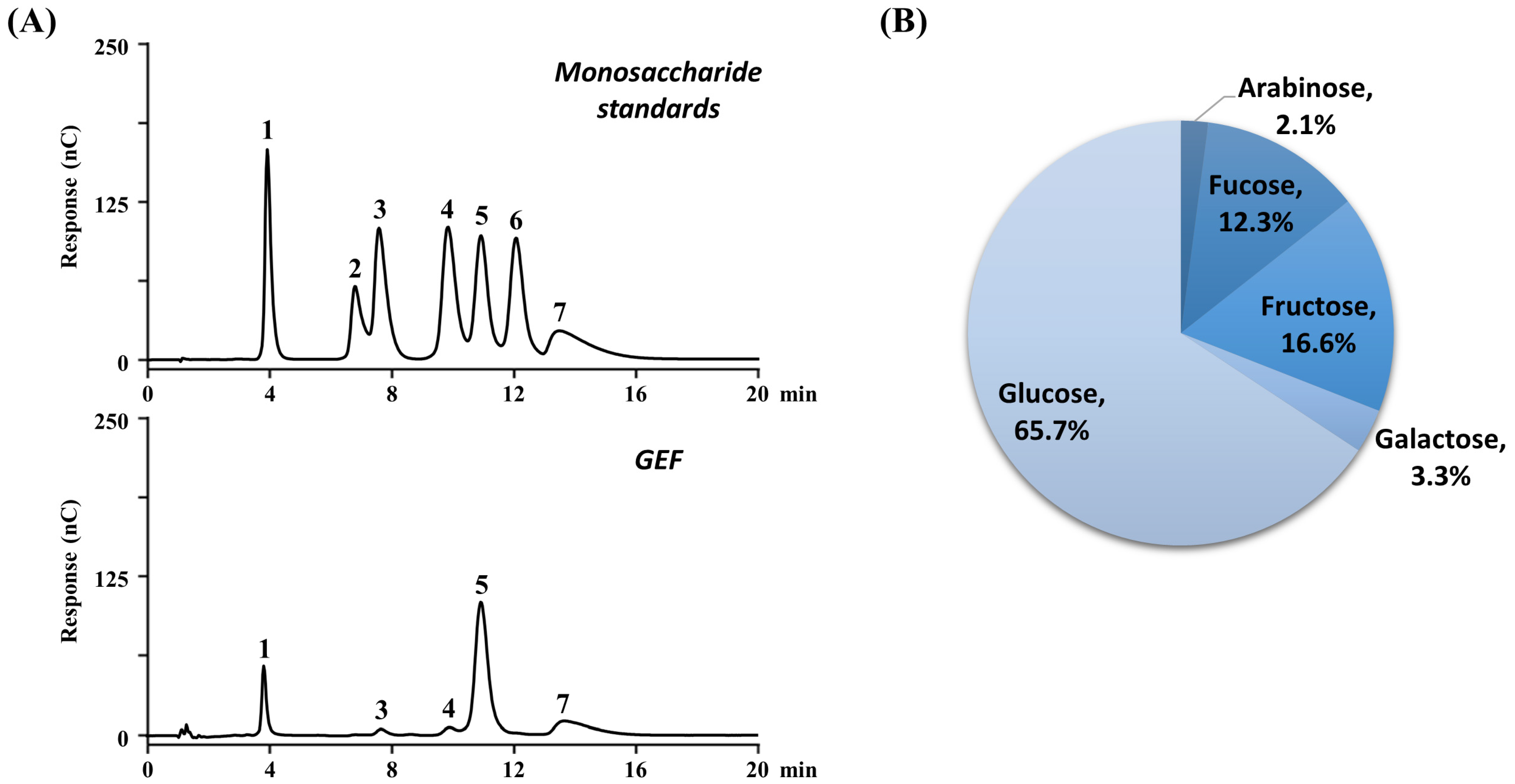
3.2. Chemical Characterization of GEF

Figure 1 shows the composition of the 17 amino acids determined using fluorescence detection after acid hydrolysis of the GEF and ME samples. A uniform composition of 16 amino acids within 10%, excluding glutamic acid (15.0%), was confirmed in ME that was obtained by a common extraction method. However, as a distinguishing characteristic of GEF, three particularly abundant amino acids were observed. The amino acids present in large quantities were, in order, two acidic amino acids—aspartic acid (25.5%) and glutamic acid (20.7%)—and one basic amino acid—arginine (17.5%). The former two were also observed in high amounts in α1-acid glycoprotein, a representative glycoprotein and immunomodulation agent [31,32].
Among the amino acids contained in GEF, essential amino acids accounted for over 16% and conditionally essential amino acids (CEAAs), such as arginine, glycine, glutamic acid, proline, serine, and tyrosine, accounted for more than 50% of the composition. Another characteristic of GEF is that it contains a large amount of CEAAs. CEAAs can be synthesized in the body but they are more important under abnormal conditions, such as illness or stress, because their synthesis is insufficient and limited [33]. In particular, glutamic acid and arginine have been reported to significantly promote the proliferation of keratinocytes and fibroblasts and are necessary for the regeneration of damaged skin [34,35,36]. In brief, GEF is expected to have biological activities in skin anti-aging as it is enriched in glycopeptides and glycoproteins and characterized by high proportions of the CEAAs (51.0%) necessary for skin and acidic/basic amino acids (67.7%), which are likely to interact with other proteins.
The monosaccharide composition of GEF was determined using an ion chromatography system after acid hydrolysis (Figure 2) and confirmed by comparing the seven monosaccharide standards. Arabinose, fructose, fucose, galactose, and glucose were identified in GEF, the most abundant of which was glucose, accounting for 65.7% of the total monosaccharides, followed by fructose (16.6%), fucose (12.3%), galactose (3.3%), and arabinose (2.1%). Glycans (oligosaccharide chains) attached to plant proteins via co- or post-translational modification are crucial to protein stability, localization, and functions, but the exact meaning of their composition and bonding is still unclear and much remains to be studied [37]. In addition, SDS-PAGE using protein standards with various molecular weights and size exclusion chromatography using maltooligosaccharides and pullulan standards were performed to estimate the molecular weight range of GEF. In both experiments, a variety of molecular weights from 3 kDa to over 200 kDa were identified in the form of connected bands or peaks, especially stronger intensities in the 6–28 kDa range.

3.3. Inhibitory Effects on the Breakdown of Elements in Skin Layers Against Photoaging

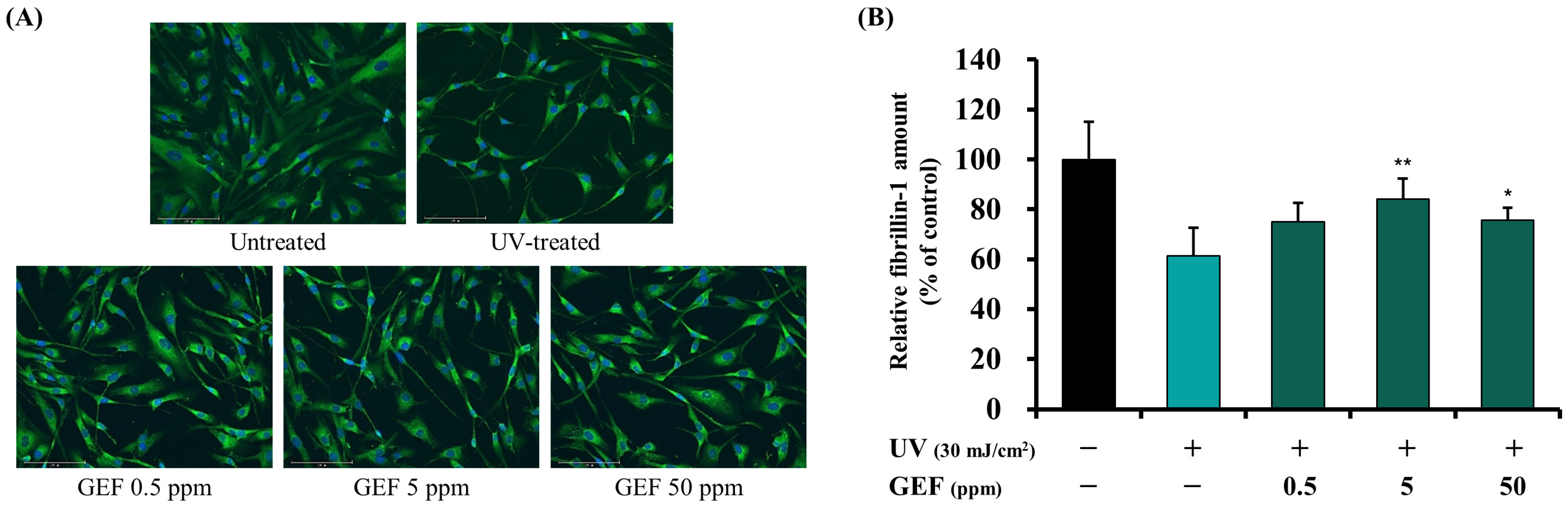
3.3.1. Effect of GEF on Fibrillin-1 Synthesis

The chemical characteristics of GEF can be summarized from three aspects; it is a peptide and protein fraction that sufficiently binds oligosaccharide chains, it has a high CEAA content, and it contains a high content of acidic/basic amino acids. Based on this, it was presumed that GEF could induce anti-aging properties when acting on skin cells such as fibroblasts and keratinocytes. First, we tested whether the synthesis of fibrillin-1 from fibroblasts was inhibited in a photoaging environment. After UV irradiation, the number of fibrillin-1 fibers (fluorescent green in Figure 3A) and the amount calculated from merging their fluorescence signals (Figure 3B) were reduced by 38.7% (the value was 61.3 ± 11.3) compared to those of the untreated group. Fibillin-1 was degraded mainly due to the activation of MMPs by UV exposure, and we also assume that it interferes with the process of fiber protein secretion from inside the fibroblasts [38].
We also investigated whether GEF treatment reduced the loss of fibrillin-1. A recovered amount of fibrillin-1 was observed at all concentrations treated and was clearly observed in the images. At the concentration of 5 ppm, the amount increased by 37.2% (the value was 84.1 ± 8.2) compared to the negative control group treated with UV light, which meant that more than half of the amount lost after UV irradiation was recovered. The amounts at the concentrations of 0.5 and 50 ppm increased by 22.5% and 23.3% (the values were 75.1 ± 7.6 and 75.6 ± 5.1), respectively, from that of the negative control group.

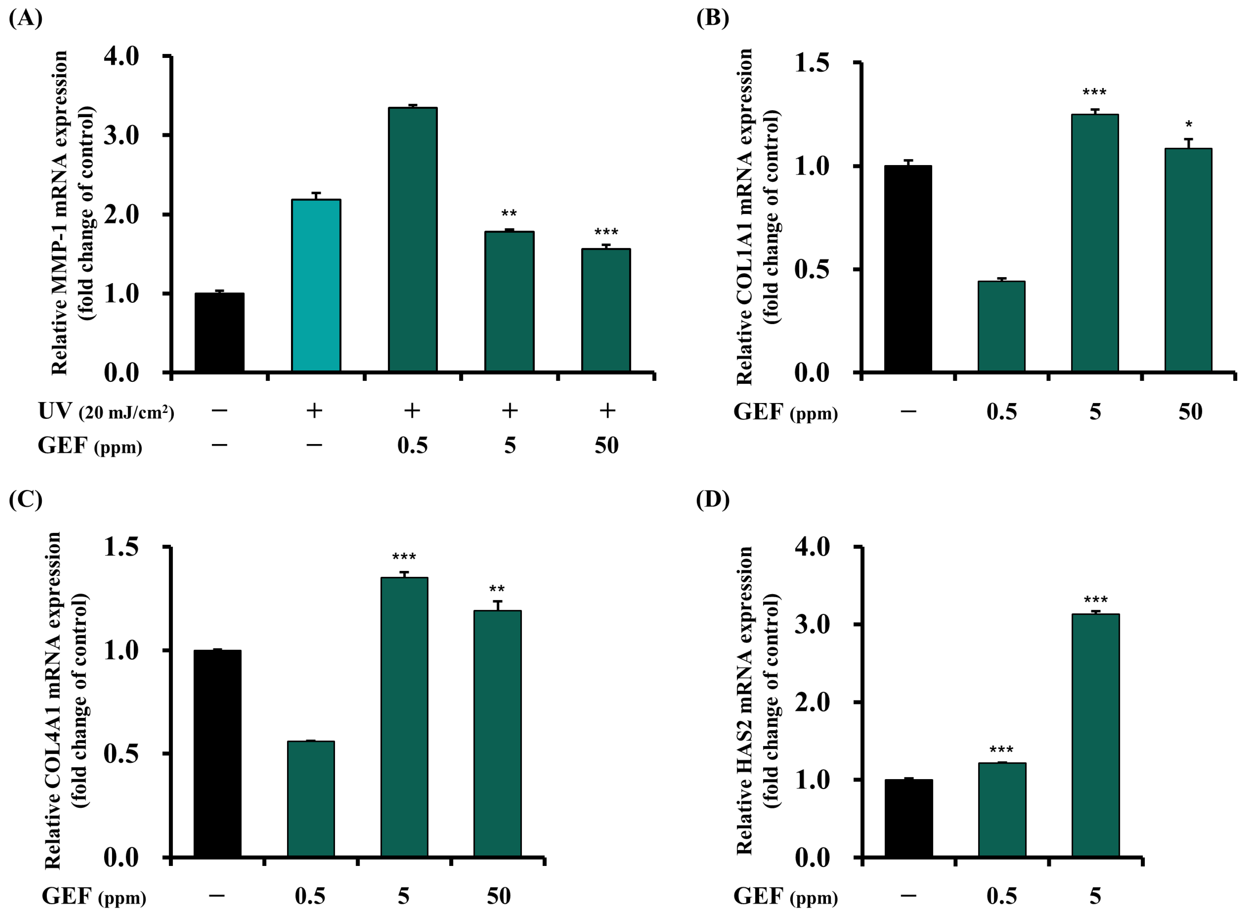
3.3.2. Inhibitory Effect of GEF on MMP-1 Gene Expression

MMPs are produced by several types of cells in the skin, such as fibroblasts and keratinocytes, and play a crucial role in proteolytic remodeling of the extracellular matrix, including collagen and elastin fibers, especially in situations promoted by ROS, UV rays, and inflammatory reactions. In these situations, raw materials that inhibit and regulate MMPs can delay and prolong the rapid breakdown of the skin layers. Among these, MMP-1 is a fibroblast collagenase that takes various types of collagen as a substrate and is the most studied type for skin anti-aging [39]. Figure 4A shows that MMP-1 gene expression is induced in HS68 cells when they are treated with UV and that GEF inhibits this expression. Compared to the untreated group, the mRNA expression of MMP-1 was 2.2-times higher (2.18 ± 0.08) in the negative control group treated with UV light. When GEF was treated with UV light, there was no effect at the concentration of 0.5 ppm, but the expression was decreased by 18.3% and 28.4% (1.78 ± 0.03 and 1.56 ± 0.05) from that of the negative control group at concentrations of 5 and 50 ppm, respectively. These results imply that GEF may delay the degradation of extracellular matrix proteins through inhibiting MMP-1 expression.

3.4. Facilitating Effects on the Synthesis of Elements in Skin Layers

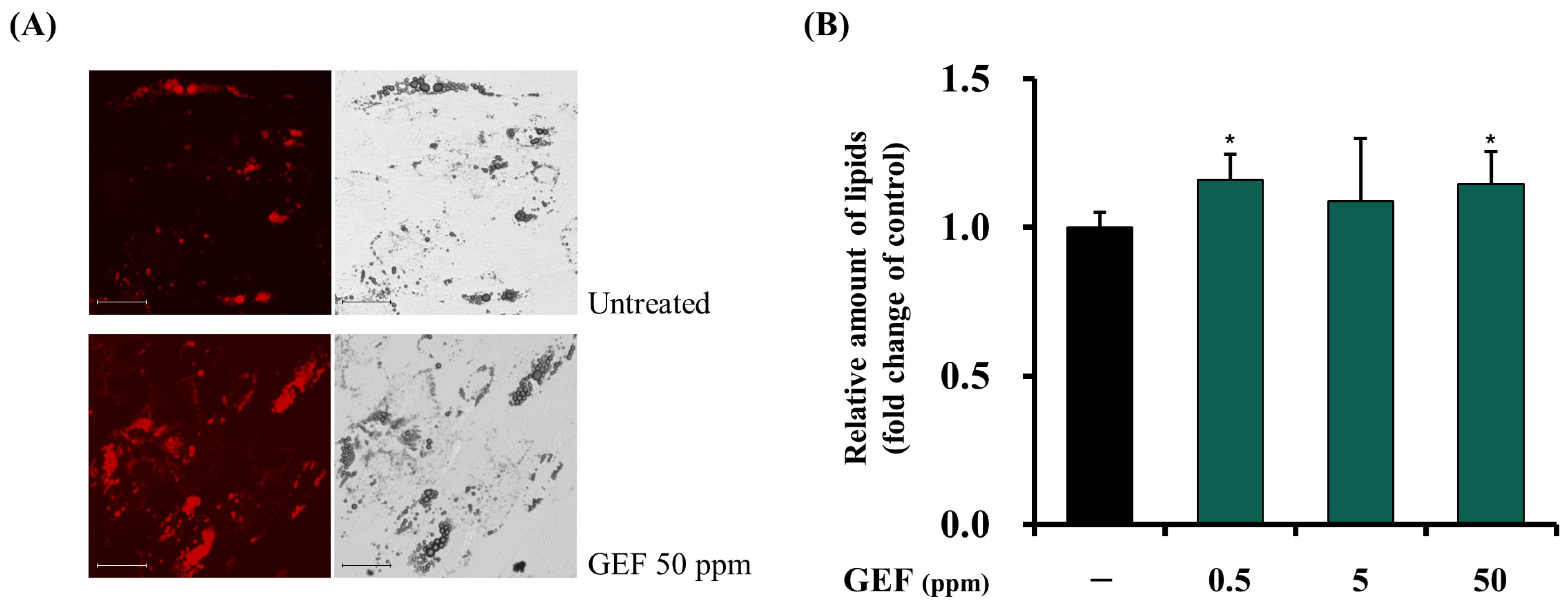
3.4.1. Facilitating Effects of GEF on Adipogenesis

Redistribution of adipose tissues within the body occurs with aging, with visceral adipose tissue increasing and subcutaneous adipose tissue decreasing. This causes visible signs of aging, such as loss of elasticity of the skin layers and the formation of wrinkles [40]. To address this problem, it is necessary to facilitate adipogenesis—the differentiation of stem cells into mature adipocytes leading to lipid accumulation—in the subcutaneous fat layer (hypodermis). Therefore, we investigated the effect of GEF on adipogenesis in ADMSCs as an anti-aging property, and the results are shown in Figure 5.
The amount of lipids produced by facilitated differentiation for 10–14 days was measured, and an increase in lipids compared to the untreated group was confirmed under a microscope (Figure 5A). As determined by measuring the absorbance, there was a tendency for production to increase by about 10–15% at the concentrations treated (1.16 ± 0.09, 1.09 ± 0.21, and 1.15 ± 0.11 in order from the lowest concentration). We have previously studied the effect of three commercial peptide mixtures in an identical experiment. The positive control in this study was rosiglitazone, an anti-diabetic drug elevating subcutaneous fat, and a significant increase in adipogenesis of about 8% was observed, with the complex showing a significant effect of about 11% [41,42]. Therefore, the effect of GEF on adipogenesis was significant considering that it is a mixture of various compounds.

3.4.2. Facilitating Effects of GEF on COL1A1, COL4A1, and HAS2 Gene Expressions

There are more than 10 different types of collagen, with the major types being types I to V. Each type is distributed in a specific part and has a specific role in the human body, particularly type I, which accounts for majority of skin collagen fibers, and type IV, which forms and supports the basement membrane of the extracellular matrix. The types are frequently studied as important collagen types in skin layers [43,44]; it has been revealed that the expressions of collagen types I and IV decrease during the intrinsic aging process, as the functions of cells decline [45]. This is why collagen synthesis decreases with age, causing wrinkles and a loss of skin elasticity. Therefore, we aimed to confirm the effect of GEF on the gene expression of collagen types I and IV (COL1A1 and COL4A1), and the results are shown in Figure 4B,C. The mRNA expressions of COL1A1 and COL4A1 showed identical trends, which were dependent on the treatment concentration. Both showed no effect at the concentration of 0.5 ppm, and the optimal expression was confirmed at a concentration of 5 ppm rather than 50 ppm. The expression increased by up to 25% (1.25 ± 0.02) in the case of COL1A1 and 35% (1.35 ± 0.03) in the case of COL4A1.
Hyaluronic acid is synthesized and degraded in the skin layers by enzymes such as hyaluronic acid synthase (HAS) and hyaluronidase. So far, there are three and six known types, respectively, and hyaluronic acid synthase 2 (HAS2) is the one that mainly mediates the synthesis of hyaluronic acid in the dermis. Thus, this is the most commonly studied type regarding skin anti-aging [46]. The facilitating effect of GEF on HAS2 gene expression is presented in Figure 4D, showing that it has the most significant effect compared to other tested gene expressions. The expression increased 1.2-fold and 3.1-fold (average values: 1.22 ± 0.02 and 3.13 ± 0.04) at concentrations of 0.5 and 5 ppm, respectively, and at 50 ppm, the cell viability of HaCaT cells was slightly less than 90% due to its high concentration. These results suggest that GEF may help in skin anti-aging through its ability to stimulate the production of certain components within the extracellular matrix of skin layers.

4. Conclusions

Inspired by the use of Gongjin-dan, the longevity medicine, a fraction enriched in glycopeptides and glycoproteins (GEF) was extracted from the modified formula for study. This fraction was distinguished from a generally prepared mixed extract (ME) with respect to the abundance of glycopeptides and glycoproteins and the high proportions of CEAAs (51.0%) and acidic/basic amino acids (67.7%). Two aspects of in vitro skin anti-aging properties were investigated. First, we investigated the fraction’s ability to inhibit the loss of dermal components due to extrinsic factors, observing an inhibition in MMP-1 gene expression and a restoration in fibrillin-1 protein, with improvements of 18–28% and 23–37%, respectively. Second, we examined the facilitating effects of GEF on dermal and hypodermal components, the synthesis of which decreases with age. As a result, the gene expressions of COL1A1 and COL4A1, HAS2 gene expression, and adipogenesis were enhanced by 25–35%, 22–213%, and 15%, respectively.
The original medicine is still in high demand as a restorative drug based on the clinical records accumulated up to the present and the rarity of animal sources such as deer antler and musk, and there is still much to be studied about its scientific background. This study is significant in that it scientifically reinterprets a traditional medicine and suggests an effective raw material for skin anti-aging in the cosmetic industry. As a follow-up to this study, it is necessary to further explore the use of purified glycopeptides or glycoproteins and understanding their mechanisms in depth.

Author Contributions

Conceptualization, G.J.L., H.J.J., H.L., N.S.S., and N.-G.K.; Methodology, G.J.L., H.L., and S.K.; Validation, G.J.L., H.J.J., H.L., N.S.S., and N.-G.K.; Formal Analysis, G.J.L., J.P., H.L., and S.K.; Investigation, G.J.L., J.P., H.J.J., T.H.K., H.L., and S.K.; Resources, G.J.L., H.J.J., and S.J.H.; Data Curation, G.J.L., J.P., and H.J.J.; Writing—Original Draft Preparation, G.J.L.; Writing—Review and Editing, G.J.L., J.P., and H.J.J.; Visualization, G.J.L.; Supervision, S.J.H.; Project Administration, N.S.S. and N.-G.K.; Funding Acquisition, N.S.S. and N.-G.K. All authors have read and agreed to the published version of the manuscript.

Funding

This research was supported by LG Household & Health Care Ltd. This research received no external funding.

Institutional Review Board Statement

Not applicable.

Informed Consent Statement

Not applicable.

Data Availability Statement

All data generated in the study are included in the published article.

Conflicts of Interest

The authors Gwang Jin Lee, Jiwon Park, Hyejin Lee, Seongsu Kang, Seung Jin Hwang, Nam Seo Son and Nae-Gyu Kang are employees of LG Household & Health Care. Hyeon Jun Jeon and Tae Heon Kim are employees of DURAE Corporation. The authors declare that this study received funding from LG Household & Health Care Ltd. The funder was not involved in the study design, collection, analysis, interpretation of data, the writing of this article or the decision to submit it for publication.

References

  1. Farage, M.A.; Miller, K.W.; Elsner, P.; Maibach, H.I. Intrinsic and Extrinsic Factors in Skin Ageing: A Review. Int. J. Cosmet. Sci. 2008, 30, 87–95. [Google Scholar] [CrossRef] [PubMed]
  2. Flament, F.; Prunel, A.; Keufer, B.; Abric, A.; Wang, Y.; Reni, A.; Cassier, M.; Delaunay, C. Changes in Facial Signs Due to Age and Their Respective Weights on the Perception of Age and Skin Plumpness among Differently Aged Korean Women. Ski. Res. Technol. 2021, 27, 526–536. [Google Scholar] [CrossRef] [PubMed]
  3. Langton, A.K.; Sherratt, M.J.; Griffiths, C.E.M.; Watson, R.E.B. A New Wrinkle on Old Skin: The Role of Elastic Fibres in Skin Ageing. Int. J. Cosmet. Sci. 2010, 32, 330–339. [Google Scholar] [CrossRef] [PubMed]
  4. Watson, R.E.B.; Griffiths, C.E.M.; Craven, N.M.; Shuttleworth, C.A.; Kielty, C.M. Fibrillin-Rich Microfibrils Are Reduced in Photoaged Skin. Distribution at the Dermal–Epidermal Junction. J. Investig. Dermatol. 1999, 112, 782–787. [Google Scholar] [CrossRef]
  5. Loo, Y.C.; Hu, H.-C.; Yu, S.-Y.; Tsai, Y.-H.; Korinek, M.; Wu, Y.-C.; Chang, F.-R.; Chen, Y.-J. Development on Potential Skin Anti-Aging Agents of Cosmos Caudatus Kunth via Inhibition of Collagenase, MMP-1 and MMP-3 Activities. Phytomedicine 2023, 110, 154643. [Google Scholar] [CrossRef]
  6. Fuller, B. Role of PGE-2 and Other Inflammatory Mediators in Skin Aging and Their Inhibition by Topical Natural Anti-Inflammatories. Cosmetics 2019, 6, 6. [Google Scholar] [CrossRef]
  7. Kim, H.-W.; Jung, Y.-A.; Yun, J.-M.; Kim, Y.; Kim, S.-A.; Suh, S.-I.; Ryoo, Y.-W. Effects of Poly-L-Lactic Acid on Adipogenesis and Collagen Gene Expression in Cultured Adipocytes Irradiated with Ultraviolet B Rays. Ann Dermatol 2023, 35, 424–431. [Google Scholar] [CrossRef]
  8. Gaur, M.; Dobke, M.; Lunyak, V.V. Mesenchymal Stem Cells from Adipose Tissue in Clinical Applications for Dermatological Indications and Skin Aging. Int. J. Mol. Sci. 2017, 18, 208. [Google Scholar] [CrossRef]
  9. Park, K.M.; Yoo, Y.J.; Ryu, S.; Lee, S.H. Nelumbo nucifera Leaf Protects against UVB-Induced Wrinkle Formation and Loss of Subcutaneous Fat through Suppression of MCP3, IL-6 and IL-8 Expression. J. Photochem. Photobiol. B Biol. 2016, 161, 211–216. [Google Scholar] [CrossRef]
  10. Papakonstantinou, E.; Roth, M.; Karakiulakis, G. Hyaluronic Acid: A Key Molecule in Skin Aging. Derm. -Endocrinol. 2012, 4, 253–258. [Google Scholar] [CrossRef]
  11. Shang, L.; Li, M.; Xu, A.; Zhuo, F. Recent Applications and Molecular Mechanisms of Hyaluronic Acid in Skin Aging and Wound Healing. Med. Nov. Technol. Devices 2024, 23, 100320. [Google Scholar] [CrossRef]
  12. Rayon, C.; Lerouge, P.; Faye, L. The Protein N-Glycosylation in Plants. J. Exp. Bot. 1998, 49, 1463–1472. [Google Scholar] [CrossRef]
  13. Baik, K.K.; Song, W.-Y.; Song, D.K.; Yun, J.; Jang, J.H.; Oh, J.Y.; Lee, M.-J.; Go, E.; Lee, K.J.; Roh, E.; et al. Protective Effects of Sesame Glycoproteins on Ultraviolet-Induced Skin Aging: In Vitro and In Vivo Studies. Pharmaceuticals 2024, 17, 1306. [Google Scholar] [CrossRef] [PubMed]
  14. Kim, J.-Y.; Lee, Y.-M.; Park, J.-P.; Lim, K.-T.; Lee, S.-J. Phytoglycoprotein Isolated from Dioscorea Batatas Decne Promotes Intestinal Epithelial Wound Healing. Chin. J. Nat. Med. 2020, 18, 738–748. [Google Scholar] [CrossRef] [PubMed]
  15. Kim, S.J.; Park, S.Y.; Hong, S.-M.; Kwon, E.-H.; Lee, T.-K. Skin Whitening and Anti-Corrugation Activities of Glycoprotein Fractions from Liquid Extracts of Boiled Sea Cucumber. Asian Pac. J. Trop. Med. 2016, 9, 1002–1006. [Google Scholar] [CrossRef]
  16. Hong, S.-S.; Lee, J.; Lee, J.-S.; Lee, H.-W.; Kim, H.-G.; Lee, S.-K.; Park, B.-K.; Son, C.-G. The Traditional Drug Gongjin-Dan Ameliorates Chronic Fatigue in a Forced-Stress Mouse Exercise Model. J. Ethnopharmacol. 2015, 168, 268–278. [Google Scholar] [CrossRef]
  17. Choi, J.-Y.; Choi, B.; Kwon, O.; Seo, C.-S.; Kim, A.-R.; Shin, H.; Kim, K. Efficacy and Safety of Herbal Medicine Gongjin-Dan and Ssanghwa-Tang in Patients with Chronic Fatigue: A Randomized, Double-Blind, Placebo-Controlled, Clinical Trial. Integr. Med. Res. 2024, 13, 101025. [Google Scholar] [CrossRef]
  18. Kim, H.; Jeon, W.; Hong, J.; Lee, J.; Yeo, C.; Lee, Y.; Baek, S.; Ha, I. Gongjin-Dan Enhances Neurite Outgrowth of Cortical Neuron by Ameliorating H2O2-Induced Oxidative Damage via Sirtuin1 Signaling Pathway. Nutrients 2021, 13, 4290. [Google Scholar] [CrossRef]
  19. Kang, J.-W.; Cho, H.-E.; Choi, H.-M.; Lee, I.-C. Anti-Wrinkle Properties of Angelica Gigas Nakai Root Extracts Using Mineral-Rich Water. J. Cosmet. Dermatol. 2023, 22, 328–334. [Google Scholar] [CrossRef]
  20. Yue, Z.; Liu, H.; Liu, M.; Wang, N.; Ye, L.; Guo, C.; Zheng, B. Cornus Officinalis Extract Enriched with Ursolic Acid Ameliorates UVB-Induced Photoaging in Caenorhabditis elegans. Molecules 2024, 29, 2718. [Google Scholar] [CrossRef]
  21. Cuong, V.T.; Chen, W.; Shi, J.; Zhang, M.; Yang, H.; Wang, N.; Yang, S.; Li, J.; Yang, P.; Fei, J. The Anti-Oxidation and Anti-Aging Effects of Ganoderma lucidum in Caenorhabditis elegans. Exp. Gerontol. 2019, 117, 99–105. [Google Scholar] [CrossRef] [PubMed]
  22. Xiong, D.; Yu, L.-X.; Yan, X.; Guo, C.; Xiong, Y. Effects of Root and Stem Extracts of Asparagus Cochinchinensis on Biochemical Indicators Related to Aging in the Brain and Liver of Mice. Am. J. Chin. Med. 2011, 39, 719–726. [Google Scholar] [CrossRef] [PubMed]
  23. Lee, M.-J.; Jeong, N.-H.; Jang, B.-S. Antioxidative Activity and Antiaging Effect of Carrot Glycoprotein. J. Ind. Eng. Chem. 2015, 25, 216–221. [Google Scholar] [CrossRef]
  24. Jang, B.-S.; Lee, M.-J.; Jeong, N.-H. Preparation and Availability Analysis of Glycoprotein from Canola Meal. J Food Sci Technol 2021, 58, 377–382. [Google Scholar] [CrossRef]
  25. Zhou, Y.; Ma, Y.; Li, L.; Yang, X. Purification, Characterization, and Functional Properties of a Novel Glycoprotein from Tartary Buckwheat (Fagopyrum tartaricum) Seed. Food Chem. 2020, 309, 125671. [Google Scholar] [CrossRef]
  26. Wingfield, P. Protein Precipitation Using Ammonium Sulfate. Curr. Protoc. Protein Sci. 1998, 13, A.3F.1–A.3F.8. [Google Scholar] [CrossRef]
  27. Masuko, T.; Minami, A.; Iwasaki, N.; Majima, T.; Nishimura, S.-I.; Lee, Y.C. Carbohydrate Analysis by a Phenol–Sulfuric Acid Method in Microplate Format. Anal. Biochem. 2005, 339, 69–72. [Google Scholar] [CrossRef]
  28. Deepachandi, B.; Weerasinghe, S.; Andrahennadi, T.P.; Karunaweera, N.D.; Wickramarachchi, N.; Soysa, P.; Siriwardana, Y. Quantification of Soluble or Insoluble Fractions of Leishmania Parasite Proteins in Microvolume Applications: A Simplification to Standard Lowry Assay. Int. J. Anal. Chem. 2020, 2020, 6129132. [Google Scholar] [CrossRef]
  29. Wallis, D.D.; Tan, F.K.; Kielty, C.M.; Kimball, M.D.; Arnett, F.C.; Milewicz, D.M. Abnormalities in Fibrillin 1–Containing Microfibrils in Dermal Fibroblast Cultures from Patients with Systemic Sclerosis (Scleroderma). Arthritis Rheum. 2001, 44, 1855–1864. [Google Scholar] [CrossRef]
  30. Manetti, M.; Romano, E.; Rosa, I.; Fioretto, B.S.; Praino, E.; Guiducci, S.; Iannone, F.; Ibba-Manneschi, L.; Matucci-Cerinic, M. Systemic Sclerosis Serum Steers the Differentiation of Adipose-Derived Stem Cells Toward Profibrotic Myofibroblasts: Pathophysiologic Implications. J. Clin. Med. 2019, 8, 1256. [Google Scholar] [CrossRef]
  31. Schmid, K.; Kaufmann, H.; Isemura, S.; Bauer, F.; Emura, J.; Motoyama, T.; Ishiguro, M.; Nanno, S. Structure of A1-Acid Glycoprotein. The Complete Amino Acid Sequence, Multiple Amino Acid Substitutions, and Homology with the Immunoglobulins. Biochemistry 1973, 12, 2711–2724. [Google Scholar] [CrossRef] [PubMed]
  32. Fournier, T.; Medjoubi-N, N.; Porquet, D. Alpha-1-Acid Glycoprotein. Biochim. Et Biophys. Acta (BBA) Protein Struct. Mol. Enzymol. 2000, 1482, 157–171. [Google Scholar] [CrossRef]
  33. Thalacker-Mercer, A.; Riddle, E.; Barre, L. Protein and Amino Acids for Skeletal Muscle Health in Aging. In Advances in Food and Nutrition Research; Toldrá, F., Ed.; Academic Press: Cambridge, MA, USA, 2020; Volume 91, pp. 29–64. [Google Scholar]
  34. Fujiwara, T.; Kanazawa, S.; Ichibori, R.; Tanigawa, T.; Magome, T.; Shingaki, K.; Miyata, S.; Tohyama, M.; Hosokawa, K. L-Arginine Stimulates Fibroblast Proliferation through the GPRC6A-ERK1/2 and PI3K/Akt Pathway. PLoS ONE 2014, 9, e92168. [Google Scholar] [CrossRef] [PubMed]
  35. Jara, C.P.; de Andrade Berti, B.; Mendes, N.F.; Engel, D.F.; Zanesco, A.M.; Pereira de Souza, G.F.; de Medeiros Bezerra, R.; de Toledo Bagatin, J.; Maria-Engler, S.S.; Morari, J.; et al. Glutamic Acid Promotes Hair Growth in Mice. Sci Rep 2021, 11, 15453. [Google Scholar] [CrossRef]
  36. Kwon, K.C.; Won, J.G.; Seo, J.H.; Kwon, O.S.; Kim, E.H.; Kim, M.-S.; Park, S.-W. Effects of Arginine Glutamate (RE:Pair) on Wound Healing and Skin Elasticity Improvement after CO2 Laser Irradiation. J. Cosmet. Dermatol. 2022, 21, 5037–5048. [Google Scholar] [CrossRef]
  37. Strasser, R.; Seifert, G.; Doblin, M.S.; Johnson, K.L.; Ruprecht, C.; Pfrengle, F.; Bacic, A.; Estevez, J.M. Cracking the “Sugar Code”: A Snapshot of N- and O-Glycosylation Pathways and Functions in Plants Cells. Front. Plant Sci. 2021, 12, 640919. [Google Scholar] [CrossRef]
  38. Katsuyama, Y.; Yamawaki, Y.; Sato, Y.; Muraoka, S.; Yoshida, M.; Okano, Y.; Masaki, H. Decreased Mitochondrial Function in UVA-Irradiated Dermal Fibroblasts Causes the Insufficient Formation of Type I Collagen and Fibrillin-1 Fibers. J. Dermatol. Sci. 2022, 108, 22–29. [Google Scholar] [CrossRef]
  39. Kähäri, V.-M.; Saarialho-Kere, U. Matrix Metalloproteinases in Skin. Exp. Dermatol. 1997, 6, 199–213. [Google Scholar] [CrossRef]
  40. Liu, M.; Lu, F.; Feng, J. Aging and Homeostasis of the Hypodermis in the Age-Related Deterioration of Skin Function. Cell Death Dis 2024, 15, 443. [Google Scholar] [CrossRef]
  41. Khoo, N.K.H.; Fazzari, M.; Chartoumpekis, D.V.; Li, L.; Guimaraes, D.A.; Arteel, G.E.; Shiva, S.; Freeman, B.A. Electrophilic Nitro-Oleic Acid Reverses Obesity-Induced Hepatic Steatosis. Redox Biol. 2019, 22, 101132. [Google Scholar] [CrossRef]
  42. Kang, S.; Jun, S.-H.; Lee, J.; An, M.J.; Kang, N.-K. In Vitro Efficacies of Complex of Cedrol and Three Peptides, and Wrinkle Improvements and Lips Volumization effects of Applied Formulations. J. Soc. Cosmet. Sci. Korea 2023, 49, 365–373. [Google Scholar]
  43. Yan, Y.; Quan, H.; Guo, C.; Qin, Z.; Quan, T. Alterations of Matrisome Gene Expression in Naturally Aged and Photoaged Human Skin In Vivo. Biomolecules 2024, 14, 900. [Google Scholar] [CrossRef] [PubMed]
  44. Holwerda, A.M.; van Loon, L.J.C. The Impact of Collagen Protein Ingestion on Musculoskeletal Connective Tissue Remodeling: A Narrative Review. Nutr Rev 2022, 80, 1497–1514. [Google Scholar] [CrossRef] [PubMed]
  45. Lago, J.C.; Puzzi, M.B. The Effect of Aging in Primary Human Dermal Fibroblasts. PLoS ONE 2019, 14, e0219165. [Google Scholar] [CrossRef]
  46. Shinozaki, Y.; Ikarashi, N.; Tabata, K.; Miyazawa, A.; Kon, R.; Sakai, H.; Hosoe, T. Expression Analysis of Genes Important for Maintaining Skin Function in a Senescence-Accelerated Mouse Prone Model. Geriatr. Gerontol. Int. 2023, 23, 951–957. [Google Scholar] [CrossRef]
Figure 1. Amino acid composition of (A) GEF and (B) ME. GEF, glycopeptide- and glycoprotein-enriched fraction; ME, mixed extract; Asp, aspartic acid; Glu, glutamic acid; Ser, serine; His, histidine; Gly, glycine; Thr, threonine; Cit, citrulline; Arg, arginine; Ala, alanine; GABA, gamma-aminobutyric acid; Tyr, tyrosine; Val, valine; Phe, phenylalanine; Ile, isoleucine; Leu, leucine; Lys, lysine; Pro, proline. Details of the determination of amino acids are described in Section 2.5.3.
Cosmetics 12 00091 g001
Figure 2. Monosaccharide composition of GEF determined using high-pH anion-exchange chromatography with pulsed amperometric detection (HPAEC-PAD). GEF, glycopeptide- and glycoprotein-enriched fraction. (A) Chromatograms of the following seven monosaccharide standards and five identified monosaccharides in GEF: (1) fucose, (2) rhamnose, (3) arabinose, (4) galactose, (5) glucose, (6) xylose, and (7) fructose. (B) Proportion of each monosaccharide in the total. Details of chromatographic separation are described in Section 2.5.2.
Cosmetics 12 00091 g002
Figure 3. Effect of GEF on fibrillin-1 synthesis in HS68 cells. GEF, glycopeptide- and glycoprotein-enriched fraction. (A) Fluorescence microscopy images obtained by staining for fibrillin-1 using immunocytochemical analysis after treatment with UV B irradiation (30 mJ/cm2) and/or GEF (green, fibrillin-1 fibers; blue, nuclei; scale bar: 275 μm). (B) Amount of fibrillin-1 calculated by merging the fluorescence signals using Image J software. Details are described in Section 2.6.2. The indicated concentrations of GEF are based on the solute, and the statistical significance of the data (presented as the mean and standard deviation, n = 3) was determined using ANOVA (* p < 0.05; ** p < 0.01).
Cosmetics 12 00091 g003
Figure 4. Effects of GEF on mRNA expression of the factors influencing skin anti-aging. GEF, glycopeptide- and glycoprotein-enriched fraction. (A) Effect of GEF on matrix metalloproteinase-1 (MMP-1) gene expression in HS68 cells treated with UV B irradiation (20 mJ/cm2). (B,C) Effect of GEF on collagen type I alpha 1 (COL1A1) and collagen type IV alpha 1 (COL4A1) gene expressions in HS68 cells. (D) Effect of GEF on hyaluronic acid synthase 2 (HAS2) gene expression in HaCaT cells. mRNA levels were analyzed using qRT-PCR, as described in Section 2.6.3. The indicated concentrations of GEF are based on the solute, and the statistical significance of the data (presented as the mean and standard deviation, n = 3) was determined using ANOVA (* p < 0.05; ** p < 0.01; *** p < 0.001).
Cosmetics 12 00091 g004
Figure 5. Effect of GEF on adipogenesis in human adipose-derived mesenchymal stem cells (ADMSCs). GEF, glycopeptide- and glycoprotein-enriched fraction. ADMSCs treated with GEF were cultured for more than 10 days to differentiate them into mature adipocytes, and then the total lipids accumulated in the cells were stained using oil red O (ORO). The amount of lipids produced was determined by eluting the dye with isopropanol and measuring the absorbance at 515 nm. Details are described in Section 2.6.2. (A) Microscopy images of stained lipids by ORO (scale bar: 100 μm) and (B) the absorbance of eluted ORO. The indicated concentrations of GEF are based on the solute, and the statistical significance of the data (presented as the mean and standard deviation, n = 3) was determined using ANOVA (* p < 0.05).
Cosmetics 12 00091 g005
Table 1. List of primer sequences used for qRT-PCR.
Target mRNAForward PrimerReverse Primer
GAPDHCATGTTCGTCATGGGGTGAACCAAGTGATGGCATGGACTGTGGTCAT
MMP-1CTGGCCACAACTGCCAAATGCTGTCCCTGAACAGCCCAGTACTT
COL1A1GCTACTACCGGGCTGATGATACCAGTCTCCATGTTGCAGA
COL4A1CTGCCTGGAGGAGTTTAGAAGGAACATCTCGCTCCTCTCTATG
HAS2AATCCAGCTCTTCTACCGGGCTTGGCGGGAAGTAAACTCG
Table 2. Total carbohydrate and protein contents of GEF and ME a.
Content bGEFME
Total carbohydrate2219 (44.4%)1824 (36.5%)
Total protein683 (13.7%)398 (8.0%)
a GEF, glycopeptide- and glycoprotein-enriched fraction; ME, mixed extract. Details for the preparation of samples and the quantification are described in Section 2.3, Section 2.4 and Section 2.5. The total concentration of the samples (0.5%) and the mixing ratio of the extracts or fractions obtained from the five raw materials are the same. bResults are expressed in ppm (wt% of the solute).
Table 3. Identification of glycopeptides and glycoproteins in GEF a.
ProteinConcentration
(mg/mL)
Absorbance
(550 nm)
Blank00.296
Lysozyme2.50.333
Bovine serum albumin2.50.413
Ovalbumin2.50.573
apo-Transferrin2.50.798
Fetuin2.55.593
α1-Acid glycoprotein2.55.274
GEF2.513.148
a GEF, glycopeptide- and glycoprotein-enriched fraction. Details of the identification of glycopeptides and glycoproteins are described in Section 2.5.1.
Disclaimer/Publisher’s Note: The statements, opinions and data contained in all publications are solely those of the individual author(s) and contributor(s) and not of MDPI and/or the editor(s). MDPI and/or the editor(s) disclaim responsibility for any injury to people or property resulting from any ideas, methods, instructions or products referred to in the content.

