Article

Natural Mineral Water–Plant Extract Combinations as Potential Anti-Aging Ingredients: An In Vitro Evaluation

by
Carolina P. Gomes
1,2,
Ana S. Oliveira
1,2,
Joana Rolo
1,2,
Tayse F. F. da Silveira
3,
Rita Palmeira de Oliveira
1,2,4,
Maria José Alves
3,5,
Paula Plasencia
3 and
Ana Palmeira de Oliveira
1,2,4,*
1
RISE-Health, Department of Medical Sciences, Faculty of Health Sciences, University of Beira Interior, Av. Infante D. Henrique, 6200-506 Covilhã, Portugal
2
Faculty of Health Sciences, University of Beira Interior, Av. Infante D. Henrique, 6200-506 Covilhã, Portugal
3
CIMO, LA SusTEC, Instituto Politécnico de Bragança, Campus de Santa Apolónia, 5300-253 Bragança, Portugal
4
Labfit-HPRD: Health Products Research and Development Lda, UBIMEDICAL Estrada Municipal 506, 6200-284 Covilhã, Portugal
5
AquaValor—Centro de Valorização e Transferência de Tecnologia da Água—Associação, Rua Doutor Júlio Martins n.°1, 5400-342 Chaves, Portugal
*
Author to whom correspondence should be addressed.
Cosmetics 2025, 12(3), 113; https://doi.org/10.3390/cosmetics12030113
Submission received: 18 April 2025 / Revised: 16 May 2025 / Accepted: 20 May 2025 / Published: 28 May 2025
(This article belongs to the Special Issue Feature Papers in Cosmetics in 2025)

Abstract

:
Natural mineral waters (NMWs) and plant extracts have long been valued for their therapeutic properties and skin benefits. This study investigated, in vitro, the role of five Portuguese NMWs (A-E), combined with plant extracts from five species (Ficus carica L., Rubus idaeus L., Vaccinium myrtillus, Cistus ladanifer and Thymus x citriodorus) as bioactive ingredients. Antioxidant capacity was assessed using the 2,2-Diphenyl-1-picrylhydrazyl (DPPH) method. Cellular biocompatibility was evaluated in fibroblasts (L929) and macrophages (RAW 264.7). Skin-repairing and anti-senescence properties were evaluated in L929 cells through the scratch-wound method and β-galactosidase assay. Superoxide dismutase (SOD) was quantified using a commercial kit, and lipopolysaccharide-induced reactive oxygen species (ROS) were quantified using a fluorescent probe (H2DCFDA) in RAW 264.7. The results highlighted the beneficial impact of extracts combined with NMWs. An increase in antioxidant capacity of up to 90% was observed in mixtures comprising Ficus carica L., compared with NMWs alone. In contrast, mixtures with Cistus ladanifer showed promising anti-aging potential, with a 40% decrease in senescent cells and a 33% ROS reduction. Rubus idaeus L. extract produced an increase in cell migration capacity (up to 50%), depending on the NMW. This study highlights the potential synergism of natural ingredients with plant extracts for anti-aging.

1. Introduction

In recent years, consumer environmental awareness has changed, and the preference for natural products has gradually increased, with consumers choosing more environmentally friendly cosmetics [1,2]. In addition, there is a growing preference for products with minimal synthetic additives to reduce adverse effects on health and the environment [3]. Among the various types of cosmetics, anti-aging products are one of the most popular among consumers as they promote younger-looking skin [4]. The process of skin aging involves the combination of both intrinsic factors, such as genetic and biological aging, and extrinsic factors, such as UV radiation, pollution, and lifestyle choices, which influence skin aging [5]. This wide range of mechanisms allows for different targets when designing an antiaging product. Natural compounds and herbal-derived formulations have been popularized due to their bioactive properties, which act through different mechanisms on several signaling pathways for skin aging [6].
Natural mineral waters (NMWs) are some of the natural ingredients featured in cosmetic formulations, attracting interest for their health benefits. The history of NMWs dates to antiquity, with the Greeks attributing healing properties to these waters, while the Romans integrated thermal baths into their daily lives [7]. NMWs originated underground and have distinct characteristics: they emerge as springs, retain bacteriological purity, and provide various health benefits [8,9]. Today, scientific research has supported some of the mechanisms behind their beneficial health properties, enhancing our understanding of these natural resources and, consequently, their value [10,11]. Studies have reported that NMWs can positively influence skin physiology by enhancing hydration, supporting the skin barrier, and reducing oxidative stress, key mechanisms involved in skin aging [12]. In this context, NMWs have been increasingly incorporated into cosmetic formulations not only for their soothing and anti-inflammatory effects, but also for their potential application in anti-aging skincare. In fact, a recent review highlighted that NMWs exhibit antioxidant, anti-inflammatory, and barrier-repairing properties, which are especially beneficial for sensitive and aging skin, with these findings supporting their role as active ingredients in formulations aimed for these applications [13].
In addition to NMWs, other natural resources have been explored for their potential in promoting skin health [14]. Since ancient times, plants have been vital resources for healthcare and fragrance ingredients, and over the years, more of their bioactive properties have been recognized [15,16]. According to the World Health Organization, around 80% of the global population relies on plant-based medicinal products, especially in developing countries [17]. Recently, the use of agro-industrial waste products to produce extracts for various applications has emerged as a sustainable strategy, enabling the valorization of surplus plant materials, thereby promoting circular economy practices [18,19]. These waste products still contain bioactive compounds recognized for their antioxidant properties, among other possible bioactivities [20]. Also, hydrolates, by products of essential oil production, have been investigated for their bioactive properties retained from the solubilization of the most hydrophilic volatile compounds in the condensation water generated during the steam distillation process [21,22,23]. Several studies have demonstrated that plant-based extracts possess significant anti-aging potential, primarily due to their rich content in polyphenols, flavonoids, and terpenoids, which act through multiple mechanisms [24,25]. Extracts derived from species such as Cistus ladanifer, Rubus idaeus, and Vaccinium myrtillus, which are also used in this study, have shown promising results in attenuating cellular senescence and promoting skin regeneration both in vitro and in vivo [26,27,28]. This evidence supports their relevance as possible active ingredients in cosmetic formulations targeting skin aging.
Given the growing interest in natural resources and circular economy, in this study, we aimed to maximize the benefits of NMWs, combining them with plant hydrolates or agro-industrial residues. Based on previous studies from our team, we selected four natural mineral waters from the central region of Portugal and one from the northern region, considering their biological active properties relevant to skin health. NMWs were combined with two hydrolates, Cistus ladanifer (rockrose), known for its healing properties [29] and Thymus x citriodorus (lemon thyme), recognized for its antiseptic and antimicrobial properties [30]. Additionally, we also combined NMWs with extracts obtained from the agro-industrial wastes of Vaccinium myrtillus, Ficus carica L., and Rubus idaeus L. We aimed to evaluate their ability to reduce reactive oxygen species, the number of senescent cells, and superoxide dismutase activity, and assess their cytotoxicity, antioxidant capacity and skin regenerating capacity. By comparing the results of the mixtures with those of the individual ingredients, we aimed to identify promising synergistic or additive combinations with anti-aging properties.

2. Materials and Methods

2.1. Extracts, Hydrolates, and Natural Mineral Waters Understudy

2.1.1. Plant and Agro-Industrial Residue Extracts

Thymus x citriodorus (batch: 08TC21) and Cistus ladanifer (batch: 6) hydrolates were obtained from two Portuguese companies, namely Ervitas CatitasTM (Borba, Portugal) and Proentia® (Proença-a-Nova, Portugal), respectively.
Aqueous extracts were produced from Vaccinium myrtillus, Ficus carica L., and Rubus idaeus L. agroindustrial wastes at the Mountain Research Center (CIMO), Polytechnic Institute of Bragança (IPB). Vaccinium myrtillus aerial parts were collected in Baião, Portugal, while leaves of Ficus carica L. were obtained from the company “Mó de Cima” (Lisbon, Portugal).
The plant extracts were produced according to ([31] Briefly, 150 mL of ethanol/water 80:20 (v/v) was added to 1 g of freeze-dried sample and subjected to ultrasound-assisted extraction (Qsonica, Q500, ultrasonic processor sonicator, 20 kHz, Newtown, CT, USA). The extraction was carried out for 10 min, with cycles of 30 s intercalated by pauses of 10 s, at 75% intensity of 500 W. An ice bath was used to avoid sample overheating. Following extraction, the samples were filtered, evaporated under reduced pressure (Büchi R-114, rotary evaporator; Büchi B-480, waterbath, and Büchi B-721, vacuum controller system, Flawil, Switzerland) at 37 °C until ethanol was entirely removed. The aqueous extract was frozen, lyophilized and stored protected from light until use. For cell assays, the extracts were used to prepare stock solutions (concentration of 8 mg/mL) in Mili-Q water.
Information about the tested hydrolates and agro-industrial waste extracts and their chemical composition is provided in Table 1.

2.1.2. Natural Mineral Waters

Five natural mineral waters were used in this study, four from the central region of Portugal and one from the northern region were used for water collection. Each NMW was collected from the boreholes of Thermal Centres in sterile collection bottles (500 mL; Deltalab, Barcelona, Spain), following the previously described procedure [34]. The samples were transported to the laboratory under the recommended conditions of the American Public Health Association (APHA), American Water Works Association (AWWA) and Water Environment Federation (WEF), described in Standard Methods for the Examination of Water and Wastewater (Washington, DC, USA) [34] and kept refrigerated in the dark until use. To maintain confidentiality, each water sample was assigned a letter code, which was used throughout this study. Waters A, B, and C are sulfuric bicarbonate sodic NMWs, water D is a silicated NMW, and water E is classified as a sodic bicarbonate gasocarbonic NMW (Table 2).

2.2. In Vitro Safety Assessment Through MTT Assay: Cell Biocompatibility in L929 and RAW 264.7 Cell Lines

Cell biocompatibility was assessed in two distinct cell lines: mouse macrophages (RAW 264.7) and fibroblasts (L929). RAW 264.7 (ATCC® TIB-71™, Manassas, VA, USA) cells were cultured in DMEM supplemented with 10% (v/v) non-inactivated fetal bovine serum (FBS), 100 μg/mL streptomycin, 100 U/mL penicillin, 4.5 g/L D-glucose, and 1.5 g/L sodium bicarbonate. L929 (ATCC® CCL-1™, Manassas, VA, USA) cells were cultured in DMEM supplemented with 10% (v/v) heat-inactivated FBS, 100 U/mL penicillin, 100 μg/mL streptomycin, 1.5 g/L sodium bicarbonate, 25 mM D-glucose (Sigma-Aldrich®, St. Louis, MO, USA), and 35.9 mM sodium bicarbonate (Sigma-Aldrich®, St. Louis, MO, USA). Both cell lines were maintained in a humidified atmosphere containing 95% air and 5% CO2 at 37 °C, according to ATCC guidelines.
Cell viability was determined using the MTT assay (3-(4,5-dimethylthiazol-2-yl)-2,5-diphenyl tetrazolium bromide), according to ISO/EN 10993-5: Biological protocol evaluation of medical devices—Part 5: Tests for in vitro cytotoxicity, with adaptations for testing the mixtures [38]. RAW 264.7 cells were seeded at approximately 2.5 × 10⁴ cells per well and L929 at 1 × 10⁴ cells per well in 96-well plates. After 24 h of incubation, the cells were exposed to different dilutions of NMWs and plant extracts for an additional 24 h. Agro-industrial residues were tested at concentrations ranging from 0.125 to 2 (mg/mL), and hydrolates in the range of 1.56–25% (v/v). NMWs were used in the same concentration range of the hydrolates, from 1.56 to 25% (v/v). Following treatment, a solution of MTT (1 mg/mL in incomplete culture medium) was added to each well, and the plates were incubated in the dark at 37 °C for 4 h. The MTT medium was removed, and propane-2-ol was added to dissolve the formazan crystals. Absorbance was measured using an xMarkTM microplate absorbance spectrophotometer from Bio-Rad (Hercules, CA, USA) at an absorbance of 570 nm, with a reference wavelength of 630 nm. The condition with the highest concentration of each natural ingredient that proved to be biocompatible with both cell lines was selected and used in the following experiments.

2.3. Efficacy Testing

2.3.1. DPPH Assay for Antioxidant Activity

The mixture’s antioxidant potential was evaluated by assessing the ability to scavenge the 2,2-diphenyl-1-picrylhydrazyl (DPPH) free radical using a previously described protocol [39,40] with adaptations for testing the mixtures. In this assay, mixtures comprising NMWs and plant extracts, as well as the isolated ingredients, were evaluated. Each ingredient, either alone or combined, was mixed with a solution of DPPH (0.05 mg/mL) in methanol in equal volumes. After adding DPPH, the plates were placed in a light-free environment and incubated at room temperature for 30 min. After incubation, absorbance readings were taken using a microplate reader at 517 nm (xMarkTM Microplate Absorbance Spectrophotometer, Bio-Rad, Hercules, CA, USA). A DPPH solution was used as a negative control, while ascorbic acid acted as a positive control for antioxidant potential, Antioxidant Activity Index (AAI), which expresses the antioxidant activity, was calculated by dividing the final DPPH concentration by the estimated IC50, as described in the following equation:
A A I = D P P H   f i n a l   c o n c e n t r a t i o n S a m p l e   I C 50
Based on the results, the individual extracts or mixtures were classified as denoting a poor antioxidant activity (AAI < 0.5); moderate antioxidant activity (0.5 < AAI < 1.0); and strong antioxidant activity (1.0 < AAI < 2.0) as described in [41].

2.3.2. Senescence-Associated β-Galactosidase in L929 Cells

Cell senescence was assessed using a commercially available β-galactosidase staining kit (Cell Signaling Technology, Danvers, MA, USA). Fibroblast cells (L929) were seeded at a density of 1 × 10⁴ per well in 12-well plates for 24 h. To induce senescence, the cells were treated with 300 μM hydrogen peroxide (H2O2) for 2 h. After removing the stimuli, the cells were washed and left to recover for 72 h in DMEM medium and either in the individual ingredients or the respective mixtures, at the previously selected biocompatible concentrations. In addition to the ingredients/mixtures, the following controls were included: a control medium (highest expected senescence) and a dilution of control consisting of cell medium and Milli-Q water in the same proportion used in the combination of extract and NMW. Following recovery, the cells were fixed and incubated with a β-galactosidase staining solution at 37 °C in a CO2-free environment for 16 h, following the manufacturers’ instructions. The next day, the cells were examined under a microscope to detect the blue color, indicative of senescent cells. Eight fields were randomly photographed per each well, and quantitative analysis was performed using Image J software version 1.53 (National Institutes of Health, Bethesda, Rockville, MD, USA) for macOS. [42]. The results were expressed as the percentage of senescent cells relative to the total number of cells analyzed, allowing for a comparative assessment of the different conditions.

2.3.3. Cell Migration Assay in L929: Skin Regenerating Potential

The potential of the mixtures to promote skin regeneration was investigated using the scratch wound assay with L929 cells, as previously described [43]. Cells were seeded at a density of 2.5 × 10⁵ cells per well in 12-well plates in 1 mL DMEM medium supplemented with 10% (v/v) FBS, 25 mM glucose, 35.9 mM sodium bicarbonate, 100 U/mL penicillin and 100 µg/mL streptomycin at 37 °C and 5% CO2 humidified atmosphere. After 16 h of incubation, a wound was made by scratching using a p200 pipette tip. The mixtures and individual ingredients were prepared in low-FBS medium (2% v/v) and were incubated with the cells for 12 h. In addition to the mixtures at the defined concentrations, controls were included: a control consisting only of control medium (maximum cell migration) and a control for the dilution factor consisting of cell medium and Milli-Q water (to replace the testing compound) in the same proportion used in the preparation of each mixture. Photographs of the wounded area were taken immediately after the scratch (t = 0 h) and again after 12 h. The wound area was quantified using Image J software version 1.53 (National Institutes of Health, Bethesda, Rockville, MD, USA) for macOS, and the wound closure was determined using the following equation.
W o u n d   c l o s u r e   =   100 % o p e n   w o u n d   a r e a   a t   t 12 h o p e n   w o u n d   a r e a   a t   t 0 h × 100
The skin regenerating potential of each mixture or individual ingredient was determined by normalizing control (cells exposed to culture medium only).

2.3.4. Reactive Oxygen Species Quantification in RAW 264.7 Cells

The ability of mixtures to reduce lipopolysaccharide (LPS)-induced reactive oxygen species (ROS) was evaluated in macrophages using the 2′,7′- dichlorofluorescein diacetate probe (H2DCFDA, Sigma). RAW 264.7 cells were seeded at a density of 2.5 × 10⁴ cells per well in a 96-well plate in DMEM culture medium without phenol red, supplemented with 10% non-inactivated fetal bovine serum (FBS), 25 mM glucose, 17.95 mM sodium bicarbonate, 100 U/mL penicillin, and 100 μg/mL streptomycin. The cells were maintained at 37 °C and 5% CO2 for 24 h. After incubation, the mixtures prepared in culture medium without phenol red at the selected biocompatible concentrations were added. A work solution of LPS (10 μg/mL) was freshly prepared, and 10 μL was added to each well (final concentration 1 μg/mL). After 24 h, the stimuli were removed, and the cells were washed with 100 μL of Hanks balanced salt solution (HBSS). A mixture containing 5 μM H2DCFDA and 0.5 μg/mL Hoechst prepared in culture medium was added to each well. The plate was incubated at 37 °C for 45 min. Before reading, the solution was carefully removed, and 100 μL of HBSS was added to each well. SpectraMax Gemini EM microplate reader quantified fluorescence intensity, with excitation/emission wavelengths of 485/530 nm for H2DCFDA and 350/461 nm for Hoechst. In addition to the mixtures at the defined concentrations, controls were included: a negative control for ROS production (medium without LPS), positive control—culture medium with LPS (maximum expected ROS production), and a dilution control consisting of cell medium and Milli-Q water at the same proportion used in each mixture. The results were normalized to the positive control for ROS production, while the control without LPS indicated the baseline of ROS production.

2.3.5. Superoxide Dismutase (SOD) Activity in RAW 264.7 Cells

Superoxide dismutase (SOD) enzyme activity was quantified in RAW 264.7 cell line, as previously described [11]. The cells were seeded at a density of 7 × 10⁷ cells per well in 6-well plates. After 24 h of incubation, the cells were exposed to the mixtures at selected concentrations and lipopolysaccharide (LPS, at 1 μg/mL) to induce SOD expression. After 24 h, total protein was extracted using RIPA Buffer (150 mM NaCl, 50 mM Tris-HCl (pH 8.0), 1% Nonidet P-40, 0.5% (v/v) sodium deoxycholate, and 0.1% v/v SDS). SOD activity was quantified using the SOD Assay Kit-WST (Sigma-Aldrich), which measures the reduction in the substrate WST-1 with a superoxide anion at 37 °C, by spectrophotometrically measuring at 450 nm. The SOD activity values were then adjusted by the total amount of protein present in each sample. In addition to the mixtures and individual ingredients, the following controls were included: negative control (culture medium) and a dilution control consisting of medium + Milli-Q water (to replace the testing compound) at the same proportion used to prepare each mixture. The results obtained were normalized in relation to the positive control (Control + LPS) and expressed as the relative percentage of SOD activity.

2.4. Data Analises: Statistical Approach

All experiments were performed in at least duplicate across two independent experiments. The results were expressed as percentages relative to the control condition. One-way ANOVA was conducted to compare the mixtures with other conditions or controls, followed by Dunnett’s multiple comparisons test. A p-value < 0.05 was considered as statistical significance. All analyses were conducted using GraphPad Prism version 9.5.0 (GraphPad Software, San Diego, CA, USA) for macOS.

3. Results and Discussion

Natural mineral waters (NMWs) have been historically known for their therapeutic properties. Previous studies have demonstrated that their mineral compositions can have a positive impact on skin health, increasing hydration, providing essential minerals, and improving the skin’s barrier function [44]. Specifically, regarding their anti-aging properties, research on NMWs has shown improvements in skin elasticity and a reduction in signs of aging after treatment [13]. Like NMWs, plant extracts have been extensively studied for their bioactivity, targeting skin aging through various mechanisms: reducing oxidative stress, modulating inflammation, inhibiting aging-related enzymes, and regulating cellular senescence [45,46].
In this work, we studied different combinations of Portuguese NMWs and plant extracts (hydrolats and agro-industrial residues) through an in vitro approach focused on skin aging, that consisted of two segments: initially, we evaluated the safety of the mixtures via cytotoxicity assays on L929 and RAW 264.7 cells, and subsequently, we studied their effectiveness, which included antioxidant activity (DPPH assay), potential for skin regeneration and their effect on ROS production and SOD activity. This assessment provided a view of how NMWs and plant extracts can target skin aging by enhancing cellular function, promoting regeneration, and reducing oxidative stress.

3.1. Assessing Biocompatibility in L929 and RAW 264.7 Cell Lines and Selection of Optimal Concentrations for Cellular Efficacy Testing

As the skin ages, there is a decline in cellular function, decreased collagen production, increased oxidative stress, and accumulation of cellular damage, leading to visible aging signs such as wrinkles, fine lines, and loss of elasticity [47,48]. In this study, we used the L929 fibroblast cell line, commonly used to investigate skin regeneration due to the vital role of fibroblasts in preserving skin structure and integrity. We also used RAW 264.7 macrophage cells to examine inflammatory responses and oxidative stress, factors that are significant in the context of skin aging.
Cell viability was evaluated using the MTT method on the L929 and RAW 264.7 cell lines. A total of 25 mixtures were created by combining each NMW with either hydrolates from medicinal plants or extracts from agro-industrial residues. A mixture was deemed biocompatible at a given concentration if cell viability exceeded 70%. Table 3 summarizes the selected concentrations for each mixture based on the highest biocompatible concentration, which comprises the maximum amount of extract and NMW. These selected concentrations were applied in the remaining cellular efficacy experiments.
The mixtures were assessed using the checkerboard method, which is commonly employed in microbiology [45]. This method involves crossing two different ingredients to evaluate their potential combined cytotoxic effect and has also been applied in previous studies with similar objectives [49,50]. Upon analyzing the results for both lines, we identified a concentration that was biocompatible for both. We noted distinct cytotoxicity profiles. Hydrolates emerged as more biocompatible compared to agro-industrial waste extracts, probably because of their reduced concentration of active compounds, which favors their application in different cosmetic products [51,52]. NMWs also showed low cytotoxicity, confirming their safety, as previously reported [10]. The mixtures that presented higher biocompatibility were those containing Cistus ladanifer and Thymus x citriodorus hydrolates, as denoted in Table 3, due to the lower cytotoxicity of both types of natural ingredients.

3.2. Assessing Antioxidant Activity

The DPPH free radical scavenging method was used to determine the antioxidant capacity of the mixtures and isolated ingredients. Considering the value of the antioxidant activity indices (AAI, Table 4), the mixtures were classified as low, moderate, strong, or very strong antioxidants. The IC50 represents the concentration required to reduce DPPH radical activity by 50%, with lower values indicating higher antioxidant capacity.
The efficacy studies started with the antioxidant activity of the mixtures using the DPPH scavenging method. Our analysis introduced a novel approach by applying two distinct calculations, accounting for the volume of NMW and the amount of extract used. NMWs alone presented a low antioxidant capacity, as was expected, with high IC50 values. The antioxidant capacity of the extracts and hydrolates was variable. Agro-industrial waste extracts, such as Vaccinium myrtillus, Ficus carica L., and Rubus idaeus L., demonstrated higher antioxidant activity (lower IC50 values) compared to hydrolates from C. ladanifer and T. citriodorus, likely due to the greater concentration and diversity of bioactive compounds like phenolics and flavonoids, denoted of antioxidant potential [53]. Phenolics and flavonoids are considered potent antioxidants due to their chemical structures, which give them the ability to neutralize free radicals and reduce oxidative stress [54,55]. The weaker antioxidant activity of hydrolates can be attributed to their lower concentration of active compounds derived from their production process through steam distillation; as they are diluted aqueous solutions of the more hydrophilic volatile components isolated during distillation, they have a lower content of organic and probably bioactive molecules [30,56,57].
Overall, we observed that the antioxidant capacity increased in mixtures involving NMWs C and B with the two hydrolates in the study, demonstrating that the combination of these ingredients can be beneficial.
When the agro-industrial waste extracts were combined with NMWs, a substantial reduction in the IC50 values of the NMWs was observed. This effect was particularly evident in mixtures containing extracts with strong antioxidant activity, suggesting that the potent free radical scavenging properties of the extracts significantly enhanced the overall antioxidant capacity of the mixtures.

3.3. Overview of the Efficacy Results Using Cell Models

Table 5 presents the results obtained for all the mixtures studied in the cellular efficacy in vitro assays. The mixtures were compared with both the NMW and extracts alone, as well as controls, and categorized as presenting statistically significant results compared with one of the ingredients or a statistically significant difference compared to all conditions. The subsections below highlight the most significant results from each efficacy test related to each bioactivity.
As shown in Table 5, some mixtures caused a statistically significant improvement in the parameters compared to the individual ingredients studied. The subsections below highlight the most significant results from each efficacy test. For example, in the results from the senescence test and ROS production, the combination of Cistus ladanifer with the different NMW produced overall positive results compared to the other extracts. In the migration assay, the Rubus idaeus L. combination produced the most interesting results, while in the SOD activity assay, Thymus x citriodorus hydrolate showed better results when compared with different extracts. In several mixtures, no significant changes were observed, which indicates that some combinations did not affect the results of the individual ingredients. For the purpose of this work, we highlighted the most promising mixtures.

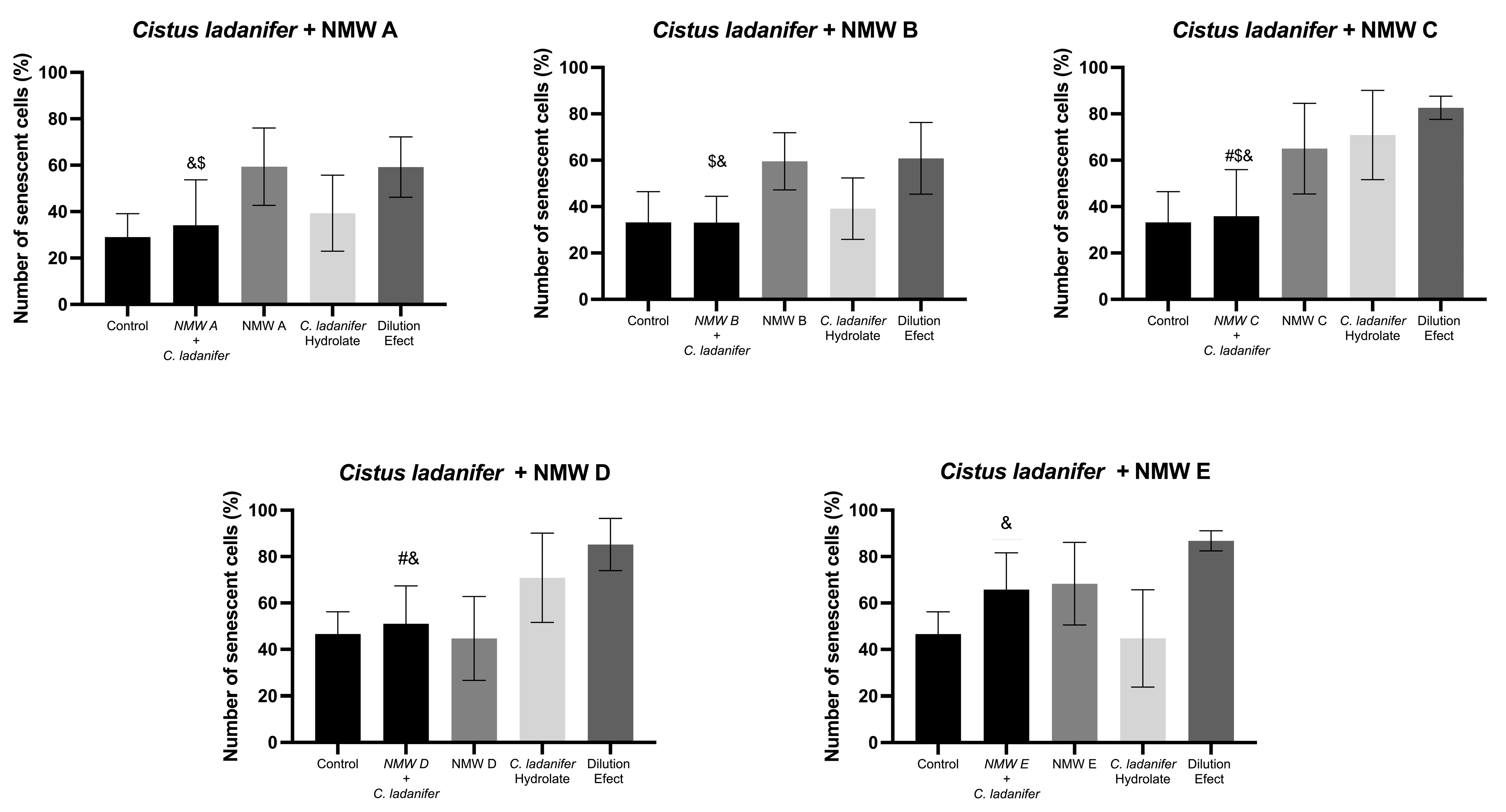
3.4. Senescence-Associated β-Galactosidase Senescence in L929 Cell Line

Regarding the senescence results, none of the mixtures showed a significant decrease in the number of senescent cells compared to all the conditions tested. However, several mixtures showed improvements when compared to the isolated ingredients.
Mixtures of NMW and Cistus ladanifer consistently showed increased efficacy compared to one of the ingredients alone. Figure 1 shows the impact of combinations of Cistus ladanifer with different NMWs on the cell senescence. However, all the mixtures showed a statistically significant improvement compared to the dilution control, which is particularly promising as it indicates that the combinations studied decreased the number of senescent cells. Combinations of this hydrolate with NMW A, B, C, and D showed significant reductions in the number of senescent cells, with statistically relevant results in contrast with the combinations with NMW E. Based on the hydrolyte’s chemical profile previously described, the composition of Cistus ladanifer hydrolate is characterized by the major compounds E-pinocarveol, borneol and terpinene-4-ol, that have recognized antioxidant and anti-inflammatory properties [29]. These compounds play a key role in modulating oxidative stress, a critical factor in inducing cellular senescence [55,58]. As shown in previous studies, terpenic compounds such as borneol can modulate senescence pathways by reducing the activation of p53/p21 and p16INK4a/Rb [59].
With the remaining conditions, this decrease in activity may be due to the loss of the predominance of the extract’s activity when diluted in water, which causes it to lose its activity [60].

3.5. Skin Regenerating Potential of the Mixtures

The skin regenerating capacity of the mixtures and isolated ingredients was assessed on the L929 fibroblast cell line, by evaluating cell migration. The mixtures showed varying degrees of efficacy, with the mixtures containing Rubus idaeus L. extract standing out for their superior migratory capacity compared with the remaining conditions tested. This profile is presented in Figure 2, which shows the effect of combinations of Rubus idaeus L. with different NMWs on cell migration capacity in L929 cells.
In general, it can be seen that the combinations with NMW A, NMW D, and NMW E show significant results compared with both isolated ingredients, NMW and R. idaeus extract, and trend for higher cell migration compared with the control, suggesting a possible benefit combination of these mixtures. In contrast, combinations with NMW B, C, and E showed a less pronounced migratory response, still with combinations comprising NMWs C and E, producing an increased effect when compared to the isolated ingredient. Notably, in the cases of NMW D, an increase in migratory capacity was observed relative to the dilution control, further supporting their potential role in enhancing cell migration.
Previous studies have characterized the chemical composition of Rubus idaeus agro-industrial waste extracts. These extracts are rich in phenolic compounds, with galloyl-HHDP-glucose being the most abundant, and an important contribute to their biological activity [33]. Numerous studies indicate that phenolic-rich extracts can enhance cell migration, a result linked to their antioxidant effects and the regulation of cellular pathways related to tissue regeneration [61]. Phenolic compounds present in Rubus idaeus, such as anthocyanins, have also been previously associated with the activation of the PI3K/Akt pathway, promoting cell migration in different cell lines [62,63]. Silica is another compound known for its structural benefits in skin regeneration, contributing to collagen synthesis, enhancing skin elasticity, and promoting wound healing by supporting fibroblast function and extracellular matrix formation [64]. Among the NMWs studied, NMW D exhibited the highest silica concentration. The improved skin regenerating potential seen in mixtures involving NMW D could suggest a possible contribution of silica to this outcome. In addition to silica, other ionic constituents may also contribute to the observed bioactivities. For instance, bicarbonates, prevalent in NMWs B and C, help maintain physiological pH in skin cells [65]. Sulfates, abundant in NMWs A-C, have been associated with keratinocyte differentiation. Studies have shown that sulfate compounds play crucial roles in regulating cell proliferation and differentiation in the epidermis, suggesting that the sulfates present in NMWs may contribute to these processes [66,67]. Sodium plays a key role in cellular hydration and osmotic balance, which can influence cell viability and migration [68]. The combination of these elements likely contributes synergistically to the observed antioxidant and regenerative properties of the mixtures. These results reinforce the potential of the mixtures to promote skin regeneration, attributed to the synergistic combination of the structural benefits of silica and the antioxidant and anti-inflammatory properties provided by the extract’s rich composition of phenolic compounds [30,69].
Cistus ladanifer hydrolate also promoted cell migration, suggesting its potential as a key ingredient in anti-aging formulations, corroborating previous studies with this hydrolate [29]. However, when this ingredient was combined with the NMW under study, the migratory capacity of cells decreased, probably consequent to the extracts dilution.

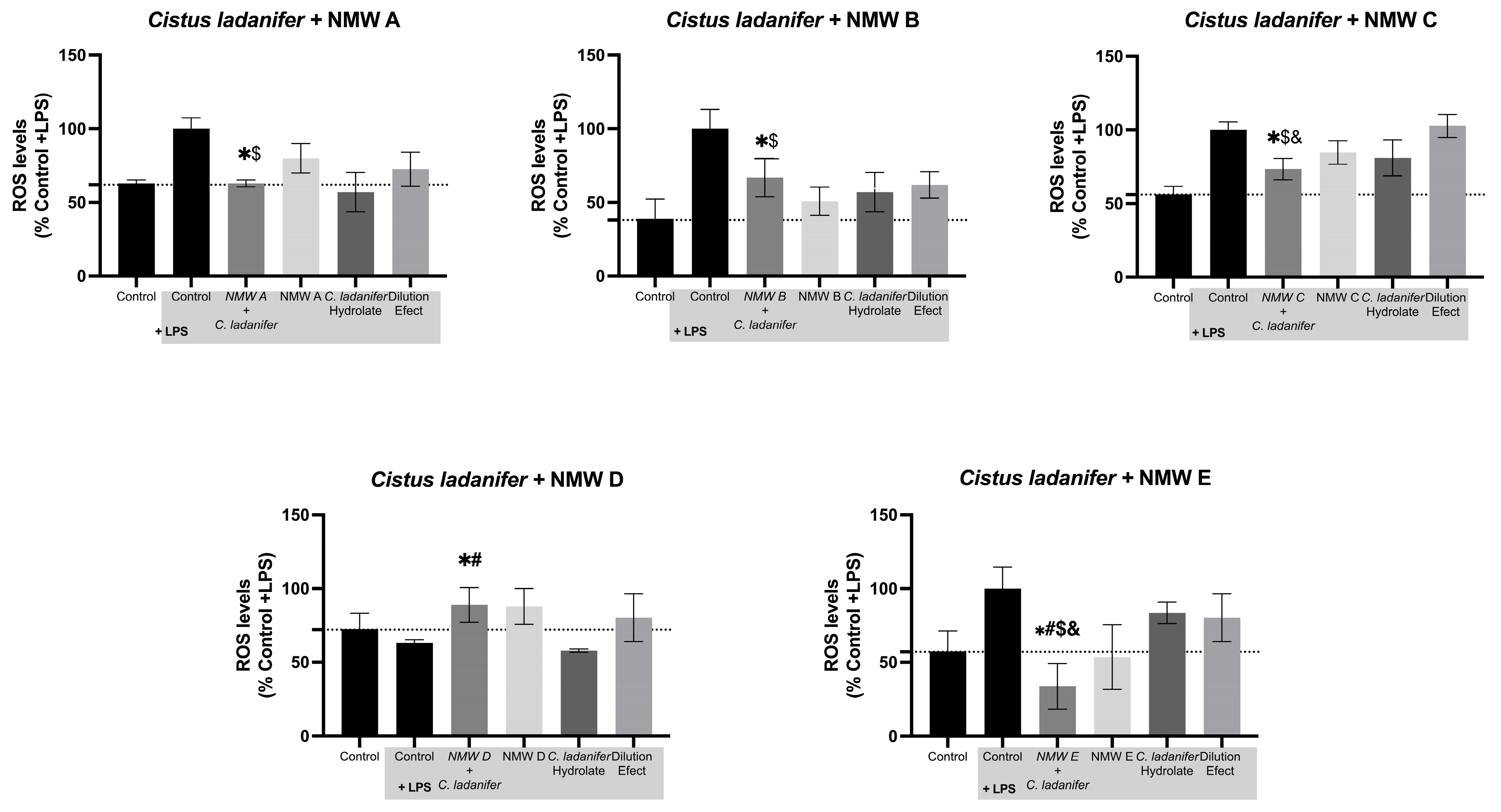
3.6. Reactive Oxygen Species Quantification

The quantification of reactive oxygen species (ROS) was performed on RAW 264.7 macrophage cells to evaluate the reversion of oxidative stress prompted by different plant extracts and their mixture with NMWs. This analysis was based on the ability of these extracts and mixtures to reduce ROS levels. During this test, we observed that the mixtures containing Cistus ladanifer hydrolate showed the most promising results (see Figure 3).
The combination of Cistus ladanifer with NMW C resulted in a significant reduction in ROS levels compared to NMW C and hydrolat alone, and the control group, reversing ROS levels to their basal state, and indicating a strong antioxidant capacity of this mixture. Cistus ladanifer contains a wide variety of bioactive compounds, such as oxygenated monoterpenes, which play a key role in modulating ROS, such as E-pinocarveol, borneol, and terpinene-4-ol [29,70], which have been described as effective in modulating oxidative stress through radical-scavenging activity and regulation of redox-sensitive cellular pathways [71]. The reduction in ROS levels observed in some mixtures may also be related to a decrease in cellular senescence, since oxidative stress plays a central role in activating pro-senescence pathways p53/p21 and p16INK4a/Rb, as discussed above [59].
The results observed for mixture E are particularly interesting, since this condition contains the highest amount of Cistus ladanifer hydrolate (12.5 per cent), suggesting that this component may contribute significantly to the observed effect. This indicates that the hydrolate alone may play a role in modulating ROS levels, possibly due to the presence of volatile compounds with radical-scavenging and anti-inflammatory properties. In this combination, NMW E—rich in bicarbonate and sulfate—may enhance redox. Similarly, in mixture A, which also showed a significant reduction in ROS, the presence of sulfur and bicarbonate in NMW A may synergize with the monoterpenes of Cistus ladanifer to support intracellular antioxidant mechanisms. In mixture C, sulfate and sodium, abundant in NMW C, may contribute to maintaining osmotic balance and modulating cellular stress responses. These observations reinforce the hypothesis that the antioxidant effects observed in mixtures A, C, and E result from an interaction between the mineral composition of the waters and the phytochemical profile of the plant extract.

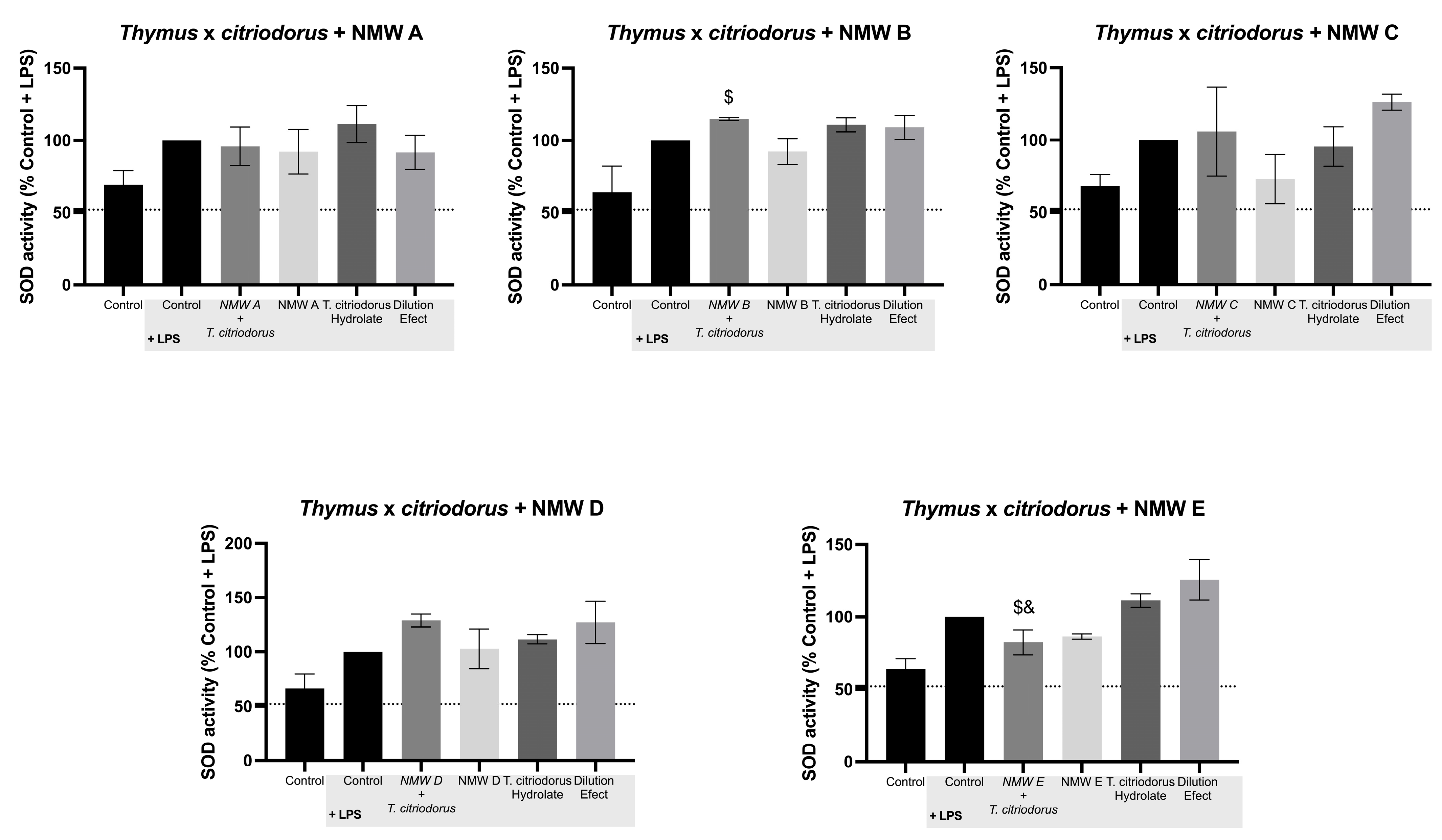
3.7. Effect on Superoxide Dismutase (SOD) Activity

Maintaining our focus on oxidant stress we also quantified the superoxide dismutase (SOD) activity in RAW 264.7 cells. When exposed to the LPS (pro-oxidant stimuli used to generate ROS), the cells presented a higher enzyme activity, when compared to the basal control group.
Overall, the addition of the mixtures did not produce significant differences in SOD activity when compared to the other conditions tested. Still, mixtures involving Thymus x citriodorus demonstrated positive results, producing an increase in the cellular antioxidant responses. The mixture of NMW B shows a significant increase in SOD activity compared to the extract and NMW alone or the control groups, as can be seen in Figure 4. SOD is a key enzyme in the defense against oxidative stress, and its increase suggests that this mixture not only directly reduces free radicals but also strengthens the cells’ endogenous antioxidant response [72]. The volatile fraction of Thymus x citriodorus is predominantly composed of oxygenated monoterpenes, such as 1,8-cineole(43.9%), α-terpineol (21.1%) and borneol (11.5%), which have antioxidant properties and can contribute to the activity of the superoxide dismutase (SOD) enzyme, protecting cells against oxidative stress [29]. As proven in previous studies, these compounds have demonstrated the ability to modulate the cellular antioxidant response and reduce the damage caused by reactive oxygen species [73]. Furthermore, the presence of specific minerals in the NMWs, particularly bicarbonates and sulfates, may have contributed to the observed synergistic effect by enhancing the antioxidant activity of the hydrolate [74].

3.8. Integrated Discussion

The NMWs studied originated from different regions of Portugal, each characterized by different mineral compositions. NMWs A, B, and C are sulfur, bicarbonate, and sodium-rich waters, and these minerals play crucial roles in skin health. Sulfur is known for its antimicrobial and antioxidant properties, bicarbonate helps maintain the skin’s pH balance, and sodium aids in moisture retention [75]. NMW D is rich in sulfates, known for their detoxifying effects, which can help purify the skin [76], while NMW E contains significant quantities of silicates, which are beneficial for skin elasticity [77].
The different mineral profiles of the NMWs contributed to the variations in bioactivity that were observed [13,78]. The highest biocompatibility of certain mixtures could be attributed to the presence of minerals that support cellular function. In fact, NMWs A, B, and C, which are rich in these minerals, showed low levels of cytotoxicity, especially when combined with Cistus ladanifer and R. idaeus extracts. Similarly, the antioxidant properties of some mixtures may be linked to minerals known for their role in oxidative stress reduction, such as sulfur, highly present in NMW A, B and C, which is recognized for its ability to neutralize free radicals and support cellular antioxidant defenses. This aligns with our findings, where combinations involving NMW A showed a significant reduction in ROS levels, particularly when combined with Cistus ladanifer. Additionally, the presence of bicarbonates in NMW B and C may contribute to maintaining cellular redox balance, as observed in the increased superoxide dismutase (SOD) activity in mixtures involving Thymus x citriodorus.
The highest biocompatibility of certain mixtures could be attributed to the presence of minerals that support cellular health and function. These combinations likely benefit from the presence of sulfur, bicarbonates and sodium, which are known to contribute to cellular homeostasis, osmotic balance, and redox regulation [65,67,68]. Similarly, the antioxidant properties of some mixtures may be linked to sulfur, highly present in NMWs A, B, and C, which is recognized for its ability to neutralize free radicals and support intracellular antioxidant defenses [78].
Given the promising results observed, additional validation in more complex biological systems could be pursued. However, the present study was designed as an in vitro initial screening to assess the bioactivity and safety of the proposed combinations. Further in vivo studies in human volunteers, may help confirm the efficacy of these mixtures under physiological conditions, provided that they comply with the ethical and regulatory requirements applicable to cosmetic ingredients.

4. Conclusions

These results provide valuable information about the anti-aging potential of NMWs combined with other natural ingredients as plant extracts. Their biocompatibility, skin regeneration potential, potential to reduce ROS levels and increase SOD activity show that these mixtures can be used as active ingredients in anti-aging formulations. Notably, some combinations were able to reduce ROS levels by up to 33% (NMW A + Cistus ladanifer), decreased senescent cell percentages by approximately 40% (NMW A + Cistus ladanifer), and increased migration capacity of over 50% (NMW A + Rubus idaeus), compared to control conditions. Particularly, the ability of these mixtures to reduce ROS and senescent cells and to increase SOD activity, support their role in reducing oxidative stress, a key factor in the aging process. The mineral compositions of NMWs and the bioactive compounds in the extracts/hydrolates contribute to their efficacy, and the results of this research emphasize the importance of selecting the right combination. Although the relationships between the different ingredients are complex, since both NMWs and plant extracts/hydrolates are composed of a large number of individual compounds, future studies could further explore the molecular mechanisms underlying these effects and assess which key players contribute to the observed activities. Moreover, given their performance and natural origin, these combinations may be considered for future application in dermocosmetic formulations targeting skin aging.

Author Contributions

C.P.G.: investigation; methodology, formal analysis and writing—original draft preparation; A.S.O. methodology, validation, investigation and writing—review and editing; J.R.: conceptualization, methodology, validation and writing—review and editing; T.F.F.d.S.: investigation, resources, validation and writing—review and editing; R.P.d.O.: validation and writing—review and editing; M.J.A.: Conceptualization, writing—review and editing, project administration and funding acquisition; P.P.: methodology, resources, validation and writing—review; A.P.d.O.: conceptualization, validation, writing—review and editing, supervision and project administration. All authors have read and agreed to the published version of the manuscript.

Funding

This work was supported by Projeto Aquae Vitae—Água Termal como Fonte de Vida e Saúde [PD20-00003], a Project funded under the Promove program of Fundação La Caixa, in partnership with BPI and FCT. This work was also developed within the scope of the CICS-UBI projects [UIDB/00709/2020] and [UIDP/00709/2020], financed by national funds through the Portuguese Foundation for Science and Technology/MCTES (FCT). Promove program of Fundação La Caixa also provided financial support to ASO and CPG through research scholarships. This work was also supported by national funds through FCT/MCTES (PIDDAC): CIMO, UIDB/00690/2020 (https://doi.org/10.54499/UIDB/00690/2020 (accessed on 17 April 2025)) and UIDP/00690/2020 (https://doi.org/10.54499/UIDP/00690/2020 (accessed on 17 April 2025)); and SusTEC, LA/P/0007/2020 (https://doi.org/10.54499/LA/P/0007/2020 (accessed on 17 April 2025)).

Institutional Review Board Statement

Not applicable.

Informed Consent Statement

Not applicable.

Data Availability Statement

Data are contained within the article.

Acknowledgments

The authors acknowledge the enrolled thermal centers for providing the natural mineral water samples studied in this work.

Conflicts of Interest

The funders had no role in the design of this study; in the collection, analyses, or interpretation of data; in the writing of the manuscript, or in the decision to publish the results.

References

  1. Ferreira, M.S.; Magalhães, M.C.; Oliveira, R.; Sousa-Lobo, J.M.; Almeida, I.F. Trends in the Use of Botanicals in Anti-Aging Cosmetics. Molecules 2021, 26, 3584. [Google Scholar] [CrossRef] [PubMed]
  2. Fonseca-Santos, B.; Corrêa, M.A.; Chorilli, M. Sustainability, natural and organic cosmetics: Consumer, products, efficacy, toxicological and regulatory considerations. Braz. J. Pharm. Sci. 2015, 51, 17–26. [Google Scholar] [CrossRef]
  3. Ahmed, I.A.; Mikail, M.A.; Zamakshshari, N.; Abdullah, A.S.H. Natural anti-aging skincare: Role and potential. Biogerontology 2020, 21, 293–310. [Google Scholar] [CrossRef]
  4. Farage, M.A.; Miller, K.W.; Elsner, P.; Maibach, H.I. Intrinsic and extrinsic factors in skin ageing: A review. Int. J. Cosmet. Sci. 2008, 30, 87–95. [Google Scholar] [CrossRef] [PubMed]
  5. Chermahini, S.H.; Adibah, F.; Majid, A.; Sarmidi, M.R. Cosmeceutical Value of Herbal Extracts as Natural Ingredients and Novel Technologies in Anti-Aging. J. Med. Plants Res. 2011, 5, 3074–3077. [Google Scholar]
  6. Martins, I.; Almeida, D.; Yoon, I.-S.; Costa, E.F.; Magalhães, W.V.; Di Stasi, L.C. Recent Advances in Herbal-Derived Products with Skin Anti-Aging Properties and Cosmetic Applications. Molecules 2022, 27, 7518. [Google Scholar] [CrossRef]
  7. Gianfaldoni, S.; Tchernev, G.; Wollina, U.; Roccia, M.G.; Fioranelli, M.; Gianfaldoni, R.; Lotti, T. History of the Baths and Thermal Medicine. Open Access Maced. J. Med. Sci. 2017, 5, 566. [Google Scholar] [CrossRef]
  8. Araujo, A.R.T.S.; Sarraguça, M.C.; Ribeiro, M.P.; Coutinho, P. Physicochemical fingerprinting of thermal waters of Beira Interior region of Portugal. Env. Geochem. Health 2017, 39, 483–496. [Google Scholar] [CrossRef]
  9. Oliveira, A.S.; Vaz, C.V.; Silva, A.; Correia, S.; Ferreira, R.; Breitenfeld, L.; Martinez-de-Oliveira, J.; Palmeira-de-Oliveira, R.; Pereira, C.; Cruz, M.T.; et al. In vitro evaluation of potential benefits of a silica-rich thermal water (Monfortinho Thermal Water) in hyperkeratotic skin conditions. Int. J. Biometeorol. 2020, 64, 1957–1968. [Google Scholar] [CrossRef]
  10. Silva, A.; Oliveira, A.S.; Vaz, C.V.; Correia, S.; Ferreira, R.; Breitenfeld, L.; Martinez-de-Oliveira, J.; Palmeira-de-Oliveira, R.; Pereira, C.M.F.; Palmeira-de-Oliveira, A.; et al. Anti-inflammatory potential of Portuguese thermal waters. Sci. Rep. 2020, 10, 1–13. [Google Scholar] [CrossRef]
  11. Vaz, C.V.; Oliveira, A.S.; Silva, A.; Cortes, L.; Correia, S.; Ferreira, R.; Breitenfeld, L.; Martinez-de-Oliveira, J.; Palmeira-de-Oliveira, R.; Pereira, C.F.; et al. Protective role of Portuguese natural mineral waters on skin aging: In Vitro evaluation of anti-senescence and anti-oxidant properties. Int. J. Biometeorol. 2022, 66, 2117–2131. [Google Scholar] [CrossRef] [PubMed]
  12. Proksch, E.; Nissen, H.P.; Bremgartner, M.; Urquhart, C. Bathing in a magnesium-rich Dead Sea salt solution improves skin barrier function, enhances skin hydration, and reduces inflammation in atopic dry skin. Int. J. Dermatol. 2005, 44, 151–157. [Google Scholar] [CrossRef] [PubMed]
  13. Figueiredo, A.C.; Rodrigues, M.; Mourelle, M.L.; Araujo, A.R.T.S. Thermal Spring Waters as an Active Ingredient in Cosmetic Formulations. Cosmetics 2023, 10, 27. [Google Scholar] [CrossRef]
  14. Yasin, Z.A.M.; Ibrahim, F.; Rashid, N.N.; Razif, M.F.M.; Yusof, R. The Importance of Some Plant Extracts as Skin Anti-aging Resources: A Review. Curr. Pharm. Biotechnol. 2017, 18, 864–876. [Google Scholar] [CrossRef]
  15. Arct, J.; Pytkowska, K. Flavonoids as components of biologically active cosmeceuticals. Clin. Dermatol. 2008, 26, 347–357. [Google Scholar] [CrossRef]
  16. De Lima Cherubim, D.J.; Martins, C.V.B.; Fariña, L.O.; da Silva de Lucca, R.A. Polyphenols as natural antioxidants in cosmetics applications. J. Cosmet. Dermatol. 2020, 19, 33–37. [Google Scholar] [CrossRef]
  17. Subramani, R.; Narayanasamy, M.; Feussner, K.D. Plant-derived antimicrobials to fight against multi-drug-resistant human pathogens. 3 Biotech 2017, 7, 172. [Google Scholar] [CrossRef] [PubMed]
  18. Enciso-Martínez, Y.; Zuñiga-Martínez, B.S.; Ayala-Zavala, J.F.; Domínguez-Avila, J.A.; González-Aguilar, G.A.; Viuda-Martos, M. Agro-Industrial By-Products of Plant Origin: Therapeutic Uses as well as Antimicrobial and Antioxidant Activity. Biomolecules 2024, 14, 762. [Google Scholar] [CrossRef]
  19. Vodnar, D.C.; Călinoiu, L.F.; Dulf, F.V.; Ştefănescu, B.E.; Crişan, G.; Socaciu, C. Identification of the bioactive compounds and antioxidant, antimutagenic and antimicrobial activities of thermally processed agro-industrial waste. Food Chem. 2017, 231, 131–140. [Google Scholar] [CrossRef]
  20. Filho, A.V.; Avila, L.B.; Lacorte, D.H.; Martiny, T.R.; Rosseto, V.; Moraes, C.C.; Dotto, G.L.; Carreno, N.L.V.; da Rosa, G.S. Brazilian Agroindustrial Wastes as a Potential Resource of Bioative Compounds and Their Antimicrobial and Antioxidant Activities. Molecules 2022, 27, 6876. [Google Scholar] [CrossRef]
  21. Gonçalves, S.; Caramelo, A. The Role of Elderberry Hydrolate as a Therapeutic Agent in Palliative Care. Antioxidants 2025, 14, 233. [Google Scholar] [CrossRef] [PubMed]
  22. Serra, D.; Cruciani, S.; Garroni, G.; Sarais, G.; Kavak, F.F.; Satta, R.; Montesu, M.A.; Floris, M.; Ventura, C.; Maioli, M. Effect of Helichrysum italicum in Promoting Collagen Deposition and Skin Regeneration in a New Dynamic Model of Skin Wound Healing. Int. J. Mol. Sci. 2024, 25, 4736. [Google Scholar] [CrossRef] [PubMed]
  23. Valente, J.V.; Palmeira-de-Oliveira, R.; Guiomar, L.; Vaz, C.V.; Rolo, J.; Gaspar, C.; Oliveira, A.S.; Caramelo, D.; Breitenfeld, L.; Gonçalves, J.C.; et al. Humulus lupulus aqueous extract and hydrolate as a potential ingredient for cosmetics: Chemical characterization and in vitro antimicrobial, cytotoxicity, antioxidant and anti-inflammatory assessment. Fitoterapia 2024, 175, 105861. [Google Scholar] [CrossRef] [PubMed]
  24. Ayu, K.; Budiasih, S.; Gede, I.; Handrean, E. The Anti-Aging Properties of Panax Ginseng: A Narrative Review of Mechanisms and Clinical Evidence. Int. J. Health Pharm. (IJHP) 2025, 5, 157–164. [Google Scholar] [CrossRef]
  25. Sun, J.; Jiang, Y.; Fu, J.; He, L.; Guo, X.; Ye, H.; Yin, C.; Li, H.; Jiang, H. Beneficial Effects of Epigallocatechin Gallate in Preventing Skin Photoaging: A Review. Molecules 2024, 29, 5226. [Google Scholar] [CrossRef]
  26. Nobile, V.; Dudonné, S.; Kern, C.; Roveda, G.; Garcia, C. Antiaging, Brightening, and Antioxidant Efficacy of Fermented Bilberry Extract (Vaccinium myrtillus): A Randomized, Double-Blind, Placebo-Controlled Trial. Nutrients 2024, 16, 2203. [Google Scholar] [CrossRef]
  27. Gaweł-Bęben, K.; Kukula-Koch, W.; Hoian, U.; Czop, M.; Strzępek-Gomółka, M.; Antosiewicz, B. Characterization of Cistus × incanus L. and Cistus ladanifer L. Extracts as Potential Multifunctional Antioxidant Ingredients for Skin Protecting Cosmetics. Antioxidants 2020, 9, 202. [Google Scholar] [CrossRef]
  28. Rattanawiwatpong, P.; Wanitphakdeedecha, R.; Bumrungpert, A.; Maiprasert, M. Anti-aging and brightening effects of a topical treatment containing vitamin C, vitamin E, and raspberry leaf cell culture extract: A split-face, randomized controlled trial. J. Cosmet. Dermatol. 2020, 19, 671–676. [Google Scholar] [CrossRef]
  29. Oliveira, A.S.; Rolo, J.; Gaspar, C.; Ramos, L.; Cavaleiro, C.; Salgueiro, L.; Palmeira-de-Oliveira, R.; Teixeira, J.P.; Martinez-de-Oliveira, J.; Palmeira-de-Oliveira, A. Thymus mastichina (L.) L. and Cistus ladanifer L. for skin application: Chemical characterization and in vitro bioactivity assessment. J. Ethnopharmacol. 2023, 302, 115830. [Google Scholar] [CrossRef]
  30. Oliveira, A.S.; Rolo, J.; Gaspar, C.; Ramos, L.; Cavaleiro, C.; Salgueiro, L.; Palmeira-de-Oliveira, R.; Teixeira, J.P.; Martinez-de-Oliveira, J.; Palmeira-de-Oliveira, A. Chemical characterization and bioactive potential of Thymus × citriodorus (Pers.) Schreb. preparations for anti-acne applications: Antimicrobial, anti-biofilm, anti-inflammatory and safety profiles. J. Ethnopharmacol. 2022, 287, 114935. [Google Scholar] [CrossRef]
  31. Plasencia, P.; Heleno, S.A.; Finimundy, T.; Carocho, M.; Calhelha, R.C.; Añibarro-Ortega, M.; Alves, M.J.; Oludemi, T.; Quidiongo, N.; Barreiro, F.; et al. Recovery of High Valuable Bioactive Molecules from Vaccinium myrtillus L. Bioresidues. Waste Biomass Valorization 2023, 14, 2873–2884. [Google Scholar] [CrossRef]
  32. Shiraishi, C.S.H.; Zbiss, Y.; Roriz, C.L.; Dias, M.I.; Prieto, M.A.; Calhelha, R.C.; Alves, M.J.; Heleno, S.A.; V., d.C.M.; Carocho, M.; et al. Fig Leaves (Ficus carica L.): Source of Bioactive Ingredients for Industrial Valorization. Processes 2023, 11, 1179. [Google Scholar] [CrossRef]
  33. Plasencia, P.; Finimundy, T.C.; Carocho, M.; Calhelha, R.C.; Añibarro-Ortega, M.; Pires, T.C.S.P.; Barreiro, F.; Garcia, P.A.; Barros, L.; Heleno, S.A.; et al. Extraction of Bioactive Compounds from Rubus Idaeus Bioresidues: A Full Screening on Phenolic Composition and Bioactive Potential. Waste Biomass Valorization 2025, 16, 737–747. [Google Scholar] [CrossRef]
  34. Rice, E.W.; Baird, R.B.; Eaton, A.D.; Clesceri, L.S.; Bridgewater, L. (Eds.) Standard Methods for the Examination of Water and Wastewater; American Public Health Association American Water Works Association Water Environment Federation: Washington, DC, USA, 2012. [Google Scholar]
  35. Hidrogenoma. Available online: https://hidrogenoma.dgeg.gov.pt/ (accessed on 15 February 2025).
  36. Oliveira, A.S.; Vaz, C.V.; Silva, A.; Ferreira, S.S.; Correia, S.; Ferreira, R.; Breitenfeld, L.; Martinez-de-Oliveira, J.; Palmeira-de-Oliveira, R.; Pereira, C.; et al. Chemical signature and antimicrobial activity of Central Portuguese Natural Mineral Waters against selected skin pathogens. Environ. Geochem. Health 2020, 42, 2039–2057. [Google Scholar] [CrossRef]
  37. Da Silveira, T.F.F.; Echegaray, N.; Guimarães, R.; Lemos, A.; Pires, T.C.S.P.; Ferreira, I.C.F.R.; Alves, M.J.; Barros, L. Unveiling the impact of thermal water in German chamomile infusions: Effects on phenolic compounds, antimicrobial and antioxidant properties. Food Chem. 2025, 463, 141481. [Google Scholar] [CrossRef]
  38. Biological Evaluation of Medical Devices—Part 5: Tests for In Vitro Cytotoxicity. ISO 10993-5:2009. Available online: https://www.iso.org/standard/36406.html (accessed on 10 May 2023).
  39. Molyneux, P. The use of the stable free radical diphenylpicryl-hydrazyl (DPPH) for estimating antioxidant activity. Songklanakarin J. Sci. Technol. 2004, 26, 211–219. [Google Scholar]
  40. JP-DM Protocol and Undefined 2012 Procedure: Preparation of DPPH Radical, and Antioxidant Scavenging Assay. Researchgate.netJM PrietoDPPH Microplate Protocol, Researchgate.Net. 2012. Available online: https://www.researchgate.net/profile/Jose-Prieto-10/post/Can_anyone_explain_the_DPPH_method_for_antioxidant_activity_in_details/attachment/59d61efac49f478072e9772d/AS%3A271744332435456%401441800305338/download/Prieto+DPPH+protocol.pdf (accessed on 10 May 2025).
  41. Scherer, R.; Godoy, H.T. Antioxidant activity index (AAI) by the 2,2-diphenyl-1-picrylhydrazyl method. Food Chem. 2009, 112, 654–658. [Google Scholar] [CrossRef]
  42. Schindelin, J.; Arganda-Carreras, I.; Frise, E.; Kaynig, V.; Longair, M.; Pietzsch, T.; Preibisch, S.; Rueden, C.; Saalfeld, S.; Schmid, B.; et al. Fiji: An open-source platform for biological-image analysis. Nat. Methods 2012, 9, 676–682. [Google Scholar] [CrossRef]
  43. Pascoal, A.S.P.; Moura, M.J.; Ferreira, P.C.N.; Ramos, M.L.C.; Salavessa, M.A.N.; Duarte, B.P.M. A Conceptual Engineering Approach to Developing a Bio-Based Hair Mask. Cosmetics 2025, 12, 45. [Google Scholar] [CrossRef]
  44. Mac-Mary, S.; Creidi, P.; Marsaut, D.; Courderot-Masuyer, C.; Cochet, V.; Gharbi, T.; Guidicelli-Arranz, D.; Tondu, F.; Humbert, P. Assessment of effects of an additional dietary natural mineral water uptake on skin hydration in healthy subjects by dynamic barrier function measurements and clinic scoring. Ski. Res. Technol. 2006, 12, 199–205. [Google Scholar] [CrossRef]
  45. Lestari, U.; Muhaimin, M.; Chaerunisaa, A.Y.; Sujarwo, W. Anti-Aging Potential of Plants of the Anak Dalam Tribe, Jambi, Indonesia. Pharmaceuticals 2023, 16, 1300. [Google Scholar] [CrossRef] [PubMed]
  46. Verma, A.; Gour, H.; Sagar, V.; Kumar, R. Medicinal Plants as Source of Anti-Ageing Agents: A Review. World J. Pharm. Pharm. Sci. 2023, 12, 946–957. [Google Scholar] [CrossRef]
  47. Papaccio, F.; D’arino, A.; Caputo, S.; Bellei, B. Focus on the Contribution of Oxidative Stress in Skin Aging. Antioxidants 2022, 11, 1121. [Google Scholar] [CrossRef] [PubMed]
  48. Varani, J.; Dame, M.K.; Rittie, L.; Fligiel, S.E.; Kang, S.; Fisher, G.J.; Voorhees, J.J. Decreased Collagen Production in Chronologically Aged Skin: Roles of Age-Dependent Alteration in Fibroblast Function and Defective Mechanical Stimulation. Am. J. Pathol. 2006, 168, 1861–1868. [Google Scholar] [CrossRef] [PubMed]
  49. Falcão, S.I.; Duarte, D.; Diallo, M.; Santos, J.; Ribeiro, E.; Vale, N.; Vilas-Boas, M. Improvement of the In Vitro Cytotoxic Effect on HT-29 Colon Cancer Cells by Combining 5-Fluorouacil and Fluphenazine with Green, Red or Brown Propolis. Molecules 2023, 28, 3393. [Google Scholar] [CrossRef]
  50. Tun, J.O.; Salvador-Reyes, L.A.; Velarde, M.C.; Saito, N.; Suwanborirux, K.; Concepcion, G.P. Synergistic Cytotoxicity of Renieramycin M and Doxorubicin in MCF-7 Breast Cancer Cells. Mar. Drugs 2019, 17, 536. [Google Scholar] [CrossRef]
  51. Aćimović, M.G.; Tešević, V.V.; Smiljanić, K.T.; Cvetković, M.T.; Stanković, J.M.; Kiprovski, B.M.; Sikora, V.S. Hydrolates: By-products of essential oil distillation: Chemical composition, biological activity and potential uses. Adv. Technol. 2020, 9, 54–70. [Google Scholar] [CrossRef]
  52. Pavlović, D.; Grigorov, M.; Martinović, M.; Tasić-Kostov, M. Hydrolates: From Waste Products to Potential Cosmetic Actives. Arch. Pharm. 2021, 71, S86. [Google Scholar]
  53. Oğuz, İ.; Oğuz, H.İ.; Attar, Ş.H.; Sönmez, D.A.; Çelik, H.; Yaşa Kafkas, N.E. Preferable Berry Fruits for Tolerance to Global Climate Change and Dry Conditions; IntechOpen Limited: London, UK, 2023. [Google Scholar] [CrossRef]
  54. Huyut, Z.; Beydemir, Ş.; Gülçin, I. Antioxidant and Antiradical Properties of Selected Flavonoids and Phenolic Compounds. Biochem. Res. Int. 2017, 2017, 7616791. [Google Scholar] [CrossRef]
  55. Rice-Evans, C.A.; Miller, N.J.; Paganga, G. Antioxidant properties of phenolic compounds. Trends Plant Sci. 1997, 2, 152–159. [Google Scholar] [CrossRef]
  56. Cid-Pérez, T.S.; Ávila-Sosa, R.; Ochoa-Velasco, C.E.; Rivera-Chavira, B.E.; Nevárez-Moorillón, G.V. Antioxidant and Antimicrobial Activity of Mexican Oregano (Poliomintha longiflora) Essential Oil, Hydrosol and Extracts from Waste Solid Residues. Plants 2019, 8, 22. [Google Scholar] [CrossRef] [PubMed]
  57. Değirmenci, H.; Erkurt, H. Relationship between volatile components antimicrobial antioxidant properties of the essential oil hydrosol extracts of Citrus aurantium L. flowers. J. Infect. Public. Health 2020, 13, 58–67. [Google Scholar] [CrossRef] [PubMed]
  58. Alves-Silva, J.M.; Gonçalves, M.J.; Silva, A.; Cavaleiro, C.; Cruz, M.T.; Salgueiro, L. Chemical Profile Anti-Microbial Anti-Inflammaging Activities of Santolina rosmarinifolia L. Essential Oil from Portugal. Antibiotics 2023, 12, 179. [Google Scholar] [CrossRef]
  59. Warman, D.J.; Jia, H.; Kato, H. Effects of Thyme (Thymus vulgaris L.) Essential Oil on Aging-Induced Brain Inflammation and Blood Telomere Attrition in Chronologically Aged C57BL/6J Mice. Antioxidants 2023, 12, 1178. [Google Scholar] [CrossRef]
  60. Faria, A.; Oliveira, J.; Neves, P.; Gameiro, P.; Santos-Buelga, C.; de Freitas, V.; Mateus, N. Antioxidant Properties of Prepared Blueberry (Vaccinium myrtillus) Extracts. J. Agric. Food Chem. 2005, 53, 6896–6902. [Google Scholar] [CrossRef]
  61. Tsakiroglou, P.; Vandenakker, N.E.; Del Bo’, C.; Riso, P.; Klimis-Zacas, D. Role of Berry Anthocyanins and Phenolic Acids on Cell Migration and Angiogenesis: An Updated Overview. Nutrients 2019, 11, 1075. [Google Scholar] [CrossRef]
  62. Costa, G.; Francisco, V.; Lopes, M.C.; Cruz, M.T.; Batista, M.T. Intracellular signaling pathways modulated by phenolic compounds: Application for new anti-inflammatory drugs discovery. Curr. Med. Chem. 2012, 19, 2876–2900. [Google Scholar] [CrossRef]
  63. Li, G.; Li, Y.Y.; Sun, J.E.; Lin, W.H.; Zhou, R.X. ILK–PI3K/AKT pathway participates in cutaneous wound contraction by regulating fibroblast migration and differentiation to myofibroblast. Lab. Investig. 2016, 96, 741–751. [Google Scholar] [CrossRef] [PubMed]
  64. Wu, H.; Li, F.; Wang, S.; Lu, J.; Li, J.; Du, Y.; Sun, X.; Chen, X.; Gao, J.; Ling, D. Ceria nanocrystals decorated mesoporous silica nanoparticle based ROS-scavenging tissue adhesive for highly efficient regenerative wound healing. Biomaterials 2018, 151, 66–77. [Google Scholar] [CrossRef]
  65. Dugué, C.; Baiyasi, M.; Jackson, S.; Tolliver, S.; Daveluy, S. Baking Soda and the Skin: A Review of Baking Soda in Dermatology. J. Integr. Dermatology. 2024. Available online: https://www.jintegrativederm.org/article/122501-baking-soda-and-the-skin-a-review-of-baking-soda-in-dermatology (accessed on 16 May 2025).
  66. Hanyu, O.; Nakae, H.; Miida, T.; Higashi, Y.; Fuda, H.; Endo, M.; Kohjitani, A.; Sone, H.; Strott, C.A. Cholesterol sulfate induces expression of the skin barrier protein filaggrin in normal human epidermal keratinocytes through induction of RORα. Biochem. Biophys. Res. Commun. 2012, 428, 99–104. [Google Scholar] [CrossRef] [PubMed]
  67. Kitazawa, K.; Nadanaka, S.; Kadomatsu, K.; Kitagawa, H. Chondroitin 6-sulfate represses keratinocyte proliferation in mouse skin, which is associated with psoriasis. Commun. Biol. 2021, 4, 1–15. [Google Scholar] [CrossRef] [PubMed]
  68. Hołyńska-Iwan, I.; Szewczyk-Golec, K. Analysis of changes in sodium and chloride ion transport in the skin. Sci. Rep. 2020, 10, 18094. [Google Scholar] [CrossRef] [PubMed]
  69. Lopez-Corona, A.V.; Valencia-Espinosa, I.; González-Sánchez, F.A.; Sánchez-López, A.L.; Garcia-Amezquita, L.E.; Garcia-Varela, R. Antioxidant, Anti-Inflammatory and Cytotoxic Activity of Phenolic Compound Family Extracted from Raspberries (Rubus idaeus): A General Review. Antioxidants 2022, 11, 1192. [Google Scholar] [CrossRef]
  70. Porres-Martínez, M.; González-Burgos, E.; Carretero, M.E.; Gómez-Serranillos, M.P. Major selected monoterpenes α-pinene and 1,8-cineole found in Salvia lavandulifolia (Spanish sage) essential oil as regulators of cellular redox balance. Pharm. Biol. 2015, 53, 921–929. [Google Scholar] [CrossRef]
  71. Wojtunik-Kulesza, K.A.; Kasprzak, K.; Oniszczuk, T.; Oniszczuk, A. Natural Monoterpenes: Much More than Only a Scent. Chem. Biodivers. 2019, 16, e1900434. [Google Scholar] [CrossRef]
  72. Younus, H. Therapeutic Potentials of Superoxide Dismutase. Int. J. Health Sci. 2018, 12, 88. [Google Scholar]
  73. Božovic, M.; Pirolli, A.; Ragno, R. Mentha suaveolens Ehrh. (Lamiaceae) Essential Oil and Its Main Constituent Piperitenone Oxide: Biological Activities and Chemistry. Molecules 2015, 20, 8605–8633. [Google Scholar] [CrossRef]
  74. Costantino, M.; Giuberti, G.; Caraglia, M.; Lombardi, A.; Misso, G.; Abbruzzese, A.; Ciani, F.; Lampa, E. Possible antioxidant role of SPA therapy with chlorine-sulphur-bicarbonate mineral water. Amino Acids 2009, 36, 161–165. [Google Scholar] [CrossRef]
  75. Baker, P.; Huang, C.; Radi, R.; Moll, S.B.; Jules, E.; Arbiser, J.L. Skin Barrier Function: The Interplay of Physical, Chemical, and Immunologic Properties. Cells 2023, 12, 2745. [Google Scholar] [CrossRef]
  76. Carbajo, J.M.; Maraver, F. Sulphurous Mineral Waters: New Applications for Health. Evid.-Based Complement Altern. Med. 2017, 2017, 8034084. [Google Scholar] [CrossRef] [PubMed]
  77. Almeida, C.; Madeira, A.; Marto, J.; Graça, A.; Pinto, P.; Ribeiro, H. Monfortinho Thermal Water-Based Creams: Effects on Skin Hydration, Psoriasis, and Eczema in Adults. Cosmetics 2019, 6, 56. [Google Scholar] [CrossRef]
  78. Cacciapuoti, S.; Luciano, M.A.; Megna, M.; Annunziata, M.C.; Napolitano, M.; Patruno, C.; Scala, E.; Colicchio, R.; Pagliuca, C.; Salvatore, P.; et al. The Role of Thermal Water in Chronic Skin Diseases Management: A Review of the Literature. J. Clin. Med. 2020, 9, 3047. [Google Scholar] [CrossRef] [PubMed]
Figure 1. Effect of mixtures containing Cistus ladanifer hydrolate and NMWs A–E on β-galactosidase senescence in fibroblasts. Four conditions were evaluated: the mixture, the respective NMW, the extracts, and controls (culture medium and dilution effect). The results are expressed as a percentage of the total number of cells counted in each condition. Error bars indicate the mean ± SEM of independent tests. # p < 0.05 relative to extract, $ p < 0.05 relative to NMW and & < 0.05 relative to dilution control. Only statistics involving the mixture when compared with the remaining ingredients or controls are represented.
Cosmetics 12 00113 g001
Figure 2. Effect of mixtures of plant extract R. idaeus with NMWs A–E on cell migration potential in L929 cells after 12 h. Four conditions were evaluated: the mixture, the respective NMW, the extracts, and controls (culture medium and dilution effect). The results are expressed as a percentage of the control group. Error bars indicate the mean ± SEM of independent tests. # p < 0.05 relative to extract, and & < 0.05 relative to dilution control and $ p < 0.05 relative to NMW. Only statistics involving the mixture when compared with the remaining ingredients or controls are represented.
Cosmetics 12 00113 g002
Figure 3. Effect of mixtures of Cistus ladanifer hydrolate with natural mineral waters A–E on ROS levels in macrophages. Four conditions were evaluated: the mixture, the respective NMW and extracts, and controls (culture medium with and without LPS and dilution effect). The results are expressed as a percentage of the control + LPS group. Error bars indicate the mean ± SEM of independent tests. * p < 0.05 represents a statistically significant difference relative to control; # p < 0.05 relative to extract, and & < 0.05 relative to dilution control and $ p < 0.05 relative to NMW. Only statistics involving the mixture when compared with the remaining ingredients or controls are represented.
Cosmetics 12 00113 g003
Figure 4. Effect of mixtures of natural mineral water A–E with Thymus x citriodorus hydrolate in SOD activity in macrophages. Four conditions were evaluated: the mixture, the respective NMW and extracts, and controls (culture medium with and without LPS and dilution effect). The results are expressed as a percentage of the control + LPS group. Error bars indicate the mean ± SEM of independent tests. & < 0.05 relative to dilution control and $ p < 0.05 relative to NMW. Only statistics involving the mixture when compared with the remaining ingredients or controls are represented.
Cosmetics 12 00113 g004
Table 1. Major compounds identified in the extracts under study.
SpeciesCommon NameSourceMajor CompoundsReference
Cistus ladaniferRockroseProentia® (Proença-a-Nova, Portugal)E-pinocarveol (25.3%), borneol (14.0%), terpinene-4-ol (9.7%)[29]
Ficus carica L. FigCIMO-IPB (Bragança, Portugal)Apigenin-C-hexoside-C-pentoside, Quercetin-O-deoxyhexosyl-hexoside [32]
Rubus idaeus L. RaspberryCIMO-IPB (Bragança, Portugal)Galloyl-HHDP-glucose, Procyanidin dimer, Procyanidin trimer, Methyl ellagic acid, hexoside[33]
Thymus x citriodorusLemon thymeErvitas Catitas™ (Borba, Portugal)1,8-cineole (43.9%), α-terpineol (21.1%), borneol (11.5%)[29]
Vaccinium myrtillusBlueberryCIMO-IPB (Bragança, Portugal)3-O-Cafeoylquinic acid, 5-O-Cafeoylquinic acid, Procyanidin trimer[31]
Table 2. Information on Main characteristics of the studied natural mineral waters. information retrieved from Hidrogenoma [35].
CodeMain CompositionSecondary CompositionMajor Compounds *PHReference
ASodium Bicarbonate, SulfuricCarbonated,
Fluoridated,
Sulfhydrated
SiO2 (↑), Na+ (↑),HCO3 (↑)8.7[36]
BSodium Bicarbonate, SulfuricCarbonated,
Fluoridated,
Sulfhydrated
HCO3 (↑↑),Na+ (↑), SiO2 (↑)8.8[36]
CSulfate, Bicarbonate, SodiumSulfhydratedHCO3 (↑↑), Na+ (↑), SiO2 (↓)8.3[36]
DSilicateSodium bicarbonateH3SiO4 (↓), SiO2 (↓),HCO3 (↓)5.6[36]
ESodium Bicarbonate, GasocarbonateFluoridatedHCO3 (↑↑), Na+ (↑↑), SiO2 (↑)6.9[37]
* The components are listed in descending order of predominance in the ionic composition of the water. (↑↑) indicates a concentration of more than 100 mg/L; (↑) between 50 and 100 mg/L; (↓) less than 50 mg/L. The information on the ionic composition was retrieved from the analytical reports provided by the participating Thermal Centres, as referenced in the original study [38,39].
Table 3. Overview of the optimal biocompatible concentrations, coincident for L929 and RAW 264.7 cell lines, for each mixture based on the highest concentration of NMW (% v/v) and extract (mg/mL) or hydrolate (% v/v).
ExtractCombination with NMW ACombination with NMW BCombination with NMW CCombination with NMW DCombination with NMW E
Cistus ladanifer25 (% v/v) NMW + 3.13 (% v/v) hyd12.5 (% v/v) NMW + 3.13 (% v/v) hyd25 (% v/v) NMW + 6.25 (% v/v) hyd12.5 (% v/v) NMW + 6.25 (% v/v) hyd25 (% v/v) NMW + 12.5 (% v/v) hyd
Ficus carica L.12.5 (% v/v) NMW + 2 mg/mL Ext12.5 (% v/v) NMW + 2 mg/mL Ext25 (% v/v) NMW + 1 mg/mL Ext12.5 (% v/v) NMW + 1 mg/mL Ext12.5 (% v/v) NMW + 2 mg/mL Ext
Rubus idaeus L.12.5 (% v/v) NMW + 1 mg/mL Ext25 (% v/v) NMW + 1 mg/mL Ext12.5 (% v/v) NMW + 0.25 mg/mL Ext12.5 (% v/v) NMW + 1 mg/mL Ext25 (% v/v) NMW + 1 mg/mL Ext
Thymus x citriodorus12.5 (% v/v) NMW + 3.13 (% v/v) hyd25 (% v/v) NMW + 3.13 (% v/v) hyd12.5 (% v/v) NMW + 6.25 (% v/v) hyd25 (% v/v) NMW + 12.5 (% v/v) hyd25 (% v/v) NMW + 12.5 (% v/v) hyd
Vaccinium myrtillus12.5 (% v/v) NMW + 0.25 mg/mL Ext25 (% v/v) NMW + 0.25 mg/mL Ext12.5 (% v/v) NMW + 0.25 mg/mL Ext12.5 (% v/v) NMW + 0.5 mg/mL Ext25 (% v/v) NMW + 0.25 mg/mL Ext
Hyd—hydrolate.
Table 4. Concentrations required to reduce DPPH radical activity by 50% (IC50). In each cell, the first values refers to the NMW IC50 combined with extract IC50 (NMW IC50 + Extract IC50). Different gray scales represent different antioxidant activity indexes (AAI) with the darker the scale, the higher the AAI, thus the stronger antioxidant activity. The IC50value of ascorbic acid is 2.59 ± 0.23, with an AAI of 10.09 ± 1.23. Dark gray represents an AAI > 2 and a very strong antioxidant capacity; lighter gray a 1 < AAI < 2 and a strong antioxidant capacity; white represents an AAI < 0.5 and a low antioxidant capacity.
Sulfuric Bicarbonate SodicSilicatedSodium, Bicarbonate,
Gasocarbonic
NMW ANMW BNMW CNMW DNMW EW/o NMW (Extract IC50)
Cistus ladanifer34.760 * + 34.760 *19.770 * + 19.770 *23.920 * + 23.920 *53.630 * + 53.630 *40.583 * + 40.583 *26.000 *
Ficus carica L.11.220 + 0.0806.144 + 0.0496.201 + 0.04486.566 + 0.052516.703 * + 0.1330.041
Rubus idaeus L.16.540 * + 0.0108.137 + 0.00610.85 + 0.00811.470 + 0.00920.932 * + 0.0160.006
Thymus x citriodorus37.770 * + 37.770 *24.230 * + 24.230 *31.050 * + 31.050 *40.820 * + 40.820 *51.846 * + 51.846 *36.980 *
Vaccinium myrtillus8.750 + 0.0105.576 + 0.0045.482 + 0.00414.910 + 0.0119.836 + 0.0070.004
W/o extract
(NMW IC50)
36.840 *25.260 *61.200 *67.540 *38.799 *-
* Indicates a statistically significant difference compared to ascorbic acid (p < 0.05); w/o—without.
Table 5. Representative table of the results obtained in the cellular efficacy in vitro tests. The results are categorized using the following symbology: ↓ indicates a statistically significant reduction (p < 0.05) compared with one of the ingredients, ↑ indicates improved result (p < 0.05) compared with one of the ingredients, ↑↑ indicates improved activity compared with two ingredients, ↑↑↑ indicates improved activity compared with three conditions, ✓ indicates improved activity compared with all conditions and—indicates no significant changes. The most interesting results resulting from specific combinations are highlighted in gray.
SenescenceMigrationROS ProductionSOD Activity
Extract ABCDEABCDEABCDEABCDE
NMW
Cistus ladanifer↑↑↑↑↑↑↑↑↑↑↑
Ficus carica L. ↑↑↑↑↑
Rubus idaeus L. ↑↑↑↑↑↑↑
Thymus x citriodorus↑↑↑↑↑↑
Vaccinium myrtillus↑↑↑↑
Disclaimer/Publisher’s Note: The statements, opinions and data contained in all publications are solely those of the individual author(s) and contributor(s) and not of MDPI and/or the editor(s). MDPI and/or the editor(s) disclaim responsibility for any injury to people or property resulting from any ideas, methods, instructions or products referred to in the content.

