Abstract
This study explores the potential of ginseng-derived peptides (GPs) as multifunctional bioactive agents for skincare. Unlike previous research into ginseng saponins and polysaccharides, we identified that ginseng extracts containing water-soluble small molecules and polypeptides exhibit potent antioxidant, anti-inflammatory, and anti-aging properties. In vitro assays revealed that ginseng peptide extract (GPE) reduced reactive oxygen species (ROS) and inflammatory cytokines (IL-6, TNF-α, IL-1β) in RAW264.7 macrophages while enhancing collagen synthesis in human skin fibroblasts (HSFs). Validation using 3D epidermal and dermal models further confirmed GPE’s ability to mitigate UV-induced damage, restore skin barrier proteins (filaggrin, loricrin), and increase collagen content. In addition, we screened 19 candidate peptides from ginseng extract using machine learning and prioritized their interaction with skin aging and inflammation-related targets. Three peptides (QEGIYPNNDLYRPK, VDCPTDDATDDYRLK, and ADEVVHHPLDKSSEVE) demonstrated significant collagen-promoting, antioxidant, and anti-inflammatory effects in cellular models. These findings highlight the efficacy of computational approaches in identifying natural bioactive ingredients, positioning ginseng peptides as promising candidates for innovative cosmeceutical formulations targeting inflammaging and skin rejuvenation.
1. Introduction
In inflammatory aging in various pathological conditions, senescent cells and macrophages release cytokines such as IL-1β, IL-6, and TNF-α [1,2,3,4,5,6], which are integral to the senescence-associated secretory phenotype (SASP) and the regulation of inflammatory responses [7,8,9,10]. In skin-related diseases, SASPs not only promote the occurrence of chronic inflammation but also induce normal cells to enter the aging process, forming a vicious cycle of inflammation and aging [11,12]. For example, the SASP secreted by senescent skin cells can disrupt the skin microenvironment, exacerbate inflammation, impair epidermal barrier function, and accelerate the aging of fibroblasts [13,14].
Ginseng (Panax ginseng C.A. Meyer) has attracted significant attention for its potential anti-inflammatory and anti-aging effects [15,16,17]. It contains a variety of chemical constituents, including polysaccharides, saponins, sterols, and organic acids, which have been shown to combat fatigue [18], exert anti-inflammatory and anti-aging effects, regulate blood glucose levels [19], enhance cardiac function [20], and possess anti-tumor activities [21,22]. In recent years, with the advancement of biotechnology, there has been a growing interest in exploring the applications of ginseng beyond its traditional medicinal and culinary uses. For instance, based on its role in hair regrowth following chemotherapy, ginseng-derived products have been incorporated into shampoos, which are designed to nourish the hair and mitigate hair loss [23]. Additionally, the antioxidant and anti-aging effects of ginsenosides, along with the moisturizing properties of ginseng polysaccharides and their melanin inhibition, support their use in skincare products targeting aging, hydration, and whitening. However, current research and development efforts have primarily focused on saponins and polysaccharides, with relatively limited and less in-depth studies on ginseng peptides (GPs).
To address the underexplored bioactive components of ginseng, this study systematically investigated the molecular mechanisms underlying the anti-inflammatory and anti-aging effects of ginseng peptide extract (GPE) with in vitro 3D skin models. Additionally, computational biology approaches were employed to identify and predict bioactive peptides within the GPE, with a focus on their potential therapeutic and cosmetic applications in skin anti-aging and anti-inflammatory interventions. This integrated experimental and computational strategy aims to elucidate the functional mechanisms of GPE and expand its utility in biomedical and skincare contexts.
2. Materials and Methods
2.1. Network Pharmacology
2.1.1. Screening Targets of Ginseng Compounds
Using Traditional Chinese Medicine Systems Pharmacology (TCMSP) databases (https://tcmsp-e.com/tcmsp.php, accessed on 13 October 2024) [24] to search for the main components of ginseng, the screening conditions were selected as oral bioavailability OB ≥ 30% and DL resistance ≥ 0.18, and the main compound components in ginseng were identified and their targets were obtained.
2.1.2. Screening of GPs
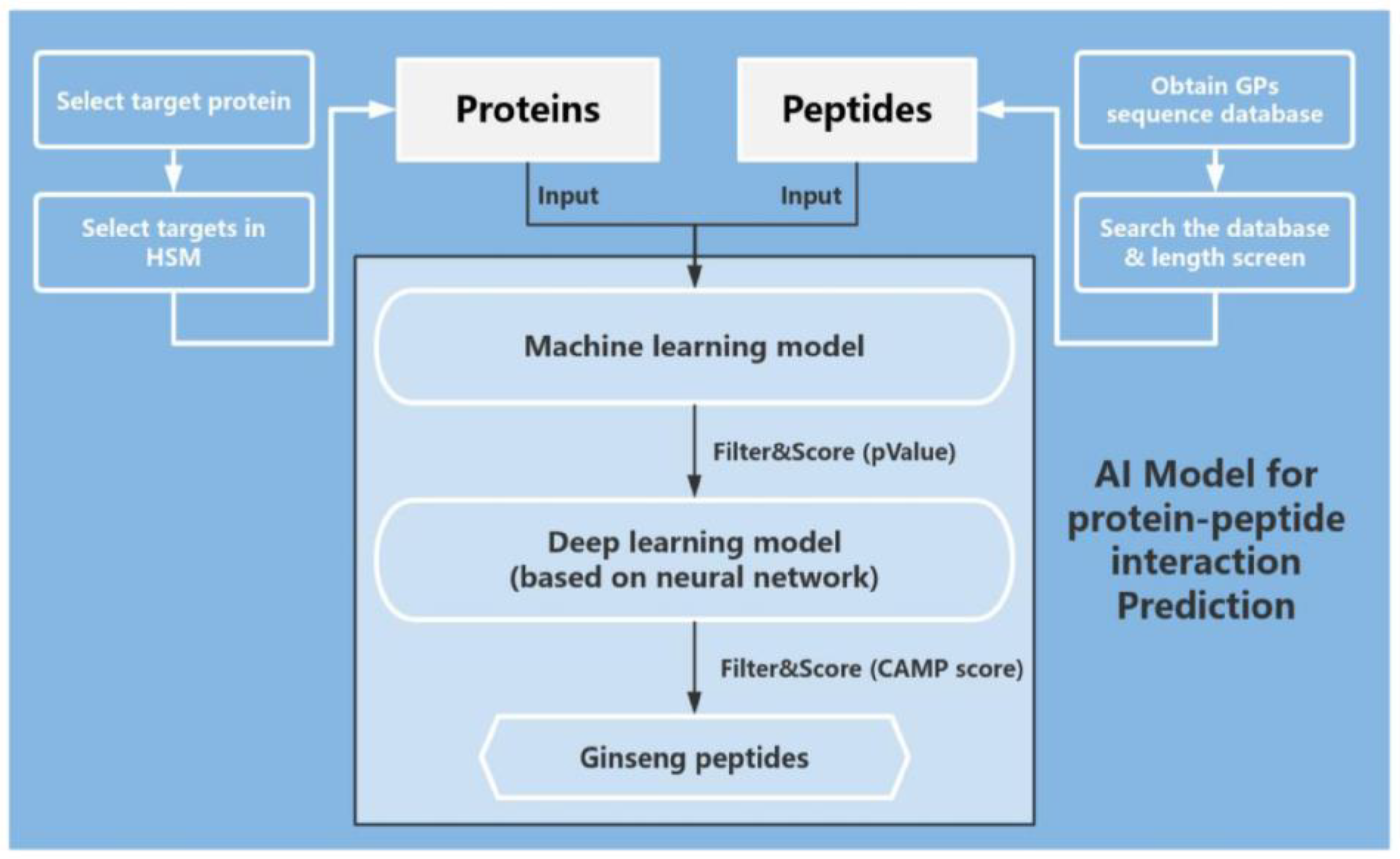
The GP database is sourced from the literature on pan ginseng and de novo sequencing. First, we selected peptides with a length ≤ 16. Next, relevant target proteins with keywords such as antioxidant, collagen promoting, proliferation promoting, aging, inflammation, and inflammaging in the HSM model were used to predict the interaction probability between GPs and target proteins in this model. The top 50 peptides with the highest probability were selected based on their scores, and each peptide retained the results of three optimal peptide target protein interactions. We used the CAMP model to predict the 150 results obtained below again and selected the results with a score of ≥50 based on the results given by the CAMP model (Figure 1).
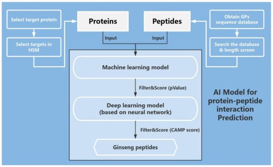
Figure 1.
Flowchart of AI prediction to screen for peptides with anti-aging activity. Using HSM and CAMP models to predict protein–peptide interaction.
2.1.3. Disease Targets Screened and Targeted with Network Pharmacological Analysis
We screened inflammaging targets using the GeneCards database (https://www.genecards.org, accessed on 14 October 2024) [25] and the online Mendelian inheritance in man (OMIM) database (https://www.omim.org/, accessed on 14 October 2024) [26], intersecting with targets of ginseng compounds and peptides, and inputting these potential targets into the Search Tool for the Retrieval of Interacting Genes/Proteins (STRING) database (https://cn.string-db.org/, accessed on 20 October 2024) [27] to obtain protein–protein interaction (PPI) network information. The network was visualized using Cytoscape 3.7.2 software. Finally, we analyzed the Kyoto Encyclopedia of Genes and Genomes (KEGG) and gene ontology (GO) functions using the Metascape database (https://metascape.org/gp/index.html, accessed on 21 October 2024) [28,29].
2.2. Preparation of the Ginseng Peptide Extract
Ginseng peptide extract was provided by Harvest Biotech, Zhejiang, Co., Ltd. Black ginseng was ground into a fine powder using a mill. The powder was then dissolved in water at a solid-to-liquid ratio of 1:20. The mixture was soaked overnight at 50 °C with a stirring rate of 120 rpm. After soaking, samples were taken from the solution. Next, sodium hydroxide was added to adjust the pH to 8.2. Subsequently, 2% (w/v) of alkaline protease (1 kg per 50 L of solution) was added in small portions to the black ginseng solution, which was then transferred to a reactor. The mixture was subjected to enzymatic hydrolysis at 50 °C with a stirring rate of 130 rpm for 2.5 h. Post hydrolysis, samples were again taken. Then, 0.4% (w/v) of flocculant (200 g per 50 L of solution) was added to the hydrolyzed solution, followed by static settling overnight at 4 °C. The mixture was then filtered using a plate and frame filter press. After filtration, 2% (w/v) of preservative (a mixture of phenoxyethanol, 1,2-hexanediol, and 1,2-pentanediol) was incorporated into the solution. Finally, the solution was sterilized by filtering it through a 0.22 µm PVDF filter cartridge. The resulting solution was stored at 4 °C away from light until further use.
2.3. Free Radical Scavenging Assessment
2.3.1. DPPH Assay
The 2,2-Diphenyl-1-picrylhydrazyl (DPPH) assay is the most commonly used antioxidant assay for plant extract that measures the ability to act as free radical scavengers or hydrogen donors. Glutathione was used as a standard in the DPPH assay, concentrations were 0, 0.125, 0.25, 0.5, 1, 2, and 4 mg/mL, dissolved and diluted with ddH2O. The DPPH solution (50 μg/mL) was dissolved with 95% ethanol. For the sample group (As), 150 μL of DPPH solution and 50 μL of glutathione solution were added. For the control group (Ac), 50 μL of glutathione solution and 150 μL of 95% ethanol solution were added. For the blank group (Ab), 150 μL of DPPH solution and 50 μL of ddH2O were added. When testing the sample, GPE was used instead of the standard, with all other procedures remaining unchanged. After thorough mixing, the samples were reacted in the dark at room temperature for 30 min. The absorbance values were measured at 517 nm with a microplate reader. Finally, the calculation was performed using the following formula (Theorem 1).
Free radical scavenging rate (%) for DPPH = [1 − (As − Ac)/Ab] × 100%
Theorem 1.
Calculation formula of DPPH free radical scavenging rate. In this formula, As, Ac, and Ab represent the absorbance values of the sample group, the control group, and the blank group, respectively.
2.3.2. ABTS Assay
The 2,2′-azino-bis (3-ethylbenzothiazoline-6-sulfonate) diammonium salt (ABTS) radical scavenging assay is also a widely used antioxidant assay for plant extracts or peptides, evaluating the ability to neutralize free radicals via electron donation or radical cation decolorization. Glutathione was used as a standard in the ABTS assay, and concentrations were 0, 0.125, 0.25, 0.5, 1, 2, and 4 mg/mL, dissolved and diluted with ddH2O. For ABTS working solution preparation, we mixed ABTS solution (7 mM) and potassium persulfate solution (2.45 mM) in equal amounts and allowed the mixture to react in the dark for 12 h. After, we diluted it with PBS (10 mM, pH 7.4) to an absorbance value of 0.7 (734 nm). Next, we mixed 150 μL of ABTS working solution with 10 μL of glutathione. When testing the sample, GPE (different concentrations of GPE were diluted by PBS) was used instead of the standard for the sample group (As), and the PBS group was used as a blank group (Ab). All other procedures remained unchanged. After reacting in the dark at room temperature for 6 min, the absorbance values were measured at 734 nm with a microplate reader. Finally, the calculation was performed using the following formula (Theorem 2).
Free radical scavenging rate (%) for ABTS = (Ab − As)/Ab × 100%
Theorem 2.
Calculation formula of ABTS free radical scavenging rate. In this formula, As and Ab represent the absorbance values of the sample group and the blank group.
2.4. Cell Inflammation Model Based on RAW264.7
2.4.1. Cell Culture of RAW264.7
Normal RAW264.7 cells (Hunan Fenghui Biotechnology Co., Ltd., Changsha, China) were cultured on high-glucose DMEM (Wisent Biotechnology Co., Ltd., Nanjing, China) supplemented with 10% FBS (Gibco, Carlsbad, CA, USA) and 1% penicillin/streptomycin (Shanghai BasalMedia Technologies Co., Ltd., Shanghai, China). The cell cultures were maintained in a humidified incubator at 5% CO2 and 37 °C. When RAW264.7 cells in T25 cell culture flasks grew to a density above 90%, 1:3 passages were performed.
When the cells reached confluency and were in good condition (with less than 5% differentiated cells), we collected the cells and seeded them at varying cell densities into corresponding culture plates for subsequent experiments.
2.4.2. Cell Viability Assay
After overnight culturing in a 96-well plate (1 × 10⁴ cells/well, 100 µL complete medium/well), the cells were treated with GPE (0, 5, 10, 25, 50, 75, 100 and 150 μg/mL) using a complete medium (containing 5% FBS) for 24 h. Thereafter, 10 µL of cck-8 (Biosharp, Hefei, China) solution was added to each well and incubated at 37 °C for 1 h, and cell-free wells were used as a blank group. Finally, the absorbance of each well was recorded at 450 nm using a multimode plate reader (PerkinElmer, Waltham, MA, USA). The OD values of each group were subtracted from the average value of the blank group, which was the cell viability of each group. The viability of 0 μg/mL GPE group was assessed as 1 (100%).
2.4.3. Nitric Oxide (NO) Production Assay
The RAW264.7 cells were cultured overnight in 24-well plates (5 × 10⁴ cells/well, 500 μL medium/well), then co-stimulated with GPE (0, 50, 100 μg/mL) or GPs (0, 10, 25 μg/mL), and 200 ng/mL of LPS (Sigma-Aldrich, St. Louis, MO, USA) using a complete medium (containing 5% FBS) for 24 h. Untreated cells were used as the blank group, while cells treated with LPS but without GPE or GPs were used as the model group, and cells treated with LPS and positive drug (50 μg/mL Dexamethasone) were used as the positive group. The culture supernatant from each well was collected at the end of the experiments and used to measure NO production. The NO production was determined using a commercial NO assay kit (Beyotime Biotechnology, Shanghai, China) according to the manufacturer’s instructions. We mixed 50 µL of cell culture medium and 50 µL of Griess reagents I and II and reacted them in a 96-well plate at room temperature for 10 min. Finally, the absorbance of each well was recorded at 540 nm using a multimode plate reader.
2.4.4. Reactive Oxygen Species (ROS) Measurement
The RAW264.7 cells were cultured overnight in 24-well plates (5 × 10⁴ cells/well, 500 μL medium/well), then co-stimulated with GPE (0, 50, 100 μg/mL) and 200 ng/mL LPS using a complete medium (containing 5% FBS) for 24 h. Then, we removed the medium and washed the cells with 500 μL of PBS at least two times. Next, we used an ROS assay kit (Beijing Solarbio Science & Technology Co., Ltd., Beijing, China) to measure ROS levels. The signal intensity of Ex488/Em525 was then detected using a multimode reader and was observed and photographed under a fluorescence microscope.
2.4.5. RNA Extraction and RT-qPCR
The RAW264.7 cells were cultured overnight in 12-well plates (2 × 10⁵ cells/well, 1 mL medium/well), then co-stimulated with GPE (0, 50, 100 μg/mL) and 200 ng/mL of LPS using a complete medium (containing 5% FBS) for 24 h. After, we removed the DMEM and washed the cells with 1 mL of PBS at least two times. Then, we extracted RNA using a Quick-RNA Microprep Kit (Genstone biotech, Beijing, China) and stored it at −80 °C. Subsequently, reverse transcription was performed using a cDNA first-strand synthesis kit (Tiangen Biotech Co., Ltd., Beijing, China) and diluted with ddH2O to an appropriate concentration. RT-qPCR was performed using diluted cDNA and qPCR SYBR Green Master Mix (Cat No.11185ES08; YEASEN, Shanghai, China) on Applied Biosystems 3 Fast Real-Time PCR System (Thermo Fisher Scientific, Shanghai, China).
2.5. Histomorphology and Collagen Change Detection Based on In Vitro Skin Model
The in vitro skin model used (Guangdong BioCell Biotechnology Co., Ltd., Dongguan, China) is a skin model which included epidermal and dermal cells. We placed the models into a 6-well plate culture mold, added 3.7 mL of culture medium (#230607, Guangdong Biocell Biotechnology Co., Ltd.) to each well, incubated in a 37 °C 5% CO2 incubator (150I, Thermo) and changed the medium every day. After 2 days of cultivation, irradiation and administration, we began according to the grouping and corresponding treatment conditions. The skin models were exposed to a mixture of ultraviolet radiation A (UVA) irradiation at 30 J/cm2 (365 nm, 16.29 mW/cm2 for 30 min and 42 s, PL-S 9W/01/2P, Philips, Shanghai, China) and ultraviolet radiation B (UVB) at 50 mJ/cm2 (311 nm, 2.748 mW/cm2 for 18 s, PL-S 9W/10/2P, Philips) for 4 consecutive days, with fresh culture medium replaced before each irradiation. The medium was removed after each irradiation, and the skin models were cultured with medium (Guangdong Biocell Biotechnology Co., Ltd.) containing 1% (v/v) GPE. The positive groups were cultured with 100 μg/mL vitamin C (VC) and 7 μg/mL vitamin E (VE) instead of sample GPE. Control groups were cultured with normal medium. After 4 days of irradiation, the samples were continuously cultured with medium containing 1% (v/v) GPE or both VC and VE for another 3 days. The medium was replaced with the same fresh medium every day. After the cultivation was completed, we removed the medium in all groups, washed them with PBS solution, and wiped off the residual liquid with a sterile swab. After, we fixed skin models with 4% paraformaldehyde for 24 h.
2.5.1. H&E Staining
After 24 h of fixation, the skin models were washed with PBS and dehydrated by stepwise immersion in solutions of increasing ethanol concentration, embedded in paraffin overnight, then were sealed with dry gum and sliced into 8-μm-thick sections. Sections were stained with hematoxylin and eosin (H&E). Then, the images were taken under the microscope.
2.5.2. Masson Staining
Paraffin sections obtained in Section 2.5.1 were dewaxed and stained using a Masson staining kit (YK2223, Shaanxi Yike biotechnology Service Co., Ltd., Xi’an, China) according to the manufacturer’s instructions. Then, the images were taken under a microscope to observe and analyze.
2.5.3. Immunofluorescence
After fixation with 4% paraformaldehyde, tissue embedding and tissue sectioning were performed. After deparaffinization and hydration of the baked slices, the paraffin sections were placed in a 0.01 M sodium citrate antigen repair solution and subjected to high-pressure repair. After cooling, the slices were removed and washed with PBS solution (three times, 5 min each). One drop of 3% H2O2 was added to each slice, and they were incubated at room temperature for 30 min, then washed with PBS (three times, 5 min each). We added serum homologous to the secondary antibody and sealed it at 37 °C for 60 min. We added primary antibody working solution dropwise, incubated overnight at 4 °C, and washed with PBS (three times, 5 min each). Next, we added the secondary antibody working solution dropwise and incubated at room temperature for 1 h, before washing with PBS (three times, 5 min each). After the completion of the secondary antibody incubation, we washed with PBS (three times, 5 min each). Then, we shook off the PBS solution attached to the glass slide, added 100 μL of Hoechst working solution dropwise to each slice, and incubated at room temperature for 5 min. We then washed with PBS (three times, 5 min each). We wiped off the PBS solution with absorbent and sealed it with a drop of quenching inhibitor. Finally, we viewed it under a fluorescence microscope (BX43, Olympus, Excitation wavelength: 488 nm, Emission wavelength: 520 nm) and took photos within 24 h. The following primary antibodies were used: anti-collagen I (#72026, Cell Signaling Technology, Danvers, MA, USA, dilution 1:200), anti-collagen III (22734-1-AP, Proteintech, Wuhan, China, dilution 1:200), anti-collagen IV (ab6311, Abcam, Cambridge, UK, dilution 1:200), anti-collagen XVII (ab186415, Abcam, dilution 1:200) and goat anti-rabbit IgG (ab150077, Abcam, dilution 1:500).
2.6. Tissue Morphological Changes, Detection of Filaggrin (FLG), Loricrin (LOR) and Transglutaminase-1 (TGM1) Content Based on 3D Epidermal Skin Model
The EpiKutis 3D skin model (Guangdong BioCell Biotechnology Co., Ltd.) is another skin model. Unlike in vitro skin models used in Section 2.5, this model only included epidermal cells. It was utilized to evaluate the effects of GPE on cellular barrier function in epidermal layer. 3D skin models were transferred to a 6-well plate with 0.9 mL EpiGrowth medium added in advance according to the grouping. WY14643 is a kind of PPARα agonist, that has anti-inflammatory properties, promoting barrier function in skin cells. In the positive group, 50 μM WY14643 were used instead of sample GPE. Control groups were cultured with normal medium. The positive control group and experimental group were irradiated with UVB (600 mJ/cm2). After irradiation, the four groups of epidermal models were cultured in a 37 °C 5% CO2 incubator for 24 h, and then fixed with 4% paraformaldehyde for 24 h. We performed H&E staining and immunofluorescence detection (the same as described in Section 2.5.2 and Section 2.5.3), took photos under a microscope for observation, collected images, and conducted analysis. The following primary antibodies were used: anti-FLG (ab218397, Abcam, dilution 1:200), anti-LOR (ab198994, Abcam, dilution 1:500), anti-TGM1 (12912-3-AP, Proteintech, China, dilution 1:200), goat anti-rabbit IgG (ab150077, Abcam, dilution 1:500) and goat anti-mouse IgG (ab150117, Abcam, dilution 1:500).
2.7. Biological Assay and Method Based on HSF Cell Inflammaging Model and UV-Induced Senescence Model
2.7.1. Cell Culture of THP-1 and HSF
Normal HSF cells (NewgainBio, Wuxi, China) were cultured on high-glucose DMEM (Wisent Biotechnology Co., Ltd., Nanjing, China) supplemented with 10% FBS (Gibco, Carlsbad, CA, USA) and 1% penicillin/streptomycin (Shanghai BasalMedia Technologies Co., Ltd., Shanghai, China). When HSF cells in T25 cell culture flasks grew to a density above 90%, 1:3 or 1:2 passages were performed. THP-1 cells (Baidi Biotech Ltd., Jiaxing, China) were cultured on RPMI-1640 medium (Servicebio, Wuhan, China) supplemented with 10% FBS (Gibco, Carlsbad, CA, USA) and 1% penicillin/streptomycin (Shanghai BasalMedia Technologies Co., Ltd.). The cell cultures were maintained in a humidified incubator at 5% CO2 and 37 °C. When THP-1 cells in T75 cell culture flasks grew to a density above 90%, 1:2 passages were performed.
2.7.2. Induction of Aging to HSF
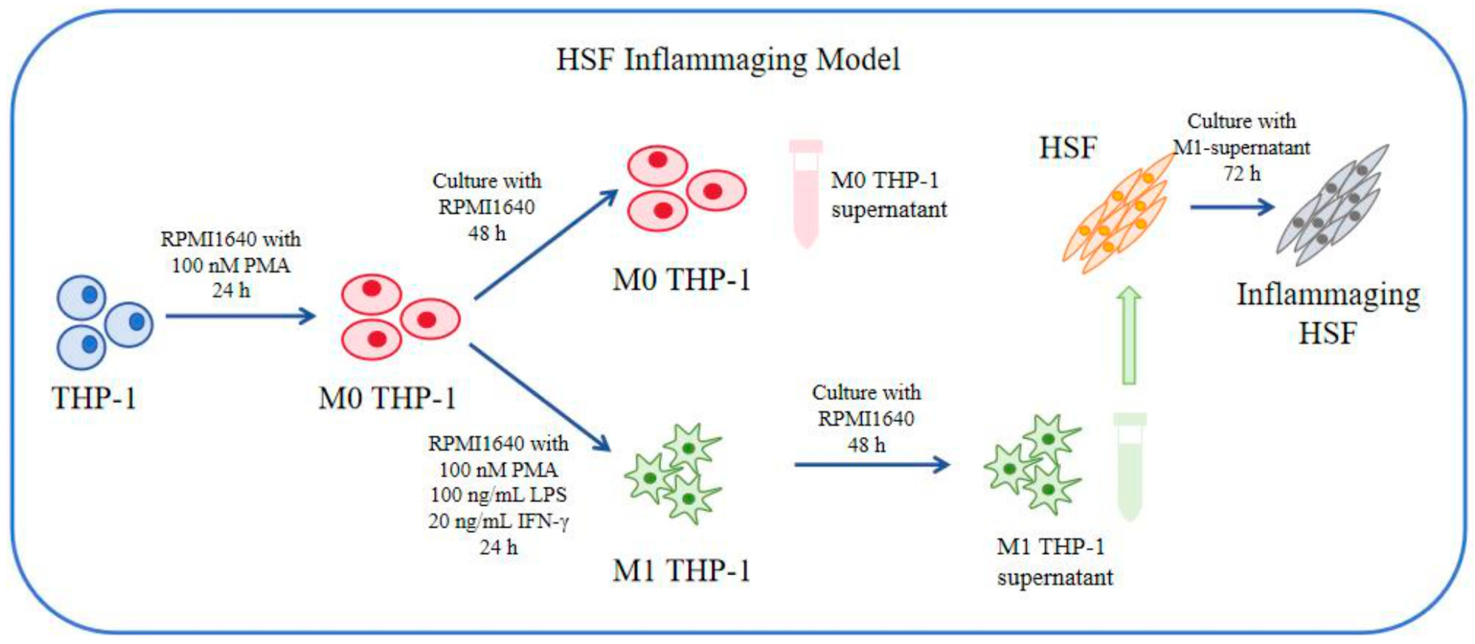
When THP-1 cells were cultured to at least two T75 cells, we then prepared the RPMI1640 complete medium containing 100 nM PMA, which was used to inoculate the cells into a 6-well plate (1 × 10⁶ cells/well) and cultured them for 24 h. We prepared the RPMI1640 complete culture medium containing 100 nM PMA, 100 ng/mL LPS, and 20 ng/mL IFN-γ, removed the original medium, added the newly prepared complete culture to each well, and continued processing for 24 h to obtain M1 type THP-1 cells. Next, we removed the culture medium, washed it with PBS three times, added new 1640 complete culture medium, incubated it for 48 h, collected the cell culture supernatant, and stored it at −80 °C or proceeded to the next experiment. It was later used to induce the inflammaging model of HSF cells (Figure 2).
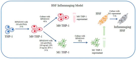
Figure 2.
Flowchart of induction of inflammaging model of HSF cells.
For this model, HSF cells were seeded onto a 12-well plate (5 × 10⁴ cells/well), and after 24 h, the original culture medium was removed. The THP-1 cell culture supernatant was used to prepare the specified concentration of drug (0, 0.01%, 0.05% GPE, or 0, 10, 25 μg/mL ginseng peptide) or positive drug (50 μg/mL TGF-β1) and stimulated for 72 h. After, we collected the culture supernatant from each well and stored it at −80 °C or performed the next experiment.
For the UV-induced aging model, HSF cells were seeded onto a 12-well plate (4 × 10⁴ cells/well). After 24 h, the original culture medium was removed and washed twice with PBS. After 15 J of UVA irradiation, PBS was discarded and replaced with medium (containing 2% FBS) containing the specified concentration of drugs (0, 10, 25 μg/mL ginseng peptide) or positive drug (50 μg/mL TGF-β1) for further incubation for 24 h. After, we collected the culture supernatant from each well and stored it at −80 °C or performed the next experiment.
2.8. Determination of Inflammatory Cytokines (IL-6, TNF-α) and Collagen I by ELISA
The concentration of IL-6, TNF-α and collagen I were determined using IL-6 ELISA kit (EK206, MultiSciences, Hangzhou, China), TNF-α ELISA kit (EK282, MultiSciences, Hangzhou, China) and collagen I ELISA kit (Cat No. 97044ES96, YEASEN, Shanghai, China) according to the manufacturer’s instructions and measured the absorbance at 450 nm/630 nm using a microplate reader.
2.9. Statistical Analysis
GraphPad Prism 9.5 software (GraphPad Software, Inc., La Jolla, CA, USA) was used for statistical analyses. Data were expressed as mean values ± SD of almost three independent experiments. A Student’s t-test was performed to compare the differences between the two groups, and one-way analysis of variance (ANOVA) was performed to compare the differences between multiple groups, followed by Dunnett’s test as the post hoc test (* p < 0.05, ** p < 0.01, *** p < 0.001). p < 0.05 was regarded as statistically significant, while p < 0.01 was regarded as a highly significant difference.
3. Results
3.1. Network Pharmacology Predicts the Anti-Aging and Anti-Inflammatory Efficacy of Ginseng
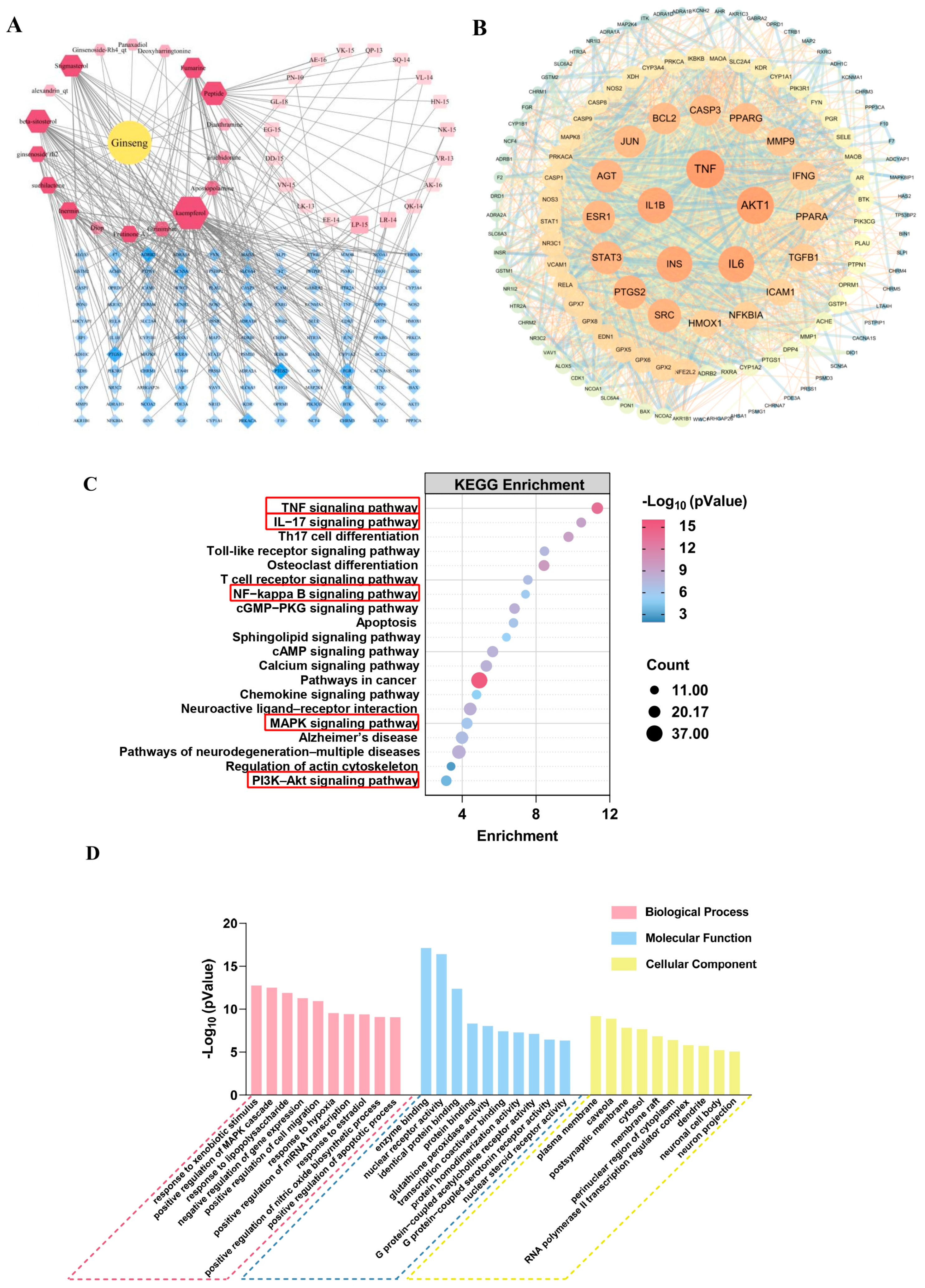
We first conducted a preliminary network pharmacology analysis of reported ginseng-soluble activities (including small molecules and peptides) and collected targets for various skin diseases. Specifically, we used the TCMSP [24] database for screening the small molecules and their regulatory targets in ginseng, while the identified GPs and their potential corresponding targets were obtained using mass spectrum data for ginseng peptides [30] and regulatory target prediction through HSM and CAMP models. We retrieved skin anti-aging and repair-related targets through the GeneCards [25] and OMIM databases [26], removed duplicates, and intersected them with the potential targets of ginseng, resulting in a total of 121 potential targets.
After integrating the potential targets of ginseng compounds and GPs, a network relationship was constructed using Cytoscape 3.7.2, resulting in a network comprising 159 nodes and 256 interaction edges. Then, we used the STRING database [27] to construct a key target network, which consisted of 134 nodes and 1895 interacting edges (Figure 3A). Using Cytoscape 3.7.2 software to analyze the PPI network, the top five targets with the highest degree values were obtained: TNF, AKT1, IL-6, INS, and IL-1β. This suggests that ginseng may alleviate inflammaging through these main targets (Figure 3B).

Figure 3.
Visualization of interacting targets in the active components of ginseng and senescence: (A) complex target pathway network of ginseng components against senescence. The deep-red hexagonal nodes represent the small molecule components in ginseng, the pink square nodes represent the calculated ginseng peptides, and the blue node is the potential target; (B) PPI network of the selected core objectives; (C) KEGG pathway analysis of senescence targets. The horizontal axis represents fold enrichment, the depth of the bubble color represents the enrichment degree of the pathway, and the bubble size represents the number of genes enriched in this pathway. The red boxes represent the main signaling pathways; (D) GO enrichment of ginseng against senescence. The ordinate indicates the degree of enrichment of the pathway, expressed in the form of −Log10 (p Value).
We performed GO functional annotation and KEGG pathway analysis on 159 targets of the PPI network using the Metascape database [28]. Then, the top 20 targets were visualized as bubble plots (Figure 3C), and it was found that ginseng may alleviate inflammaging through the TNF, IL-17, NF kappa B, MAPK, and PI3K-Akt signaling pathways. In the biological processes (BPs), ginseng mainly has a great influence on MAPK cascade reaction, response to lipopolysaccharide (LPS), and negative regulation of gene expression and nitric oxide biosynthesis process. Regarding the molecular functions (MFs), the functions of ginseng pharmaceutical components are mainly related to enzyme binding, protein binding, and transcription coactivator binding. The targets in the cellular components (CCs) are closely related to the plasma membrane and cytosol (Figure 3D).
Based on the analysis of the results from network pharmacology and computational biology, we hypothesize that ginseng may exert anti-inflammatory and anti-aging effects by down-regulating the expression of pro-inflammatory cytokines such as IL-6, IL-1β, and TNF-α.
3.2. In Vitro Evaluation of the Antioxidative and Anti-Inflammatory Effects of GPE
To verify the anti-inflammatory effects of GPE, we first assessed its antioxidant activity and anti-inflammatory activities. We initially obtained GPE through enzymatic hydrolysis, which mainly included water-soluble small molecules and water-soluble polypeptides. The results showed that the half-maximal scavenging rates were 5.13% and 4.91%, respectively, indicating that the GPE possesses good free radical scavenging ability (Figure 4A,B).
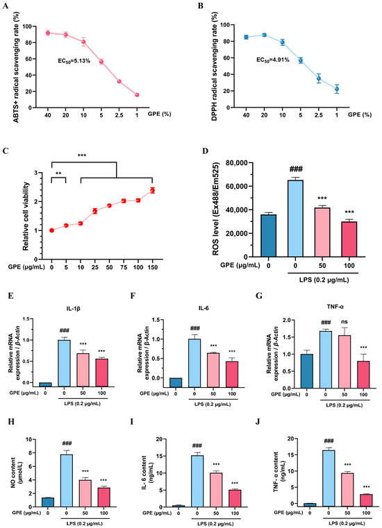
Figure 4.
In vitro evaluation of the antioxidative and anti-inflammatory effects of GPE in RAW264.7 cell model. (A) ABTS+ radical scavenging rate; (B) DPPH radical scavenging rate; EC50: half-maximal effective concentration; (C) cell viability calculated by the cck-8 assay after incubation of 24 h with different concentrations of GPE. The viability of untreated group was assessed as 1 (100%); (D) ROS levels after 24 h of treatment with different concentration of GPE; (E–G) changes in IL-1β, IL-6, and TNF-α gene expression evaluated by RT-qPCR; (H) changes in NO content; (I,J) release of IL-6 and TNF-α obtained by ELISA. Values are expressed as mean ± SD (n = 3). ANOVA was used for all statistical analyses and Dunnett’s test was used for the post hoc test. Untreated cells were used as the blank group, while cells treated with LPS but without GPE were used as the model group. ### p < 0.001 vs. blank group; ** p < 0.01 vs. control group; *** p < 0.001 vs. control group; ns, not significant between the indicated groups.
Next, we evaluated the effects of GPE on the viability of RAW264.7 cells. The results showed that as the concentration of GPE increased (0–150 μg/mL), cell viability increased in a dose-dependent manner. This indicates that GPE promotes the viability of RAW264.7 cells within a certain concentration range and may have a positive impact on cell growth (Figure 4C). Subsequently, we further determined the antioxidative and anti-inflammatory effects of GPE in the RAW264.7 model. The ROS experimental results indicated that GPE could effectively reduce the intracellular ROS levels in RAW264.7 cells, which may also be one of the important mechanisms of its anti-inflammatory action (Figure 4D). Gene expression analysis found that GPE significantly inhibited the gene expression levels of IL-6, IL-1β, and TNF-α (Figure 4E–G). Additionally, the cytokine analysis results showed that the levels of IL-6 and TNF-α in cells treated with GPE were significantly reduced, while the levels of NO were also significantly decreased (Figure 4H–J). The experimental results indicated that GPE has significant antioxidative and anti-inflammatory effects.
3.3. Systematic Evaluation of the Anti-Inflammaging Effects of GPE
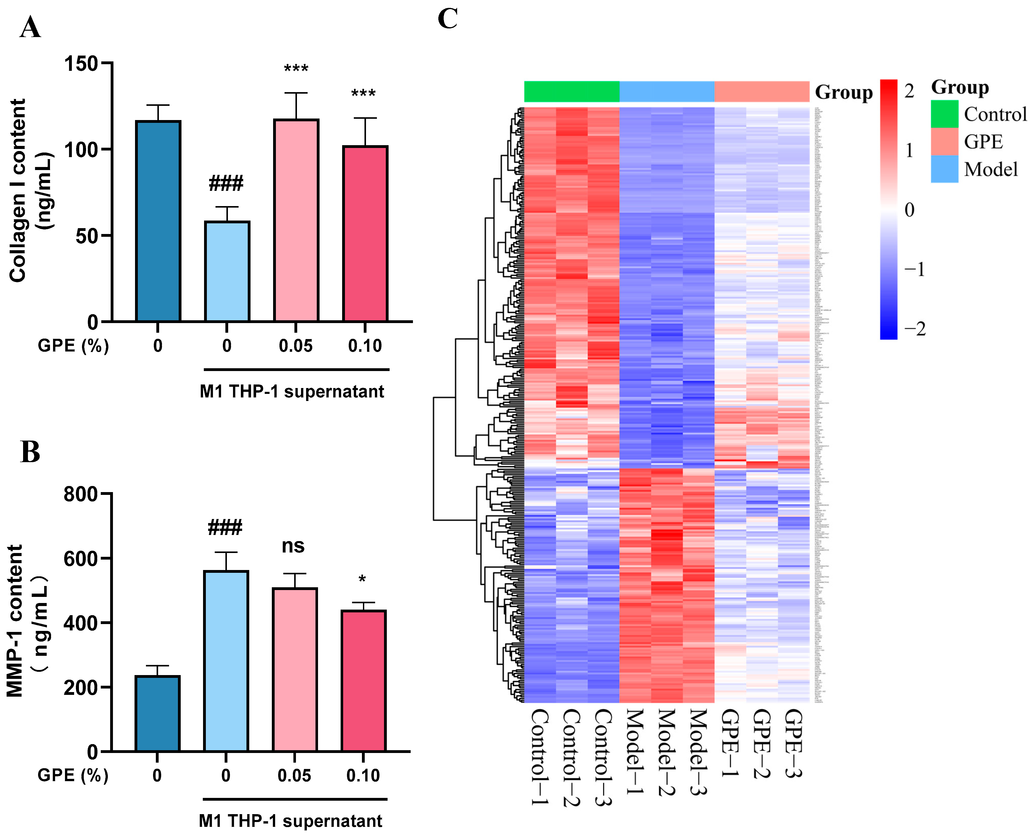
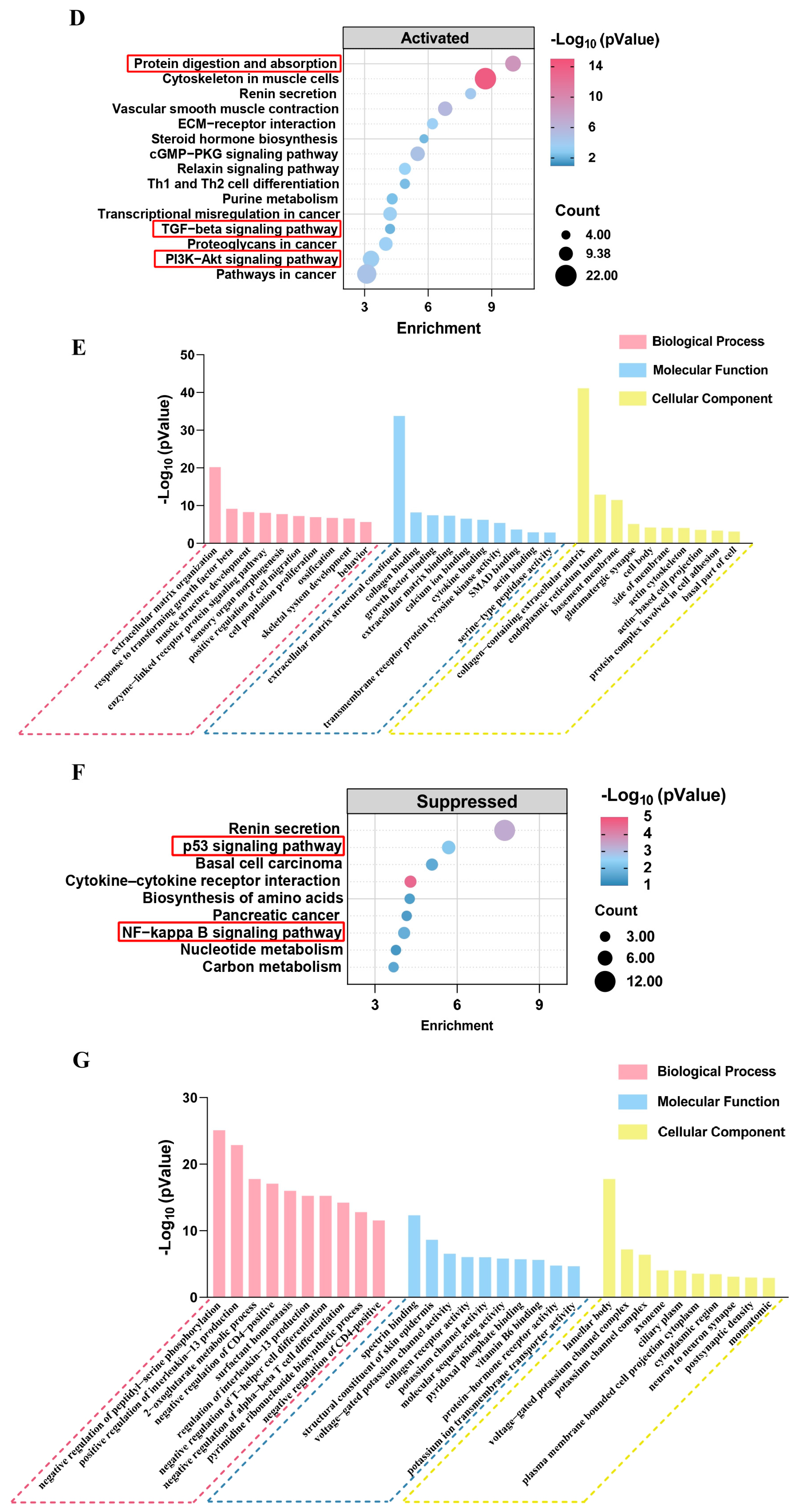
To verify whether GPE can alleviate skin cell senescence by reducing inflammatory responses, we utilized the HSF inflammaging model to assess the anti-aging efficacy of GPE. The HSF inflammaging model is an HSF cell senescence model induced by inflammatory cytokines produced by M1-type THP-1 cells. Compared with the use of a single inflammatory cytokine to induce an aging model, it can simulate cellular aging caused by changes in complex inflammatory environments in the skin more realistically [31,32]. In this inflammaging model, we found that the content of collagen I in the supernatant of the model group (cells treated with M1 THP-1 supernatant without GPE) significantly decreased, while the content of MMP-1 significantly increased. However, after the addition of GPE, the levels of collagen I and MMP-1 in the supernatant tended to return to normal (Figure 5A,B).
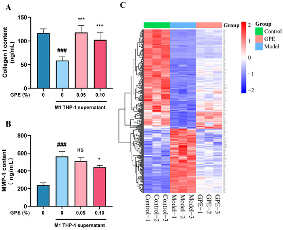
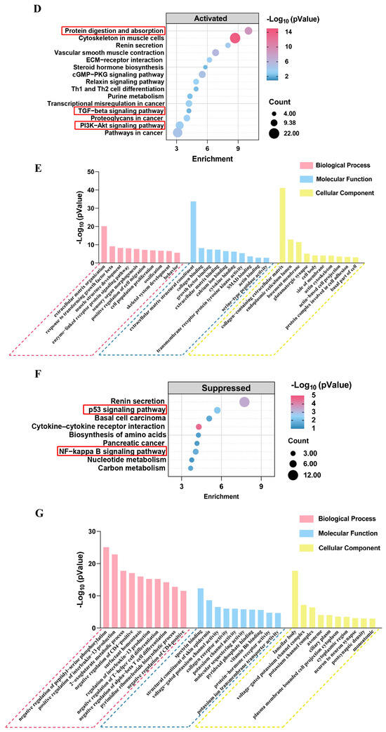
Figure 5.
Evaluation of the anti-aging effect of GPE on inflammaging HSF cells. (A,B) inflammaging HSF cells were treated with different doses of GPE for 24 h and ELISA was used to detect collagen I and MMP-1 content change. Untreated cells were used as the blank group, while cells treated with THP-1 supernatant but without GPE were used as the model group. Values are expressed as mean ± SD (n = 3). ANOVA was used for all statistical analyses and Dunnett’s test was used for the post hoc test. ### p < 0.001 vs. Control group; * p < 0.05, *** p < 0.001 vs. Model group; ns, not significant between the indicated groups (vs. Model group); (C) heat map of differentially expressed genes in transcriptome sequencing. The red and blue blocks represent high and low expression levels of genes, respectively; (D,E) KEGG pathway analysis and GO enrichment of activated genes; (F,G) KEGG pathway analysis and GO enrichment of suppressed genes. In KEGG enrichment analysis, the horizontal axis represents fold enrichment, the depth of bubble color represents the enrichment degree of the pathway, and the bubble size represents the number of genes enriched in this pathway. The red boxes represent the main signaling pathways. In GO enrichment analysis, the ordinate indicates the degree of enrichment of the pathway, expressed in the form of −Log10 (p Value).
To comprehensively investigate the differential gene expression levels of GPE in HSF cells, we conducted transcriptome sequencing. After filtering the results, a total of 203 activated genes and 132 inhibited genes were identified (Figure 5C). Using the Metascape database for GO functional annotation and KEGG pathway analysis, it was found that the activated genes were primarily enriched in protein digestion and absorption and the TGF-β and PI3K-Akt signaling pathways (Figure 5D). In the GO enrichment analysis, the enrichment items for GPE included cell population proliferation, muscle structure development, response to TGF-β, growth factor binding, cytokine binding, collagen binding, and collagen-containing extracellular matrix (Figure 5E). The suppressed genes were mainly enriched in the p53 and NF-kappa B signaling pathways (Figure 5F). In the GO enrichment analysis, the enrichment items for GPE included a structural constituent of the skin epidermis and collagen receptor activity (Figure 5G). Based on the comprehensive analysis of the transcriptome sequencing results, we determined that the anti-aging effects of GPE on HSF cells are primarily manifested in the increased synthesis of collagen. The increase in collagen content further indicates that GPE has a protective effect against aging in HSF cells induced by inflammatory factors.
3.4. Systematic Evaluation of Anti-Inflammatory Aging in Dermatological Application Using Multi-Layer Skin Models
To confirm whether GPE can protect epidermal skin cells from UV damage, we first used UV irradiation on a 3D epidermal skin model to induce damage and detected changes in tissue morphology, filaggrin (FLG), loricrin (LOR), and transglutaminase 1 (TGM1) content. H&E staining showed that the number of sunburn cells increased after UVB irradiation but significantly decreased after GPE treatment (Figure 6A,B). Immunofluorescence was then used to detect the expression levels of FLG, LOR, and TGM1. The results showed that the expression levels of FLG, LOR, and TGM1 were significantly lower in cells after UVB irradiation compared to the control group. In contrast, the levels of these proteins were significantly higher in the GPE-treated group, with enhancement rates of 95.56%, 146.15%, and 207.69%, respectively (Figure 6C–E). These data indicate that GPE can upregulate the expression of FLG, LOR, and TGM1 proteins, protect epidermal skin cells from UVB-induced damage, and possess reparative effects.
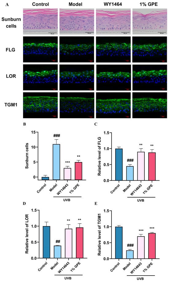
Figure 6.
Effects of GPE treatment on UV-induced EpiKutis 3D epidermal skin model damage as assessed via immunofluorescence and H&E staining. All groups (except the control group) were treated with UVB. WY14643 (pirinixic acid) was used as the positive control drug in the experiment. The results show that 1% (v/v) GPE could protect epidermal skin cells from UVB-induced damage and possess reparative effects. (A) sunburn cells were calculated and expression of filaggrin (FLG), loricrin (LOR), and transglutaminase-1 (TGM1) proteins were analyzed after treatment with 1% (v/v) GPE in 3D human skin cells using a confocal microscope. Scale bar = 100 μm; (B–E) sunburn cells and relative expression level of FLG, LOR, and TGM1 proteins were analyzed. Values are expressed as mean ± SD (n = 3). Comparisons between groups were performed using a Student’s t-test. ANOVA was used for all statistical analyses and Dunnett’s test was used for the post hoc test. ## p < 0.01, ### p < 0.001 vs. Control group; ** p < 0.01, *** p < 0.001 vs. Model group.
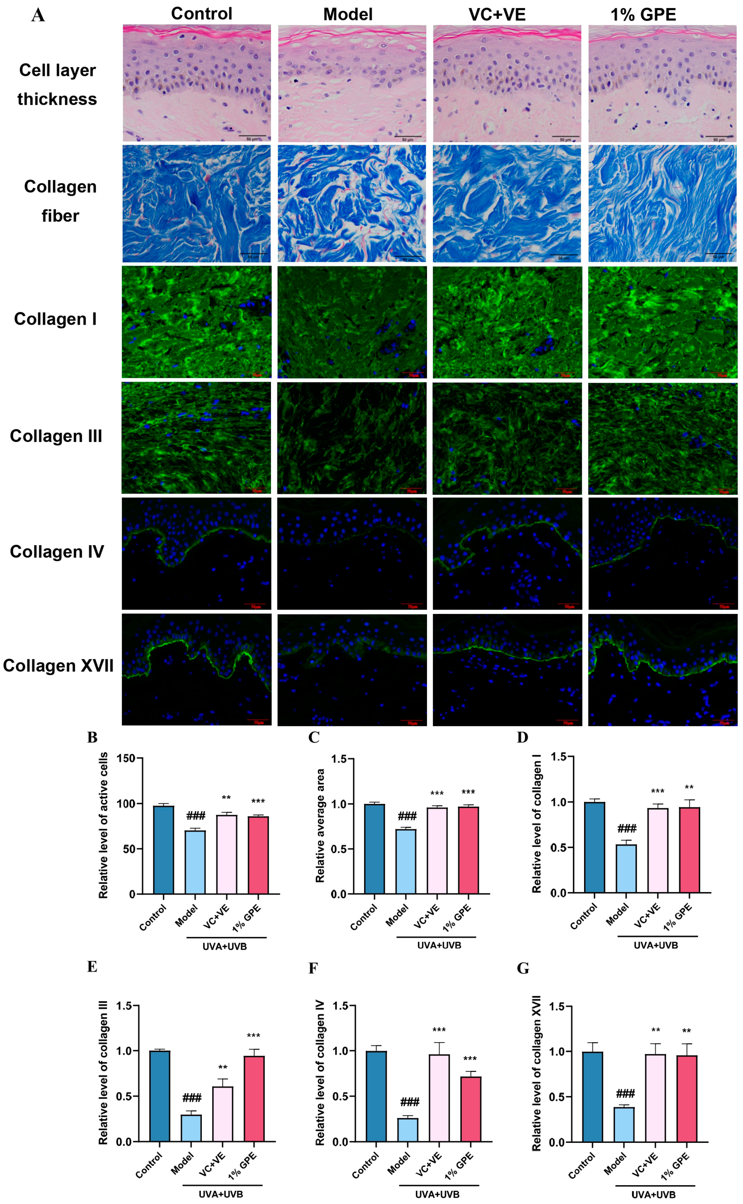
After confirming the efficacy of GPE in the epidermal cells, we further sought to verify whether GPE also has anti-aging effects on dermal skin cells. Therefore, we utilized a combination of UVA (30 J/cm2) and UVB (50 mJ/cm2) irradiation to induce an in vitro skin model. H&E staining was used to observe histological changes, revealing that the thickness of the viable skin cell layer significantly decreased after UV irradiation but increased after GPE treatment. Masson staining also showed that GPE treatment could improve the UV-induced decrease in collagen fiber content (Figure 7A–C). Subsequently, we used immunofluorescence to detect changes in collagen content in the in vitro skin model. After UV induction, the content of collagen I, collagen III, collagen IV, and collagen XVII all significantly decreased, but GPE treatment significantly increased the collagen content, with enhancement rates of 77.36%, 213.33%, 176.92%, and 146.15%, respectively (Figure 7A,D–G). This indicates that skin function improved after treatment with 1% (v/v) GPE. Based on the above results, we determined that GPE applied to the in vitro skin model significantly increases collagen content and improves skin senescence induced by UV.
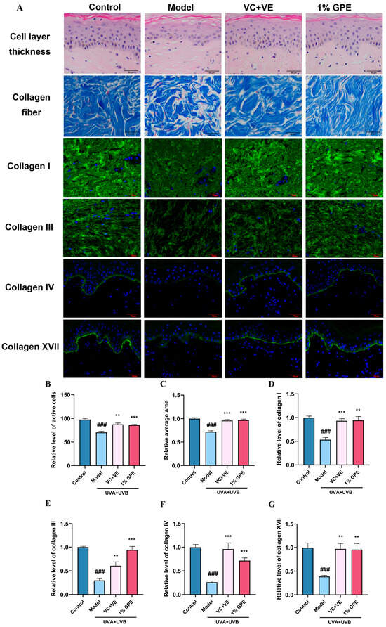
Figure 7.
Protective effect of GPE on the UV-induced in vitro skin model. All groups (except the control group) were treated with UVA and UVB. VC (vitamin C) and VE (vitamin E) were used as the positive control drug in the experiment. The results indicated that impaired skin function due to UV exposure was markedly reversed after 1% (v/v) GPE treatment. (A) protective effect of GPE was evaluated through H&E staining, Masson staining, and immunofluorescence on in vitro skin models. Scale bar = 100 μm; (B) cell layer thickness, i.e., the relative level of active cells; (C) relative content of collagen fiber; (D–G) relative content of collagen I, collagen III, collagen IV, and collagen XVII was analyzed. Values are expressed as mean ± SD (n = 3). Comparisons between groups were performed using a Student’s t-test. ANOVA was used for all statistical analyses and Dunnett’s test was used for the post hoc test. ### p < 0.001 vs. Control group; ** p < 0.01, *** p < 0.001 vs. Model group.
3.5. Prediction of GPs Based on Protein Interaction Model
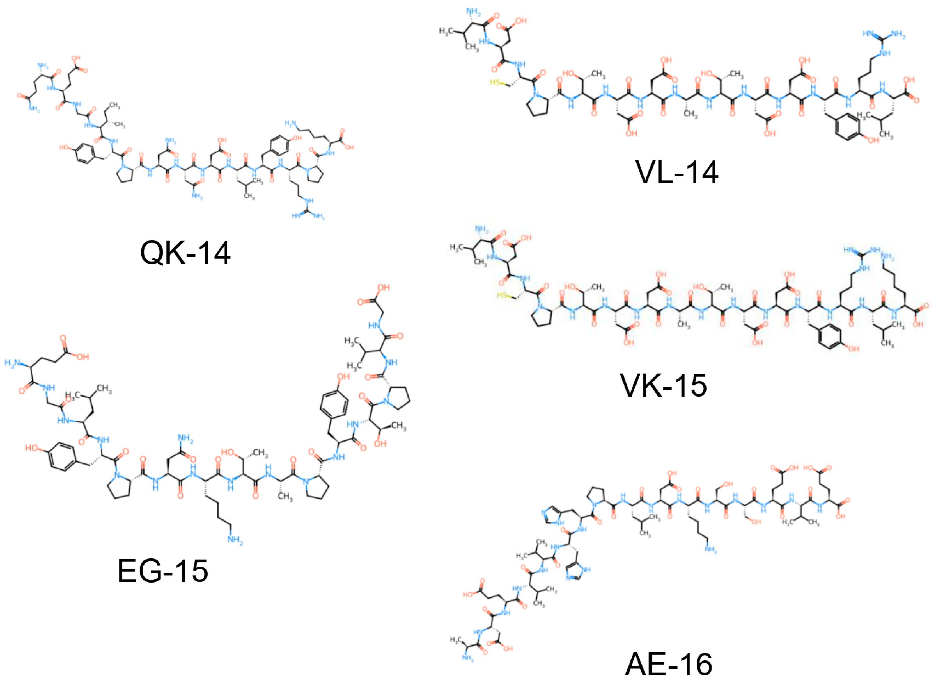
Next, we utilized the in-house customized machine learning model [33], which predicts the probability of protein–peptide interactions, to screen for bioactive peptides in GPE. We first applied the modified HSM model to the GP database [30]. Based on the scores output by the model, we selected the top 50 GPs with the highest interaction probabilities with the targets related to inflammaging in the modified HSM model [33,34]. This step provided us with a list of high-potential peptide candidates. Subsequently, we used the modified CAMP model to further predict the results from the HSM model [33,35]. We conducted an in-depth analysis of these 50 peptides using the CAMP model and, based on the predicted scores, ultimately selected peptides with interaction scores greater than 0.5. Through the predictions of these two models, we identified 19 peptides and 14 potential target proteins, providing a list of GPs with potential anti-aging and anti-inflammatory effects (Appendix A Table A1). After summarizing and screening the results, we synthesized the top five GPs with different sequences (QK-14, EG-15, VL-14, VK-15, AE-16) for subsequent experimental validation (Figure 8).
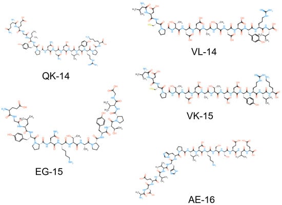
Figure 8.
Structure of the obtained GPs screened. QK-14 (QEGIYPNNDLYRPK), VL-14 (VDCPTDDATDDYRL), EG-15 (EGLYPNKTAPYTPVG), VK-15 (VDCPTDDATDDYRLK), and AE-16 (ADEVVHHPLDKSSEVE).
3.6. Evaluation of Anti-Inflammatory and Anti-Aging Effects of GPs
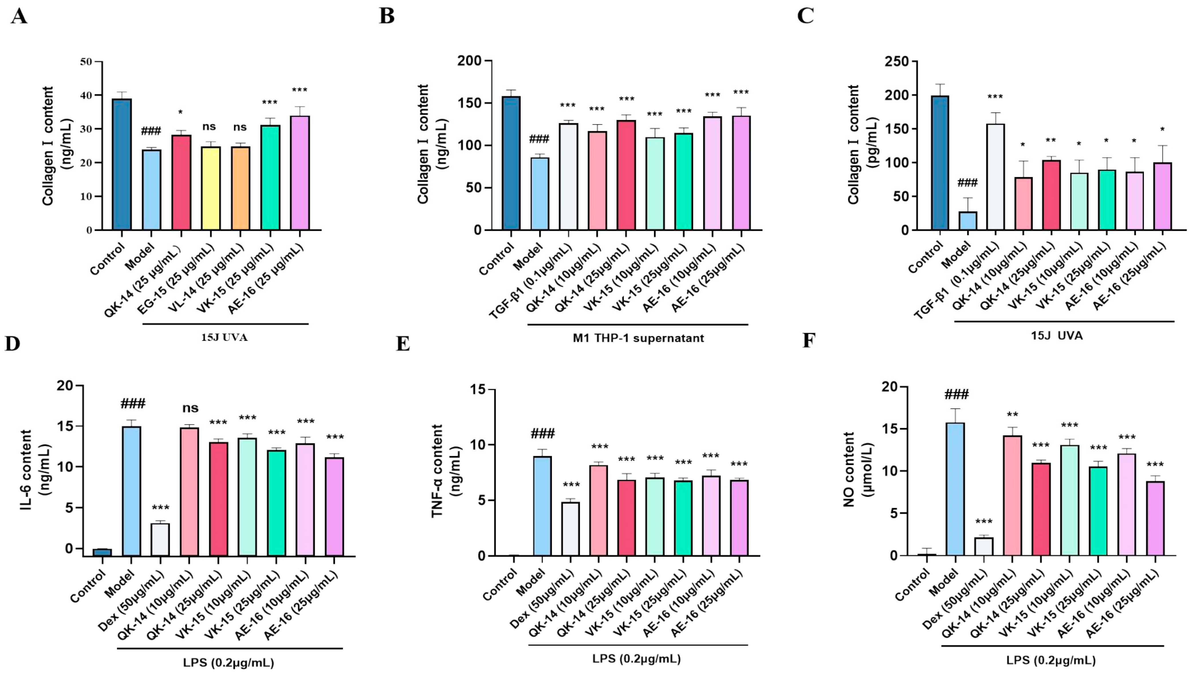
To verify whether the peptides we screened have anti-inflammatory and anti-aging effects, we selected the top five GPs (QK-14, EG-15, VL-14, VK-15, and AE-16) and conducted preliminary evaluation using a UVA-induced UV aging model. It was found that the three peptides QK-14, VK-15, and AE-16 can increase the content of collagen I in senescent cells (Figure 9A). After in-depth exploration, we found that it has anti-aging effects on cell aging caused by UV and inflammatory factors and can enhance the expression of collagen I in cells (Figure 9B,C). Applied to the RAW264.7 cell inflammation model, it was found through ELISA detection that three GPs can significantly reduce the levels of IL-6 and TNF-α produced by M1 type RAW264.7 cells, and NO detection also showed the same effect (Figure 9D–F). Based on the above results, we determined that the GPs we calculated have excellent anti-inflammatory effects and can alleviate the decrease in collagen content caused by UV or inflammatory factors in skin cells with anti-aging effects.
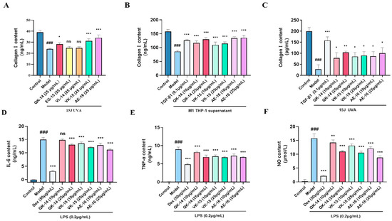
Figure 9.
Verification of the anti-inflammatory and anti-aging effects of GPs. (A) preliminary evaluation of GPs using UVA-induced HSF senescence model; (B) changes in collagen I content in the HSF cell inflammaging model after treatment with QK-14, VK-15, or AE-16 (10 or 25 μg/mL); (C) changes in collagen I content in the HSF cell UVA-induced senescence model after treatment with GPs, TGF-β1 was used as the positive control drug; (D–F) changes in IL-6, TNF-α, and NO levels in the RAW264.7 cell inflammation model treated with GPs, Dex (Dexamethasone) was used as the positive control drug in the experiment. Values are expressed as mean ± SD (n = 3). ANOVA was used for all statistical analyses and Dunnett’s test was used for the post hoc test. ### p < 0.001 vs. Control group; * p < 0.05, ** p < 0.01, *** p < 0.001 vs. Model group. ns, not significant between the indicated groups (vs. Model group).
4. Discussion
This study explores the effects of GPE on anti-inflammatory aging activity and skin protection, revealing potential mechanisms at the cellular and molecular levels. Cellular assays demonstrated that GPE enhanced cell viability within a defined concentration range, ameliorating inflammatory responses and supporting tissue repair. While in an inflammatory aging model, GPE significantly elevated collagen deposition and reduced MMP-1 expression. In vitro skin models exposed to UV irradiation showed that GPE attenuated epidermal damage and enhanced collagen synthesis. Machine learning-driven screening identified three bioactive peptides (QK-14, VK-15, AE-16) from GPE that demonstrated dual anti-inflammatory and collagen-promoting activities in both cellular senescence and inflammation models, collectively indicating GPE’s therapeutic potential in dermatoprotection and anti-aging interventions.
Ginseng (Panax ginseng C.A. Meyer) has long been recognized for its medicinal properties, including anti-inflammatory, anti-aging, and immunomodulatory effects. These properties are primarily attributed to its saponins and polysaccharides, which have been extensively studied for their therapeutic potential in various health conditions [36,37,38,39]. However, the role of ginseng peptides (GPs) in skincare has remained relatively underexplored compared to saponins and polysaccharides [30]. Our study utilizes network pharmacology and machine learning to identify and validate the bioactive peptides within GPE, demonstrating their potential to reduce inflammation and promote collagen synthesis in both in vitro and ex vivo models. This approach aligns with recent trends in computational biology, which emphasize the use of predictive models to identify bioactive compounds from natural sources [40,41,42,43,44,45].
Compared to previous studies, our work provides a more comprehensive evaluation of GPE’s effects on skin health. For instance, while earlier research has focused on the antioxidant properties of ginseng saponins [46,47,48], our study extends this by demonstrating the anti-inflammatory and collagen-promoting effects of GPE. Additionally, our use of multi-layer skin models to validate GPE’s reparative effects on UV-induced damage offers a more realistic assessment of its potential in skincare applications [49,50]. Our findings on GPE’s bioactivity are consistent with studies on other herbal peptides. For example, Ganoderma lucidum (G. lucidum, Lingzhi), commonly known as Reishi mushroom, is another herbal source that has been extensively studied for its bioactive peptides. Reishi peptides have been reported to exhibit significant anti-inflammatory, antioxidant, and immunomodulatory effects [51,52]. For example, a study identified several bioactive peptides from Reishi mushroom that demonstrated potent anti-inflammatory effects by inhibiting the production of pro-inflammatory cytokines such as IL-6 and TNF-α [51]. Another study reported that ginseng peptides could promote collagen synthesis in human skin models, thereby enhancing skin elasticity and reducing signs of aging [53]. Similarly, peptides derived from soybeans have been shown to exhibit anti-inflammatory and anti-aging effects, particularly in reducing the expression of inflammatory cytokines such as IL-6 and TNF-α [54]. Likewise, peptides from green tea or other plants have demonstrated significant antioxidant properties, which are crucial for mitigating oxidative stress-induced skin aging [55,56]. These parallels suggest that herbal peptides may share common mechanisms in promoting skin health through anti-inflammatory and antioxidative pathways.
Traditional methods of peptide discovery from natural sources often involve labor-intensive and time-consuming processes, such as extraction, purification, and in vitro and in vivo testing. These methods typically require extensive experimentation and can be limited by the complexity of natural products and the variability in their biological activities. In contrast, our study employs a combination of network pharmacology and machine learning to predict and identify bioactive peptides from ginseng extract. This computational approach offers an advantage over traditional methods: by using predictive models, we can rapidly screen and prioritize potential bioactive peptides, significantly reducing the time and resources required for experimental validation [57], and network pharmacology allows us to integrate multiple data sources, providing a more holistic view of their therapeutic potential [58].
Notably, compared with other computational screening methods, our computational approach has several unique features and advantages: (1) We combined multiple algorithms, including the HSM model [34] and the CAMP model [35], to enhance the accuracy and reliability of peptide prediction. The HSM model provided a preliminary screening of potential peptides, while the CAMP model further refined the selection by predicting protein–peptide interactions [45]. (2) Our approach not only identified bioactive peptides but also predicted their potential targets and pathways involved in anti-inflammatory and anti-aging effects. This comprehensive analysis can identify potential targets and pathways involved in the bioactivity of various peptides, facilitating a deeper understanding of their mechanisms of action [59]. (3) Our computational framework can be easily adapted to screen peptides from other herbal sources, making it a versatile tool for identifying bioactive compounds from natural products.
However, our study also has some limitations: (1) the bioactivity of the identified peptides was only suggested in vitro, and our 3D experimental models do not fully replicate the complexity of human skin [60]. Therefore, clinical trials are also needed to confirm GPE’s efficacy and safety in human subjects, and more comprehensive models are needed to validate the bioactivity of ginseng peptides; (2) the discovery of bioactive peptides in GPE offers new possibilities for cosmeceuticals against inflammaging. Future research should explore the specific molecular mechanisms of GPE’s effects, such as related signaling pathways; (3) additionally, studying the synergistic effects of GPE with other bioactive compounds could enhance its therapeutic potential, which can determine optimal combination and dosages for skincare formulations [61].
As computational biology evolves, the virtual screening methods used here can be improved. Better machine learning algorithms and predictive models could increase the accuracy and efficiency of identifying bioactive peptides from natural sources [62]. This would allow our research approach to be applied more broadly to the discovery of bioactive compounds in various natural ingredients. Moreover, future research could focus on the structural optimization and modification of the identified ginseng peptides using various computational approaches to enhance their efficacy, stability, and bioavailability for practical applications in skincare and therapeutic formulations [63,64,65].
5. Conclusions
In conclusion, our study provides a comprehensive evaluation of GPE’s potential as an anti-inflammatory and anti-aging agent for skincare. By integrating network pharmacology, machine learning, and experimental validation, we have identified key ginseng peptides that demonstrate significant bioactivity. These findings not only expand the current understanding of ginseng’s therapeutic potential but also offer new directions for the development of innovative skincare formulations. Future research should aim to address the limitations identified here and further explore the clinical applications of ginseng peptides in skin health.
Author Contributions
Conceptualization, Z.X., W.L., F.Z., S.K. and J.L.; methodology, F.Z., W.X., Z.X. and P.T.; software, W.X. and D.L.; validation, Z.X., P.T. and X.Z.; formal analysis, D.L. and X.Y.; investigation, Z.X.; resources, J.L., Q.S. and X.L.; data curation, Z.X. and F.Z.; writing—original draft preparation, Z.X.; writing—review and editing, J.L.; visualization, Z.X.; supervision, S.K. and W.L.; project administration, S.K. and W.L.; funding acquisition, Q.S., X.L. and W.L. All authors have read and agreed to the published version of the manuscript.
Funding
This research was funded by The Zhejiang Provincial Natural Science Foundation of China (LQ23H160002); The Collaborative Project with China Resources Research Institute of Science & Technology (ILH24001); The fund of Yangtze Delta Region Institute of Tsinghua University, Zhejiang (LZZLX23C002); and The Key Research and Development Program of Shihezi City (No. 2024SF08).
Institutional Review Board Statement
Not applicable.
Informed Consent Statement
Not applicable.
Data Availability Statement
The data presented in this study are available in this article. Other data that support the findings of this study are available upon request from the corresponding author.
Conflicts of Interest
W.L. was employed by Beijing Yiqing Daily Chemical Co., Ltd. F.Z., S.K., and J.L. were employed by Harvest Biotech (Zhejiang) Co., Ltd. The remaining authors declare that the research was conducted in the absence of any commercial or financial relationships that could be construed as potential conflicts of interest.
Abbreviations
The following abbreviations are used in this article:
| ABTS+ | 2,2′-azino-bis (3-ethylbenzothiazoline-6-sulfonate) diammonium salt |
| BP | biological processes |
| CC | cellular components |
| COL1A1 | Collagen type I alpha 1 |
| Dex | Dexamethasone |
| DPPH | 2,2-diphenyl-1-picrylhydrazyl |
| ELISA | Enzyme-linked immunosorbent assay |
| FLG | Filaggrin |
| GO | Gene ontology |
| GP | Ginseng peptides |
| GPE | Ginseng peptide extract |
| HSM | Hierarchical statistical mechanical modeling |
| KEGG | Kyoto Encyclopedia of Genes and Genomes |
| LOR | Loricrin |
| MF | molecular functions |
| NO | Nitric oxide |
| OMIM | Online Mendelian inheritance in man |
| PPI | Protein–protein interaction |
| ROS | Reactive oxygen species |
| RT-qPCR | Quantitative real-time polymerase chain reaction |
| SASP | senescence-associated secretory phenotype |
| STRING | Search Tool for the Retrieval of Interacting Genes/Proteins |
| TGM1 | Transglutaminase-1 |
| TCMSP | Traditional Chinese Medicine Systems Pharmacology |
Appendix A

Table A1.
Sequence of GPs, keywords of targets, and predicted scores of modified HSM and CAMP models.
Table A1.
Sequence of GPs, keywords of targets, and predicted scores of modified HSM and CAMP models.
| Peptide_Sequence | Keywords | Protein | HSM Score (p_Value) | CAMP Score |
|---|---|---|---|---|
| LNQEGIYPNNDLYRP | proliferation | BIN1 | 0.000222856 | 0.99666637 |
| LNQEGIYPNNDLYR | Inflammation, proliferation | FGR | 0.000571771 | 0.9769991 |
| QEGIYPNNDLYRPK | Proliferation, proliferation | FGR | 0.0013086 | 0.97212684 |
| LNQEGIYPNNDLYRP | inflammation | PSTPIP1 | 0.000271744 | 0.94917411 |
| ANLLHKLEETLGMNDK | Inflammation, proliferation, oxidat | TP53BP2 | 0.0000043 | 0.908577681 |
| LNQEGIYPNNDLYRP | inflammation | BTK | 0.000251922 | 0.89676905 |
| VDCPTDDATDDYR | proliferation | FYN | 0.000111128 | 0.8797974 |
| NQEGIYPNNDLYRPK | Inflammation, proliferation | FGR | 0.00017643 | 0.86943609 |
| HLNAVPEIDFTKNEN | inflammation | PSTPIP1 | 0.000113509 | 0.86510551 |
| LNQEGIYPNNDLYRP | Inflammation, oxidat | PIK3R1 | 0.000212876 | 0.84001458 |
| VDCPTDDATDDYRL | proliferation | FYN | 0.000110712 | 0.83449197 |
| LNQEGIYPNNDLYR | inflammation | BTK | 0.00057089 | 0.8144055 |
| LNQEGIYPNNDLYR | oxidat | NCF4 | 0.000371933 | 0.79551464 |
| SEYVLTDINVCVNQ | inflammation | BTK | 0.000883059 | 0.79394358 |
| VDCPTDDATDDYR | Inflammation, proliferation | FGR | 0.00016603 | 0.7798081 |
| LNQEGIYPNNDLYR | Inflammation, oxidat | PIK3R1 | 0.000545315 | 0.77899241 |
| QEGIYPNNDLYRP | collagen | WWC1 | 0.000382538 | 0.76215649 |
| VDCPTDDATDDYRLK | proliferation | FYN | 0.0000267 | 0.73349589 |
| ADEVVHHPLDKSSEVE | Inflammation, proliferation, oxidat | JIP1 | 0.0000021 | 0.730936706 |
| PEIDFTKNEN | inflammation | BTK | 0.000421379 | 0.72908759 |
| VDCPTDDATDDYRL | Inflammation, proliferation | FGR | 0.000133978 | 0.72363502 |
| NQEGIYPNNDLYRPK | oxidat | NCF4 | 0.000182778 | 0.71487856 |
| GVQKTEVEATSTVPAQKL | Inflammation, proliferation, oxidat | JIP1 | 0.0000027 | 0.708695173 |
| EGLYPNKTAPYTPVG | Inflammation, proliferation | FGR | 0.000300853 | 0.69255173 |
| DKSSEVETTDRGLFD | proliferation | VAV1 | 0.0000597 | 0.685672045 |
| VQVLEGNGGVGTIKN | proliferation | ARHGAP26 | 0.0000169 | 0.6738382 |
| LNQEGIYPNNDLYRP | oxidat | NCF4 | 0.000162998 | 0.64596671 |
| QEGIYPNNDLYRPK | Inflammation, proliferation | ITK | 0.001193209 | 0.62135625 |
| LTVTPEEPVVVEK | Inflammation, proliferation | FGR | 0.000264268 | 0.59293336 |
| VQVLEGNGGVGTIKN | Inflammation, proliferation | FGR | 0.0000117 | 0.5275951 |
| EGLYPNKTAPYTPVG | oxidat | SGR | 0.000258785 | 0.52582133 |
| EHTNTEDKQFWEHE | inflammation | BTK | 0.0000917 | 0.50152653 |
References
- Hofseth, L.J.; Ying, L. Identifying and Defusing Weapons of Mass Inflammation in Carcinogenesis. Biochim. Biophys. Acta (BBA)-Rev. Cancer 2006, 1765, 74–84. [Google Scholar] [CrossRef] [PubMed]
- Barton, G.M. A Calculated Response: Control of Inflammation by the Innate Immune System. J. Clin. Investig. 2008, 118, 413–420. [Google Scholar] [CrossRef]
- Davies, L.C.; Jenkins, S.J.; Allen, J.E.; Taylor, P.R. Tissue-Resident Macrophages. Nat. Immunol. 2013, 14, 986–995. [Google Scholar] [CrossRef] [PubMed]
- Zhao, Y.; Zhao, N.; Kollie, L.; Yang, D.; Zhang, X.; Zhang, H.; Liang, Z. Sasanquasaponin from Camellia Oleifera Abel Exerts an Anti-Inflammatory Effect in RAW 264.7 Cells via Inhibition of the NF-κB/MAPK Signaling Pathways. Int. J. Mol. Sci. 2024, 25, 2149. [Google Scholar] [CrossRef]
- Wu, T.; Zhang, J.; Geng, M.; Tang, S.-J.; Zhang, W.; Shu, J. Nucleoside Reverse Transcriptase Inhibitors (NRTIs) Induce Proinflammatory Cytokines in the CNS via Wnt5a Signaling. Sci. Rep. 2017, 7, 4117. [Google Scholar] [CrossRef] [PubMed]
- Wang, Y.; Miao, X.; Jiang, Y.; Wu, Z.; Zhu, X.; Liu, H.; Wu, X.; Cai, J.; Ding, X.; Gong, W. The Synergistic Antitumor Effect of IL-6 Neutralization with NVP-BEZ235 in Hepatocellular Carcinoma. Cell Death Dis. 2022, 13, 146. [Google Scholar] [CrossRef]
- Ross, E.A.; Devitt, A.; Johnson, J.R. Macrophages: The Good, the Bad, and the Gluttony. Front. Immunol. 2021, 12, 708186. [Google Scholar] [CrossRef]
- van Furth, R.; Cohn, Z.A.; Hirsch, J.G.; Humphrey, J.H.; Spector, W.G.; Langevoort, H.L. The Mononuclear Phagocyte System: A New Classification of Macrophages, Monocytes, and Their Precursor Cells. Bull. World Health Organ. 1972, 46, 845–852. [Google Scholar]
- Fu, J.; Lu, Z.; Wu, G.; Yang, Z.; Wu, X.; Wang, D.; Nie, Z.; Sheng, Q. Gastrodia elata Specific miRNA Attenuates Neuroinflammation via Modulating NF-κB Signaling Pathway. Int. J. Neurosci. 2024, 134, 1652–1662. [Google Scholar] [CrossRef]
- Chen, Q.; Kang, J.; Fu, C. The Independence of and Associations among Apoptosis, Autophagy, and Necrosis. Signal Transduct. Target. Ther. 2018, 3, 18. [Google Scholar] [CrossRef]
- Acosta, J.C.; Banito, A.; Wuestefeld, T.; Georgilis, A.; Janich, P.; Morton, J.P.; Athineos, D.; Kang, T.-W.; Lasitschka, F.; Andrulis, M.; et al. A Complex Secretory Program Orchestrated by the Inflammasome Controls Paracrine Senescence. Nat. Cell Biol. 2013, 15, 978–990. [Google Scholar] [CrossRef] [PubMed]
- Barbé-Tuana, F.; Funchal, G.; Schmitz, C.R.R.; Maurmann, R.M.; Bauer, M.E. The Interplay between Immunosenescence and Age-Related Diseases. Semin. Immunopathol. 2020, 42, 545–557. [Google Scholar] [CrossRef]
- Li, X.; Li, C.; Zhang, W.; Wang, Y.; Qian, P.; Huang, H. Inflammation and Aging: Signaling Pathways and Intervention Therapies. Signal Transduct. Target. Ther. 2023, 8, 239. [Google Scholar] [CrossRef]
- Li, Y.; Tian, X.; Luo, J.; Bao, T.; Wang, S.; Wu, X. Molecular Mechanisms of Aging and Anti-Aging Strategies. Cell Commun. Signal. 2024, 22, 285. [Google Scholar] [CrossRef] [PubMed]
- Attele, A.S.; Wu, J.A.; Yuan, C.-S. Ginseng Pharmacology. Biochem. Pharmacol. 1999, 58, 1685–1693. [Google Scholar] [CrossRef] [PubMed]
- Yun, T.K. Brief Introduction of Panax Ginseng CA Meyer. J. Korean Med. Sci. 2001, 16, S3. [Google Scholar] [CrossRef]
- Xia, P.; Mao, Y.; Liang, Z. Two Important Ginseng Plants in Southeast Asia: A Systematic Review of Their Traditional Uses, Botany, Phytochemistry, and Pharmacology. Acta Physiol. Plant. 2022, 44, 105. [Google Scholar] [CrossRef]
- Lu, G.; Liu, Z.; Wang, X.; Wang, C. Recent Advances in Panax Ginseng C.A. Meyer as a Herb for Anti-Fatigue: An Effects and Mechanisms Review. Foods 2021, 10, 1030. [Google Scholar] [CrossRef]
- Hernández-García, D.; Granado-Serrano, A.B.; Martín-Gari, M.; Naudí, A.; Serrano, J.C. Efficacy of Panax Ginseng Supplementation on Blood Lipid Profile. A Meta-Analysis and Systematic Review of Clinical Randomized Trials. J. Ethnopharmacol. 2019, 243, 112090. [Google Scholar] [CrossRef]
- Fan, W.; Huang, Y.; Zheng, H.; Li, S.; Li, Z.; Yuan, L.; Cheng, X.; He, C.; Sun, J. Ginsenosides for the Treatment of Metabolic Syndrome and Cardiovascular Diseases: Pharmacology and Mechanisms. Biomed. Pharmacother. 2020, 132, 110915. [Google Scholar] [CrossRef]
- Li, J.; Li, F.; Jin, D. Ginsenosides Are Promising Medicine for Tumor and Inflammation: A Review. Am. J. Chin. Med. 2023, 51, 883–908. [Google Scholar] [CrossRef] [PubMed]
- Kim, Y.-J.; Zhang, D.; Yang, D.-C. Biosynthesis and Biotechnological Production of Ginsenosides. Biotechnol. Adv. 2015, 33, 717–735. [Google Scholar] [CrossRef]
- Truong, V.-L.; Keum, Y.-S.; Jeong, W.-S. Red Ginseng Oil Promotes Hair Growth and Protects Skin against UVC Radiation. J. Ginseng Res. 2021, 45, 498–509. [Google Scholar] [CrossRef]
- Ru, J.; Li, P.; Wang, J.; Zhou, W.; Li, B.; Huang, C.; Li, P.; Guo, Z.; Tao, W.; Yang, Y.; et al. TCMSP: A Database of Systems Pharmacology for Drug Discovery from Herbal Medicines. J. Cheminform. 2014, 6, 13. [Google Scholar] [CrossRef] [PubMed]
- Stelzer, G.; Rosen, N.; Plaschkes, I.; Zimmerman, S.; Twik, M.; Fishilevich, S.; Stein, T.I.; Nudel, R.; Lieder, I.; Mazor, Y.; et al. The GeneCards Suite: From Gene Data Mining to Disease Genome Sequence Analyses. Curr. Protoc. Bioinform. 2016, 54, 1–30. [Google Scholar] [CrossRef]
- Hamosh, A.; Amberger, J.S.; Bocchini, C.; Scott, A.F.; Rasmussen, S.A. Online Mendelian Inheritance in Man (OMIM®): Victor McKusick’s Magnum Opus. Am. J. Med. Genet. Part A 2021, 185, 3259–3265. [Google Scholar] [CrossRef] [PubMed]
- Szklarczyk, D.; Kirsch, R.; Koutrouli, M.; Nastou, K.; Mehryary, F.; Hachilif, R.; Gable, A.L.; Fang, T.; Doncheva, N.T.; Pyysalo, S.; et al. The STRING Database in 2023: Protein–Protein Association Networks and Functional Enrichment Analyses for Any Sequenced Genome of Interest. Nucleic Acids Res. 2023, 51, D638–D646. [Google Scholar] [CrossRef]
- Zhou, Y.; Zhou, B.; Pache, L.; Chang, M.; Khodabakhshi, A.H.; Tanaseichuk, O.; Benner, C.; Chanda, S.K. Metascape Provides a Biologist-Oriented Resource for the Analysis of Systems-Level Datasets. Nat. Commun. 2019, 10, 1523. [Google Scholar] [CrossRef]
- Lu, M.; Xu, X.; Xi, B.; Dai, Q.; Li, C.; Su, L.; Zhou, X.; Tang, M.; Yao, Y.; Yang, J. Molecular Network-Based Identification of Competing Endogenous RNAs in Thyroid Carcinoma. Genes 2018, 9, 44. [Google Scholar] [CrossRef]
- Ye, X.; Zhao, N.; Yu, X.; Han, X.; Gao, H.; Zhang, X. Extensive Characterization of Peptides from Panax ginseng C.A. Meyer Using Mass Spectrometric Approach. Proteomics 2016, 16, 2788–2791. [Google Scholar] [CrossRef]
- Horiba, S.; Kami, R.; Tsutsui, T.; Hosoi, J. IL-34 Downregulation—Associated M1/M2 Macrophage Imbalance Is Related to Inflammaging in Sun-Exposed Human Skin. JID Innov. 2022, 2, 100112. [Google Scholar] [CrossRef] [PubMed]
- Xu, Y.; Liu, Y.; Li, J.; Li, Y.; Xu, L.; Dong, K.; Lin, X.; Zhang, T. Unveiling a Novel In-Vitro Model of Skin Inflammaging. Front. Med. 2025, 12, 1556680. [Google Scholar] [CrossRef] [PubMed]
- Li, X.-H.; Su, W.-R.; Wang, F.-F.; Li, K.; Zhu, J.-Y.; Zhu, S.-Y.; Kang, S.-N.; He, C.-F.; Li, J.-X.; Lin, X. Computational Biology in Topical Bioactive Peptide Discovery for Cosmeceutical Application: A Concise Review. Biomed. Eng. Commun. 2023, 2, 14. [Google Scholar] [CrossRef]
- Cunningham, J.M.; Koytiger, G.; Sorger, P.K.; AlQuraishi, M. Biophysical Prediction of Protein–Peptide Interactions and Signaling Networks Using Machine Learning. Nat. Methods 2020, 17, 175–183. [Google Scholar] [CrossRef]
- Lei, Y.; Li, S.; Liu, Z.; Wan, F.; Tian, T.; Li, S.; Zhao, D.; Zeng, J. A Deep-Learning Framework for Multi-Level Peptide–Protein Interaction Prediction. Nat. Commun. 2021, 12, 5465. [Google Scholar] [CrossRef]
- Zheng, Y.; Shao, R.; Xia, P.; Liang, Z.; Yan, K. Activity and Function Studies of the Promoter Cis-Acting Elements of the Key Enzymes in Saponins Biosynthesis of DS from Panax Notoginseng. Protoplasma 2022, 259, 163–171. [Google Scholar] [CrossRef]
- Su, J.; Su, Q.; Hu, S.; Ruan, X.; Ouyang, S. Research Progress on the Anti-Aging Potential of the Active Components of Ginseng. Nutrients 2023, 15, 3286. [Google Scholar] [CrossRef]
- Fan, W.; Fan, L.; Wang, Z.; Mei, Y.; Liu, L.; Li, L.; Yang, L.; Wang, Z. Rare Ginsenosides: A Unique Perspective of Ginseng Research. J. Adv. Res. 2024, 66, 303–328. [Google Scholar] [CrossRef]
- Zhou, G.; Wang, C.-Z.; Mohammadi, S.; Sawadogo, W.R.; Ma, Q.; Yuan, C.-S. Pharmacological Effects of Ginseng: Multiple Constituents and Multiple Actions on Humans. Am. J. Chin. Med. 2023, 51, 1085–1104. [Google Scholar] [CrossRef]
- Xie, W.-Y.; Ji, Z.-H.; Ren, W.-Z.; Zhao, P.-S.; Wei, F.-H.; Hu, J.; Yuan, B.; Gao, W. Wheat Peptide Alleviates DSS-Induced Colitis by Activating the Keap1–Nrf2 Signaling Pathway and Maintaining the Integrity of the Gut Barrier. Food Funct. 2024, 15, 5466–5484. [Google Scholar] [CrossRef]
- Wang, W.; Lin, H.; Shen, W.; Qin, X.; Gao, J.; Cao, W.; Zheng, H.; Chen, Z.; Zhang, Z. Optimization of a Novel Tyrosinase Inhibitory Peptide from Atrina Pectinata Mantle and Its Molecular Inhibitory Mechanism. Foods 2023, 12, 3884. [Google Scholar] [CrossRef] [PubMed]
- Yin, H.; Zhang, S.; Yue, H.; Wang, M.; Zeng, J.; Wu, W.; Wang, J.; Zheng, H.; Xue, C.; Zhao, Y.-T. Isolation, Identification and in Silico Analysis of Two Novel Cytoprotective Peptides from Tilapia Skin against Oxidative Stress-Induced Ovarian Granulosa Cell Damage. J. Funct. Foods 2023, 107, 105629. [Google Scholar] [CrossRef]
- Zhu, Q.; Xue, J.; Wang, P.; Wang, X.; Zhang, J.; Fang, X.; He, Z.; Wu, F. Identification of a Novel ACE Inhibitory Hexapeptide from Camellia Seed Cake and Evaluation of Its Stability. Foods 2023, 12, 501. [Google Scholar] [CrossRef]
- Yuxiu, Z.; Haisheng, L.; Lei, D.; Jialong, G.; Wenhong, C.; Xiaoming, Q.; Zhongqin, C.; Huina, Z.; Saiyi, Z. A Novel Tyrosinase Inhibitory Peptide Obtained from Sipunculus Nudus Gelatin Hydrolysate: Preparation, Identification, and Action Mechanism. LWT 2024, 202, 116293. [Google Scholar] [CrossRef]
- Feifei, W.; Wenrou, S.; Sining, K.; Siyu, Z.; Xiaolei, F.; Junxiang, L.; Congfen, H.; Xuhui, L. A Novel Functional Peptide, Named EQ-9 (ESETRILLQ), Identified by Virtual Screening from Regenerative Cell Secretome and Its Potential Anti-Aging and Restoration Effects in Topical Applications. Peptides 2023, 169, 171078. [Google Scholar] [CrossRef]
- Baik, I.-H.; Kim, K.-H.; Lee, K.-A. Antioxidant, Anti-Inflammatory and Antithrombotic Effects of Ginsenoside Compound K Enriched Extract Derived from Ginseng Sprouts. Molecules 2021, 26, 4102. [Google Scholar] [CrossRef]
- Ratan, Z.A.; Haidere, M.F.; Hong, Y.H.; Park, S.H.; Lee, J.-O.; Lee, J.; Cho, J.Y. Pharmacological Potential of Ginseng and Its Major Component Ginsenosides. J. Ginseng Res. 2021, 45, 199–210. [Google Scholar] [CrossRef]
- Wu, H.; Pei, H.; Liu, J.; Zeng, J.; Liu, S.; Chen, W.; He, Z.; Du, R. Protective Effect of Total Saponins of Ginseng Stems and Leaves (GSLS) on Chlorpyrifos-Induced Brain Toxicity in Mice through the PTEN/PI3K/AKT Axis. Aging 2022, 14, 8982–8999. [Google Scholar] [CrossRef]
- Yang, J.-E.; Ngo, H.T.T.; Hwang, E.; Seo, S.A.; Park, S.W.; Yi, T.-H. Dietary Enzyme-Treated Hibiscus Syriacus L. Protects Skin against Chronic UVB-Induced Photoaging via Enhancement of Skin Hydration and Collagen Synthesis. Arch. Biochem. Biophys. 2019, 662, 190–200. [Google Scholar] [CrossRef]
- Bai, D.; Wang, Z.; Xiao, Y.; Liu, T.; Pu, Y.; Sun, H.; Wang, M.; Guo, C.; Zhang, J. Transdermal Delivery of Elastin Peptide Assisted by Betaine-Based Deep Eutectic Solvent to Ameliorate Skin Photoaging. Biomater. Adv. 2024, 163, 213965. [Google Scholar] [CrossRef]
- Aursuwanna, T.; Noitang, S.; Sangtanoo, P.; Srimongkol, P.; Saisavoey, T.; Puthong, S.; Reamtong, O.; Karnchanatat, A. Investigating the Cellular Antioxidant and Anti-Inflammatory Effects of the Novel Peptides in Lingzhi Mushrooms. Heliyon 2022, 8, e11067. [Google Scholar] [CrossRef] [PubMed]
- Meng, M.; Wang, L.; Yao, Y.; Lin, D.; Wang, C.; Yao, J.; Sun, H.; Liu, M. Ganoderma Lucidum Polysaccharide Peptide (GLPP) Attenuates Rheumatic Arthritis in Rats through Inactivating NF-κB and MAPK Signaling Pathways. Phytomedicine 2023, 119, 155010. [Google Scholar] [CrossRef]
- Zhu, N.; Xu, M.-H.; Li, Y. Bioactive Oligopeptides from Ginseng (Panax Ginseng Meyer) Suppress Oxidative Stress-Induced Senescence in Fibroblasts via NAD+/SIRT1/PGC-1α Signaling Pathway. Nutrients 2022, 14, 5289. [Google Scholar] [CrossRef]
- Wen, L.; Bi, H.; Zhou, X.; Jiang, Y.; Zhu, H.; Fu, X.; Yang, B. Structure Characterization of Soybean Peptides and Their Protective Activity against Intestinal Inflammation. Food Chem. 2022, 387, 132868. [Google Scholar] [CrossRef]
- Kanlayavattanakul, M.; Khongkow, M.; Klinngam, W.; Chaikul, P.; Lourith, N.; Chueamchaitrakun, P. Recent Insights into Catechins-Rich Assam Tea Extract for Photoaging and Senescent Ageing. Sci. Rep. 2024, 14, 2253. [Google Scholar] [CrossRef] [PubMed]
- Mo, Q.; You, S.; Fu, H.; Wang, D.; Zhang, J.; Wang, C.; Li, M. Purification and Identification of Antioxidant Peptides from Rice Fermentation of Lactobacillus Plantarum and Their Protective Effects on UVA−Induced Oxidative Stress in Skin. Antioxidants 2022, 11, 2333. [Google Scholar] [CrossRef] [PubMed]
- Zhang, H.; Saravanan, K.M.; Wei, Y.; Jiao, Y.; Yang, Y.; Pan, Y.; Wu, X.; Zhang, J.Z.H. Deep Learning-Based Bioactive Therapeutic Peptide Generation and Screening. J. Chem. Inf. Model. 2023, 63, 835–845. [Google Scholar] [CrossRef]
- Feifei, W.; Wenrou, S.; Jinyue, S.; Qiaochu, D.; Jingjing, L.; Jin, L.; Junxiang, L.; Xuhui, L.; Xiao, L.; Congfen, H. Anti-ageing Mechanism of Topical Bioactive Ingredient Composition on Skin Based on Network Pharmacology. Int. J. Cosmet. Sci. 2025, 47, 134–154. [Google Scholar] [CrossRef]
- Zhang, M.; Zhou, J.; Wang, X.; Wang, X.; Ge, F. DeepBP: Ensemble Deep Learning Strategy for Bioactive Peptide Prediction. BMC Bioinform. 2024, 25, 352. [Google Scholar] [CrossRef]
- Jing, C.; Guo, J.; Li, Z.; Xu, X.; Wang, J.; Zhai, L.; Liu, J.; Sun, G.; Wang, F.; Xu, Y.; et al. Screening and Research on Skin Barrier Damage Protective Efficacy of Different Mannosylerythritol Lipids. Molecules 2022, 27, 4648. [Google Scholar] [CrossRef]
- Kim, J.-H.; Lee, R.; Hwang, S.-H.; Choi, S.-H.; Kim, J.-H.; Cho, I.-H.; Lee, J.I.; Nah, S.-Y. Ginseng and Ginseng Byproducts for Skincare and Skin Health. J. Ginseng Res. 2024, 48, 525–534. [Google Scholar] [CrossRef] [PubMed]
- Gupta, R.; Srivastava, D.; Sahu, M.; Tiwari, S.; Ambasta, R.K.; Kumar, P. Artificial Intelligence to Deep Learning: Machine Intelligence Approach for Drug Discovery. Mol. Divers. 2021, 25, 1315–1360. [Google Scholar] [CrossRef] [PubMed]
- Wang, L.; Wang, N.; Zhang, W.; Cheng, X.; Yan, Z.; Shao, G.; Wang, X.; Wang, R.; Fu, C. Therapeutic Peptides: Current Applications and Future Directions. Signal Transduct. Target. Ther. 2022, 7, 48. [Google Scholar] [CrossRef]
- Zhang, Q.-Y.; Yan, Z.-B.; Meng, Y.-M.; Hong, X.-Y.; Shao, G.; Ma, J.-J.; Cheng, X.-R.; Liu, J.; Kang, J.; Fu, C.-Y. Antimicrobial Peptides: Mechanism of Action, Activity and Clinical Potential. Mil. Med. Res. 2021, 8, 48. [Google Scholar] [CrossRef]
- Song, G.; Sun, Y.; Liu, T.; Zhang, X.; Zeng, Z.; Wang, R.; Li, P.; Li, C.; Jiang, G. Transdermal Delivery of Cu-Doped Polydopamine Using Microneedles for Photothermal and Chemodynamic Synergistic Therapy against Skin Melanoma. Chem. Eng. J. 2021, 426, 130790. [Google Scholar] [CrossRef]


Disclaimer/Publisher’s Note: The statements, opinions and data contained in all publications are solely those of the individual author(s) and contributor(s) and not of MDPI and/or the editor(s). MDPI and/or the editor(s) disclaim responsibility for any injury to people or property resulting from any ideas, methods, instructions or products referred to in the content. |
© 2025 by the authors. Licensee MDPI, Basel, Switzerland. This article is an open access article distributed under the terms and conditions of the Creative Commons Attribution (CC BY) license (https://creativecommons.org/licenses/by/4.0/).