Communication

Jatrorrhizine Isolated from Phellodendron amurense Improves Collagen Homeostasis in CCD-986sk Human Dermal Fibroblast Cells

Department of Nutrition and Food Science, University of Maryland, College Park, MD 20742, USA
Cosmetics 2025, 12(2), 70; https://doi.org/10.3390/cosmetics12020070
Submission received: 5 March 2025 / Revised: 28 March 2025 / Accepted: 7 April 2025 / Published: 9 April 2025
(This article belongs to the Section Cosmetic Dermatology)

Abstract

:
Jatrorrhizine is one of the major bioactive compounds found in Phellodendron amurense. Previous studies have reported various health benefits of jatrorrhizine, but little is known about its effect on skin health. In this study, jatrorrhizine isolated from Phellodendron amurense was used to determine the impact on collagen homeostasis in CCD-986sk human dermal fibroblast cells. Jatrorrhizine did not show toxicity of up to 10 μM in CCD-986sk cells. Jatrorrhizine induced procollagen and hyaluronic acid synthesis by increasing the gene expression of collagen type I alpha 2, TIMP metallopeptidase inhibitor 1, transforming growth factor beta 1, and hyaluronan synthase 2. In addition, jatrorrhizine treatment inhibited the gene expression of matrix metallopeptidase 1 and matrix metallopeptidase 9 by increasing tissue inhibitors of metalloproteinase. Our results suggest that jatrorrhizine has the potential for application in therapeutic and cosmetic products to improve collagen homeostasis and prevent wrinkle formation.

1. Introduction

Phellodendron amurense is a tree found in the northeastern Asia countries of Korea, Japan, and China [1]. This tree has been commonly used for traditional medicinal purposes due to its high content of bioactive compounds. These bioactive compounds include phenolic compounds, alkaloids, terpenes, and flavonoids that have been widely known to be beneficial to human health [2,3]. Among these bioactive compounds in Phellodendron amurense, jatrorrhizine is a major alkaloid which has been known to have health beneficial effects including anti-inflammation effect [4,5]. However, little is known about its anti-aging properties, particularly in skin aging.
Human skin structure is composed of collagen, elastin, and hyaluronic acid [6]. In particular, collagen is one of the key components of the extracellular matrix (ECM) by serving as the primary structural protein to maintain the skin elasticity [7,8,9]. During skin aging, the structure of the skin becomes weaker as collagen, elastin, and hyaluronic acid breaks down and disrupts the strong bond between these components [10]. In addition, this process can occur by external stimulants such as ultraviolet rays in sunlight. Thus, it is crucial to understand the cellular interactions between skin components. However, skin aging studies face challenges due to the complex biology of human skin and experimental models that represent or are translatable to human skin are limited. Therefore, researchers have been using cellular models for studies on skin aging, including CCD-986sk human dermal fibroblasts cells.
CCD-986sk human dermal fibroblasts are widely used for studies on skin aging, extracellular matrix (ECM), and wound healing. CCD-986sk cells can be used to understand the interactions between the key structural components of the skin, such as collage, elastin, and hyaluronic acid, which maintains the skin strength and elasticity to prevent wrinkle formation. Skin aging in CCD-986sk cells can be achieved by causing damage to the cell with ultraviolet ray B (UV-B) irradiation. Thus, these characteristics of CCD-986sk cells provide advantages in screening compounds that can regulate the components of the skin matrix to improve wrinkle formation [11,12,13,14]. In this study, we have determined the potential anti-wrinkle effect by maintaining collagen homeostasis and the working mechanisms of jatrorrhizine isolated from Phellodendron amurense using CCD-986sk fibroblast. The findings from this study can provide insight into the role of jatrorrhizine in skin aging and emphasize its potential as a natural anti-aging agent. As the demand for natural compounds for skin health increases, this study is expected to contribute to dermatological research field and may support the development of new functional cosmetics, particularly targeting wrinkle prevention.

2. Materials and Methods

2.1. Materials

Phellodendron amurense was purchased from a herb market (Daegu, Republic of Korea) and jatrorrhizine was isolated at a purity of ≥99%. Human dermal fibroblast CCD-986sk was purchased from ATCC (Manassas, VA, USA). (3-(4,5-dimethylthiazolyl-2)-2, 5-diphenyltetrazolium bromide (MTT) assay reagent was purchased from Sigma-Aldrich Co. (St. Louis, MO, USA). Fetal bovine serum (FBS) was purchased from HyClone Laboratories Inc (Logan, UT, USA). All primers were purchased from Thermo Fisher Scientific (Waltham, MA, USA). All other chemicals were purchased from Fischer Scientific (Pittsburgh, PA, USA).

2.2. Jatrorrhizine Isolation and Identification

Phellodendron amurense extract was prepared as in our previous study [4,5]. Phellodendron amurense was extracted with 60% ethanol in a shaking incubator for 24 h at room temperature. Open column chromatography was performed using 5 g of freeze-dried Phellodendron amurense extract with a dextran gel Sephadex LH-20 column (12 × 120 cm). The elution was carried out with 60% ethanol followed by water or 100% ethanol. The elution rate of the column was 9.82 mL/min, and fractions of 15 mL were collected per tube. For the second fractionation, chromatography was performed using a Mixed-Mode Chromatography ligand (MCL) gel column (4.5 × 50 cm). The elution solvent had an elution rate of 0.74 mL/min. The compounds isolated from column chromatography were developed with silica gel thin-layer chromatography (TLC) (5 × 5 cm) with benzene:ethyl formate:formic acid (1:7:2) based on Matsuo and Ito (1978) and Hostettmann, K. and Hostettmann, M. (1982) with modifications [15,16]. For identification, compounds were reacted with 10% H2SO4 on silica gel TLC. The purity of isolated compound was confirmed to be above 99% with HPLC analysis (Supplementary Figure S1). The yellow-red acicular crystal form isolate was used to perform 1H-NMR and 13C-NMR and analyzed using Fourier Transform Nuclear Magnetic Resonance (600 MHz FT-NMR, Bruker Avance Neo 600, Burker Co., Billerica, MA, USA). The analysis identified the isolate as jatrorrhizine (Supplementary Figure S1), which matched with previous studies [17,18].

2.3. Cell Culture and Maintenance

Dulbecco’s Modified Eagle Medium (DMEM) with 10% FBS and 1% penicillin/streptomycin (100 U/mL) was used as media for culturing and CCD-986sk cells were incubated in 5% CO2 incubator at 37 °C.

2.4. Determination of Cytotoxicity

To determine cytotoxicity, human dermal fibroblast CCD-986sk was seeded to 48 well plate at 5 × 10³ cells/well and incubated in 5% CO2 incubator at 37 °C for 24 h. Jatrorrhizine was treated at concentration of 5–100 µM for 48 h. After treatment, 50 μL MTT solution was treated to each well at 5 mg/mL and incubated for 4 h. After incubation, supernatant was removed and 150 µL of dimethyl sulfoxide was added for 10 min. The optical density (OD) was measured at 540 nm using plate reader model SunriseTM (Tecan, Männedortf, Switzerland). Cytotoxicity was calculated relative to the control group.

2.5. UV-B Irradiation and Treatment

Human dermal fibroblast CCD-986sk cells were seeded at 1 × 10⁶ cells/plate in 10 cm Petri dish. After washing with phosphate-buffered saline (PBS) 3 times, 1 mL of PBS was added to the plate and cells were irradiated with 312 nm UV-B (20 mJ/cm2) for 1 min using UV-B irradiator (Model Bio-Sun, Vilber, Marine, France) [19]. While the control group was not irradiated with UV-B, treatment groups were irradiated with UV-B. After UV-B irradiation, jatrorrhizine was treated by each treatment group in different concentrations, except the control group, for 48 h and incubated at 37 °C in a 5% CO2 incubator.

2.6. Gene Expression

Ribospin RNA extraction kit, GeneAll (Seoul, Republic of Korea) was used to extract wrinkle-associated genes to measure mRNA expression. qPCRBIO cDNA Synthesis Kit, PCR Biosystems Ltd. (London, UK) was used to synthesize cDNA from extracted mRNA. cDNA was diluted with Tris/ethylenediamine tetra-acetic acid (EDTA) buffer and used for analysis using PCRmax Eco 48 Real-time PCR system, Cole-Parmer (Vernon Hills, IL, USA). PCR conditions and primer sequences for wrinkle-associated genes are shown in Table 1.

2.7. Statistical Analysis

All data are presented as mean ± standard deviation and analyzed with one-way ANOVA followed by Duncan’s multiple range test using SPSS 23 for windows (Statistical Package for Social Science, Chicago, IL, USA). Data were considered significant when p < 0.05 compared to control group.

3. Results and Discussion

3.1. Cytotoxicity and Jatrorrhizine and UV-B Irradiation Recovery in CCD-986sk Fibroblast Cells

To determine the toxicity of jatrorrhizine treatment on CCD-986sk cells, MTT assay was used to measure viability of cells. Our result showed that cell viability decreased in dose-dependent manner by showing approximately 90% viability at 20 µM and lower than approximately 87% at 30 µM in CCD-986sk cells (Figure 1B). This suggested that jatrorrhizine showed cell toxicity at concentrations above 20 µM. Based on these results, we have used jatrorrhizine up to 10 µM in CCD-986sk cells. Furthermore, cellular recovery was observed in UV-B irradiation CD-986sk cells with jatrorrhizine treatment at 10 µM (Figure 1C).

3.2. Jatrorrhizine Increase Procollagen Production and Inhibits Collagen Degradation in CCD-986sk Cells

Collagen is a key protein that forms extracellular matrix (ECM) and maintains the skin’s elasticity. It is produced by fibroblast cells and abundant throughout the body [20]. When the fibroblast cell is stimulated with UV-B irradiation, the expression of matrix metalloproteinases (MMPs) increases, which leads to collagen degradation. Among many MMPs, MMP-1 degrades collagen and MMP-9 degrades gelatin. In contrast, UV-B irradiation decreases TIMP-1 expression, which decreases the ability to suppress the activity of MMPs to prevent collagen degradation [21]. Thus, we irradiated UV-B to CCD-986sk cells with jatrorrhizine treatment to determine the changes in collagen type I alpha 2 (COLIA2), MMP-1, MMP-9, and TIMP-1 expression.
COL1A2 encodes the alpha-2 chain of type 1 collagen, which is a procollagen that is essential to collagen synthesis. It is a key structural protein that provides strength and elasticity to the skin by forming fibrils in the dermis to maintain skin firmness. A reduced expression of COL1A2 leads to reduced collagen synthesis, which can result in forming wrinkles by thinning of the dermis and loss of elasticity. Our result showed that gene expression of COL1A2 was significantly recovered up to 32% compared to 16% of the control group (Figure 2A). This suggests that jatrorrhizine was able to increase the expression of COL1A2 and recover the collagen synthesis process.
MMP-1 and MMP-9 are enzymes that promote the degradation of ECM components and are synthesized by the transcription factor activating protein-1 (AP-1) [22]. Over 10 similar enzymes form the MMP family, including MMP-1 and MMP-9, which are zinc-containing metalloproteinases that can degrade structural proteins of ECM, particularly collagen and form wrinkles [23,24]. MMP-1 is known to be one of the major collagenases, which degrade the binding site of the collagen synthesized from type 1 pro-collagen and prioritize degrading the fibrous collagen that maintains the tensile strength [9,25,26]. MMP-9 is a gelatinase that further degrades collagen fibers that were degraded from MMP-1, which leads to further development of wrinkle formation and reduction in elasticity [24]. For these reasons, they are commonly considered as the biomarkers of skin aging. Our results showed that the treatment of jatrorrhizine significantly reduced the expression of both MMP-1 and MMP-9 gene expression (Figure 2B,C), which was increased due to UV-B irradiation. Gene expression of MMP-1 was inhibited by approximately 17% and MMP-9 by 33% compared to the control group. These results suggest that jatrorrhizine can inhibit the expression of MMP-1 and MMP-9.
TIMPs are known to be the inhibitor of MMPs in normal skin tissue [27]. Among the four known TIMPs, TIMP-1 specifically inhibits MMP-1 and MMP-9 to inhibit collagen degradation [28]. In our current study, the expression of TIMP-1 was recovered by up to approximately 39% with jatrorrhizine treatment compared to 13% of the control group (Figure 2D). Thus, this suggests that jatrorrhizine recovers TIMP-1 expression when damaged with UV-B irradiation.
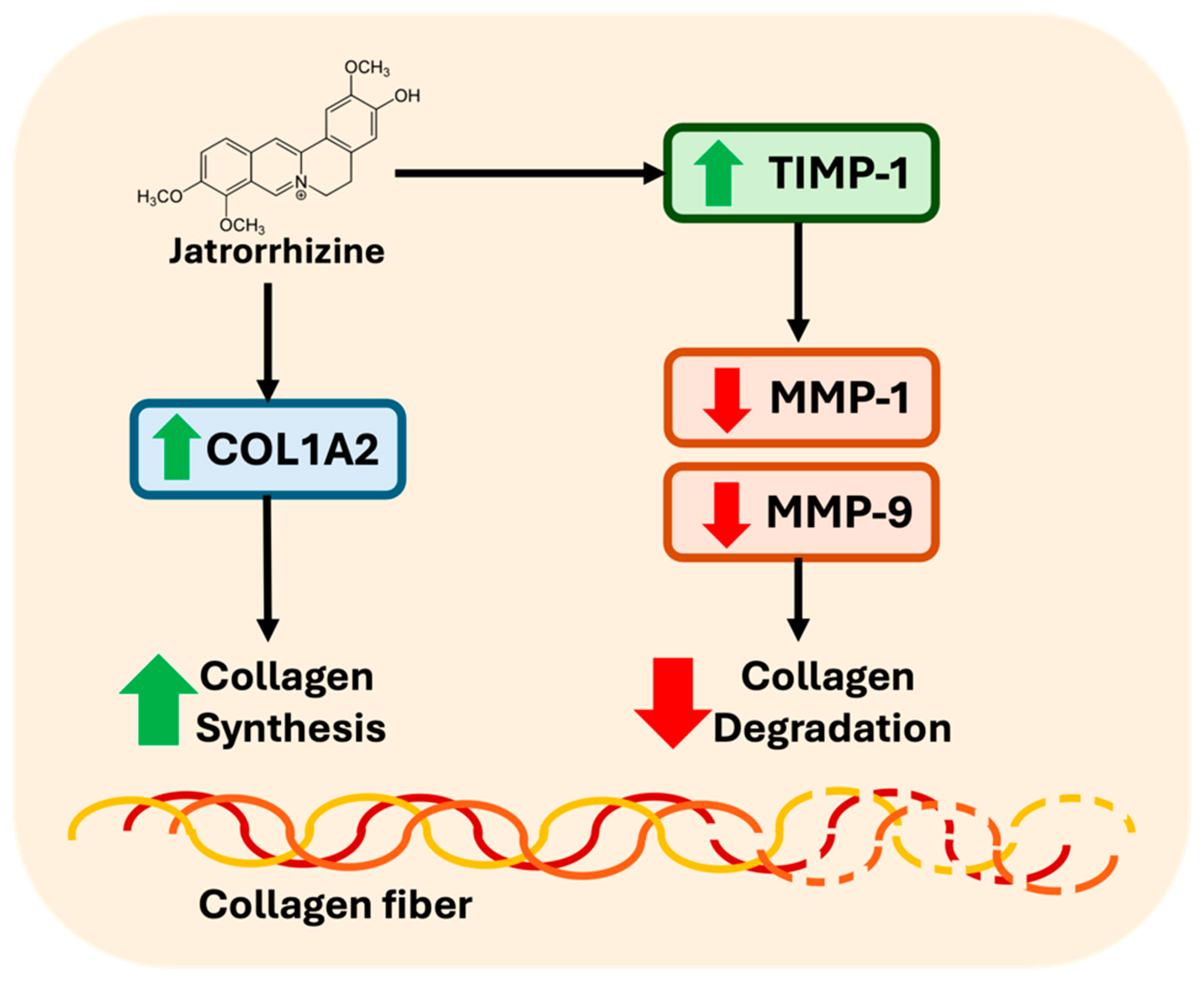
These results suggest that jatrorrhizine recovers the expression of COL1A2 to promote collagen synthesis. In addition, TIMP-1 expression was recovered to inhibit the activity of MMPs, which may have enhanced preventive effects on collagen degradation. This matched with the previous finding of the interactions between MMP-1, MMP-9, and TIMP-1 [29,30]. Previous studies have reported that UV-B irradiated fibroblast CCD-986sk cells increased the expression of MMP-1 and MMP-9 while decreasing TIMP-1 expression, which leads to significant collagen degradation as TIMP-1 fails to inhibit MMP-1 and MMP-9 activity [31,32,33]. While jatrorrhizine is suggested to maintain collagen homeostasis in CCD-986sk cells, further studies using additional cellular model or in vivo model are suggested to understand the clear working mechanisms of jatrorrhizine.

3.3. Jatrorrhizine Increases the Expression of TGFB1 and HAS2 in CCD-986sk Cells

TGFB1 encodes TGF-ß, which is one of the key factors in regulating cell differentiation, proliferation, and growth in extracellular connective tissues. It is also known to regulate collagen synthesis, which is crucial in maintaining the skin structure [34]. The activation of TGF-ß1 is known to induce collagen type-1 [35]. Furthermore, TGF-ß is known to suppress MMP-1, which can prevent collagen degradation [36]. Our result showed that jatrorrhizine treatment increased the expression of TGFB1 in UVB stimulated CCD-986sk cells (Figure 3A). Jatrorrhizine treatment at 10 µM showed the expression of TGFß1 over 64%, which indicates a significant recovery from UVB damage. This suggests that jatrorrhizine can increase the expression of TGFB1 to enhance the collagen synthesis process while adding a more preventive effect on collagen degradation by inhibiting MMP-1 activity.
While collagen is one of the major key components to form the complex skin structure, hyaluronic acid (HA) is another key component of the skin structure by maintaining skin hydration, elasticity, and volume, due to its unique ability to retain water. However, over time, the ability to synthesize HA decreases, which leads to reduced moisture retention and cause skin dryness, loss of elasticity, and wrinkle formation. Hyaluronan synthase 2 (HAS2) is responsible for HA synthesis. Thus, maintaining HAS2 expression level to prevent the decline in HA in the skin structure is another key to preventing skin aging, particularly wrinkle formation. Our result showed that UV-B irradiation significantly reduced the gene expression of HAS2 expression (Figure 3B). However, jatrorrhizine treatment significantly recovered HAS2 expression in a dose-dependent manner (Figure 3B). Compared to 11% of HAS2 expressions in the control group, jatrorrhizine treatment at 5 µM recovered HAS2 expression by up to approximately 20%, while at 10 µM was approximately 30%.
The result from the current study indicates that jatrorrhizine increases collagen synthesis and inhibits collagen degradation, which can potentially show a dual effect that can enhance collagen homeostasis (Figure 4). While our study suggests the working mechanisms for collagen homeostasis of jatrorrhizine, additional studies with more complex models will be needed to further understand the working mechanism. Furthermore, more studies on determining the bioavailability of jatrorrhizine are needed. It has been previously reported that jatrorrhizine has poor permeability and bioavailability with oral consumption [37,38]. However, under pathological conditions, the bioavailability and absorption of jatrorrhizine has significantly increased [38]. This may suggest that oral consumption of jatrorrhizine may be inefficient, but direct application to the target tissue under pathological conditions, such as skin wounds, can maximize the bioavailability and take advantage of its health beneficial effect.
While many natural compounds have been reported with health benefits, many of these natural compounds can show toxicity at higher doses. Previously, quercetin has been reported to show beneficial health effects such as lowering blood pressure or antioxidative effects but can cause toxicity in the liver or kidney at high doses [39,40]. Also, epigallocatechin gallate (EGCG) is a well-known antioxidant with various health benefits but it is known to cause hepatotoxicity at high concentrations [41]. Interestingly, jatrorrhizine did not show cytotoxicity up to 100 µM in mouse B16F10 melanoma cell, which suggests that different tissue or experimental models can show different responses to treatment [42]. This suggests that identifying the adequate concentration for health beneficial effects without potential toxicity is crucial. Thus, our study identified the adequate concentration to take advantage of jatrorrhizine on enhancing collagen homeostasis without toxicity.
In conclusion, jatrorrhizine maintained collagen homeostasis by regulating collagen synthesis and collagen degradation in UV-B irradiated CCD-986sk cells. Jatrorrhizine is suggested to increase collagen synthesis by upregulating COL1A2, and to reduce collagen degradation by inhibiting AP-1-mediated MMP1/MMP9 transcription and upregulation of TIMP-1 expression. In addition, jatrorrhizine can contribute to maintaining the skin structure by increasing HA synthesis. These findings from the current study suggest that jatrorrhizine can potentially be used for therapeutic or cosmetic products for anti-wrinkle and skin structure recovery purposes.

Supplementary Materials

The following supporting information can be downloaded at: https://www.mdpi.com/article/10.3390/cosmetics12020070/s1, Figure S1: Chemical structure and (A) 1H-NMR, (B) 13C-NMR spectrum, and (C) purity of jatrorrhizine isolated from Phellodendron amurense. (A) H-NMR (600 MHz, DMSO-d6) showed, 9.86 (1H, s, H-8), 8.98 (1H, s, H-13), 8.19 (1H, d, J = 2 Hz, H-12), 8.02 (1H, d, J = 2 Hz, H-11), 7.70 (1H, s, H-1), 6.87 (1H, s, H-4), 4.93 (2H, d, J = 6.2 Hz, H-6), 4.10 (3H, s, -OCH3), 4.07 (3H, s, -OCH3), 3.95 (3H, s, 2-OCH3), 3.16 (2H, t, J = 3.92 Hz, H-5). (B) C-NMR (600 MHz, DMSO-d6) showed, 150.3 (C-9), 148.3 (C-8), 145.7 (C-2), 144.0 (C-10), 138.6 (C-13α), 133.7 (C-12α), 129.3 (C-1α), 127.2 (C-13), 123.7 (C-12), 121.6 (C-4a), 119.8 (C-11), 118.1 (C-8α), 115.4 (C-4), 109.9 (C-1), 62.3 (C-6), 57.5 (9-OCH3), 56.7 (10-OCH3), 55.8 (2-OCH3), 26.2 (C-5). The NMR spectra of the isolate from Phellodendron amurense was identified as jatrorrhizine. (C) The HPLC analysis was performed using a Waters 2795 separations module (Waters Co., Milford, MA, USA) equipped with a Kintex 5 μm C18 100 Å column (4.6 × 100 mm, Phenomenex, Torrance, CA, USA). The column was maintained at 30 °C with a 1 mL/min flow rate. The mobile phase consisted of solvent A (0.1% phosphoric acid in water) and solvent B (0.1% phosphoric acid in acetonitrile) with a gradient of 7:3 for 20 min, 4:6 for 5.1 min, 0:10 for 10.1 min, and back to 7:3 for 5 min. Detection was carried out using a photo diode array detector set at 280 nm.

Funding

This research received no external funding.

Institutional Review Board Statement

Not applicable.

Informed Consent Statement

Not applicable.

Data Availability Statement

The datasets used during the current study are available from the corresponding author upon request.

Conflicts of Interest

The authors declare that the research was conducted in the absence of any commercial or financial relationships that could be construed as a potential conflict of interest. The funders had no role in the design of the study; in the collection, analyses or interpretation of data; in the writing of the manuscript; or in the decision to publish the results.

References

  1. Read, R.A.; Zasada, J.C. Phellodendron amurense Rupr.: Amur corktree; U.S. Department of Agriculture: Washington DC, USA, 2008. [Google Scholar]
  2. Aryal, B.; Raut, B.K.; Bhattarai, S.; Bhandari, S.; Tandan, P.; Gyawali, K.; Sharma, K.; Ranabhat, D.; Thapa, R.; Aryal, D.; et al. Potential therapeutic applications of plant-derived alkaloids against inflammatory and neurodegenerative diseases. Evid.-Based Complement. Altern. Med. 2022, 2022, 7299778. [Google Scholar] [CrossRef] [PubMed]
  3. Dey, P.; Kundu, A.; Kumar, A.; Gupta, M.; Lee, B.M.; Bhakta, T.; Dash, S.; Kim, H.S. Chapter 15—Analysis of alkaloids (indole alkaloids, isoquinoline alkaloids, tropane alkaloids). In Recent Advances in Natural Products Analysis; Sanches Silva, A., Nabavi, S.F., Saeedi, M., Nabavi, S.M., Eds.; Elsevier: Amsterdam, The Netherlands, 2020; pp. 505–567. [Google Scholar] [CrossRef]
  4. Cho, Y.-J. Anti-inflammatory effect of jatrorrhizine from Phellodendron amurense in lipopolysaccharide-stimulated Raw264.7 Cells. J. Appl. Biol. Chem. 2011, 54, 114–119. [Google Scholar] [CrossRef]
  5. Cho, Y.-J. Characteristics of cosmetic with whitening compounds from Phellodendron amurense. J. Appl. Biol. Chem. 2011, 54, 108–113. [Google Scholar] [CrossRef]
  6. Brenneisen, P.; Sies, H.; Scharffetter-Kochanek, K. Ultraviolet-B irradiation and matrix metalloproteinases: From induction via signaling to initial events. Ann. N. Y. Acad. Sci. 2002, 973, 31–43. [Google Scholar] [CrossRef] [PubMed]
  7. Kavitha, O.; Thampan, R.V. Factors influencing collagen biosynthesis. J. Cell. Biochem. 2008, 104, 1150–1160. [Google Scholar] [CrossRef]
  8. Talwar, H.S.; Griffiths, C.E.; Fisher, G.J.; Hamilton, T.A.; Voorhees, J.J. Reduced type I and type III procollagens in photodamaged adult human skin. J. Investig. Dermatol. 1995, 105, 285–290. [Google Scholar] [CrossRef] [PubMed]
  9. Vincenti, M.P.; Brinckerhoff, C.E. Transcriptional regulation of collagenase (MMP-1, MMP-13) genes in arthritis: Integration of complex signaling pathways for the recruitment of gene-specific transcription factors. Arthritis Res. Ther. 2002, 4, 157. [Google Scholar] [CrossRef]
  10. Galvez-Martin, P.; Soto-Fernandez, C.; Romero-Rueda, J.; Cabañas, J.; Torrent, A.; Castells, G.; Martinez-Puig, D. A novel hyaluronic acid matrix ingredient with regenerative, anti-aging and antioxidant capacity. Int. J. Mol. Sci. 2023, 24, 4774. [Google Scholar] [CrossRef]
  11. Ellenrieder, V.; Alber, B.; Lacher, U.; Hendler, S.F.; Menke, A.; Boeck, W.; Wagner, M.; Wilda, M.; Friess, H.; Büchler, M.; et al. Role of MT-MMPs and MMP-2 in pancreatic cancer progression. Int. J. Cancer 2000, 85, 14–20. [Google Scholar] [CrossRef]
  12. Jenkins, G. Molecular mechanisms of skin ageing. Mech. Ageing Dev. 2002, 123, 801–810. [Google Scholar] [CrossRef]
  13. Kim, Y.A.; Kim, D.H.; Yu, J.M.; Bin Park, C.; Park, T.S.; Park, B.J. Anti-wrinkle effects of extracts and solvent fractions from Nymphoides peltate on CCD-986sk. J. Appl. Biol. Chem. 2017, 60, 357–362. [Google Scholar] [CrossRef]
  14. Puizina-Ivić, N. Skin aging. Acta Dermatovenerol Alp Pannonica Adriat 2008, 17, 47–54. [Google Scholar] [PubMed]
  15. Matsuo, T.; Ito, S. The chemical structure of Kaki-tannin from immature fruit of the Persimmon (Diospyros kaki L.). Agric. Biol. Chem. 1978, 42, 1637–1643. [Google Scholar] [CrossRef]
  16. Hostettmann, K.; Hostettmann, M. Isolation techniques for flavonoids. In The Flavonoids; Harborn, J.B., Mabry, T.J., Eds.; Springer: Boston, MA, USA, 1982. [Google Scholar] [CrossRef]
  17. Li, Y.; He, W.; Liu, J.; Sheng, F.; Hu, Z.; Chen, X. Binding of the bioactive component Jatrorrhizine to human serum albumin. Biochim. Biophys. Acta 2005, 1722, 15–21. [Google Scholar] [CrossRef] [PubMed]
  18. Parmar, V.S.; Bracke, M.E.; Philippe, J.; Wengel, J.; Jain, S.C.; Olsen, C.E.; Bisht, K.S.; Sharma, N.K.; Courtens, A.; Sharma, S.K.; et al. Anti-invasive activity of alkaloids and polyphenolics in vitro. Bioorganic Med. Chem. 1997, 5, 1609–1619. [Google Scholar] [CrossRef]
  19. Lee, K.-E.; Mun, S.; Pyun, H.-B.; Kim, M.-S.; Hwang, J.-K. Effects of macelignan isolated from Myristica fragrans (Nutmeg) on expression of matrix metalloproteinase-1 and type I procollagen in UVB-irradiated human skin fibroblasts. Biol. Pharm. Bull. 2012, 35, 1669–1675. [Google Scholar] [CrossRef]
  20. Ricard-Blum, S.; Ruggiero, F. The collagen superfamily: From the extracellular matrix to the cell membrane. Pathol. Biol. 2005, 53, 430–442. [Google Scholar] [CrossRef]
  21. Jang, Y.-A.; Yeom, B.-S.; Kim, S.-G.; Lee, J.-T. Evaluation of whitening activity and wrinkle inhibitory effect of ethanol extracts of Nelumbinis Rhizomatis Nodus. J. Life Sci. 2019, 29, 1192–1199. [Google Scholar]
  22. Lee, Y.-R.; Noh, E.-M.; Han, J.-H.; Kim, J.-M.; Hwang, J.-K.; Hwang, B.-M.; Chung, E.-Y.; Kim, B.-S.; Lee, S.-H.; Lee, S.J.; et al. Brazilin inhibits UVB-induced MMP-1/3 expressions and secretions by suppressing the NF-κB pathway in human dermal fibroblasts. Eur. J. Pharmacol. 2012, 674, 80–86. [Google Scholar] [CrossRef]
  23. Dragsbæk, K.; Neergaard, J.; Hansen, H.; Byrjalsen, I.; Alexandersen, P.; Kehlet, S.; Bay-Jensen, A.-C.; Christiansen, C.; Karsdal, M. Matrix metalloproteinase mediated type I collagen degradation—An independent risk factor for mortality in women. EBioMedicine 2015, 2, 723–729. [Google Scholar] [CrossRef]
  24. Nikolov, A.; Popovski, N. Role of gelatinases MMP-2 and MMP-9 in healthy and complicated pregnancy and their future potential as preeclampsia biomarkers. Diagnostics 2021, 11, 480. [Google Scholar] [CrossRef] [PubMed]
  25. Svensson, R.B.; Hassenkam, T.; Grant, C.A.; Magnusson, S.P. Tensile properties of human collagen fibrils and fascicles are insensitive to environmental salts. Biophys. J. 2010, 99, 4020–4027. [Google Scholar] [CrossRef]
  26. Vincenti, M.P.; White, L.A.; Schroen, D.J.; Benbow, U.; Brinckerhoff, C.E. Regulating expression of the gene for matrix metalloproteinase-1 (collagenase): Mechanisms that control enzyme activity, transcription, and mrna stability. Crit. Rev. Eukaryot. Gene Expr. 1996, 6, 391–411. [Google Scholar] [CrossRef] [PubMed]
  27. Moore, C.S.; Crocker, S.J. An alternate perspective on the roles of TIMPs and MMPs in pathology. Am. J. Pathol. 2012, 180, 12–16. [Google Scholar] [CrossRef]
  28. Brew, K.; Nagase, H. The tissue inhibitors of metalloproteinases (TIMPs): An ancient family with structural and functional diversity. Biochim. Biophys. Acta 2010, 1803, 55–71. [Google Scholar] [CrossRef]
  29. Boon, L.; Ugarte-Berzal, E.; Martens, E.; Fiten, P.; Vandooren, J.; Janssens, R.; Blanter, M.; Yu, K.; Boon, M.; Struyf, S.; et al. Citrullination as a novel posttranslational modification of matrix metalloproteinases. Matrix Biol. 2021, 95, 68–83. [Google Scholar] [CrossRef] [PubMed]
  30. Bosch, R.; Philips, N.; Suárez-Pérez, J.A.; Juarranz, A.; Devmurari, A.; Chalensouk-Khaosaat, J.; González, S. Mechanisms of photoaging and cutaneous photocarcinogenesis, and photoprotective strategies with phytochemicals. Antioxidants 2015, 4, 248–268. [Google Scholar] [CrossRef]
  31. Brenneisen, P.; Oh, J.; Wlaschek, M.; Wenk, J.; Briviba, K.; Hommel, C.; Herrmann, G.; Sies, H.; Scharffetter-Kochanek, K. Ultraviolet B wavelength dependence for the regulation of two major matrix-metalloproteinases and their inhibitor TIMP-1 in human dermal fibroblast. Phtochem. Photobiol. 1996, 64, 877–885. [Google Scholar] [CrossRef]
  32. Kim, Y.-J.; Lee, E.-H.; Cho, E.-B.; Kim, D.-H.; Kim, B.-O.; Kang, I.-K.; Jung, H.-Y.; Cho, Y.-J. Protective effect of galangin against UVB irradiation-induced photo-aging in CCD-986sk human skin fibroblasts. Appl. Biol. Chem. 2019, 62, 40. [Google Scholar] [CrossRef]
  33. Park, H.J.; Cho, J.H.; Hong, S.H.; Kim, D.H.; Jung, H.Y.; Kang, I.K.; Cho, Y.J. Whitening and anti-wrinkle activities of ferulic acid isolated from Tetragonia tetragonioides in B16F10 melnoma and CCD-986sk fibroblast cells. J. Nat. Med. 2018, 72, 127–135. [Google Scholar] [CrossRef]
  34. Zhang, Y.; Alexander, P.B.; Wang, X.-F. TGF-β Family signaling in the control of cell proliferation and survival. Cold Spring Harb. Perspect. Biol. 2017, 9, a022145. [Google Scholar] [CrossRef] [PubMed]
  35. Pan, X.; Chen, Z.; Huang, R.; Yao, Y.; Ma, G. Transforming growth factor β1 induces the expression of collagen type I by DNA methylation in cardiac fibroblasts. PLoS ONE 2013, 8, e60335. [Google Scholar] [CrossRef]
  36. Yuan, W.; Varga, J. Transforming growth factor-β repression of matrix metalloproteinase-1 in dermal fibroblasts involves Smad3. J. Biol. Chem. 2001, 276, 38502–38510. [Google Scholar] [CrossRef]
  37. Cui, H.-M.; Zhang, Q.-Y.; Wang, J.-L.; Chen, J.-L.; Zhang, Y.-L.; Tong, X.-L. Poor permeability and absorption affect the activity of four alkaloids from Coptis. Mol. Med. Rep. 2015, 12, 7160–7168. [Google Scholar] [CrossRef]
  38. Zhong, F.; Chen, Y.; Chen, J.; Liao, H.; Li, Y.; Ma, Y. Jatrorrhizine: A review of sources, pharmacology, pharmacokinetics and toxicity. Front. Pharmacol. 2021, 12, 783127. [Google Scholar] [CrossRef]
  39. Aghababaei, F.; Hadidi, M. Recent advances in potential health benefits of quercetin. Pharmaceuticals 2023, 16, 1020. [Google Scholar] [CrossRef]
  40. Chen, R.; Lin, J.; Hong, J.; Han, D.; Zhang, A.D.; Lan, R.; Fu, L.; Wu, Z.; Lin, J.; Zhang, W.; et al. Potential toxicity of quercetin: The repression of mitochondrial copy number via decreased POLG expression and excessive TFAM expression in irradiated murine bone marrow. Toxicol. Rep. 2014, 1, 450–458. [Google Scholar] [CrossRef]
  41. Sergi, C.M. Epigallocatechin-3-Gallate Toxicity in Children: A Potential and Current Toxicological Event in the Differential Diagnosis with Virus-Triggered Fulminant Hepatic Failure. Front. Pharmacol. 2019, 10, 1563. [Google Scholar] [CrossRef] [PubMed]
  42. Cho, J.; Lee, E.-H.; Cho, Y.-J.; Park, Y. Jatrorrhizine, an alkaloid isolated from Phellodendron amurense, reduces melanogenesis in mouse B16F10 melanoma cells. NFS J. 2025, 38, 100221. [Google Scholar] [CrossRef]
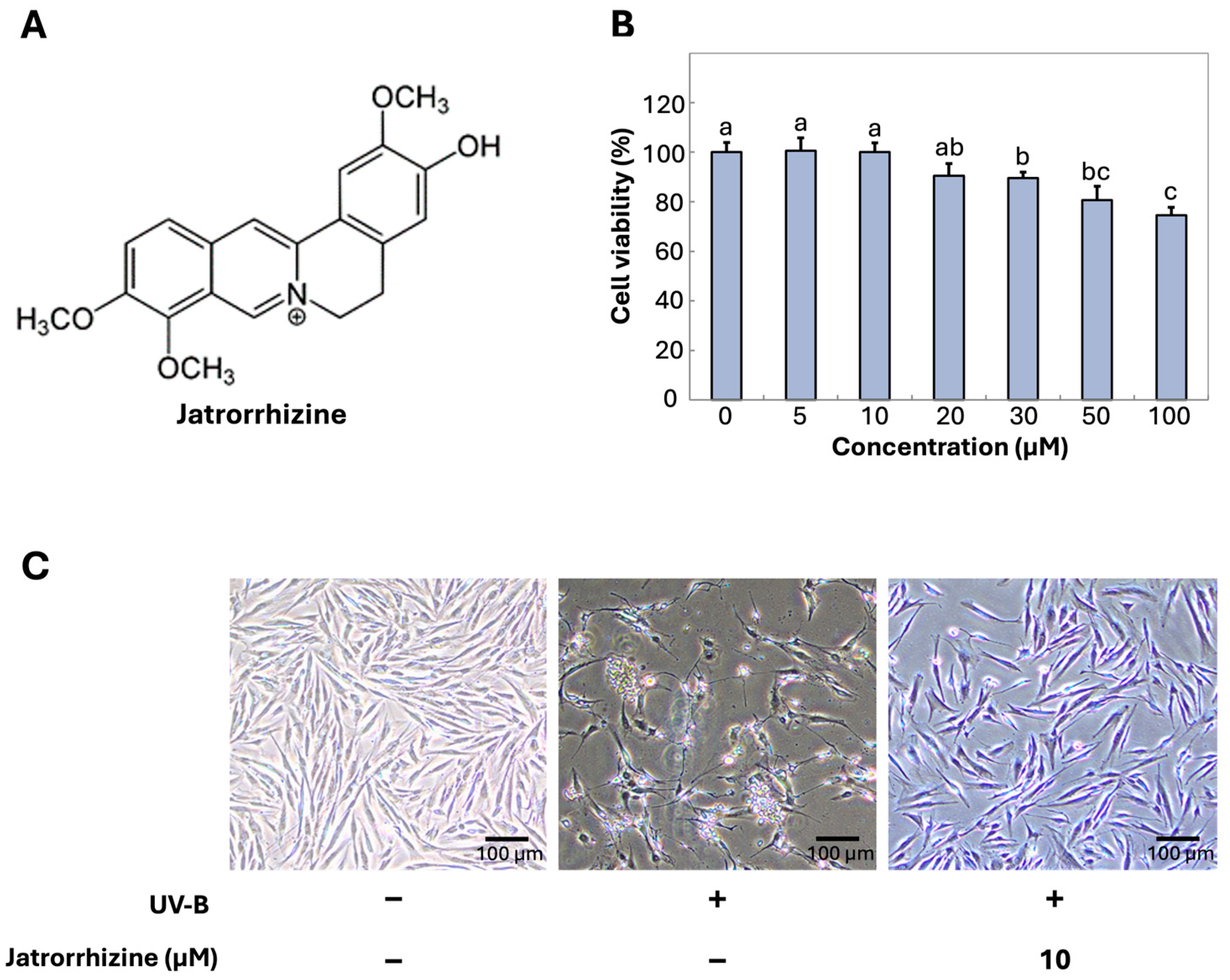
Figure 1. The effect of (A) jatrorrhizine on (B) cell viability in CCD-986sk human skin fibroblast cells and (C) recovery effect when irradiated with UV-B. (B) MTT assay was performed by seeding CCD-986sk cells in serum-free medium for 24 h and jatrorrhizine was treated at various concentration (5, 10, 20, 25, 50, 100 μM) for 48 h. (C) Representative images of UV-B irradiated CCD-986sk cells with jatrorrhizine treatment. Data presented in (B) are mean ± SD of three independent experiments (n = 9). Data with different letters are significant (p < 0.05).
Cosmetics 12 00070 g001
Figure 2. Jatrorrhizine recovered the expression of collagen synthesis-associated genes and inhibited collagen degradation-associated genes in UV-B irradiated CCD-986sk cells. Gene expression of (A) Collagen type I alpha 2 (COLIA2), (B) matrix metalloproteinases-1 (MMP-1), (C) matrix metalloproteinases-9 (MMP-9), and (D) TIMP metallopeptidase inhibitor 1 (TIMP-1). CCD-986sk cells were irradiated with UV-B at 312 nm UV-B (20 mJ/cm2) for 1 min and treated jatrorrhizine for 48 h. Data are presented mean ± SD (n = 3). Means with different letters are significant (p < 0.05).
Cosmetics 12 00070 g002
Figure 3. Jatrorrhizine recovered mRNA expression of (A) TGFB1 and (B) HAS2 in UV-B irradiated CCD-986sk cells. CCD-986sk cells were irradiated with UV-B at 312 nm UV-B (20 mJ/cm2) for 1 min and treated jatrorrhizine for 48 h. Data are presented mean ± SD (n = 3). Means with different letters are significant (p < 0.05).
Cosmetics 12 00070 g003
Figure 4. Suggested mechanisms of collagen homeostasis by jatrorrhizine. Jatrorrhizine targets COL1A2 to increase collagen synthesis. MMP-1 and MMP-9 are downregulated by TIMP-1 to inhibit collagen degradation.
Cosmetics 12 00070 g004
Table 1. The primer sequences and real-time PCR conditions for collagen homeostasis-associated genes.
GeneAccession No.PrimerSequence (5′-3′)Amplicon Size (bp)
COL1A2NM_000089.3ForwardAGAAACACGTCTGGCTAGGAG105
ReverseGCATGAAGGCAAGTTGGGTAG
MMP-1NM_002421.4ForwardTGGGAGGCAAGTTGAAAAGC135
ReverseCATCTGGGCTGCTTCATCAC
MMP-9NM_004994.3ForwardCCTGGGCAGATTCCAAACCT172
ReverseGTACACGCGAGTGAAGGTGA
TIMP-1NM_003254.3ForwardCTTCTGCAATTCCGACCTCGT79
ReverseACGCTGGTATAAGGTGGTCTG
TGFB1NM_000660.6ForwardCAATTCCTGGCGATACCTCAG86
ReverseGCACAACTCCGGTGACATCAA
HAS2NM_005328.3ForwardGAGGACGACTTTATGACCAAGAGC121
ReverseTAAGCAGCTGTGATTCCAAGGAGG
β-actinNM_007393.4ForwardCGTGCGTGACATCAAAGAGAA137
ReverseGCTCGTTGCCAATAGTGATGA
GeneReal-Time PCR Condition
COL1A295 °C for 5 min, followed by undergoing 40 cycles of 95 °C for 10 s, 60 °C for 20 s,
followed by each 95 °C, 60 °C, 95 °C for 15 s
MMP-1
MMP-9
TIMP-1
TGFB1
HAS2
ß-actin
Disclaimer/Publisher’s Note: The statements, opinions and data contained in all publications are solely those of the individual author(s) and contributor(s) and not of MDPI and/or the editor(s). MDPI and/or the editor(s) disclaim responsibility for any injury to people or property resulting from any ideas, methods, instructions or products referred to in the content.