Article

Exploring the Potential Anti-Inflammatory and Wound-Healing Proprieties of Cepaea hortensis Snail Mucin

1
Biosciences, Environment and Health Laboratory, Faculty of Sciences—Tetouan, University Abdelmalek Essaâdi, Tetouan 93000, Morocco
2
Central Pharmacy University Hospital Center Mohamed VI, Route de Rabat KM 17 BP 398, Geznaya, Tanger 90100, Morocco
3
Biotechnology and Biomolecules Engineering Research Team (BBERT), Faculty of Science and Technology—Tangier, University Abdelmalik Essaâdi, Tetouan 93000, Morocco
4
Laboratory of Life and Health Sciences, Faculty of Medicine and Pharmacy, Route de Rabat km 15 Gzenaya BP 365 Tanger, University Abdelmalek Essaâdi, Tetouan 9300, Morocco
5
Universidade Católica Portuguesa, CBQF—Centro de Biotecnologia e Química Fina—Laboratório Associado, Escola Superior de Biotecnologia, Rua Diogo Botelho 1327, 4169-005 Porto, Portugal
6
LEPABE—Laboratory for Process Engineering, Environment, Biotechnology and Energy, Faculty of Engineering, University of Porto (FEUP), Rua Dr. Roberto Frias, s/n, 4200-465 Porto, Portugal
7
AliCE—Associate Laboratory in Chemical Engineering, Faculty of Engineering, University of Porto (FEUP), Rua Dr. Roberto Frias, s/n, 4200-465 Porto, Portugal
*
Author to whom correspondence should be addressed.
Cosmetics 2023, 10(6), 170; https://doi.org/10.3390/cosmetics10060170
Submission received: 18 October 2023 / Revised: 4 December 2023 / Accepted: 7 December 2023 / Published: 14 December 2023

Abstract

:
Long before its popularization as a skincare ingredient, snail slime was reported to have a variety of health-promoting attributes. Its medicinal use involved the treatment of ailments, such as anthrax, hernias, stomach pain, chest pain, as well as tuberculosis. The main aim of this study was to investigate the anti-inflammatory and wound-healing properties of snail slime from Cepaea hortensis (Müller, 1774) (brown garden snail). The slime was extracted by a mild method, and the anti-inflammatory characteristics were determined by croton-oil-induced mouse-ear edema. The histological study showed that the snail slime exerts its action at the level of inflammatory mediators, explaining the drastic decrease of edema (59% in comparison to the 47% of indomethacin). The daily application of 50 µL (15 mg/kg) of snail slime on excision wounds in rabbits proved to induce an almost full tissue repair after 24 days of treatment (87.80–92.7% wound closure in width and length, respectively). Analysis of the slime’s safety aspect allowed affirming the nontoxicity of snail slime on both the skin and eyes.

1. Introduction

Inflammation is a complex physiological process involving multiple cellular and molecular mediators [1]. At the cellular level, macrophages and mast cells play a central role in initiating the inflammatory response through the secretion of proinflammatory cytokines such as TNFα, IL-1β and IL-6 [2]. These cytokines then activate other cell types such as T lymphocytes and stimulate the production of vasodilator mediators such as histamine and prostaglandins [3]. At the molecular level, the NF-κB and MAPK signaling pathways play a pivotal role in amplifying and regulating the inflammatory signal [4]. More recently, many research studies have evaluated the potential anti-inflammatory activity of natural products. Those biomolecules are now in the spotlight of many research groups. They aim to discover new avenues for the development of drugs against a wide range of diseases, particularly cancers and inflammatory diseases. Biomolecules are obtained from natural sources such as plants, microorganisms or animals (insects, invertebrates, etc.) [5].
Mollusks are renowned for their abundance of active ingredients, allowing them to act at multiple levels of the organism. Land snails are among the terrestrial mollusks exploited in pharmacology. These gastropods have already undergone biological screenings that have revealed effective therapeutic potential [6,7,8]. One of the most promising ingredients produced by snails is a mucoid structure widely known as snail mucus, identified as an adhesive lubricant that allows snails to adhere to various surfaces, maintain hydration of their body and protect themselves against mechanical damage and predators [9,10].
This product has been researched by various researchers due to its abundance of organic ingredients that confer multiple biological activities to snail meat, including anti-inflammatory, healing, antimicrobial, antioxidant, anti-tyrosinase, anti-tumor, anti-aging and regenerative properties [11,12,13,14,15,16]. These activities are due to the presence of several chemically identified compounds in snail slime, including proteins, glycosaminoglycans, glycoproteins, polyphenols, fatty acids, glycolic acid, vitamins, allantoin, minerals and carbohydrates [17], but several factors are involved in this composition, including the species studied and their different functional requirements, as well as environmental factors such as temperature, humidity, soil conditions and food supply [18,19].
Furthermore, this study was therefore conducted with the aim of improving and highlighting the value of snail secretions from the species Cepaea hortensis (Müller, 1774) (garden snail) cultivated in Morocco, by evaluating its toxicity and anti-inflammatory and healing activities through the application of various in vivo and in vitro techniques.
In addition, studies on the bioactivity of secretions from the garden snail Cepaea hortensis remain little documented. However, further characterization of its specific components is needed. The identification of new metabolites from C. hortensis could contribute to the discovery of further therapeutic prospects from sustainable local resources, and the molecular mechanism of action of this species deserves further study.

2. Materials and Methods

2.1. Snail Crude Material

The garden snail Cepaea hortensis (Müller, 1774) was chosen as the study model, and specimens were gathered during the spring months from various locations in Eastern Morocco. The animals were housed in plastic containers within a laboratory setting at a temperature of 25 ± 1 °C and provided with a diet consisting of lettuce, cucumber and carrot.
The slime extraction procedure involved gently crushing the snails with a spatula to minimize harm while eliciting the production of slime. The collected slime was then transferred to tubes and stored at 4 °C in a refrigerator.

2.2. Animals

2.2.1. Evaluation of Dermal and Ocular Toxicity and Analysis of Wound-Healing Activity

Between 30 and 40 Albino-strain rabbits with a body weight ranging from 900 to 1300 g were used. The animals were held in individual cages. The temperature of the experimental room was adjusted to 20 ± 3 °C for the rabbits, with a humidity level of around 50 to 60%. Artificial lighting was applied with an alternating 12 h of light and 12 h of darkness. The experimental models were provided with standard laboratory animal feed and drank drinking water ad libitum.

2.2.2. Evaluation of Anti-Inflammatory Activity

Albino mice weighing approximately 28 to 32 g were selected as the study models for this experiment. The animals were raised in the biology laboratory of the Faculty of Science and Technology of Tangier under standard lighting conditions (12 h of white light, 12 h of darkness) and temperature conditions (25 ± 1 °C). All experimental animals received standard food and tap water ad libitum as a part of their diet.

2.3. Dermal Toxicity Evaluation

A cutaneous toxicity assessment was conducted according to the method described in OCDE [20]. Briefly, the right flank of 6 young rabbits was shorn, and 3 parallel scarifications of the epidermis were made. Twenty-four hours later, a 0.5 mL dose of snail slime was applied to the wound area and later covered by a gauze pad and fastened with a nonirritating Band-Aid. The intact left flanks served as controls. The patch was removed after 4 h of exposure, and symptoms of erythema and edema were observed at various time points (immediately following removal, after 30 min and at 24, 48 and 72 h).

2.4. Ocular Toxicity Evaluation

The ocular toxicity evaluation was performed following the method provided in OCDE [21]. The test was carried out on 3 rabbits with indefective and intact eyes. A decimal milliliter of slime was applied directly into the conjunctival sac of the right eye of each rabbit. Successive clinical observations of the eyeballs were made after one hour, then one, two, three and four to seven days after instillation, with the noninstilled eye serving as a control model to assess the possible irritation of the treated eye.

2.5. Topical Anti-Inflammatory Activity Assessment

The evaluation of the anti-inflammatory activity of snail slime was performed using the croton-oil edema model of the mouse ear. Three batches of 10 mice were weighed and labeled. Cutaneous inflammation was induced through the application of 5 µL of the croton-oil solution to the inner surface of the auricle of the right ear, pre-anesthetized intraperitoneally with ketamine hydrochloride (150 mg/kg) to minimize pain and distress during the procedure. The left ear was not treated, which allowed for it to act as a control. The positive control batch received 250 µg of indomethacin, while the test batch received 5 µL of the test solution (250 µg/ear). The three batches were observed for 6 h [22] and then sacrificed by stretching the spinal cord at the cervical level. Using a button drill (diameter = 7 mm), the right and left ears were sectioned at the level of their implantation. A piece of the auricle was removed at the tip margin and immediately weighed. The inhibition rate of inflammation was calculated as follows (Equations (1) and (2)):
I n f l a m m a t i o n   i n h i b i t i o n   % = ( A . D A . D . T ) × 100 / A . D
I n h i b i t i o n   c a l c u l a t e d   f o r   e a c h   a n i m a l   % = ( A . D D . i ) × 100 / A . D
where
A.D = Average weight difference between the pieces of the right ears treated with croton oil and those of the untreated left ears of the positive control batch.
A.D.T = Average weight difference between the pieces of the right ears treated with the anti-inflammatory substance and those of the treated left ears.
D.i = Weight difference between the pieces of the right ear and those of the left ear of a chosen mouse.
The tests were performed from 10:00 am to 4:00 pm to avoid possible variations in responses resulting from circadian fluctuations in the animals’ corticosteroids.

2.6. Histological Study of the Ear Pieces

The sample was put in an embedding cassette and preserved in 10% formalin. It was then subjected to dehydration by an automaton containing 12 baths: 8 alcohol baths of gradually increasing concentration (from 70 to 100%) for 45 min; 2 xylene and hematoxylin baths for 30 min; and 2 melted paraffin baths for 1 h 30 min. The tissues were then embedded with paraffin, and fine slices made by a microtome were applied to glass slides. Afterwards, the sample was deparaffinized in an incubator overnight at 70 °C and dyed as follows: xylene for 30 min, ethanol for 10 min, water for 10 min, hematoxylin for 3 min to stain the nuclei and eosin for 1 min to stain the cytoplasm. The sample was later washed with alcohol and then put in xylene. The observation was performed under a light microscope at ×40, ×100 and ×400 magnification.

2.7. Evaluation of the In Vivo Healing Effect

The followed procedure is the one described by Pourrat [23]. Thirty albino rabbits were divided into 10 rabbits per batch and placed in individual cages as described below (Table 1).
A rectangle approximately 15 mm long and 5 mm wide was cut into the skin of the dorsolumbar region of rabbits previously anesthetized with ketamine hydrochloride (15 mg/kg). Each specimen was treated with a topical application of 50 µL (15 mg/kg of snail slime) once a day using an insulin syringe. The macroscopic observation was performed before each new application. A grading scale (4-point scale) was established to follow the evolution of the healing process. The dimensions (length and width) of the excision wounds were measured every 4 days during the 24-day trial period using a digital caliper [BAY tech, Panasonic communications Co., Ltd., Fukuoka, Japan] (accuracy 0.03 mm/0.001), and photographs were taken to document the changes. The percentage change in wound contraction was calculated by the following Equation (3):
C o n t r a c t i o n   % = ( H e a l e d   w o u n d   s u r f a c e I n i t i a l   s u r f a c e ) × 100

2.8. Statistical Analysis

All results are expressed as averages in order to simplify the presentation of data from the different experimental groups. Statistica v6.1 (Statsoft, Inc., Palo Alto, CA, USA) was used for statistical analysis [24,25].

3. Results and Discussion

3.1. Toxicological Tests

Irritation is an important element in dermopharmacy. It represents the effect that may arise after a single projection or a severe but short exposure. The results of the determination of the PDII (primary dermal irritation index) and the eye irritation index (Table 1 and Table 2) reflect the absence of vesicles, cracks or irritation and, therefore, allow us to conclude that snail slime is nonirritating and noncorrosive to the skin and eye.

3.2. Evaluation of the Anti-Inflammatory Activity of Snail Slime

After six hours of treatment, mice in the negative control group developed an edema in the right ear of 150 mg on average, thus indicating the development of edema. Meanwhile, mice in the group treated locally with 0.5 mg indomethacin showed a reduction in ear thickness, compared to those in the negative control group (79.75 mg). This corresponds to a 47% inflammation inhibition. The treatment of the mice with 2 mg of snail slime induced an even greater reduction in inflammation, compared to the mice of the negative control group: the thickness of the ear was estimated at 61.33 mg, which is the equivalent of roughly a 59% inhibition. This inhibition is moderately greater than that observed with indomethacin. Therefore, snail slime appears to have an anti-inflammatory effect that is superior to that of indomethacin. Table 3 and Figure 1 provide a comprehensive illustration of the results obtained from the experiment.
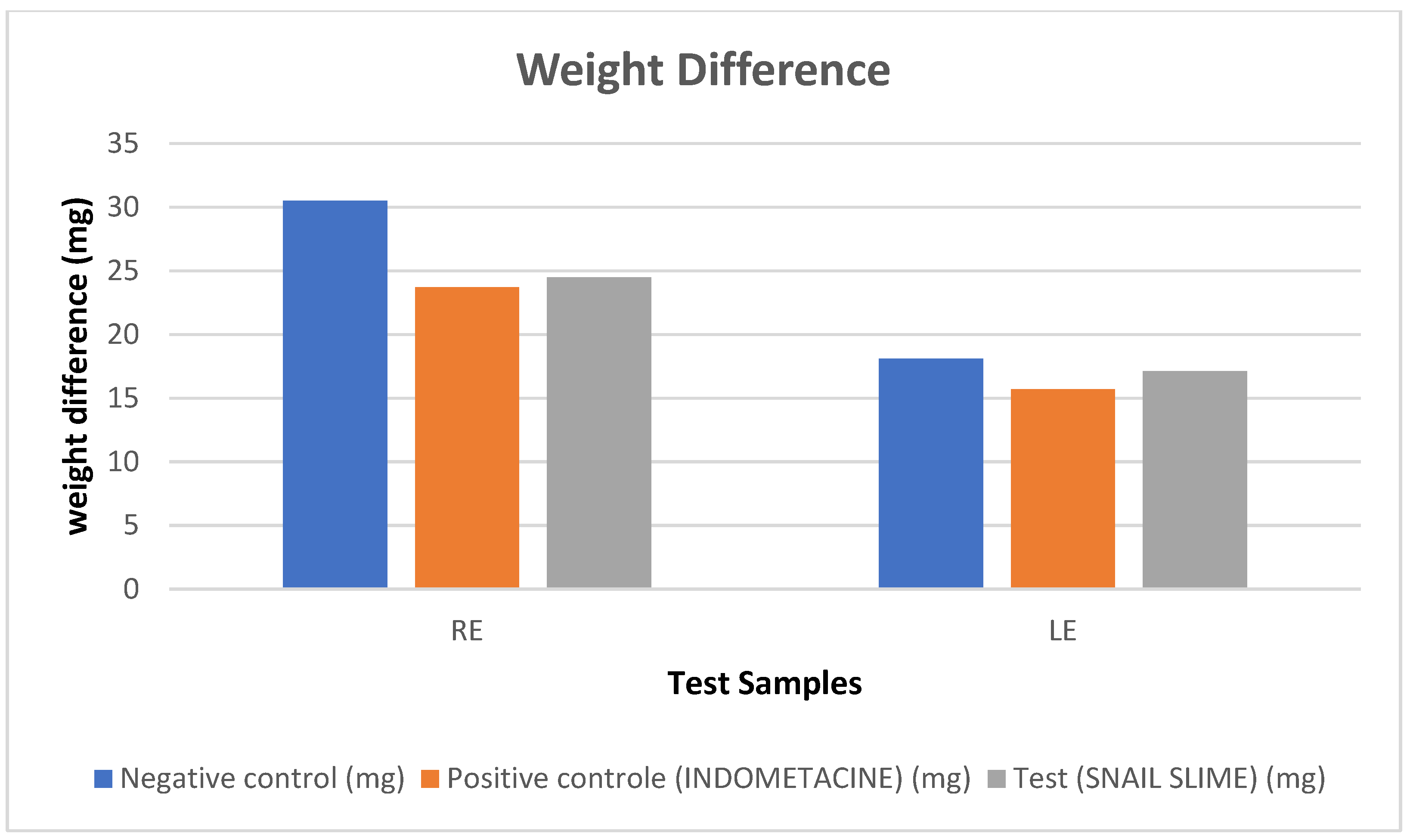
Figure 2 presents the evaluation of the anti-inflammatory effect on the weight of the left and right ears (LE and RE, respectively) of the three distinct groups of mice. The measurement of the right ear weight in this study revealed crucial information about the impact of the treatments on the mice’s ears. Batch 2 was treated with indomethacin as a positive control and had a right ear weight of 23.7 ± 0.21 mg. On the other hand, Batch 1, the negative control group (which was not treated), had a right ear weight of 30.5 ± 1.50 mg, significantly higher than that of Batch 2. Finally, Batch 3, which was treated with snail slime, had a right ear weight of 24.5 ± 2.12 mg, closer to the weight recorded for the positive control group (Batch 2). The left ear weight in the three batches of mice also showed a similar trend. The left ear weight in Batch 2 was recorded as 15.7 ± 0.48 mg, while that in Batch 1 was 18.1 ± 1.10 mg and, finally, 17.1 ± 2.28 mg in Batch 3. Although the negative control group in Batch 1 had a slightly higher weight compared to the other two batches, the differences were not as statistically different as those seen in the right ear.
The inflammatory response is an adaptive mechanism triggered in response to harmful stimuli, such as an infection or tissue aggression. It requires a fine regulation, generally beneficial, which leads to the eradication of possible pathogens and a return to the homeostasis of the injured tissue [26,27]. The main function of inflammation is to eliminate the aggressive agent and allow tissue recovery. To evaluate the effect of snail slime on acute inflammation, the croton-oil-induced ear-edema model was used in mice. This experimental model is widely used in order to screen for synthetic or natural anti-inflammatory substances with a local effect [28]. The photogenic action of croton oil is mainly due to the active ingredient 12-O-tetradecanoyl phorbol acetate (TPA), whose main mechanisms involve the induction of an inflammatory reaction characterized by a significant production of proinflammatory mediators and an increase in vascular permeability as well as edema [29]. The weight of the edema reaches its peak 6 h after applying croton oil [22].
According to the results depicted in Table 3, a 59% inflammation inhibition was recorded after treatment of the induced edema with snail slime, which is higher than the results obtained in the group treated with indomethacin (47%). These findings demonstrate the high anti-inflammatory effect of the tested slime samples. Based on our knowledge, this is the first study conducted on North Moroccan slime extracted from garden snails. However, previous authors have demonstrated the anti-inflammatory potential of slime extracted from a different type of garden snail, Helix aspersa, and the results revealed a significant reduction in the expression of cytokine levels and the complete inhibition of iNOS synthesis [30].
The anti-inflammatory effect was equally assessed in terms of weight differences between the three studied groups. The data presented in Figure 2 indicate that the treatment with snail slime may have a moderate impact on the weight of the mice’s ears, as the weight of both the left and right ear in Batch 3 is relatively close to that of the positive control group in Batch 2. However, it is noteworthy that the weight of the right ear in the negative control group (Batch 1) was significantly higher than that of the treated and positive control groups, which may reflect a different response to the experimental conditions in this batch. Further and deeper investigation is necessary to determine the exact cause of this discrepancy and to draw more definitive conclusions about the effects of snail slime on mice’s ear weight.
Considerable research has been conducted to demonstrate the potential of snail slime in reducing inflammation and its significance as a powerful barrier against skin damage. Analysis of the chemical constituents of snail-slime samples has been an integral part of studies conducted and has revealed the presence of several antioxidant enzymes such as superoxide dismutase (SOD), catalase (CAT) and glutathione peroxidase (GPX). Indeed, a study by Putranti et al. [31] proved that the presence of these enzymes neutralizes free radicals and can thus positively reduce UV-induced skin damage such as ulceration and dermal inflammation [31]. Additionally, other nonenzymatic antioxidants present in snail slime include glutathione (GSH) derivatives, whose sulfhydryl (-SH) group is extensively involved in peroxide reduction, making them efficient antioxidants. Snail slime is also characterized by an acidic combination, with the presence of acetic, citric and lactic acids, the latter being responsible for its antibacterial properties [32,33,34,35].
To our knowledge, the chemical composition of Cepaea hortensis has not been the subject of any prior research, nor has its therapeutic potential. Therefore, this study highlights the originality of the results obtained and provides an opportunity to further research to identify the exact chemical composition responsible for the demonstrated therapeutic activities. Moreover, this product is particularly interesting for future research due to its proven absence of toxicity or harmful effects on human health. In addition, the abundance of Cepaea hortensis in various parts of the world and the accessibility of its habitat offer an additional point of interest compared to other species localized in difficult-to-access areas; this accessibility suggests the conceptualization of a potentially more affordable and more reliable product.

3.3. Evaluation of Ear Tissue through Microscopic Examination

The results of the histological examination of ear tissue treated with snail slime provided support and confirmation for our initial hypothesis (Figure 3). The study revealed a minimal amount of vascular congestion and a resolution of edema, as well as a regular appearance of the epidermis and normal cartilage and subcutaneous tissue. Furthermore, a reduction in the number of inflammatory cells was more apparent in the batch treated with snail slime compared to the positive control group. This reduction was characterized by a decrease in the concentration of inflammatory cells around blood vessels, suggesting that our product has a significant impact on the mediators responsible for inflammation.
The findings from the histological study are particularly noteworthy because they provide insight into the mechanism of action of snail slime in reducing inflammation. The reduced presence of inflammatory cells around blood vessels highlights the potential of snail slime as an anti-inflammatory agent. These results support the idea that snail slime has a direct impact on the underlying causes of inflammation and not just the symptoms, which is a significant advancement in our understanding of its therapeutic potential. Moreover, the regular appearance of the epidermis and normal cartilage and subcutaneous tissue suggest that snail slime has a relatively low risk of adverse effects, making it a promising candidate for further research and development as a potential treatment option for various inflammatory conditions.

3.4. Evaluation of the In Vivo Healing Effect

Snail slime, being a natural substance, took time to act effectively, as opposed to what we witnessed in the positive control batch. Nevertheless, on the 24th day, the rate of healing reached a maximum level in the snail-slime group: 89.66 ± 2.60% in width and 87.76 ± 3.18% in length (Figure 4). The average snail-slime healing rate increased significantly, especially in the period between D4 and D24, reaching levels from 52.05 ± 4.00% to 89.66 ± 2.60% in length and 12.98 ± 10.10% to 87.76 ± 3.18% in width. On D4, the average width of the negative control group was significantly lower (negative value) than that of the snail-slime experiment. The same is plausible for the positive control group.
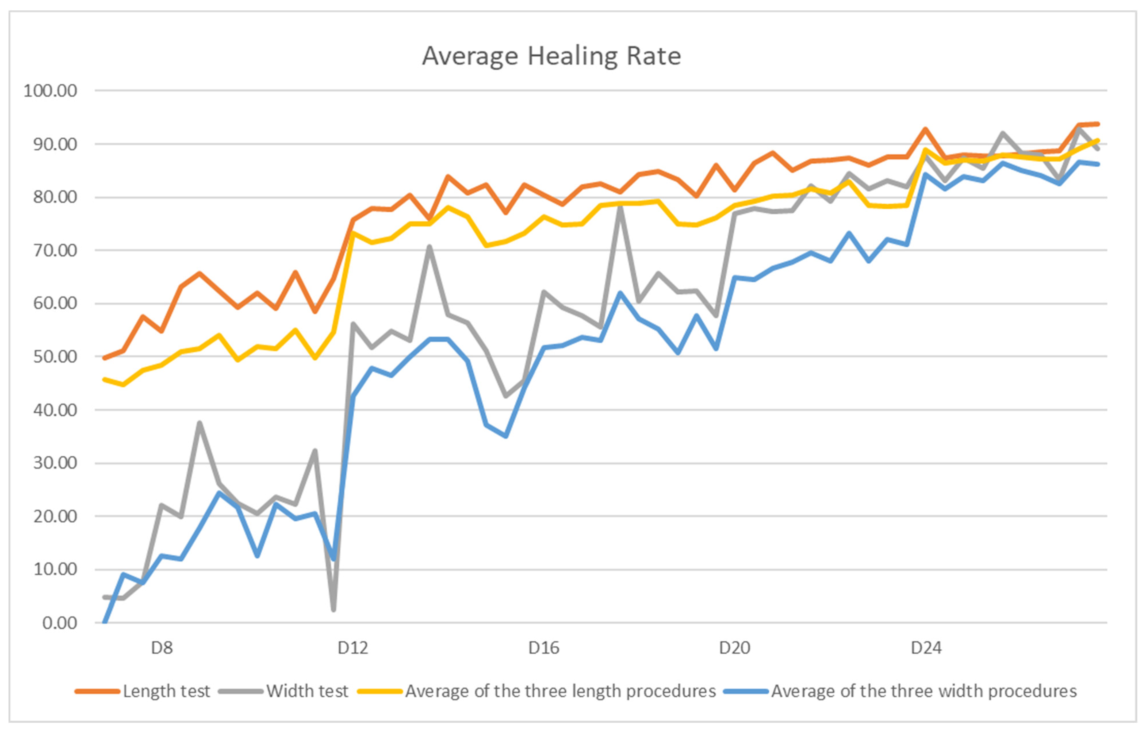
The results of the snail-slime treatment were thoroughly evaluated and compared with the other treatment groups, such as the positive and negative control groups (Figure 5). The findings showed that the snail-slime treatment had a healing rate that was very much in line with the average of the other treatments. In terms of length, the healing rate of the snail-slime batch was found to be optimal and exceeded expectations. Additionally, the width healing rate was well within the average range, making it a promising and effective alternative to other treatments.
Figure 6 shows the wound-healing process in the negative control (none treated), positive control (Madecassol) and test groups (snail slime). Rabbits in the negative control group showed impaired wound healing which persisted till day 8. On the other hand, the application of Madecassol and snail slime revealed a decrease in wound severity and an increase in size in the slime-treated group after 8 days. On day 12, the wounds treated with slime showed significant healing progress and a large decrease in size. The effect continued to persist and progress till the end of the treatment period, where no visible wound traces were detected on a macroscopic level.
Wound healing involves several physiological pathways such as hemostatic and inflammatory mechanisms, cell migration and proliferation, protein synthesis, wound contraction, as well as tissue remodeling or reconstitution [36]. The speed and quality of wound healing varies, depending on the general condition of the injured organism, the etiology of the wound, age and gender and the occurrence or absence of infection and genetic factors that may or may not predispose to wound-healing disorders [37]. In this study, the efficacy of snail slime in wound healing was evaluated, and the results showed an effectiveness in promoting wound healing in mice without adverse effects on their health. On the 24th day of treatment, the rate of healing reached a maximum level in the snail-slime group, and similar results were reported in the literature [38]. Moreover, the lack of adverse effects and normal weight gain in the mice during the treatment period is a promising indication of the safety and effectiveness of snail slime. Therefore, this study suggests that snail slime extracted from Cepaea hortensis (Müller, 1774) has potential as a natural alternative in wound-healing therapy.

4. Conclusions

The results of this study revealed that snail mucus has a therapeutic effect in reducing inflammation and promoting wound healing. Moreover, the toxicity tests revealed no signs of skin or eye irritation. The obtained results are in line with the literature data showing the antioxidant, antibacterial and pro-healing effects reported for other snail mucus.
Based on these results, snail mucus could be a promising candidate for medicinal exploitation as a cream or gel to harness the therapeutic benefits of snail mucus safely and effectively after appropriate clinal studies.
Further research is needed to elucidate the underlying mechanisms of action and assess longer-term toxicity. Identification of the active metabolites would enable the development of standardized formulations. This would contribute to a better understanding of the medical potential of snail slime and its use as a natural therapeutic agent.

Author Contributions

Conceptualization, A.L. (Adnane Louajri), A.L. (Amin Laglaoui) and F.E.; methodology, A.L. (Adnane Louajri), A.L. (Amin Laglaoui) and F.E.; validation, J.M.R., N.E.A. and A.L. (Adnane Louajri); formal analysis, A.L. (Adnane Louajri); investigation, F.E.; resources, A.L. (Amin Laglaoui) and A.L. (Adnane Louajri); data curation, F.E., N.E.A. and J.M.R. writing—original draft preparation, F.E., H.B. and S.H.; writing—review and editing, F.E., S.H.; N.E.A. and J.M.R.; visualization, H.B. and N.E.A. supervision, A.L. (Adnane Louajri), N.E.A. and J.M.R.; project administration, A.L. (Adnane Louajri); funding acquisition, A.L. (Amin Laglaoui), A.L. (Adnane Louajri), F.E., N.E.A. and J.M.R. All authors have read and agreed to the published version of the manuscript.

Funding

This research received no external funding.

Institutional Review Board Statement

The study was conducted in accordance with the Declaration of Helsinki, and approved by the Moroccan Ethic Committee for Animal Research (MECAR) UAE-FMP-01/2022, Approval Period: 20 January 2022 to 20 January 2025.

Informed Consent Statement

Not applicable.

Data Availability Statement

The data presented in this study are available on request from the corresponding author. The data are not publicly available due to the preparation of another paper.

Acknowledgments

The authors would like to thank COST Action 18101 SOURDOMICS—Sourdough biotechnology network towards novel, healthier and sustainable food and bioprocesses (https://sourdomics.com/; https://www.cost.eu/actions/CA18101/, accessed on 4 August 2023), where the author N.E.A. is a member of the working groups 3, 4 and 7, and the author J.M.R. is the Chair and Grant Holder Scientific Representative and is supported by COST (European Cooperation in Science and Technology) (https://www.cost.eu/, accessed on 4 August 2023). COST is a funding agency for research and innovation networks.

Conflicts of Interest

The authors declare no conflict of interest.

References

  1. Libby, P. Inflammatory Mechanisms: The Molecular Basis of Inflammation and Disease. Nutr. Rev. 2007, 65, 140–146. [Google Scholar] [CrossRef]
  2. Abdulkhaleq, L.; Assi, M.; Abdullah, R.; Zamri-Saad, M.; Taufiq-Yap, Y.; Hezmee, M. The crucial roles of inflammatory mediators in inflammation: A review. Vet. World 2018, 11, 627–635. [Google Scholar] [CrossRef] [PubMed]
  3. Mastrangelo, F.; Ronconi, G.; Kritas, S.; Conti, P. Stimulated mast cells release inflammatory cytokines: Potential suppression and therapeutical aspects. J. Biol. Regul. Homeost. Agents 2018, 32, 1355–1360. [Google Scholar]
  4. Kaminska, B. MAPK signalling pathways as molecular targets for anti-inflammatory therapy—From molecular mechanisms to therapeutic benefits. Biochim. Biophys. Acta 2005, 1754, 253–262. [Google Scholar] [CrossRef] [PubMed]
  5. Sheela, M.K.; Sasikala, M.S.; Padmini, M.R.; Sivaranjani, M.D.; Shobana, M.M. Biomolecules; Associationofindian Biologists Publications: New Delhi, India, 2021; ISBN 978-81-950231-2-7. [Google Scholar]
  6. Bonnemain, B. Helix and Drugs: Snails for Western Health Care From Antiquity to the Present. Evid.-Based Complement. Altern. Med. 2005, 2, 25–28. [Google Scholar] [CrossRef] [PubMed]
  7. Ghosh, A.K.; Hirasawa, N.; Lee, Y.S.; Kim, Y.S.; Shin, K.H.; Ryu, N.; Ohuchi, K. Inhibition by acharan sulphate of angiogenesis in experimental inflammation models. Br. J. Pharmacol. 2002, 137, 441–448. [Google Scholar] [CrossRef] [PubMed]
  8. Kim, H.S.; Lee, Y.H.; Lee, Y.R.; Im, S.-A.; Lee, J.K.; Kim, Y.S.; Sim, J.S.; Choi, H.S.; Lee, C.K. Activation of professional antigen presenting cells by acharan sulfate isolated from giant african snail, achatina fulica. Arch. Pharmacal. Res. 2007, 30, 866–870. [Google Scholar] [CrossRef]
  9. Dhiman, V.; Pant, D. Human health and snails. J. Immunoass. Immunochem. 2021, 42, 211–235. [Google Scholar] [CrossRef]
  10. Gabriel, U.I.; Mirela, S.; Ionel, J. Quantification of mucoproteins (glycoproteins) from snails mucus, Helix aspersa and Helix Pomatia. J. Agroaliment. Process. Technol. 2011, 17, 410–413. [Google Scholar]
  11. Gubitosa, J.; Rizzi, V.; Fini, P.; Fanelli, F.; Sibillano, T.; Corriero, N.; Cosma, P. Chitosan/snail slime films as multifunctional platforms for potential biomedical and cosmetic applications: Physical and chemical characterization. J. Mater. Chem. B 2023, 11, 2638–2649. [Google Scholar] [CrossRef]
  12. Truchuelo, M.T.; Vitale, M. A cosmetic treatment based on the secretion of Cryptomphalus aspersa 40% improves the clinical results after the use of nonablative fractional laser in skin aging. J. Cosmet. Dermatol. 2020, 19, 622–628. [Google Scholar] [CrossRef]
  13. Atta, S.A.; Ibrahim, A.M.; Megahed, F.A.K. In-Vitro Anticancer and Antioxidant Activities of Eremina desertorum (Forsskal, 1775) Snail Mucin. Asian Pac. J. Cancer Prev. APJCP 2021, 22, 3467–3474. [Google Scholar] [CrossRef]
  14. Ho, C.-Y.; Hu, D.-W.; Chen, B.-R.; Yang, C.-C.; Yao, C.-H.; Ni, T.; Ho, W.-C.; Chen, Y.-S.; Tu, C.-Y.; Chang, W.-C.; et al. Snail Mucus Enhances Chemosensitivity of Triple-negative Breast Cancer Via Activation of the Fas Pathway. Anticancer Res. 2022, 42, 845–855. [Google Scholar] [CrossRef]
  15. Ricci, A.; Gallorini, M.; Feghali, N.; Sampò, S.; Cataldi, A.; Zara, S. Snail Slime Extracted by a Cruelty Free Method Preserves Viability and Controls Inflammation Occurrence: A Focus on Fibroblasts. Molecules 2023, 28, 1222. [Google Scholar] [CrossRef]
  16. Rizzi, V.; Gubitosa, J.; Fini, P.; Nuzzo, S.; Agostiano, A.; Cosma, P. Snail slime-based gold nanoparticles: An interesting potential ingredient in cosmetics as an antioxidant, sunscreen, and tyrosinase inhibitor. J. Photochem. Photobiol. B Biol. 2021, 224, 112309. [Google Scholar] [CrossRef]
  17. Di Filippo, M.F.; Di Matteo, V.; Dolci, L.S.; Albertini, B.; Ballarin, B.; Cassani, M.C.; Passerini, N.; Gentilomi, G.A.; Bonvicini, F.; Panzavolta, S. Effectiveness of Snail Slime in the Green Synthesis of Silver Nanoparticles. Nanomaterials 2022, 12, 3447. [Google Scholar] [CrossRef]
  18. Noothuan, N.; Apitanyasai, K.; Panha, S.; Tassanakajon, A. Snail mucus from the mantle and foot of two land snails, Lissachatina fulica and Hemiplecta distincta, exhibits different protein profile and biological activity. BMC Res. Notes 2021, 14, 138. [Google Scholar] [CrossRef]
  19. El-Zawawy, N.A.; Mona, M.M. Antimicrobial efficacy of Egyptian Eremina desertorum and Helix aspersa snail mucus with a novel approach to their anti-inflammatory and wound healing potencies. Sci. Rep. 2021, 11, 24317. [Google Scholar] [CrossRef]
  20. OCDE. Essai n° 404: Effet Irritant/Corrosif Aigu sur la Peau; OCDE: Paris, France, 2015. [Google Scholar]
  21. OCDE. Essai n° 405: Effet Irritant/Corrosif Aigu sur les Yeux; OCDE: Paris, France, 2023. [Google Scholar]
  22. Tubaro, A.; Dri, P.; Delbello, G.; Zilli, C.; Della Loggia, R. The Croton oil ear test revisited. Agents Actions 1986, 17, 347–349. [Google Scholar] [CrossRef]
  23. Arvouet-Grand, A.; Lejeune, B.; Bastide, P.; Pourrat, A.; Privat, A.M.; Legret, P. Étude de la cicatrisation des plaies chez le lapin et le rat. J. PharmBelg. 1993, 48, 171–178. [Google Scholar]
  24. De Sá, J.P.M. Applied Statistics Using SPSS, Statistica, MatLab and R; Springer Science & Business Media: Berlin/Heidelberg, Germany, 2007; SPIN: 11908944. [Google Scholar]
  25. Hourfane, S.; Mechqoq, H.; Errajouani, F.; Rocha, J.M.; El Aouad, N. In Vitro and In Silico Evaluations of Boswellia carterii Resin Dermocosmetic Activities. Cosmetics 2022, 9, 131. [Google Scholar] [CrossRef]
  26. Nathan, C. Points of control in inflammation. Nature 2002, 420, 846–852. [Google Scholar] [CrossRef]
  27. Barton, G.M. A calculated response: Control of inflammation by the innate immune system. J. Clin. Investig. 2008, 118, 413–420. [Google Scholar] [CrossRef]
  28. Altinier, G.; Sosa, S.; Aquino, R.P.; Mencherini, T.; Della Loggia, R.; Tubaro, A. Characterization of Topical Antiinflammatory Compounds in Rosmarinus officinalis L. J. Agric. Food Chem. 2007, 55, 1718–1723. [Google Scholar] [CrossRef]
  29. Delaporte, R.; Sarragiotto, M.; Takemura, O.; Sanchez, G.; Nakamura, C. Evaluation of the antioedematogenic, free radical scavenging and antimicrobial activities of aerial parts of Tillandsia streptocarpa Baker–Bromeliaceae. J. Ethnopharmacol. 2004, 95, 229–233. [Google Scholar] [CrossRef]
  30. Gubitosa, J.; Rizzi, V.; Fini, P.; Laurenzana, A.; Fibbi, G.; Veiga-Villauriz, C.; Fanelli, F.; Fracassi, F.; Onzo, A.; Bianco, G.; et al. Biomolecules from snail mucus (Helix aspersa) conjugated gold nanoparticles, exhibiting potential wound healing and anti-inflammatory activity. Soft Matter 2020, 16, 10876–10888. [Google Scholar] [CrossRef]
  31. Putranti, I.O.; Sutrisna, E.; Ekowati, N.; Mawardi, P.; Novrial, D. The Role of Achatina Fulica Snail Slime Extract Enzymatic Antioxidants as Photoprotector in Sunburn Model Mice. Int. J. Med. Sci. Clin. Res. Stud. 2022, 02, 624–630. [Google Scholar] [CrossRef]
  32. Anju, M.; Vijayan, K. Comparison of antimicrobial properties of the slime of invasive alien pest Lissachatina fulica and an indigenous pest Cryptozona bistrialis snails. Crop Res. 2021, 56, 159–164. [Google Scholar]
  33. Cilia, G.; Fratini, F. Antimicrobial properties of terrestrial snail and slug mucus. J. Complement. Integr. Med. 2018, 15, 20170168. [Google Scholar] [CrossRef]
  34. Perpelek, M.; Tamburaci, S.; Aydemir, S.; Tihminlioglu, F.; Baykara, B.; Karakasli, A.; Havitcioglu, H. Bioactive snail mucus-slime extract loaded chitosan scaffolds for hard tissue regeneration: The effect of mucoadhesive and antibacterial extracts on physical characteristics and bioactivity of chitosan matrix. Biomed. Mater. 2021, 16, 065008. [Google Scholar] [CrossRef]
  35. Swastini, I.G.A.A.P.; Widnyana, A.N.K.P. Antimicrobial properties of snail slime (Achatina fulica) on the growth of actinobacilus actinomycetemcomitans bacteria causing periodontitis. J. Glob. Pharma Technol. 2009, 11, 170–174. [Google Scholar]
  36. Monaco, J.L.; Lawrence, W.T. Acute wound healing. Clin. Plast. Surg. 2003, 30, 1–12. [Google Scholar] [CrossRef]
  37. Guo, S.; DiPietro, L.A. Factors Affecting Wound Healing. J. Dent. Res. 2010, 89, 219–229. [Google Scholar] [CrossRef]
  38. Song, Y.; Cui, Y.; Hao, L.; Zhu, J.; Yi, J.; Kang, Q.; Huang, J.; Lu, J. Wound-healing activity of glycoproteins from white jade snail (Achatina fulica) on experimentally burned mice. Int. J. Biol. Macromol. 2021, 175, 313–321. [Google Scholar] [CrossRef]
Figure 1. Percentage of anti-inflammatory inhibition in treated ear edemas with snail slime and indomethacin as the positive control and nontreated mice as the negative control.
Cosmetics 10 00170 g001
Figure 2. Weight differences between right and left ear pieces.
Cosmetics 10 00170 g002
Figure 3. Results of the histological study in all three batches under a microscope. The observations were done under a light microscope at ×40, ×100 and ×400 magnification, and these images were obtained at a GX400 magnification.
Cosmetics 10 00170 g003
Figure 4. Wound-healing rates for all three atches.
Cosmetics 10 00170 g004
Figure 5. Comparison of the average healing rate of the three tests and the healing rate of snail slime.
Cosmetics 10 00170 g005
Figure 6. The visual representation of the healing process in the three groups of rabbits.
Cosmetics 10 00170 g006
Table 1. Primary dermal irritation index.
Rabbit n°TimeErythema and Eschar (0: Absence)Edema (0: Absence)
Scarred AreaUnscarred AreaScarred AreaUnscarred Area
130 min0000
24 h0000
72 h0000
230 min0000
24 h0000
72 h0000
330 min0000
24 h0000
72 h0000
430 min0000
24 h0000
72 h0000
530 min0000
24 h0000
72 h0000
630 min0000
24 h0000
72 h0000
Table 2. Determination of eye irritation indices.
Evaluation of Damage1 h24 h48 h
Rabbit Number123123123
ConjunctivaA: chemosis000000000
B: lachrymation000000000
C: enanthem000000000
(a + b + c) × 2000000000
IrisDirect photomotor reflex of the pupilNNNNNNNNN
A: congestion degree000000000
A × 5000000000
CorneaA: degree of opacity000000000
B: area reached000000000
A × b × 5000000000
Ulceration000000000
Area reached000000000
Granulation000000000
Individual Eye irritation Index (I.E.I)000000000
Score per zoneConjunctiva Iris
Cornea
0
0
0
Conjunctiva Iris
Cornea
0
0
0
Conjunctiva Iris
Cornea
0
0
0
Average Eye Index (A.E.I)000
Table 3. Percentage inhibition of edema in treated animals.
Administered Dose (µL in Ear)Number of AnimalsAverage Weight Difference (mg)Edema
Inhibition %
Negative control batch (µL)5101500%
Positive control (indomethacin) (µg)501079.7547%
Test batch (snail slime) (mg)151061.3359%
Disclaimer/Publisher’s Note: The statements, opinions and data contained in all publications are solely those of the individual author(s) and contributor(s) and not of MDPI and/or the editor(s). MDPI and/or the editor(s) disclaim responsibility for any injury to people or property resulting from any ideas, methods, instructions or products referred to in the content.