Article

Citrulluside T, Isolated from the Citrullus lanatus Stem, Inhibits Melanogenesis in α-MSH-Induced Mouse B16F10 Cells

Department of Chemistry and Cosmetics, Jeju Inside Agency and Cosmetic Science Center, Jeju National University, Jeju 63243, Republic of Korea
*
Author to whom correspondence should be addressed.
These authors contributed equally to this work.
Cosmetics 2023, 10(4), 108; https://doi.org/10.3390/cosmetics10040108
Submission received: 23 June 2023 / Revised: 16 July 2023 / Accepted: 16 July 2023 / Published: 21 July 2023
(This article belongs to the Special Issue Feature Papers in Cosmetics in 2023)

Abstract

:
With the increasing number of cosmetic consumers emphasizing value consumption and sustainability, upcycling has gained attention as a solution to agricultural by-products, which are the main culprits of environmental problems. In this study, we isolated citrulluside T with whitening activity from discarded Citrullus lanatus stems and investigated the anti-melanogenic effect of citrulluside T and the underlying mechanisms. We found that citrulluside T did not exhibit cytotoxicity up to a concentration of 90 μM and significantly reduced the melanin content and intracellular tyrosinase activity in B16F10 cells. In addition, citrulluside T inhibited the expression of melanogenic enzymes such as tyrosinase, tyrosinase-related protein (TRP)-1, and TRP-2, as well as melanin synthesis via cAMP-dependent protein kinase (PKA)/cAMP response element-binding protein (CREB)-mediated downregulation of microphthalmia-associated transcription factor (MITF), a key transcription factor in melanogenesis. Furthermore, we found that citrulluside T exerted its anti-melanogenic effect by downregulating the β-catenin protein and upregulating phosphorylated β-catenin. Finally, we confirmed that citrulluside T was safe for skin through skin irritation tests on 33 subjects, suggesting its applicability as a protective agent against hyperpigmentation for topical applications such as cosmetics and ointments.

1. Introduction

As environmental protection and sustainability have emerged as important topics around the world, food consumption is shifting to value consumption, and research on the value utilization of waste that enables upcycling of by-products is actively underway. Upcycling is a combination of upgrading and recycling and is a concept that creates high added value by applying new technologies to resources with a low product value such as by-products and waste [1,2]. In the field of cosmetics, good cosmetics that upcycle agricultural by-products are also attracting attention, and upcycling of coffee grounds is a representative example. Coffee is one of the most consumed beverages in the world, and it has been reported that coffee by-products and coffee beans are rich in bioactive compounds with great commercial value, such as those that can be utilized as active ingredients in skincare products and cosmetic formulations [3,4,5].
In addition, lilies, a favorite of the Japanese people, are mainly used for ornamental purposes, but by dissecting the roots, they can be used for medicinal purposes and thus have the advantage of adding additional value by utilizing the roots. As a traditional Korean medicine, lilies have long been used to treat lily disease, a mentally debilitating condition, as the root has the ability to stop coughing and stabilize the mind, and it is especially known to be effective in treating chronic obstructive pulmonary disease, the main symptoms of which are dry cough and shortness of breath [6,7,8]. Studies have also been reported on the whitening effect of lily root and its active ingredients [9].
The most important agricultural by-product is that of citrus fruits. The juice industry processes millions of tons of citrus per year but only utilizes the pulp, while the peel, seeds, and citrus press cake are mostly discarded. Phytochemical investigations have shown that citrus peels are an excellent source of bioactive compounds such as phenolic compounds, carotenoids, and monoterpenes, and researchers around the world have developed innovative technologies to recover phytochemicals from citrus waste. This study is about upcycling using stems discarded in the process of cultivating watermelons and supplying them to consumers [10,11,12].
Citrullus lanatus is a dicotyledonous plant in the Cucurbitaceae (cucumber) family and there are over 1000 watermelon cultivars. The vines can be up to 10 feet long, and the fruit is usually red with seeds but can also be pink or yellow, and there are seedless varieties. Citrullus lanatus contains citrulline, which has a diuretic property, and is known to be effective in sodium excretion due to its high potassium content [13,14,15]. In addition, several studies have reported antibacterial, whitening, and anti-inflammatory activity in Citrullus lanatus pulp, envelope, and vines. Citrullus lanatus stems are agricultural by-products that are not utilized for food or industrial purposes, being of great interest in terms of the utilization of unutilized agricultural resources [16,17,18]. Therefore, in this study, we manufactured polarized fractions of Citrullus lanatus stems to separate citrulluside T through column chromatography and used 1H and 13C NMR data analysis and comparison of literature values to confirm its applicability as a melanogenic inhibitor.
Melanogenesis starts with an initial reaction through tyrosinase in melanocytes, where tyrosine is converted to 3,4-dihydroxyphenylalanin (DOPA), which is then continuously converted to DOPAquinone and DOPA-chrome by DOPA oxidase enzymes. In this process, melanogenic enzymes such as tyrosinase, tyrosinase related protein-1 (TRP-1), and dopachrome tautomerase (TRP-2) are involved in melanin synthesis by catalyzing successive oxidation reactions. It is also influenced by several hormones, most notably α-melanocyte-stimulating hormone (α-MSH) secreted by the pituitary gland, which increases the expression of microphthalmia-associated transcription factor (MITF), a transcription factor that regulates melanin formation, which in turn activates various genes involved in cellular responses such as melanin production, cell proliferation, and migration. Ultimately, melanin biosynthesis is a complex process that is regulated by tyrosinase, TRP-1, TRP-2, and MITF. Therefore, it is important to demonstrate the inhibition of melanogenic enzymes, including tyrosinase, in order to verify the whitening effect [19,20,21,22].
Currently, arbutin, ascorbic acid, hydroquinone, kojic acid, azelaic acid, and linoleic acid are the most commonly known representative whitening agents that inhibit the activity of tyrosinase, but due to the emergence of human side effects caused by problems such as skin irritation, cytotoxicity, and formulation stability, the search for natural materials that can minimize these side effects and effectively inhibit factors involved in melanogenesis has been actively conducted [23,24,25]. In this study, we aimed to investigate the inhibitory effect of citrulluside T isolated from Citrullus lanatus stems on α-MSH-induced melanogenesis in mouse B16F10 melanoma cells and its mechanism to confirm its potential as a new natural whitening ingredient.

2. Materials and Methods

2.1. General Experimental Procedures

Citrullus lanatus stems from Jeju Island were washed, dried, and shredded for easy extraction. Solvents used for the extraction, fractionation, and separation of single substances in the experiments were of analytical grade. Column chromatography was performed using silica gel 60 (mesh size 0.040–0.063 mm) and Sephadex LH-20 (particle size 18–111 µm, dry) for the isolation of citrulluside T, which were purchased from Merck (Darmstadt, Germany) and Cytiva (Uppsala, Sweden), respectively. Analytical HPLC for analysis was performed on a high-performance liquid chromatography system equipped with a Waters Alliance 2489 separation module with a Waters 2695 UV/visible light detector, a quaternary pump, a column temperature control module, and a Waters 717 plus autosampler (Milford, MA, USA). Data acquisition was performed using an Empower pro data processing system (Waters Co., Milford, CT, USA). The elution conditions are shown in Table 1. The nuclear magnetic resonance (NMR) systems used for structure identification were JNM-LA 400 (FT-NMR system, JEOL Co., Tokyo, Japan) and JNM-ECX 400 (FT-NMR system, JEOL Co., Tokyo, Japan), and the NMR measurement solvent was CD3OD, a dedicated NMR solvent from Cambridge Isotope Laboratories, Inc (Tewksbury, MA, USA).

2.2. Extraction and Isolation from the Citrullus lanatus Stem

The Citrullus lanatus stem was crushed to a powder using a blender and extracted at room temperature with the addition of 70% ethanol. The extract was filtered and concentrated to give an extract (14.9 g), which was suspended in distilled water and fractionated sequentially with hexane, chloroform, ethyl acetate, and butanol. An amount of 0.4016 g of the ethyl acetate fraction was subjected to column chromatography using chloroform and methanol on a column packed with normal silica gel. The eluate obtained was subjected to compound isolation using 100% methanol on a column packed with Sephadex LH-20 to increase the purity (Figure 1). The isolated compounds were identified via NMR analysis, referring to the literature [26,27].

2.3. Cell Culture

Mouse B16F10 melanoma cells were obtained from the American Type Culture Collection (ATCC, Manassas, VA, USA) and cultured under the conditions of 37 °C and 5% CO2 with DMEM containing 1% penicillin/streptomycin and 10% FBS. The culture medium was changed every 3 days, and cells were subcultured when the number of cells grew above 80%.

2.4. Cell Viability

To investigate the effect of citrulluside T on the viability of B16F10 cells, cytotoxicity was evaluated using the MTT assay. B16F10 cells (1.5 × 10⁴ cells/mL) were seeded in 24-well plates, treated with various concentrations of citrulluside T after 24 h of preincubation, and cultured in a 37 °C, 10% CO2 cell incubator for 3 days. B16F10 cells were seeded in 24-well plates at 1.5 × 10⁴ cells/mL and incubated for 24 h; then, they were treated with citrulluside T at different concentrations from 5.63 µM to 360 µM and incubated for 72 h before the MTT assay. MTT solution (0.5 mg/mL) was added to the wells and reacted for 4 h, followed by the addition of 1 mL of DMSO to completely dissolve it, and the absorbance was measured at 540 nm. Cytotoxicity was measured by comparing the absorbance values with those of the untreated samples.

2.5. Tyrosinase Activity

B16F10 cells were seeded at 8.0 × 10⁴ cells/well in 60 mm culture plates and cultured for 24 h. Then, α-MSH (100 nM) and citrulluside T were treated simultaneously at concentrations from 22.5 μM to 90 μM and cultured for 3 days. At this time, arbutin at a concentration of 200 μM was used as a positive control. After incubation, the medium was removed, washed twice with cold 1× PBS, and lysed using RIPA buffer supplemented with protease inhibitor cocktail as the lysis buffer at 4 °C for 20 min. The supernatant was then separated via centrifugation under the following conditions: 4 °C, 1500 rpm, 20 min. The supernatant was quantified with the same amount of protein (20 μg/mL) using the BCA Quantification Kit and treated with 80 μL of 2 mg/mL L-DOPA for 1 h at 37 °C. The tyrosinase activity was then measured at 490 nm absorbance using a spectrophotometric microplate reader.

2.6. Melanin Contents

B16F10 cells were seeded in 6-well plates at 6.0 × 10⁴ cells/well and cultured for 24 h; then, they were treated with α-MSH (100 nM) and citrulluside T simultaneously at concentrations from 22.5 μM to 90 μM and incubated for 3 days. At this time, arbutin at a concentration of 200 μM was used as a positive control. After incubation, the medium was removed, washed twice with cold 1× PBS, and dissolved in 1N NaOH containing 10% DMSO at 70 °C for 20 min to measure the melanin content at 405 nm absorbance.

2.7. Western Blot

Citrulluside T and α-MSH (100 nM) were treated at different concentrations and incubated according to the respective protein expression time. After incubation, the supernatant was removed, washed twice with cold 1× PBS, and lysed at 4 °C for 20 min using RIPA buffer (in 1% protease inhibitor cocktail) as the lysis buffer. The supernatant was then separated via centrifugation under the following conditions: 4 °C, 1500 rpm, 20 min. The supernatant was quantified with the same amount of protein (30 μg/mL) using the BCA Quantification Kit, and a loading sample was prepared. The loading sample was loaded on a 10% SDS-polyacrylamide gel and transferred to a PVDF membrane. To prevent non-specific binding, the membrane was blocked with 5% skim milk (in TBS-T) for 1 h and washed. The primary antibody (1:2000) diluted in TBS-T (in Tris-buffered saline with 1% Tween 20) was shaken overnight at 4 °C. Then, after washing 6 times with TBS-T, the secondary antibody (1:1000) was reacted for 2 h at room temperature. After the reaction, the membrane was washed 6 times with TBS-T and then developed for the expression of specific proteins using an ECL kit, Chemidoc (Vilber Lourmat, Collégien, France). The expression of β-actin was used as a loading control.

2.8. Human Skin Irritation Test

The ethical and scientific validity of this study was reviewed by Dermapro’s Institutional Review Board based on the Declaration of Helsinki and was conducted in accordance with ethical principles with the voluntary consent of the subjects (IRB No. 1-220777-A-N-01-DICN23044). Thirty-three female subjects who met the exclusion and inclusion criteria participated in the overall study. The mean age of the subjects was 45.82 ± 7.84 years, with a high of 53 years and a low of 25 years. The purpose, methods, and adverse events of the study were explained to the selected subjects, and those who expressed interest in participating completed an informed consent form. The test site (back) was cleaned with 70% ethanol, and 20 μL of the test substance was applied to the test site for 24 h. The first evaluation was performed 20 min after application removal, and the second evaluation was performed 24 h later. The skin reaction results for each test substance were calculated according to the formula below. The average reactivity of each test substance calculated was determined in this way. However, according to the PCPC guidelines, if the skin reaction is graded +5, it is more likely to be an allergic reaction rather than an irritant reaction, so the maximum grade was graded +4.
Response = Grade   ×   No .   of   Responders 4   Maximum   Grade ×   n   Total   Subjects × 100 × 1 / 2

2.9. Statistical Analyses

All experimental results are expressed as the mean ± standard deviation (SD) of at least three independent experiments. Statistical analysis was performed using Student’s t-test or one-way analysis of variance using IBM SPSS (SPSS Inc. v. 20, SPSS, Armonk, NY, USA). * p < 0.1, ** p < 0.01, *** p < 0.001.

3. Results

3.1. Isolation and Structural Identification of Compounds

HPLC analysis was performed to identify the compounds isolated from the watermelon stem extract. Peaks were identified based on the HPLC retention times and UV–Vis spectra, and the chromatogram obtained at 329.7 nm wavelength identified a single peak with a retention time of 27.8 min (Figure 2). NMR was measured and interpreted to identify the chemical structure of the isolated compound from the watermelon stem extract, and with reference to the literature, the isolated compound was identified as citrulluside T.
Citrulluside T 1H-NMR (400 MHz, CD3OD) 2,3-dihydroxyisovaleroyl moiety δ 4.83 (1H, s, H-2), 1.31 (3H, s, H-4), 1.29 (3H, s, H-5) benzyl alcohol moiety 7.31 (1H, d, 8.2, H-2′), 7.06 (1H, d, 8.7, H-3′), 7.06 (1H, d, 8.7, H-5′), 7.31 (1H, d, 8.2, H-6′), 5.13 (2H, s, H-7′) caffeoyl moiety 7.05 (1H, m, H-2″), 6.79 (1H, d, 8.2, H-5″), 6.97 (1H, dd, 1.3, 8.2, H-6″), 7.64 (1H, d, 16.0, H-7″), 6.34 (1H, d, 16.0, H-8″) glucose moiety 4.95 (1H, m, H-1‴), 3.43–3.52 (4H, m, H-2‴, 3‴, 4‴, 5‴), 3.70 (1H, dd, 5.0, 11.9, Ha-6‴), 3.88 (1H, m, Hb-6‴). 13C-NMR (100 MHz, CD3OD) 2,3-dihydroxyisovaleroyl moiety δ 170.2 (C-1), 80.7 (C-2), 71.4 (C-3), 26.5 (C-4), 26.3 (C-5) benzyl alcohol moiety 130.9 (C-1′), 131.2 (C-2′), 117.7 (C-3′), 159.2 (C-4′), 117.7 (C-5′), 131.2 (C-6′), 67.7 (C-7′) caffeoyl moiety 127.7 (C-1″), 114.1 (C-2″), 146.9 (C-3″), 149.9 (C-4″), 115.3 (C-5″), 123.4 (C-6″), 148.3 (C-7″), 116.6 (C-8″), 168.6 (C-9″) glucose moiety 102.2 (C-1‴), 74.9 (C-2‴), 78.2 (C-3‴), 71.8 (C-4‴), 78.0 (C-5‴), 62.5 (C-6‴).

3.2. Inhibitory Effect of Citrulluside T on Melanogenesis in B16F10 Cells

The effects of citrulluside T on melanogenesis in B16F10 cells were determined. First, the effect of citrulluside T on cell viability was determined using the MTT assay. As shown in Figure 3a, citrulluside T was used at concentrations from 5.63 µM to 360 µM and incubated for 72 h, and the survival rate was more than 90% compared with the untreated group at concentrations below 90 µM. Therefore, the effect of citrulluside T on melanin production and tyrosinase enzyme activity occurred at concentrations below 90 µM. Figure 3b,c show that citrulluside T significantly reduced α-MSH-induced melanogenesis and tyrosinase activity in B16F10 cells in a concentration-dependent manner. At the high concentration of 90 µM, the citrulluside T treatment decreased melanin content by 19.44% compared to the negative control α-MSH treatment. In addition, tyrosinase activity also decreased by 29.48% at the high concentration of 90 µM compared to the negative control α-MSH treatment.

3.3. Effect of Citrulluside T on MITF and Melanogenic Enzymes

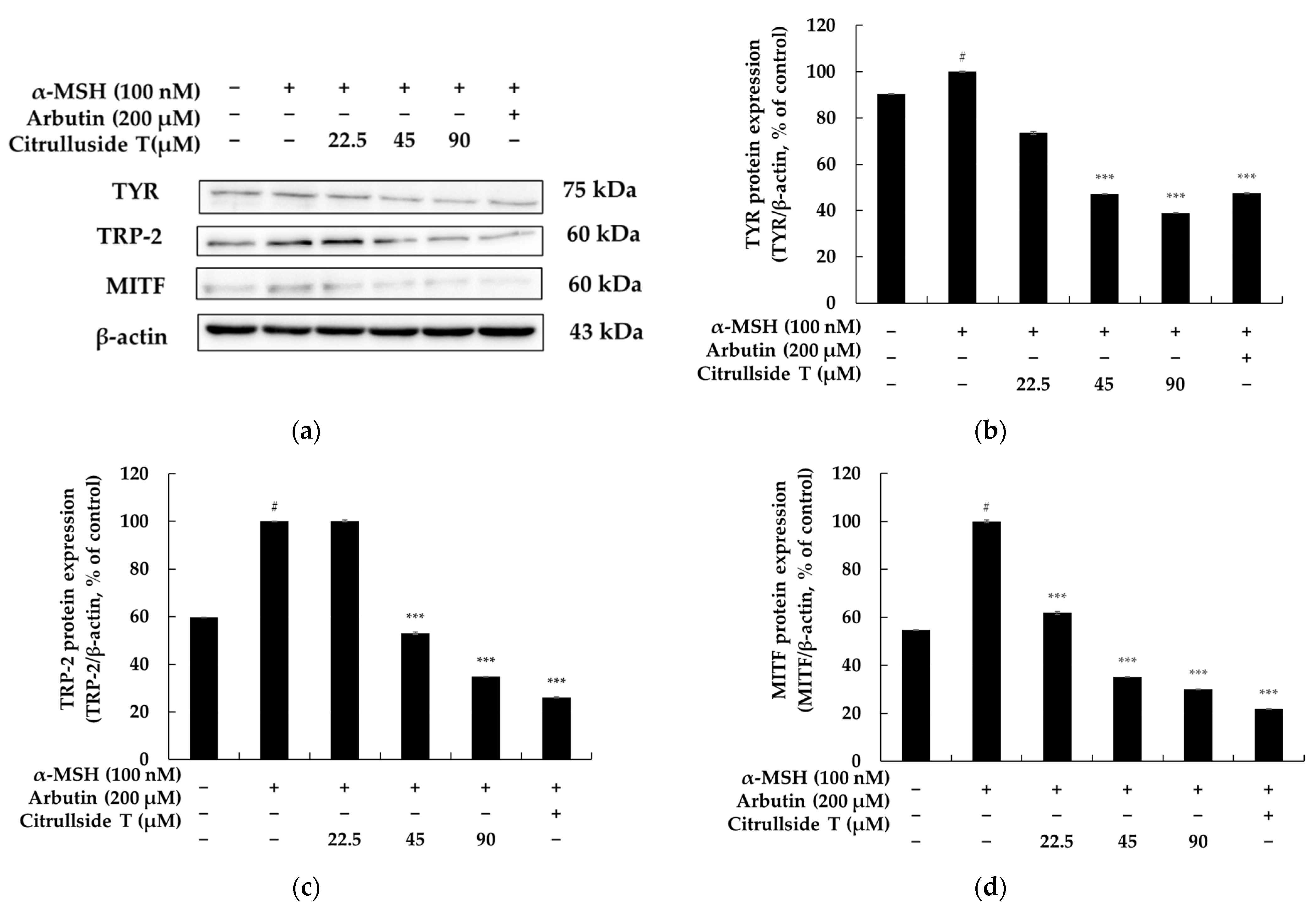
MITF transcription factors, which regulate the expression of genes that play essential roles in cell differentiation, proliferation, and survival, are transcription factors for melanogenic enzymes such as TRP-1 and TRP-2, which regulate melanin synthesis in melanocytes [19,20,21,22]. Therefore, to elucidate the depigmentation mechanism of citrulluside T in melanocytes, we evaluated its effect on the expression of melanogenic enzymes such as tyrosinase and TRP-2 in B16F10 cells. B16F10 cells were treated with citrulluside T for a specified time in the presence of α-MSH. Then, the protein levels of tyrosinase and TRP-2 were determined via a Western blot assay. Compared with α-MSH-stimulated B16F10 cells, citrulluside T significantly decreased the protein expression of melanogenic enzymes tyrosinase and TRP-2, especially at the high concentration of 90 µM: tyrosinase was decreased by about 61.08%, and TRP-2 was decreased by about 65.23%. The expression of MITF was also significantly decreased in a concentration-dependent manner and was inhibited by about 69.97% at high concentrations (Figure 4). These results confirm that citrulluside T inhibits the protein expression of tyrosinase and TRP-2 through the regulation of MITF expression in B16F10 cells, thereby reducing melanogenesis.

3.4. Effects of Citrulluside T on the cAMP/PKA Signaling Pathway

In the cAMP/PKA pathway, MC1R upon stimulation by α-MSH activates adenyl cyclase, leading to the accumulation of intracellular cAMP, which subsequently phosphorylates PKA, translocates into the nucleus, and upregulates MITF transcription through the phosphorylation of CREB [28,29]. Therefore, to determine the involvement of the cAMP/PKA signaling pathway in the inhibition of melanogenesis by citrulluside T, B16F10 cells stimulated with α-MSH were treated with citrulluside T for 24 h and the expression levels of related proteins were determined via Western blotting. As shown in Figure 5, citrulluside T significantly reduced the phosphorylation of PKA and CREB in a concentration-dependent manner. In particular, compared with α-MSH-stimulated B16F10 cells, phosphorylated PKA and CREB were reduced by 63.91% and 40.79%, respectively, at a high concentration of 90 µM. Therefore, it can be confirmed that citrulluside T downregulates the expression level of the transcriptional regulator MITF through the cAMP/PKA signaling pathway, which ultimately inhibits melanogenesis.

3.5. Effects of Citrulluside T on the Wnt/β-Catenin Signaling Pathway

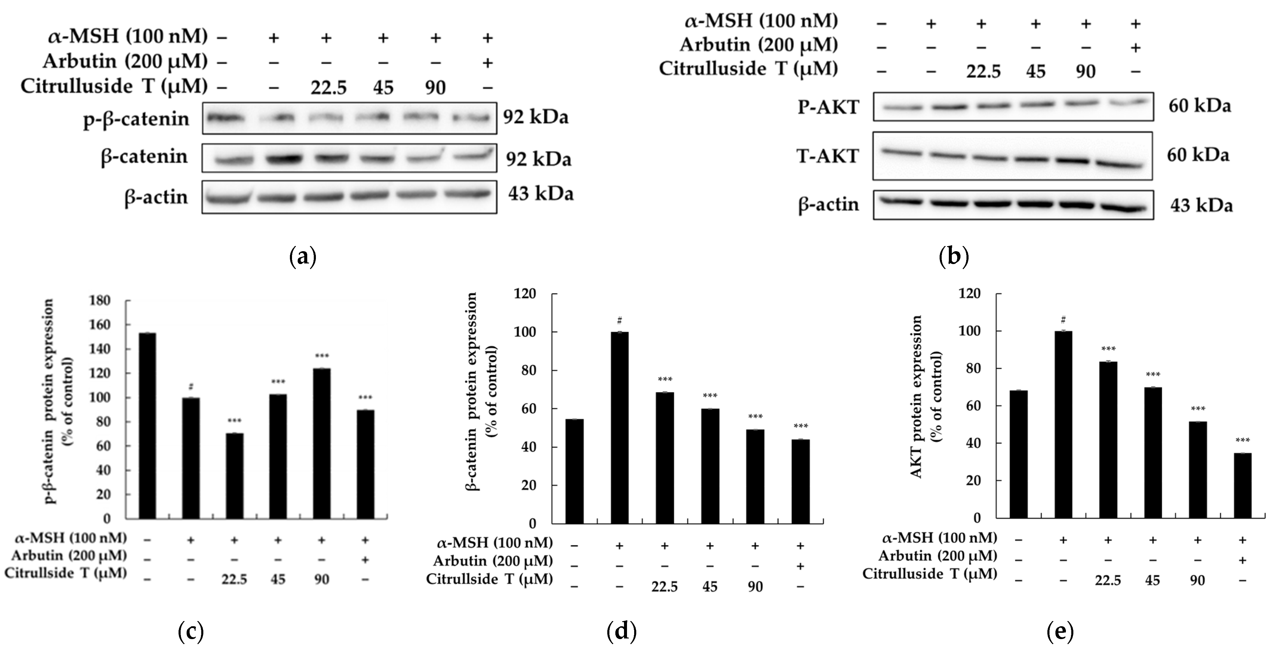
Considering that citrulluside T suppressed MITF expression in B16F10 cells, we then investigated whether β-catenin signaling is involved in this suppression. As the primary regulator of Wnt/β-catenin signaling, we first examined the impact of citrulluside T on intracellular β-catenin levels. The data show that the protein level of β-catenin was much higher in α-MSH-stimulated B16F10 cells than in the non-treatment control group, while treatment with citrulluside T remarkably and concentration-dependently decreased the intracellular β-catenin content in B16F10 cells. When Wnt proteins bind to their receptors, GSK3β is inactivated, which triggers β-catenin phosphorylation, leading to the destruction of phosphorylated β-catenin in the proteasome. As a result, β-catenin accumulates in the cytoplasm and translocates to the nucleus. Conversely, activation of β-catenin phosphorylation can interfere with its translocation to the nucleus, ultimately repressing the transcription of MITF [30,31]. As shown in Figure 6, citrulluside T caused a concentration-dependent decrease in the expression of β-catenin and a concentration-dependent increase in the phosphorylation of β-catenin. In particular, compared to α-MSH-stimulated B16F10 cells, the expression of β-catenin was decreased by about 50.93% at a high concentration of 90 µM, while p-β-catenin was increased by 23.98%. Therefore, it can be seen that citrulluside T downregulates the expression level of the transcriptional regulator MITF through the Wnt signaling pathway.

3.6. Effects of Citrulluside T on the PI3K/AKT Signaling Pathway

The PI3K/Akt signaling pathway regulates the expression of melanogenic proteins as well as melanogenesis. Specifically, activation of the PI3K/AKT pathway downregulates tyrosinase, TRP-1, and TRP-2 protein expression and the MITF transcription regulator in B16F10 cells [19,20,21,22]. Therefore, we investigated whether citrulluside T increases Akt phosphorylation. As shown in Figure 6, we found that citrulluside T increased AKT protein expression in a concentration-dependent manner while decreasing its phosphorylation. Overall, however, we found a concentration-dependent decrease in the ratio of AKT total protein to phosphorylated protein. In particular, the expression of phosphorylated AKT was reduced by about 48.42% relative to the total protein at a high concentration of 90 µM compared to B16F10 cells stimulated with α-MSH. Therefore, it can be seen that citrulluside T downregulates the expression level of MITF, a transcriptional regulator, through the negative control of the PI3K/AKT signaling pathway.

3.7. Skin Primary Irritation Test

Patches containing citrulluside T at concentrations of 45 and 90 µM were applied to the skin and kept in contact for 24 h, after which the patches were removed and the area was observed 48 h later. The results are presented in Table 1, categorizing the test substance (citrulluside T) as “none to slight” in terms of its effectiveness. The solvent, squalene, was used as a negative control for comparison.

4. Discussion

Many studies have demonstrated that melanin is essential for protecting humans from the harmful effects of UV radiation and maintaining the normal homeostasis of the skin and that melanin particles play an important role in the metabolism of skin cells. However, an excess of melanin after exposure to UV radiation can lead to abnormal skin conditions such as melasma, freckles, age spots, and café au lait spots (flat brown spots), as well as epidermal nevi and melanoma. The entire process of melanogenesis involves several steps, including melanosome formation, melanin synthesis, and melanosome transport, and requires a panel of melanogenic genes. Therefore, the use of melanogenesis inhibitors is one of the possible therapeutic approaches for the treatment of the above diseases caused by hyperpigmentation [32,33,34,35]. Therefore, we focused on the discovery of novel melanogenesis inhibitors from plant extracts.
According to the Korean government, the total watermelon cultivation area in Korea is expected to reach 12,000 ha in 2023, with a watermelon production of 470,000 tons. Therefore, the enormous generation of watermelon stems, leaves, and rinds, which are types of food waste, makes the discovery of upcycled functional materials from them an attractive study. In this study, a comprehensive investigation of the ethyl acetate fraction of the stem ethanol extract allowed us to isolate and structurally characterize a previously undescribed citrulluside T compound from the stem.
In fact, we are not the first to isolate citrulluside T. Itoh et al. [27] first isolated citrulluside H and citrulluside T from young watermelon fruits and reported that these two compounds attenuated ultraviolet B radiation-induced matrix metalloproteinase expression by scavenging reactive oxygen species produced by human skin fibroblasts. However, no studies have reported that compounds in the citrulluside family effectively inhibit melanogenesis. To determine the effect of citrulluside T on melanogenesis, we determined whether citrulluside T effectively inhibits melanogenesis and intracellular tyrosinase activity. Using Western blot analysis, we demonstrated that citrulluside T inhibits melanogenesis by regulating the expression of MITF, a transcription factor important for melanocyte development and melanogenesis. As is known, MITF regulates the transcription of melanogenesis-related genes, including tyrosinase, TRP-1, and TRP-2 [19,20,21,22]. As expected, citrulluside T downregulated the expression levels of tyrosinase, TRP-1, and TRP-2. These data indicate that the melanogenesis inhibitory effect of citrulluside T occurs primarily through downregulation of tyrosinase family genes, which is mediated through inhibition of MITF production. The melanogenic signaling pathways include PI3K/Akt signaling, GSK-3β/β-catenin signaling, and PKA/CREB signaling, which is a cAMP-dependent pathway [33]. As a result, we found that citrulluside T inhibits melanogenesis by inhibiting the phosphorylation of PKA, CREB, AKT, and GSK-3β and, conversely, activating the phosphorylation of β-catenin.
Taken together, we speculate that the effect of citrulluside T on melanogenesis is due to the inhibition of GSK-3β phosphorylation, resulting in a decrease in the accumulation of β-catenin in the cytoplasm, which in turn interferes with the translocation of β-catenin to the nucleus, where it directly binds to MITF and inhibits its transcription and the production of melanogenic enzymes [30,31]. Our data showed that citrulluside T inhibits melanogenesis by interfering with the GSK-3β/β-catenin and PKA/CREB pathways and decreasing the expression of MITF and tyrosinase family genes.
Human skin needs to be protected from environmental factors and chemicals contained in pharmaceuticals and cosmetics. To protect the general population, especially sensitive populations such as children, it is necessary to evaluate the potential of chemical ingredients contained in cosmetics and final products to cause acute skin irritation. Therefore, we conducted a skin irritation test on 33 subjects for the topical application of citrulluside T according to the OECD guidelines and finally confirmed the skin safety of citrulluside T, as shown in Table 1.
In summary, the discovery of effective natural ingredients that inhibit melanogenesis may help to control non-genetic pigment imbalance diseases and reduce the side effects of chemical drugs. Our findings suggest that the candidate compound, citrulluside T, may serve as a strategy that can be used for skin hyperpigmentation. These results suggest that citrulluside T could be used as a whitening ingredient without the risk of causing skin damage or as an ingredient in hyperpigmentation treatments such as for lentigo. Further studies are needed to evaluate the involvement of additional signaling pathways such as MAPK and PI3K in the melanin inhibitory efficacy of citrulluside T. Furthermore, inhibitor studies with the PKA inhibitor H-89, the MAPK/ERK inhibitor PD98059, the PI3K/Akt inhibitor LY294002, and the p38 MAPK inhibitor SB203580 are needed to further explore the molecular mechanisms underlying the inhibitory effects of citrulluside T on melanogenesis. Finally, in vivo studies of citrulluside T using alternative animal models such as zebrafish embryos or direct application to human skin are essential.

Author Contributions

Conceptualization, C.-G.H.; methodology, H.-M.K. and M.-Y.M.; software, H.-M.K. and M.-Y.M.; formal analysis, H.-M.K. and M.-Y.M.; investigation, H.-M.K. and M.-Y.M.; data curation, H.-M.K. and M.-Y.M.; writing—original draft preparation, C.-G.H.; writing—review and editing, C.-G.H.; supervision, C.-G.H.; project administration, C.-G.H.; funding acquisition, C.-G.H. All authors have read and agreed to the published version of the manuscript.

Funding

This research was supported by the 2023 scientific promotion program funded by Jeju National University.

Institutional Review Board Statement

This study was conducted in accordance with the Declaration of Helsinki and approved by the ethics committee of Dermapro Co., Ltd. for studies involving humans (IRB number: 1-220777-A-N-01-DICN23044).

Informed Consent Statement

Informed consent was obtained from all subjects involved in the study.

Data Availability Statement

The data presented in this study are available on request from the corresponding author.

Conflicts of Interest

The authors declare no conflict of interest.

References

  1. Sharma, C.; Deutsch, J.M. Upcycling in the context of biotechnology-based solutions for food quality, loss, and consumer perception. Curr. Opin. Biotechnol. 2023, 81, 102920. [Google Scholar] [CrossRef] [PubMed]
  2. Gil-Martín, E.; Forbes-Hernández, T.; Romero, A.; Cianciosi, D.; Giampieri, F.; Battino, M. Influence of the extraction method on the recovery of bioactive phenolic compounds from food industry by-products. Food Chem. 2022, 378, 131918. [Google Scholar] [CrossRef] [PubMed]
  3. Grigolon, G.; Nowak, K.; Poigny, S.; Hubert, J.; Kotland, A.; Waldschütz, L.; Wandrey, F. From Coffee Waste to Active Ingredient for Cosmetic Applications. Int. J. Mol. Sci. 2023, 24, 8516. [Google Scholar] [CrossRef] [PubMed]
  4. de Mello, V.; de Mesquita Júnior, G.A.; Alvim, J.G.E.; Costa, J.C.D.; Vilela, F.M.P. Recent patent applications for coffee and coffee by-products as active ingredients in cosmetics. Int. J. Cosmet. Sci. 2023, 45, 267–287. [Google Scholar] [CrossRef] [PubMed]
  5. Lestari, W.; Hasballah, K.; Listiawan, M.Y.; Sofia, S. Coffee by-products as the source of antioxidants: A systematic review. F1000Research 2022, 11, 220. [Google Scholar] [CrossRef]
  6. Zhang, H.; Chi, X.; Pan, W.; Wang, S.; Zhang, Z.; Zhao, H.; Wang, Y.; Wu, Z.; Zhou, M.; Ma, S.; et al. Antidepressant mechanism of classical herbal formula lily bulb and Rehmannia decoction: Insights from gene expression profile of medial prefrontal cortex of mice with stress-induced depression-like behavior. Genes Brain Behav. 2020, 19, e12649. [Google Scholar] [CrossRef]
  7. Chi, X.; Wang, S.; Baloch, Z.; Zhang, H.; Li, X.; Zhang, Z.; Zhang, H.; Dong, Z.; Lu, Y.; Yu, H.; et al. Research progress on classical traditional Chinese medicine formula Lily Bulb and Rehmannia Decoction in the treatment of depression. Biomed. Pharmacother. 2019, 112, 108616. [Google Scholar] [CrossRef]
  8. Lee, E.; Yun, N.; Jang, Y.P.; Kim, J. Lilium lancifolium Thunb. extract attenuates pulmonary inflammation and air space enlargement in a cigarette smoke-exposed mouse model. J. Ethnopharmacol. 2013, 26, 148–156. [Google Scholar] [CrossRef]
  9. Yoon, H.S.; Yang, K.Y.; Kim, J.E.; Kim, J.M.; Lee, N.H.; Hyun, C.G. Hypopigmenting Effects of Extracts from Bulbs of Lilium Oriental Hybrid ‘Siberia’ in Murine B16/F10 Melanoma Cells. J. Korean Soc. Food Sci. Nutr. 2014, 43, 705–711. [Google Scholar] [CrossRef]
  10. Wijesinghe, W.A.; Senevirathne, M.; Oh, M.C.; Jeon, Y.J. Protective effect of methanol extract from citrus press cakes prepared by far-infrared radiation drying on H2O2-mediated oxidative damage in Vero cells. Nutr. Res. Pract. 2011, 5, 389–395. [Google Scholar] [CrossRef]
  11. Panwar, D.; Panesar, P.S.; Chopra, H.K. Evaluation of nutritional profile, phytochemical potential, functional properties and anti-nutritional studies of Citrus limetta peels. J. Food Sci. Technol. 2023, 60, 2160–2170. [Google Scholar] [CrossRef]
  12. Phucharoenrak, P.; Muangnoi, C.; Trachootham, D. Metabolomic Analysis of Phytochemical Compounds from Ethanolic Extract of Lime (Citrus aurantifolia) Peel and Its Anti-Cancer Effects against Human Hepatocellular Carcinoma Cells. Molecules 2023, 28, 2965. [Google Scholar] [CrossRef]
  13. Kumar, S.; Konwar, J.; Purkayastha, M.D.; Kalita, S.; Mukherjee, A.; Dutta, J. Current progress in valorization of food processing waste and by-products for pectin extraction. Int. J. Biol. Macromol. 2023, 239, 124332. [Google Scholar] [CrossRef]
  14. Grumet, R.; McCreight, J.D.; McGregor, C.; Weng, Y.; Mazourek, M.; Reitsma, K.; Labate, J.; Davis, A.; Fei, Z. Genetic Resources and Vulnerabilities of Major Cucurbit Crops. Genes 2021, 12, 1222. [Google Scholar] [CrossRef]
  15. Shaik, R.S.; Zhu, X.; Clements, D.R.; Weston, L.A. Understanding invasion history and predicting invasive niches using genetic sequencing technology in Australia: Case studies from Cucurbitaceae and Boraginaceae. Conserv. Physiol. 2016, 26, cow030. [Google Scholar] [CrossRef]
  16. Burton-Freeman, B.; Freeman, M.; Zhang, X.; Sandhu, A.; Edirisinghe, I. Watermelon and L-Citrulline in Cardio-Metabolic Health: Review of the Evidence 2000–2020. Curr. Atheroscler. Rep. 2021, 23, 81. [Google Scholar] [CrossRef]
  17. Wahid, S.; Khan, R.A.; Feroz, Z.; Ikram, R. Analgesic, anti-inflammatory and toxic effects of ethanol extracts of Cucumis melo and Citrullus lanatus seeds. Pak. J. Pharm. Sci. 2020, 33, 1049–1055. [Google Scholar]
  18. Mehta, A.; Srivastva, G.; Kachhwaha, S.; Sharma, M.; Kothari, S.L. Antimycobacterial activity of Citrullus colocynthis (L.) Schrad. against drug sensitive and drug resistant Mycobacterium tuberculosis and MOTT clinical isolates. J. Ethnopharmacol. 2013, 149, 195–200. [Google Scholar] [CrossRef]
  19. Kikuchi, T.; Okada, R.; Harada, Y.; Ikushima, K.; Yamakawa, T.; Yamada, T.; Tanaka, R. Cucurbitane-type triterpenes from Citrullus lanatus (watermelon) seeds. Nat. Prod. Commun. 2013, 8, 1367–1369. [Google Scholar] [CrossRef]
  20. Kim, T.; Kang, J.K.; Hyun, C.G. 6-Methylcoumarin Promotes Melanogenesis through the PKA/CREB, MAPK, AKT/PI3K, and GSK3β/β-Catenin Signaling Pathways. Molecules 2023, 28, 4551. [Google Scholar] [CrossRef]
  21. Kim, T.; Kim, K.B.; Hyun, C.G. A 7-Hydroxy 4-Methylcoumarin Enhances Melanogenesis in B16-F10 Melanoma Cells. Molecules 2023, 28, 3039. [Google Scholar] [CrossRef] [PubMed]
  22. Kim, H.M.; Hyun, C.G. Miglitol, an Oral Antidiabetic Drug, Downregulates Melanogenesis in B16F10 Melanoma Cells through the PKA, MAPK, and GSK3β/β-Catenin Signaling Pathways. Molecules 2022, 28, 115. [Google Scholar] [CrossRef] [PubMed]
  23. Kim, T.; Hyun, C.G. Imperatorin Positively Regulates Melanogenesis through Signaling Pathways Involving PKA/CREB, ERK, AKT, and GSK3β/β-Catenin. Molecules 2022, 27, 6512. [Google Scholar] [CrossRef] [PubMed]
  24. Phacharapiyangkul, N.; Thirapanmethee, K.; Sa-Ngiamsuntorn, K.; Panich, U.; Lee, C.H.; Chomnawang, M.T. Effect of Sucrier Banana Peel Extracts on Inhibition of Melanogenesis through the ERK Signaling Pathway. Int. J. Med. Sci. 2019, 16, 602–606. [Google Scholar] [CrossRef] [PubMed]
  25. Zolghadri, S.; Beygi, M.; Mohammad, T.F.; Alijanianzadeh, M.; Pillaiyar, T.; Garcia-Molina, P.; Garcia-Canovas, F.; Munoz-Munoz, J.; Saboury, A.A. Targeting tyrosinase in hyperpigmentation: Current status, limitations and future promises. Biochem. Pharmacol. 2023, 212, 115574. [Google Scholar] [CrossRef]
  26. Gryn-Rynko, A.; Sperkowska, B.; Majewski, M.S. Screening and Structure-Activity Relationship for Selective and Potent Anti-Melanogenesis Agents Derived from Species of Mulberry (Genus Morus). Molecules 2022, 27, 9011. [Google Scholar] [CrossRef]
  27. Itoh, T.; Fujita, S.; Koketsu, M.; Hashizume, T. Citrulluside H and citrulluside T from young watermelon fruit attenuate ultraviolet B radiation-induced matrix metalloproteinase expression through the scavenging of generated reactive oxygen species in human dermal fibroblasts. Photodermatol. Photoimmunol. Photomed. 2021, 37, 386–394. [Google Scholar] [CrossRef]
  28. Ninomiya, M.; Itoh, T.; Fujita, S.; Hashizume, T.; Koketsu, M. Phenolic glycosides from young fruits of Citrullus lanatus. Phytochem. Lett. 2020, 40, 135–138. [Google Scholar] [CrossRef]
  29. Yoon, J.H.; Youn, K.; Jun, M. Discovery of Pinostrobin as a Melanogenic Agent in cAMP/PKA and p38 MAPK Signaling Pathway. Nutrients 2022, 14, 3713. [Google Scholar] [CrossRef]
  30. Tang, H.H.; Zhang, Y.F.; Yang, L.L.; Hong, C.; Chen, K.X.; Li, Y.M.; Wu, H.L. Serotonin/5-HT7 receptor provides an adaptive signal to enhance pigmentation response to environmental stressors through cAMP-PKA-MAPK, Rab27a/RhoA, and PI3K/AKT signaling pathways. FASEB J. 2023, 37, e22893. [Google Scholar] [CrossRef]
  31. Huang, H.C.; Yen, H.; Lu, J.Y.; Chang, T.M.; Hii, C.H. Theophylline enhances melanogenesis in B16F10 murine melanoma cells through the activation of the MEK 1/2, and Wnt/β-catenin signaling pathways. Food Chem. Toxicol. 2020, 137, 111165. [Google Scholar] [CrossRef]
  32. Yin, L.; Niu, C.; Liao, L.X.; Dou, J.; Habasi, M.; Aisa, H.A. An Isoxazole Chalcone Derivative Enhances Melanogenesis in B16 Melanoma Cells via the Akt/GSK3β/β-Catenin Signaling Pathways. Molecules 2017, 22, 2077. [Google Scholar] [CrossRef]
  33. Merin, K.A.; Shaji, M.; Kameswaran, R. A Review on Sun Exposure and Skin Diseases. Indian J. Dermatol. 2022, 67, 625. [Google Scholar] [CrossRef]
  34. Han, H.; Hyun, C.G. Acenocoumarol, an Anticoagulant Drug, Prevents Melanogenesis in B16F10 Melanoma Cells. Pharmaceuticals 2023, 16, 604. [Google Scholar] [CrossRef]
  35. Pillaiyar, T.; Manickam, M.; Jung, S.H. Downregulation of melanogenesis: Drug discovery and therapeutic options. Drug Discov. Today 2017, 22, 282–298. [Google Scholar] [CrossRef]
Figure 1. Isolation procedure of isolated compound from the Citrullus lanatus stem.
Cosmetics 10 00108 g001
Figure 2. HPLC chromatogram of isolated compound (UV 329.7 nm).
Cosmetics 10 00108 g002
Figure 3. Effect of citrulluside T on viability, melanin contents, and cellular tyrosinase activity in B16F10 melanoma cells. (a) Cell viability of citrulluside T in B16F10 cells. (b) Citrulluside T reduces melanin synthesis in B16F10 cells. (c) Citrulluside T inhibits tyrosinase activity in B16F10 cells. Data are expressed as the mean ± SD (n = 3). # p < 0.001 vs. untreated control group; *** p < 0.001, ** p < 0.01 vs. negative control group.
Cosmetics 10 00108 g003
Figure 4. Citrulluside T inhibits the expression of MITF and melanin synthetase. The α-MSH-treated melanoma B16F10 cells were treated with citrulluside T at different concentrations for 48 h. (a) Protein detection results. Protein expression of (b) TYR/β-actin, (c) TRP-2/β-actin, and (d) MITF/β-actin. Data are expressed as the mean ± SD (n = 3) from a single triplicate experiment using ImageJ software. # p < 0.001 vs. untreated control group; *** p < 0.001 vs. negative control group.
Cosmetics 10 00108 g004
Figure 5. Citrulluside T inhibits the expression of the cAMP/PKA signaling pathway. The α-MSH-treated melanoma B16F10 cells were treated with citrulluside T at different concentrations for 24 h. (a) Protein detection result. Protein expression of (b) p-PKA/t-PKA and (c) p-CREB/t-CREB. Equal amounts of protein loadings were confirmed by using β-actin. Data are expressed as the mean ± SD (n = 3) from a single triplicate experiment using ImageJ software. # p < 0.001 vs. untreated control group; *** p < 0.001 vs. negative control group.
Cosmetics 10 00108 g005
Figure 6. Citrulluside T inhibits the expression of the Wnt/PI3K/AKT signaling pathway. α-MSH-treated melanoma B16F10 cells were treated with citrulluside T at different concentrations for 24 and 4 h. (a,b) Protein detection results. Protein expression of (c) p-β-catenin/β-actin, (d) β-catenin/β-actin, and (e) p-AKT/t-AKT. Equal amounts of protein loadings were confirmed by using β-actin. Data are expressed as the mean ± SD (n = 3) from a single triplicate experiment using ImageJ software. # p < 0.001 vs. untreated control group; *** p < 0.001 vs. negative control group.
Cosmetics 10 00108 g006
Table 1. Results of human skin primary irritation test (n = 33).
NoSamplesNo. of
Responders
24 h48 hReaction Grade
+1+2+3+4+1+2+3+424 h48 hMean
1Citrulluside T (45 μM)0--------000
2Citrulluside T (90 μM)0--------000
3Control
(Squalene)
0--------000
Disclaimer/Publisher’s Note: The statements, opinions and data contained in all publications are solely those of the individual author(s) and contributor(s) and not of MDPI and/or the editor(s). MDPI and/or the editor(s) disclaim responsibility for any injury to people or property resulting from any ideas, methods, instructions or products referred to in the content.