Article

Natural Antioxidant-Loaded Nanoemulsions for Sun Protection Enhancement

by
Eleni Galani
1,2,
Dimitrios Galatis
1,
Kyriaki Tzoka
3,
Vassiliki Papadimitriou
1,
Theodore G. Sotiroudis
1,
Antonios Bonos
3,
Aristotelis Xenakis
1 and
Maria D. Chatzidaki
1,3,*
1
Institute of Chemical Biology, National Hellenic Research Foundation, 116 35 Athens, Greece
2
Food Chemistry & Human Nutrition, School of Food, Biotechnology and Development, Agricultural University of Athens, 118 55 Athina, Greece
3
Cleanway Ltd., Leof. Konstantinoupoleos 160, 135 61 Athina, Greece
*
Author to whom correspondence should be addressed.
Cosmetics 2023, 10(4), 102; https://doi.org/10.3390/cosmetics10040102
Submission received: 14 March 2023 / Revised: 4 July 2023 / Accepted: 6 July 2023 / Published: 14 July 2023

Abstract

:
The aim of this study was to formulate nanodispersions to encapsulate antioxidants extracted from olive mill wastewater (OMW) and phycocyanin extracted from Spirulina maxima to act as enhancers for the skin’s protection against UV radiation. For this purpose, two water-in-oil nanoemulsions were prepared using a low-energy homogenization method. Both systems were based on isopropyl myristate as the continuous phase, while water or a mixture of glycerol and water was used as the dispersed phase. Then, antioxidants extracted from OMW and phycocyanin from Spirulina maxima were encapsulated in the water core of the nanoemulsions. The empty and antioxidant-loaded systems were then structurally studied using dynamic light scattering for the detection of their droplet size and stability over time. Electron paramagnetic resonance (EPR) spectroscopy using adequate probes was applied for the characterization of the surfactants’ monolayer in the presence and absence of antioxidants. It was found that the mean droplet diameter of the emulsions was 200 nm. The nanoemulsions remained stable for over 2 months. The encapsulated antioxidants were assessed for their scavenging activity of a model stable radical by applying EPR spectroscopy. It was found that the loaded systems exhibited an increased antioxidant capacity compared with the empty ones. Finally, the most stable system was added to commercial sunscreen lotions and the overall sun protection factor (SPF) was assessed. The sunscreen lotions that contained the nanoemulsions loaded with OMW extracts or phycocyanin showed an increase in their SPF value.

1. Introduction

The sun is undeniably a source of life for the planet. Spending time in the sun has many benefits like the production of vitamin D. Nevertheless, as an effect of ozone depletion, nowadays, a much larger number of UV rays reaches us, having serious effects such as an increase in the number of skin cancer cases [1,2]. That is why the use of sunscreen is very important. The filters used in sunscreens to block UV radiation can be either mineral/physical or chemical. Sunscreens protect the skin from UV (ultraviolet) rays using chemical or mineral filters. Chemical filters absorb UV rays before they penetrate the skin, and then they release them as heat, while mineral or physical blockers act as barriers on the skin, reflecting UV light. Examples of chemical filters include oxybenzone, octinoxate, and avobenzone, while mineral sunscreens are generally derived from titanium dioxide or zinc oxide. In recent years, several concerns have been raised about the safety of those filters due to studies showing that they can be potentially harmful to both human health and the environment [3,4,5,6]. The need to decrease chemical UV filters has led scientists to search for natural alternatives that can provide certain protection against UV radiation, such as herbal extracts, antioxidants, proteins, and other biomolecules that could enhance photoprotection [7,8,9].
Nowadays, food waste is considered a cheap source of valuable compounds. Their recycling ability, while retaining their functionality as additives in different products, is gaining more and more interest due to the tremendous environmental impact. Food waste can be applied in various domains, including cosmetics. Some of them, such as antioxidants extracted from olive mill wastewater (OMW), have even been used as UV booster agents in sunscreens and cosmetic products [10].
Olive mill wastewater (OMW) is generated from olive oil extraction systems. It has high-added-value compounds, namely phenolics, pectin, and some important enzymes, while its untreated disposal to the environment causes a certain toxicity/phytotoxicity because of its high phenolic content [11]. Apart from water (83–92%), the main components of OMW are phenolic compounds, sugars, and organic acids. OMW also contains valuable resources such as mineral nutrients, especially potassium, which could potentially be reused as a fertilizer. OMW also has significant impacts when discharged directly into surface water [12]. Polyphenols are contained in OMW in high concentrations, and they are ingredients of high value since they can be used in several applications. Polyphenols are also excellent ingredients for cosmetics because of their mechanism for binding free radicals, making them very good candidates for sun protection, antiaging, and other dermo-cosmetics [13,14].
Spirulina is a microscopic photosynthetic and filamentous cyanobacterium (blue–green algae) belonging to the family Oscillatoriaceae that has been used as a food source since ancient times. Apart from its crude protein (60–70%, w/w) and vitamins (4%, w/w), spirulina is also rich in essential amino acids (EEAs), minerals, essential fatty acids (EFAs), and antioxidants. It is now used as a nutraceutical food supplement since it contains prophylactic, therapeutic nutrients, and several unexplored bioactive compounds [15]. Spirulina can also be used in cosmetics [16]. Spirulina, and algae in general, contain colored components such as carotenoids, chlorophyll, and phycobiliproteins. The major phycobiliprotein is phycocyanin [17]. Phycocyanin can be used as a coloring agent in foods (chewing gums, dairy products, etc.) and cosmetics (lipsticks, eyeliners, etc.). More recently, phycocyanin has also been found to have a photoprotective effect over ultraviolet B radiation and could be used in sun-protective products [18,19].
Nanoemulsions (NEs) are kinetically stable colloidal dispersions of two immiscible liquids, with one of the liquids being dispersed (dispersed phase) as spherical drops in the other liquid (continuous phase). NEs consist of an oil and an aqueous phase, surfactants, and possibly co-surfactants. Depending on which phase is dispersed to the other, water-in-oil (W/O) or oil-in-water (O/W) types of nanoemulsions can be formed [20]. NEs have been used for the encapsulation and delivery of bioactive compounds for several purposes, such as food applications [21,22,23], drug delivery [24,25,26,27,28], and even for the cosmetics industry [29,30].
In the literature, there are studies that deal with the use of NEs, OMW, and algae in cosmetics and sunscreen applications. More specifically, some studies have indicated that sunflower oil NEs used in conventional sunscreens can enhance the SPF value due to their smaller droplet sizes compared with traditional emulsions [31]. Additionally, systems containing spirulina or nanoemulsions containing carotenoids that are derived from algae have been shown to have an SPF-boosting effect [32,33]. Finally, antioxidants from OMW have also been applied in cosmetics in general and as SPF boosters, according to previous studies [34,35]. Furthermore, concepts such as ecodesign, circular economy, bioeconomy, and industrial symbiosis are no longer only sustainability trends or academic endeavors. They develop and materialize in policies and regulations, as seen in the Ecodesign Directive 2009/125/EC, the EU’s bioeconomy strategy and action plan, the EU Circular Economy Action Plan, and the European Green Deal, as well as in the Global Sustainable Development Goals, specifically, Goal 12 concerning responsible production and consumption.
The research in the present paper builds upon these concepts of great importance for the Mediterranean and Greek biodiversity. Taking the case of OMW, the goal is to transform them from waste to resource and close a production loop, linking the output of agricultural production to the input of the chemical industry. Why do so? Because on the one hand, OMW has high toxicity due to a high organic load. This is a chronic environmental problem in Mediterranean countries that needs to be addressed. On the other hand, this organic load can be a resource. Indeed, OMW is full of phenolic compounds some of which, such as tyrosol, hydroxytyrosol, oleuropein, and oleocanthal, are valuable not only for cosmetics, due to their antioxidant properties, but also to other sectors like pharmaceuticals and the food industry or for its use in biofuel, agriculture, enzyme production, etc. [36,37,38]. This holistic perspective where separate industries are considered within one “industrial ecosystem” not only helps reduce environmental burdens but also creates a whole new economy root, i.e., olive mills could earn from the commercial exploitation of their byproducts by offering them for sale and industry could benefit from the purchase of low-cost raw materials.
Similarly, algae such as spirulina are also of significant economic importance within a bioeconomy, i.e., they can be used as food sources or fertilizers, as well as in fish farming. They also play a key role in the reclamation of alkaline and can be used as a soil-binding agent as well as in a variety of commercial products [39]. Spirulina is economically significant because it can be quickly produced on an industrial scale using low-cost materials such as wastewater from potato processing plants, molasses, etc. [40].
In the present study, biocompatible W/O NEs were formulated for the encapsulation of antioxidants extracted from OMW and phycocyanin from Spirulina maxima with the aim to be used as components in the cosmetics industry and more specifically as sun protection enhancement agents. These systems were structurally studied in terms of their antioxidant capacity, as well as with respect to their effect on the SPF values of already-known sunscreens. These novel systems can help the cosmetics industry formulate products using more sustainable and environmentally friendly materials and reduce chemicals in their products without missing any of their effectiveness. Following this project, more biocompatible substances could be found in the light of research, and more agricultural byproducts could be used so that new economic paths are created, waste products are reduced, and more sustainable products are created. Finally, the combination of these bioactive substances with their encapsulation in NEs could significantly enhance their effectiveness due to better dispersion and endurance since they are protected from coming in direct contact with air or sunlight.

2. Materials and Methods

2.1. Materials

Ethyl acetate and n-hexane were purchased from Lab Scan (Bangkok 10330, Thailand). Folin–Ciocalteu reagent, 2,2-diphenyl-1-picrylhydrazyl (DPPH), galvinoxyl, and 5-doxyl stearic acid-free radicals were acquired from Sigma-Aldrich (Chemie GmBH, Munich, Germany). Lauroglycol 90, Plurol Oleique MB, and Labrasol ALF were kindly donated by Gattefossé (Saint-Priest, France). Isopropyl tetradecanoate 98% (IPM) was purchased from Alfa Aesar (Kandel, Germany), and glycerol was obtained from Serva (Heidelberg, Germany). Sun lotion SPF 30, sun lotion SPF 50, reference sunscreen formulation SPF 43.0 (P6), and reference sunscreen formulation SPF 63.1 (P8) were used. Olive mill wastewater was collected from olive mills in Filiatra, Messinia, Greece, and Spirulina maxima was provided by Algi S.A, Serres, Greece. During the experiments, both distilled and ultra-pure water were used.

2.2. Methods

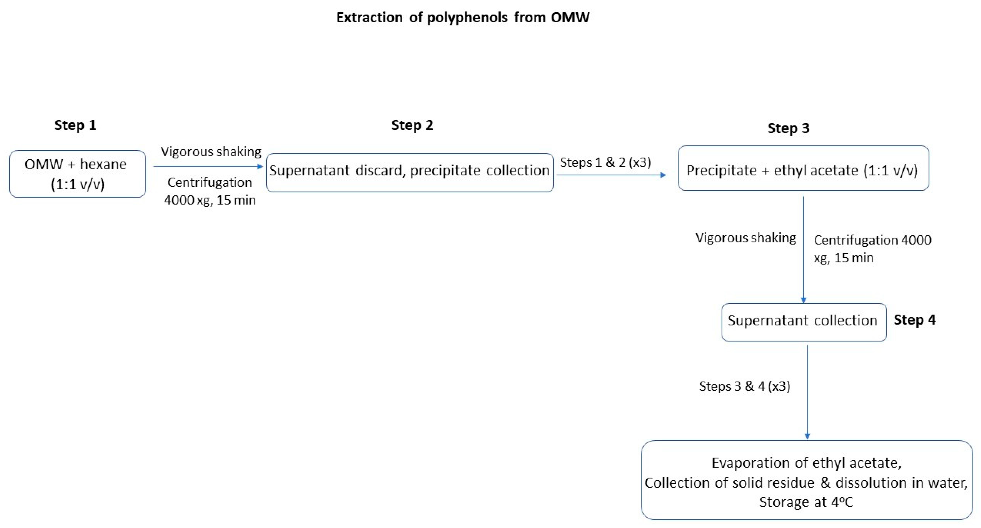
2.2.1. Extraction of Antioxidants from OMW

Wastewater samples were collected from two-phase (process without the addition of water) and three-phase (process with added water) centrifugal olive oil mills. From the three-phase olive mills, waste samples were collected from the first and final stages of production (sample 1 and sample 2, respectively). Two-phase olive mills produce waste only in one stage, and that is what was collected (sample 3). The samples were collected in plastic containers 10 L in volume (approved for food contact applications) and stored in a cold room at 4 °C. To avoid the frequent exposure of the samples to oxygen, they were divided into smaller volumes and stored at 4 °C. The extraction procedure was carried out using the liquid–liquid extraction method, as described by Leouifoudi et al. (2014). Initially, equal volumes of OMW and hexane were transferred to a flask and were vigorously shaken for 1 min, and then the content of the flask was centrifuged for 15 min at 4000× g in order to remove any organic content of the OMW. The supernatant was removed, and washing was repeated two more times by adding hexane to the precipitate. Extraction was then carried out with ethyl acetate, following the same procedure as the washing process. More specifically, the precipitate from the washing process was mixed with an equal volume of ethyl acetate, and the mixture was shaken vigorously and then centrifuged for 15 min, at 4000× g. The supernatant was collected this time in a vial, and the procedure was repeated two more times. The temperature was always kept at 25 °C. Finally, ethyl acetate was evaporated using a rotary evaporator with a bath at 45 °C, and the residues were redissolved in distilled water and stored in the refrigerator at 4 °C [41]. The procedure is also presented as a svhematic representation in Figure 1.

2.2.2. Extraction of Phycocyanin from Spirulina maxima

Spirulina maxima flakes were mixed with 50 mM of phosphate buffer (NaH2PO4:Na2HPO4), pH 7.2, in a plastic tube in a 1:10 biomass-to-buffer ratio. The tube was folded with aluminum foil, and its contents were shaken thoroughly until the flakes were fully dissolved. Afterward, the tube was put in the freezer (−4 °C) until it was solidly frozen, and then it was left outside at room temperature (25 °C) (the freeze–thaw process), and the procedure was repeated two more times to break down the cells. The tube was then left to reach room temperature, and it was centrifuged at 13,420× g at 4 °C for 20 min. The supernatant was collected and saturated with ammonium sulfate up to 30% to remove any contaminants. The vial was then left in a cold room (4 °C) under stirring for 1 h, and the mixture was centrifuged again at 13,420× g for 20 min. The supernatant was collected, and ammonium sulfate was added again to reach 70% saturation. The mixture was left under stirring at 4 °C overnight. The mixture was then centrifuged at 13,420× g at 4 °C for 30 min. The precipitate was collected and then redissolved in distilled water. The solution was transferred to dialysis membranes, which were put in beakers with distilled water. Dialysis was carried out for two days with the water being regularly changed. Finally, the phycocyanin solutions were collected and mixed with glycerol in a 1:1 v/v ratio and stored in the freezer (−6 °C). The phycocyanin solution that was going to be immediately used was not mixed with glycerol [42,43]. The procedure is also presented as a svhematic representation in Figure 2.

2.2.3. Determination of the OMW Extracts’ Total Phenol Content

In a tube, 0.5 mL of the properly diluted extract was mixed with 2.5 mL of Folin–Ciocalteu reagent (diluted 1:10 in ultrapure water) and was kept in the dark for 5 min. Afterward, 2 mL of 7.5% sodium carbonate was added, and the tubes were kept in the dark for 2 h. Absorbance was measured at 760 nm using a glass cuvette. The quantitative results were calculated using a standard curve of gallic acid and are expressed as mg of gallic acid equivalents per kg of OMW sample [44].

2.2.4. Determination of the Spirulina maxima Extract’s Phycocyanin Content

Phycocyanin concentration in the extract was calculated photometrically. After proper dilution with distilled water, the extracts’ absorption was measured at 620 and 650 nm. The concentration was calculated using the formula indicated below, where PC is the phycocyanin concentration (mg/mL), A620 is the sample’s absorption at 620 nm, and A650 is the sample’s absorption at 650 nm [32].
P C = A 620 0.474 A 650 5.34
Alongside the determination of phycocyanin concentration, the extract’s purity was also determined photometrically. The extract’s absorption was measured at 280 nm after proper dilution. The purity is determined using the ratio indicated below, where A620 is the maximum absorbance of phycocyanin, and A280 is the absorbance of total proteins [45].
E P = A 620 A 280

2.2.5. Preparation of W/O NEs Using the Low-Energy Method

Two low-energy O/W NEs (N1 and N2) were prepared using the isothermal spontaneous self-emulsification method [36] with a mixture of surfactants (Plurol Pleique, Plurol Diisostearique, Labrasol, and Dehymuls PGPH) and IPM as the continuous phase and water or a mixture of glycerol and water as the internal phase.
To formulate the NEs, the oil phase of the process was initially carried out by mixing the surfactants with IPM using a magnetic stirrer. Afterward, the aqueous phase of the process was performed with the addition of water (with or without the additives) to the oil-surfactant mixture under constant magnetic stirring. After the completion of the aqueous phase, the mixture was left under stirring for 1 h. The prepared NEs were transferred to glass bottles and stored at ambient temperature. For the loaded NEs, antioxidants extracted from OMW or phycocyanin were added at a concentration of 0.12% w/w. The compositions of the NEs used are listed in Table 1. All the ingredients used were biocompatible.

2.2.6. The Antioxidant Assessment of the OMW and Spirulina maxima Extracts via DPPH Scavenging

Briefly, 0.5 mL of the sample was mixed with 0.5 mL of ethyl acetate. Then, 0.5 mL of this mixture was added to 2 mL DPPH (70 μΜ in ethyl acetate). The solution was put in a glass cuvette, and the absorption was measured at 515 nm with time scanning. The percentage of inhibition was calculated using the formula [(Ac − As)/Ac] × 100, where Ac is the absorbance of the control sample (DPPH in ethyl acetate), and As is the absorbance of the sample at a certain time [46].

2.2.7. Determination of the Antioxidant Activity Using the Galvinoxyl Free Radical Detected via Electron Paramagnetic Resonance (EPR) Spectroscopy

EPR spectra were obtained at a room temperature of 25 °C, with a Bruker EMX EPR spectrometer operating at the X-Band. Before the analysis, the samples were prepared as described below. In a vial of 1.5 mL capacity, covered with aluminum foil, 1 mL of galvinoxyl free radical (0.25 mM in isooctane) was transferred. The vial was stored in the dark for 30 min. Subsequently, 50 μL of the galvinoxyl was removed, and 50 μL of the sample was added at a final concentration of 0.5%. The mixture was shaken and then transferred to a quartz flat aqueous sample cell and then placed in the EPR probe. As a blank sample, 50 μL of isooctane was added to the galvinoxyl free radical solution. Measurements were taken at 2, 5, 8, 10, 13, 15, 18, 20, 23, 25, 28, and 30 min. The Bruker WinEPR acquisition and processing program was used for data collection and analysis. The percentage of inhibition was calculated with the formula [(A0 − A)/A0] × 100, where A0 is the integral intensity of the EPR spectrum of each sample’s first measurement, which was considered as the zero time point, and A is the integral intensity of the EPR spectrum at a given time [47].

2.2.8. Dynamic Light Scattering (DLS) Measurements

A Zetasizer Nano ZS (ZEN3600) analyzer (Malvern Instruments Ltd., Malvern, UK) equipped with a He-Ne laser (633 nm) and non-invasive backscatter (NIBS) optics was used for droplet size and PDI measurements of the W/O NEs. The results were processed with the Malvern Zetasizer Nano software, version 6.32 (Malvern Instruments Ltd., Malvern, UK), which fits a spherical model of diffusing particles with low polydispersity. The samples were placed in a glass cuvette of 1 cm width with no dilution or further treatment. Measurements were carried out in triplicate at 25 °C.

2.2.9. Surfactant Membrane Dynamics Using Electron Paramagnetic Resonance (EPR) Spectroscopy

In this study, 5-doxyl stearic acid, a fatty acid derivative labeled at the aliphatic chain, was used to probe membrane dynamics at a certain depth.
EPR measurements were performed with an EMX Bruker EPR spectrometer at the X-Band (9.8 GHz) using a quartz flat aqueous sample cell. Instrument settings were a center field of 0.348 T, scan range of 0.01 T, receiver gain of 5.64 × 10⁴, time constant of 10.24 ms, conversion time of 5 ms, and modulation amplitude of 0.4 mT. Briefly, 5-DSA dissolved in ethanol (1 mL, 1% w/v) was placed in a plastic tube, and the ethanol was evaporated with the use of low nitrogen flow. Afterward, 1 mL of nanoemulsion was added to the tube. The contents of the tube were shaken vigorously and were left for 24 h at ambient temperature so that the spin probe could be fully incorporated.
The Bruker WinEPR acquisition and processing program was used for data collection and analysis. EPR spectra were analyzed in terms of rotational correlation time (τR) and order parameter (S), as described in detail elsewhere [23,48,49].

2.2.10. Clinical Pre-Assessment of the Sun Protection Factor (SPF)

A total of five volunteers were recruited for the study. All the volunteers fulfilled the requirements of inclusion for the study and signed the informed consent form (ICF, Human Ethics Committee 0001031910). All the volunteers were aware of the purpose and nature of the study and any foreseeable risks.
The non-inclusion criteria, which consist of the limitations on the design of the clinical study, were the following:
  • Children and persons below the local legal age of consent or >70 years;
  • Pregnant or lactating women;
  • Subjects using medication with photo-sensitizing potential;
  • Subjects using anti-inflammatory medication;
  • Subjects with systemic dermatological conditions;
  • Subjects with a history of abnormal response to the sun;
  • Subjects who used tanning beds in the previous eight weeks prior to SPF testing;
  • Subjects with sun exposure on the back area in the previous eight weeks prior to SPF testing;
  • Subjects with marks, blemishes, or nevi in the test area;
  • Subjects presenting with existing sun damage in the test area;
  • Subjects with excessive hair in the area on the test on the day of testing;
  • Subjects with skeletal protrusions and extreme areas of curvature in the test area.
The NEs that were selected to be tested for their sun protection effectiveness were incorporated in two commercial sunscreen lotions with already-tested SPF values of 30 and 50, respectively. The sunscreen lotions were produced according to the method followed by Cleanway Ltd. with the addition of the NEs in the oil phase of the lotion at concentrations of 1, 3, and 5% w/w at 70–75 °C under stirring with the use of a heating magnetic stirrer (Agimatic-N, J.P. Selecta, Barcelona, Spain) at 700 rpm. Finally, the solutions from the oil and aqueous phases were mixed, and they were homogenized using a high-shear mixer (X1000D Unidrive, Ingenieurbüro CAT, Ballrechten-Dottingen, Germany) at 2500 rpm for 5 min.
The testing procedure for the evaluation of the SPF value is described as follows:
Day 1
The irradiation of the test sites of unprotected skin without test products was carried out over randomly chosen sites on the back of the volunteers.
Day 2
Before product application, the test area was cleaned, but only by using a dry cotton pad or equivalent.
Afterward, a precise and homogenous amount (2.00 ± 0.05 mg/cm2) of the products was spread over randomly selected test sites on the back of the volunteers. The same procedure was followed with two reference sunscreen formulations, namely P6 and P8. The products were applied consistently in order to obtain a constant thickness so that the length of the UV rays’ pathway through the sample could be considered homogeneous at each point. The exposure of the test site to the sequence of UV doses started about 15–30 min after the application of the products. The standards (P6 and P8) used were in accordance with ISO 24444:2019 (E) [50]. A xenon lamp (Universal Arc Lamp Housing 66021, Xenon Lamp 1000 W, Oriel Instruments, Staford, CA, USA), connected to a suitable power supply (68820 Universal Power Supply, Oriel Instruments), was used for irradiation. After UV exposure, the standard and tested products were gently removed from the skin of the volunteers using a cotton pad.
Day 3
The minimal erythemal dose (MED) is defined as the lowest UV dose that produces the first perceptible unambiguous erythema, with defined borders appearing over most of the UV exposure area 16 to 24 h after UV exposure. The MEDs for unprotected skin (MEDu), protected skin using the testing product (tpMEDp), and protected skin using the standard sunscreen product (ssMEDp) were determined on the same day. The MEDs were assessed 20 ± 4 h after UV exposure. The MEDs were assessed visually by a trained specialist. Visual assessment was performed with sufficient and uniform illumination (>500 lux). The calculated MEDs are expressed in terms of energy/surface (μj/cm2). The individual sun protection factor (SPFi) value for a product is defined as the ratio of the MED of product-protected skin (MEDpi) and the MED of unprotected skin (MEDui) for the same subject. The SPF of the product is the arithmetic mean of all valid SPFi values obtained from all the subjects in the test, expressed to one decimal place: SPFi = MEDpi/MEDui.
Table 2 shows the SPF values as well as the accepted limits for the two reference sunscreen formulations that were used. For sunscreen products with SPF ≥ 25 but less than SPF 50, P6 and P8 reference standards were used as references standard in accordance with ISO 24444:2019 (E).

3. Results and Discussion

In the present study, antioxidants were extracted from OMW and Spirulina maxima. The extraction yield was assessed based on the quantification of the substances in the two extracts, namely polyphenols from the OMW and phycocyanin from Spirulina maxima. The extracts were studied in terms of their antioxidant capacity and then encapsulated in two NEs. Both empty and loaded NEs were structurally characterized in terms of their antioxidant capacity in the presence and absence of the aforementioned bioactives.
Two novel W/O NEs were formulated to encapsulate polyphenols and phycocyanin. The NEs were formulated using a combination of the low-energy method and high-speed homogenization. Both empty and loaded NEs were formulated, and their antioxidant capacity was determined via EPR spectroscopy. EPR spectroscopy and DLS were used to structurally define the NEs.

3.1. Determination of the OMW Extracts’ Total Phenol Content

By performing these analyses, the goal was to examine the total phenol content of the OMW extracts since the polyphenol concentration in OMW can be up to 53% [51]. More specifically, it is widely known that the most abundant antioxidant found in OMW is hydroxytyrosol, an antioxidant with a high scavenging effect against free radicals [52]. The phenolic content may also be rich in tyrosol, caffeic acid, vanillic acid, and other hydrophilic compounds found in olives [53].
For the determination of the total phenol content, the Folin–Ciocalteau method was used. The Folin–Ciocalteau reagent (FCR) is a mixture of phosphomolybdate and phosphotungstate. The reagent is not only used to measure phenols but also every reducing substrate. More specifically, in an alkaline environment, phenolic compounds oxidize, while the FCR is reduced to a mixture of cyan molybdate and tungstate oxides, which absorb at 760 nm. The concentration of the phenols is calculated with a standard curve, and it is expressed as gallic acid equivalents (GAEs).
Table 3 shows that the highest total phenol content was found in sample 1, which was collected from the first-stage waste products of the three-phase olive oil mill. Sample 2, collected from the final stage of the three-phase olive oil mill, had the second-highest phenol content, while the waste products of the two-phase olive mill contained the lowest amount of phenols (sample 3). This is due to the oil extraction process, since in three-phase olive mills, water is added, and hydrophilic polyphenols are removed from the oil. The same does not apply in two-phase olive mills, where water is not added during olive extraction. These results are supported by research concerning the production and valorization of byproducts derived from different ways of oil extraction. Indeed, it was found that the first stage of three-phase olive mills produces the richest waste in phenols, which necessitates its replacement by two-phase olive mills [54,55]. Hence, it was decided to proceed with the extraction of sample 1, since it had the highest yield among the samples, and the recovered antioxidants were encapsulated in the W/O NEs. These results are supported by other studies comparing the different kinds of olive oil extraction and the waste byproducts that each method produces [56].

3.2. Extraction of Phycocyanin from Spirulina maxima

The extraction of phycocyanin followed certain steps. Initially, the biomass was dispersed in phosphate buffer at a 1:10 (w/v) ratio, and cyanobacteria cells were broken down using the freeze–thaw method (three cycles), followed by saturation with ammonium sulfate to achieve the subsidence of the protein. Finally, dilution was carried out in order to remove other molecules so that the final sample would have a higher purity. Phycocyanin does not have a long lifetime and has great sensitivity to light [57]. Therefore, for longer storage, it was mixed with glycerol (1:1 v/v) and kept in the freezer. The freeze–thaw process was considered the simplest approach for breaking down the cells, and according to previous research, three cycles is the optimum choice [42,45]. The biomass extracted from spirulina was 10.2 mg/g, with 0.44 purity in phycocyanin.

3.3. Dynamic Light Scattering (DLS) Measurements

System N1, in which IPM was used as the continuous phase, and water was used as the internal phase, and system N2, in which IPM was used as the continuous phase, and a mixture of water and glycerol was used as the internal phase, were studied regarding the size evolution of their droplets and PDI. The NEs were prepared as both empty and containing the antioxidants extracted from OMW or phycocyanin extracted from Spirulina maxima. In Figure 3, the mean droplet diameter of the samples is presented, as well as its evolution with time. As shown in Figure 3a, all three NEs, namely the empty sample (N1 empty), the one loaded with antioxidants from OMW (N1 OMW), and the one loaded with phycocyanin (N1 Phy) at the beginning of the preparation process, had a mean droplet diameter of about 200 nm. With time, the NEs reached equilibrium, and the mean droplet diameter of all three NEs was 170 nm. The same pattern was observed for the PDI measurements of N1, which are presented in Figure 4. What is interesting is that the droplet size of the NEs containing the bioactives decreased noticeably quicker than the empty N1 emulsion. This means that the presence of additives affects the surfactant monolayer. In fact, the surface activity of phycocyanin has been found to be increased in the oil–water interface [58]. Furthermore, some studies have revealed that polyphenols can act as amphiphiles, depending on their structure, and can modify the hydrophilic/lipophilic balance of the molecules with which they interact [59,60]. Phase separation was observed for the system loaded with phycocyanin after 38 days of storage at room temperature. The same did not occur for the empty NEs and those loaded with antioxidants, which did not break for at least 60 days. The evolution of the droplets’ size was in correspondence with the PDI. For the N1 Phy system, the PDI gradually increased following the course of droplet size, up to the point of phase separation. On the other hand, N1 empty and N1 OMW had a stable PDI that did not show significant changes up to the 60 days of storage. Another phenomenon observed concerning the PDI of the N1 empty and N1 OMW systems was that it started at a higher level on the first day of preparation and decreased during the first 3 days until it reached equilibrium. This result has also been observed in other studies [61], which could be due to the high energy input that is given to the molecules to participate at the interface after homogenization, and therefore it takes some time to reach equilibrium.
N2 systems were not so stable, as they phase-separated between 12 and 14 days of storage at ambient temperature. PDI and the mean droplet diameter of all three systems gradually increased up to the point of breakdown. Another difference observed between N1 and N2 systems was that the initial PDI values of all N2 systems were lower than those of the N1 systems. In fact, glycerol has been proven to be a very effective co-surfactant that, in certain concentrations, helps to decrease the droplet size of emulsions and increase their stability [62,63,64].
In order to investigate whether we could prolong the stability of these systems, they were prepared and stored at 4 °C in the fridge. The N1 Phy system remained stable for 25 days, while the N2 systems were stable for 22 days with no signs of phase separation. The increased stability of NEs stored at 4 °C is supported by several other studies [65,66].
The stability of nanoemulsions is a topic with a very broad spectrum of results. Unlike thermodynamically stable microemulsions, NEs are only kinetically stable, meaning that their collapse is contingent upon the specific ingredients and preparation methods employed. There are different physicochemical mechanisms through which nanoemulsions may break down, including gravitational separation flocculation, coalescence, and Ostwald ripening [20]. The DLS experiments revealed that the most probable breakdown process in the empty and loaded N2 nanoemulsions was Ostwald ripening since the evolution of droplet size was observed over time [20,22]. In the case of N1 nanoemulsions, phase separation could be related to interfacial changes due to the different participation of the additives, which is attributed to the coalescence mechanism. Regarding the effect of the formulation method on stability, the preparation of highly stable nanoemulsions using the low-energy approach has been reported in the literature [47,60].

3.4. Structural Characterization Using Electron Paramagnetic Resonance (EPR) Spectroscopy

Information on the dynamics of the surfactant layer was obtained using EPR spectroscopy, with the aid of an amphiphilic spin probe using 5-DSA in both empty and loaded NEs. Notably, 5-DSA is a fatty acid derivative labeled with a N-O moiety attached to the C-5 position of the hydrocarbon chain. Due to its amphiphilic nature, it is found at the water–oil interface interacting with the surfactants. The results obtained from these experiments were in the form of three-line spectra, characteristic for all spin probes with N-O moieties (Figure 5 with EPR spectra). The values of the correlation time (τR) expressing the mobility of the 5-DSA probe and the order parameter (S) expressing the rigidity of the surfactant’s monolayer were calculated from the experimental EPR spectra, as reported in previous publications from our group [47,48]. These values are given in Table 4 for both N1 and N2 in the presence and absence of the encapsulated phycocyanin or OMW. Parameter S is given in values ranging from 0 to 1, with 0 being the extremely loose state and 1 being the most rigid state of the surfactants’ membrane. Data from Table 3 indicate that the surfactants’ membrane was relatively elastic (all values were close to 0) and did not seem to change in the presence of the encapsulated antioxidants or glycerol. This is in accordance with other encapsulated antioxidants in W/O matrices, such as microemulsions, as suggested in the literature [67].
The τR values regarding N1 exhibited a slight increase in the mobility of the probe upon the addition of both OMW and phycocyanin. This could be due to the interactions between the antioxidants and the polar heads of the polyphenols, while this was not the case for N2. In the case of N2 systems, the presence of phycocyanin and OMW appeared to decrease the τR values. Phycocyanin is a protein; hence, it is a larger molecule than polyphenols and therefore could have a greater effect on the membrane due to its structure. This reflects the results obtained from the DLS measurements, indicating a decrease in the droplets’ size when the additives were incorporated into the system. Furthermore, the presence of glycerol at the dispersed phase of the N2 systems facilitated the mobility of the spin probe for the empty systems, as it may interfere with the surfactant’s monolayer. This could be due to the well-known co-solvent ability of glycerol, as mentioned above [68,69]. The rigidity of the membrane did not seem to be affected in any of the systems, as presented in Table 4.

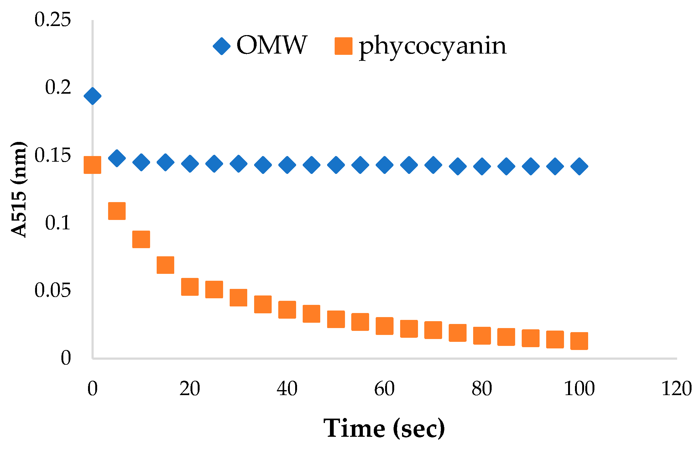
3.5. Antioxidant Assessment of the OMW and Spirulina maxima Extracts via DPPH Scavenging

The antioxidant capacity of the extracts was photometrically measured with the DPPH scavenging technique over time to observe the kinetics of the reaction between the free radicals and antioxidants of each extract. The DPPH assay was used to predict antioxidant activity via the mechanism in which antioxidants act as deterrents against lipid oxidation. Thus, the scavenging of the DPPH free radical occurs, and free radical scavenging capacity can be determined. Figure 6 shows the % inhibition of the DPPH free radical during a time period of 120 s after the addition of the sample in the DPPH solution.
Phycocyanin appeared to inhibit the free radical much more than the OMW extracts, reaching an inhibition percentage of 98.4% and 83.1%, respectively, after 100 s. The antioxidant capacity of polyphenols has been widely studied and proven to be very effective against free radicals and in particular DPPH [51,70]. Their antioxidant properties have also been studied in order to be used as natural antioxidant additives in cosmetics [35,71]. The main component that is found in OMW is hydroxytyrosol (HT) [72], while the rest consist of several flavonoids. On the other hand, phycocyanin has also been found to be effective in free radical scavenging, with multiple studies proving its potential as a natural antioxidant for food and cosmetic applications [73,74]. Phycocyanin is usually formed by polypeptide subunits (α and β), which are covalently linked to chromophores (bilins, phycocyanobilin (PCB), and phycoerythrobilin (PEB)) through thioether linkage [75]. It has been discovered that the α subunit contains one PCB, whereas the β subunit carries one PCB and one PEB [76]. While searching for mechanisms to explain phycocyanin’s antioxidant capacity, it was retorted that both apoprotein (α and β subunits) and PCB components are involved [77]. More specifically, apoprotein (subunits α and β) has been found to contribute to hydroxyl radical (OH) scavenging activity [78], and PCB scavenges most radicals [79]. It can be concluded that both phycocyanin and antioxidants from OMW are very powerful antioxidants. The difference between their scavenging ability in the present study is not very clear. Nevertheless, it could be assumed that the presence of glycerol in phycocyanin, which was used as a preservation medium, enhanced its antioxidant effect [68].

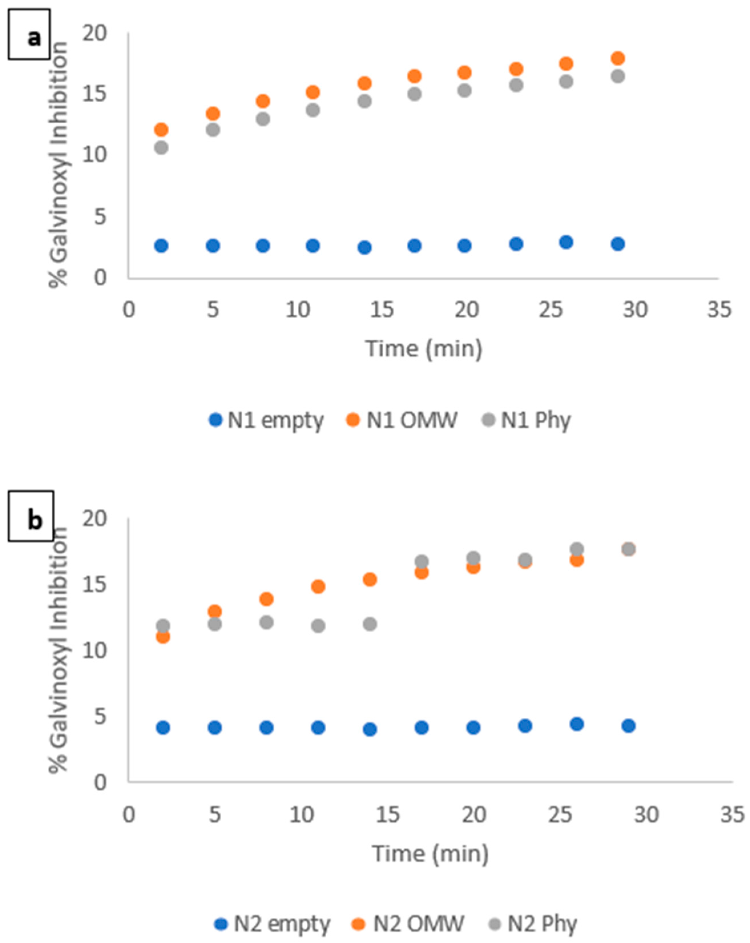
3.6. Determination of the Antioxidant Activity Using the Galvinoxyl Free Radical Detected via Electron Paramagnetic Resonance (EPR) Spectroscopy

Electron paramagnetic resonance (EPR) spectroscopy is a technique that measures free radical signals, which are directly indicative of their scavenging by antioxidants over time. The creation of free radicals on the skin can have effects that range between mild and very serious for human health. Hence, the scavenging ability of cosmetic products is of great importance and is currently under study [80,81].
The EPR spectroscopy measurements of the stable free radical galvinoxyl were carried out on both Ν1 (empty and with the additives) and Ν2 (empty and with the additives) systems. Figure 7a shows the gradual increase in the free radical’s inhibition with the addition of the additives in N1 systems. Both the antioxidants extracted from OMW and phycocyanin extracted from Spirulina maxima enhanced the sample’s antioxidant ability. In N1, the antioxidants from OMW increased the sample’s antioxidant capacity more than phycocyanin. This could be due to the chemical affinity of the water used in the aqueous phase and polyphenols, which were the main components of the OMW extracts. The results shown in Figure 7b, concerning N2 systems, do not differ significantly from those in Figure 7a. A slight increase in the N2 Phy system was observed, indicating a correlation between phycocyanin and the presence of glycerol. It has been found that glycerol acts protectively against phycocyanin degradation, and it is even used for storage [82].

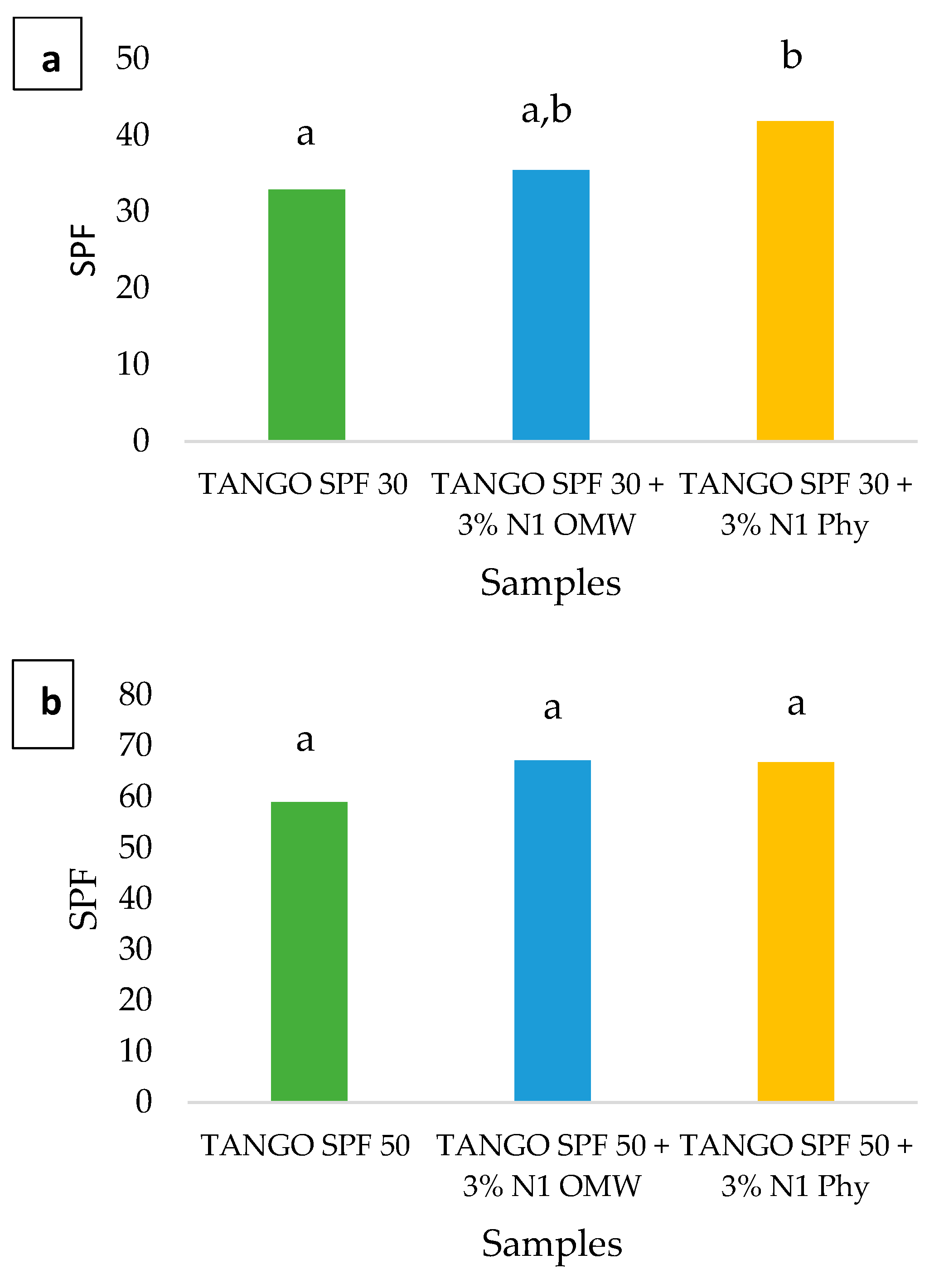
3.7. Clinical Pre-Assessment of the Sun Protection Factor (SPF)

The SPF booster efficiency of the extracts from OMW and phycocyanin from Spirulina maxima was tested via a clinical assessment of the SPF.
The SPF booster efficiency was assessed for the N1 loaded systems, namely N1 loaded with antioxidants extracted from OMW and N1 containing phycocyanin. N1 systems were chosen instead of N2 due to their increased stability at room temperature. The NE was added at three different concentrations, namely 1, 3, and 5% w/w, in the Tango Sun Lotion SPF 50. The results showed no significant differences between NE concentrations, so we decided to proceed with the 3% NE concentration for our investigation so that we could conduct the procedure with the addition of less NE, which also led to the highest efficiency. As shown in Figure 8a,b, both bioactives resulted in an increase in the SPF value of the original sun lotion. In the case of the sun lotion with an SPF value of 30, the addition of N1 OMW increased the SPF by 8%, while N1 Phy exhibited an increase of 27% when compared to the original sun lotion. For the case of sun lotion SPF 50, the obtained results were also positive for both additives. The presence of N1 OMW increased the SPF by 14%, while at the same time, N1 Phy revealed no significantly different results, increasing the SPF by 13%. The actual SPF of the original SPF 50 sun lotion is very close to the maximum effectiveness of a sun lotion, as has been proposed by the FDA [83,84]. This is probably why there was no significant variation in the additives’ booster efficiency since there was little room for further increase. A t-test was performed, and the results showed that, for the SPF 30 sun lotion, the contribution of phycocyanin was statistically significant in boosting the SPF value. OMW on the other hand, while increasing the SPF value in number, did not appear to do so in a significant way. An increase, but not statistically significant, was also observed with the additives in the SPF 50 sun lotion. The relevance or not of the statistical analysis is debatable due to the small number of test subjects, which resulted in high standard deviations.
Polyphenols, specifically the ones derived from plants, have been tested for years and have proven to be quite effective as anti-inflammatory, antioxidant, and DNA repair agents [85,86]. More recent research focuses more on the protection those substances can offer against UV radiation. Several published studies have demonstrated the effectiveness of NEs, thereby corroborating the data we obtained [34,87,88].
Phycocyanin is a naturally occurring compound of spirulina, a well-known type of cyanobacteria. The pigments of cyanobacteria, including spirulina, have been studied for their antioxidant as well as sun protective activities, giving promising results, thus supporting our claim of phycocyanin being a UV protection-booster agent [89,90].
The NEs were selected for the better delivery, bioavailability, and dispersion of the bioactives in the final product as well as for the protection they offer to the encapsulated substances, mainly against oxidation [91,92,93,94]. This capability of NEs has already been verified in numerous studies published over the years, so it can be assumed that the encapsulation of the bioactives in the NEs increases their effectiveness. Galanakis et. al. (2018) showed that the entrapment of phenols in silica particles prior to their emulsification in cosmetics improved their water resistance, which is also a very important criterion for a sunscreen’s actual effectiveness [33].

4. Conclusions

Antioxidants extracted from olive mill wastewater and phycocyanin extracted from Spirulina maxima were encapsulated in two W/O NEs. For the two NEs, IPM was used as the continuous phase, while water (N1) or a mixture of water and glycerol (N2) was used as the dispersed phase. The addition of the OMW extracts seemed to further stabilize the emulsion, while the presence of phycocyanin led to less stable systems. In N1, the antioxidants from OMW increased the sample’s antioxidant capacity more than phycocyanin. This could be due to the chemical affinity of the water, used as the aqueous phase, and the polyphenols, which were the main components of the OMW extracts. On the other hand, N2 containing OMW had similar antioxidant activity to the one containing phycocyanin. The N1 system was more stable than N2 when stored at room temperature, and it was the one that was used for the determination of the SPF boosting efficiency. The results we obtained show a significant increase in the SPF value when the two additives were present. Although both phycocyanin and OMW increased the SPF, OMW in SPF50 increased the SPF significantly.
Overall, the OMW and phycocyanin encapsulated in biocompatible NEs retained their antioxidant activity and could be used as components in cosmetic products for sun protection. The formulated systems also contained IPM, biocompatible surfactants, and glycerol, which are very commonly used ingredients in cosmetic creams since they have stabilizing and hydrating properties. OMW seemed to be very effective in boosting SPF protection, and it is also an inexpensive material, with a good extraction yield of antioxidants and no special know-how or facilities required for their extraction. Due to the aforementioned reasons, we believe NEs loaded with phycocyanin or antioxidants derived from OMW are highly promising alternatives for reducing reliance on chemical UV filters.

Author Contributions

Conceptualization, M.D.C. and A.B.; methodology, E.G., M.D.C. and T.G.S.; software, E.G.; validation, A.X., T.G.S. and A.B.; investigation, E.G., D.G. and K.T.; resources, A.B.; data curation, E.G.; writing—original draft preparation, E.G.; writing—review and editing, M.D.C.; visualization, M.D.C.; supervision, M.D.C. and A.B.; project administration, V.P.; funding acquisition, A.X. and A.B. All authors have read and agreed to the published version of the manuscript.

Funding

This research has been co-financed by the European Regional Development Fund of the European Union and Greek national funds through the Operational Program Competitiveness, Entrepreneurship, and Innovation, under the call RESEARCH–CREATE–INNOVATE, Τ2ΕΔΚ_03640.

Institutional Review Board Statement

The study was conducted in accordance with the Declaration of Helsinki and approved by the Ethics Committee of the National Kapodistrian University of Athens (protocol code 33685/5-4-2023 and date of approval 15 May 2023).

Informed Consent Statement

Informed consent was obtained from all subjects involved in the study.

Data Availability Statement

Data are available upon request.

Acknowledgments

We express our thanks to Giorgos Sotiroudis of the ICB/NHRF, for his aid during the extraction of phycocyanin. A.B. would like to thank the Laboratory of Pharmaceutical Technology, Unit of Skin Pharmacology and Cosmetology, located in Panepistimiopolis, Zografou, Athens, Greece, as well as Kapodistriaki Dermatofarmakeftiki P.C., for their collaboration in the performance of the SPF evaluation studies.

Conflicts of Interest

Kyriaki Tzoka, Antonios Bonos and Maria D. Chatzidaki the authors are employees of Cleanway Ltd. The authors declare that the research was conducted in the absence of any commercial or financial relationships that could be construed as a potential conflict of interest. The funders had no role in the design of the study; in the collection, analyses or interpretation of data; in the writing of the manuscript; or in the decision to publish the results.

References

  1. Leiter, U.; Keim, U.; Garbe, C. Epidemiology of Skin Cancer: Update 2019. Adv. Exp. Med. Biol. 2020, 1268, 123–139. [Google Scholar] [CrossRef] [PubMed]
  2. Cadet, J.; Douki, T. Formation of UV-Induced DNA Damage Contributing to Skin Cancer Development. Photochem. Photobiol. Sci. 2018, 17, 1816–1841. [Google Scholar] [CrossRef]
  3. Oral, D.; Yirun, A.; Erkekoglu, P. Safety Concerns of Organic Ultraviolet Filters: Special Focus on Endocrine-Disrupting Properties. J. Environ. Pathol. Toxicol. Oncol. 2020, 39, 201–212. [Google Scholar] [CrossRef]
  4. Chatzigianni, M.; Pavlou, P.; Siamidi, A.; Vlachou, M.; Varvaresou, A.; Papageorgiou, S. Environmental Impacts Due to the Use of Sunscreen Products: A Mini-Review. Ecotoxicology 2022, 31, 1331–1345. [Google Scholar] [CrossRef]
  5. Jesus, A.; Sousa, E.; Cruz, M.T.; Cidade, H.; Lobo, J.M.S.; Almeida, I.F. UV Filters: Challenges and Prospects. Pharmaceuticals 2022, 15, 263. [Google Scholar] [CrossRef]
  6. Levine, A. Reducing the Prevalence of Chemical UV Filters from Sunscreen in Aquatic Environments: Regulatory, Public Awareness, and Other Considerations. Integr. Environ. Assess. Manag. 2021, 17, 982–988. [Google Scholar] [CrossRef]
  7. Yarovaya, L.; Khunkitti, W. The Effect of Grape Seed Extract as a Sunscreen Booster Effect of Grape Seed Extract as a Sunscreening Booster. Songklanakarin J. Sci. Technol. 2019, 41, 708–715. [Google Scholar] [CrossRef]
  8. Aguilera, J.; Vicente-Manzanares, M.; de Gálvez, M.V.; Herrera-Ceballos, E.; Rodríguez-Luna, A.; González, S. Booster Effect of a Natural Extract of Polypodium Leucotomos (Fernblock®) That Improves the UV Barrier Function and Immune Protection Capability of Sunscreen Formulations. Front. Med. 2021, 8, 684665. [Google Scholar] [CrossRef]
  9. Radice, M.; Manfredini, S.; Ziosi, P.; Dissette, V.; Buso, P.; Fallacara, A.; Vertuani, S. Herbal Extracts, Lichens and Biomolecules as Natural Photo-Protection Alternatives to Synthetic UV Filters. A Systematic Review. Fitoterapia 2016, 114, 144–162. [Google Scholar] [CrossRef]
  10. Galanakis, C.M.; Tsatalas, P.; Galanakis, I.M. Phenols from Olive Mill Wastewater and Other Natural Antioxidants as UV Filters in Sunscreens. Environ. Technol. Innov. 2018, 9, 160–168. [Google Scholar] [CrossRef]
  11. Babić, S.; Malev, O.; Pflieger, M.; Lebedev, A.T.; Mazur, D.M.; Kužić, A.; Čož-Rakovac, R.; Trebše, P. Toxicity Evaluation of Olive Oil Mill Wastewater and Its Polar Fraction Using Multiple Whole-Organism Bioassays. Sci. Total Environ. 2019, 686, 903–914. [Google Scholar] [CrossRef] [PubMed]
  12. Rahmanian, N.; Jafari, S.M.; Galanakis, C.M. Recovery and Removal of Phenolic Compounds from Olive Mill Wastewater. JAOCS 2014, 91, 1–18. [Google Scholar] [CrossRef]
  13. Wang, T.; Zhao, J.; Yang, Z.; Xiong, L.; Li, L.; Gu, Z.; Li, Y. Polyphenolic Sunscreens for Photoprotection. Green Chem. 2022, 24, 3605–3622. [Google Scholar] [CrossRef]
  14. de Lima Cherubim, D.J.; Buzanello Martins, C.V.; Oliveira Fariña, L.; da Silva de Lucca, R.A. Polyphenols as Natural Antioxidants in Cosmetics Applications. J. Cosmet. Dermatol. 2020, 19, 33–37. [Google Scholar] [CrossRef] [PubMed]
  15. Wan, D.; Wu, Q.; Kuča, K. Chapter 42—Spirulina. In Nutraceuticals; Gupta, R.C., Ed.; Academic Press: Cambridge, MA, USA, 2016; pp. 569–583. ISBN 9780128021477. [Google Scholar] [CrossRef]
  16. Yarkent, Ç.; Gürlek, C.; Oncel, S.S. Potential of Microalgal Compounds in Trending Natural Cosmetics: A Review. Sustain. Chem. Pharm. 2020, 17, 100304. [Google Scholar] [CrossRef]
  17. Sarada, R.; Pillai, M.G.; Ravishankar, G.A. Phycocyanin from Spirulina Sp: Influence of Processing of Biomass on Phycocyanin Yield, Analysis of Efficacy of Extraction Methods and Stability Studies on Phycocyanin. Process. Biochem. 1999, 34, 795–801. [Google Scholar] [CrossRef]
  18. Jang, Y.A.; Kim, B.A. Protective Effect of Spirulina-Derived C-Phycocyanin against Ultraviolet B-Induced Damage in HaCaT Cells. Medicina 2021, 57, 273. [Google Scholar] [CrossRef]
  19. Vega, J.; Bonomi-Barufi, J.; Gómez-Pinchetti, J.L.; Figueroa, F.L. Cyanobacteria and Red Macroalgae as Potential Sources of Antioxidants and UV Radiation-Absorbing Compounds for Cosmeceutical Applications. Mar. Drugs 2020, 18, 659. [Google Scholar] [CrossRef] [PubMed]
  20. McClements, D.J. Nanoemulsions versus Microemulsions: Terminology, Differences, and Similarities. Soft Matter 2012, 8, 1719–1729. [Google Scholar] [CrossRef]
  21. Silva, H.D.; Cerqueira, M.Â.; Vicente, A.A. Nanoemulsions for Food Applications: Development and Characterization. Food Bioprocess Technol. 2012, 5, 854–867. [Google Scholar] [CrossRef] [Green Version]
  22. Golfomitsou, I.; Mitsou, E.; Xenakis, A.; Papadimitriou, V. Development of Food Grade O/W Nanoemulsions as Carriers of Vitamin D for the Fortification of Emulsion Based Food Matrices: A Structural and Activity Study. J. Mol. Liq. 2018, 268, 734–742. [Google Scholar] [CrossRef]
  23. Demisli, S.; Theochari, I.; Christodoulou, P.; Zervou, M.; Xenakis, A.; Papadimitriou, V. Structure, Activity and Dynamics of Extra Virgin Olive Oil-in-Water Nanoemulsions Loaded with Vitamin D3 and Calcium Citrate. J. Mol. Liq. 2020, 306, 112908. [Google Scholar] [CrossRef]
  24. Pathak, K.; Pattnaik, S.; Swain, K. Application of Nanoemulsions in Drug Delivery; Elsevier Inc.: Amsterdam, The Netherlands, 2018; pp. 415–433. ISBN 9780128118399. [Google Scholar] [CrossRef]
  25. Sánchez-López, E.; Guerra, M.; Dias-Ferreira, J.; Lopez-Machado, A.; Ettcheto, M.; Cano, A.; Espina, M.; Camins, A.; Garcia, M.L.; Souto, E.B. Current Applications of Nanoemulsions in Cancer Therapeutics. Nanomaterials 2019, 9, 821. [Google Scholar] [CrossRef] [Green Version]
  26. Mitsou, E.; Pletsa, V.; Sotiroudis, G.T.; Panine, P.; Zoumpanioti, M.; Xenakis, A. Development of a Microemulsion for Encapsulation and Delivery of Gallic Acid. The Role of Chitosan. Colloids Surf. B Biointerfaces 2020, 190, 110974. [Google Scholar] [CrossRef]
  27. Mitsou, E.; Kalogianni, E.P.; Georgiou, D.; Stamatis, H.; Xenakis, A.; Zoumpanioti, M. Formulation and Structural Study of a Biocompatible Water-in-Oil Microemulsion as an Appropriate Enzyme Carrier: The Model Case of Horseradish Peroxidase. Langmuir 2019, 35, 150–160. [Google Scholar] [CrossRef]
  28. Theochari, I.; Xenakis, A.; Papadimitriou, V. Nanocarriers for Effective Drug Delivery. In Smart Nanocontainers Micro Nano Technologies; Elsevier: Amsterdam, The Netherlands, 2020; pp. 315–341. [Google Scholar] [CrossRef]
  29. Mansur, M.C.P.P.R.; Campos, C.; Vermelho, A.B.; Nobrega, J.; da Cunha Boldrini, L.; Balottin, L.; Lage, C.; Rosado, A.S.; Ricci-Júnior, E.; dos Santos, E.P. Photoprotective Nanoemulsions Containing Microbial Carotenoids and Buriti Oil: Efficacy and Safety Study. Arab. J. Chem. 2020, 13, 6741–6752. [Google Scholar] [CrossRef]
  30. Faria-Silva, A.C.; Costa, A.M.; Ascenso, A.; Ribeiro, H.M.; Marto, J.; Gonçalves, L.M.; Carvalheiro, M.; Simões, S. Nanoemulsions for Cosmetic Products. Nanocosmetics 2020, 59–77. [Google Scholar] [CrossRef]
  31. Reis-Mansur, M.C.P.P.; Firmino Gomes, C.C.; Nigro, F.; Ricci-Júnior, E.; de Freitas, Z.M.F.; dos Santos, E.P. Nanotechnology as a Tool for Optimizing Topical Photoprotective Formulations Containing Buriti Oil (Mauritia flexuosa) and Dry Aloe vera Extracts: Stability and Cytotoxicity Evaluations. Pharmaceuticals 2023, 16, 292. [Google Scholar] [CrossRef]
  32. Yang, C.C.; Hung, C.F.; Chen, B.H. Preparation of coffee oil-algae oil-based nanoemulsions and the study of their inhibition effect on UVA-induced skin damage in mice and melanoma cell growth. Int. J. Nanomed. 2017, 12, 6559–6580. [Google Scholar] [CrossRef] [Green Version]
  33. Galanakis, C.M.; Tsatalas, P.; Galanakis, I.M. Implementation of Phenols Recovered from Olive Mill Wastewater as UV Booster in Cosmetics. Ind. Crops Prod. 2018, 111, 30–37. [Google Scholar] [CrossRef]
  34. Wiseva, K.O.; Widyastuti, F.; Faadhilah, H.; Wathoni, N. A review on various formulation of nanoemulsions in cosmetics with plant extracts as the active ingredients. Int. J. Appl. Pharm. 2021, 13, 34–40. [Google Scholar] [CrossRef]
  35. El-Abbassi, A.; Saadaoui, N.; Kiai, H.; Raiti, J.; Hafidi, A. Potential Applications of Olive Mill Wastewater as Biopesticide for Crops Protection. Sci. Total Environ. 2017, 576, 10–21. [Google Scholar] [CrossRef]
  36. Aissam, H.; Penninckx, M.J.; Benlemlih, M. Reduction of Phenolics Content and COD in Olive Oil Mill Wastewaters by Indigenous Yeasts and Fungi. World J. Microbiol. Biotechnol. 2007, 23, 1203–1208. [Google Scholar] [CrossRef]
  37. Hawker, M.; Wang, F.; Mouzakitis, Y.; Adamides, E.D. Techno-Economic Assessment of an Olive Mill Wastewater (OMWW) Biorefinery in the Context of Circular Bioeconomy. Eng 2022, 3, 488–503. [Google Scholar] [CrossRef]
  38. Economic Importance of Algae for NEET—Features, Types, Importance and Effects. Available online: https://www.vedantu.com/neet/economic-importance-of-algae (accessed on 31 March 2023).
  39. Pereira, A.G.; Fraga-Corral, M.; Garcia-Oliveira, P.; Otero, P.; Soria-Lopez, A.; Cassani, L.; Cao, H.; Xiao, J.; Prieto, M.A.; Simal-Gandara, J. Single-Cell Proteins Obtained by Circular Economy Intended as a Feed Ingredient in Aquaculture. Foods 2022, 11, 2831. [Google Scholar] [CrossRef] [PubMed]
  40. Leouifoudi, I.; Zyad, A.; Amechrouq, A.; Oukerrou, M.A.; Mouse, H.A.; Mbarki, M. Identification and Characterisation of Phenolic Compounds Extracted from Moroccan Olive Mill Wastewater. Food Sci. Technol. 2014, 34, 249–257. [Google Scholar] [CrossRef] [Green Version]
  41. Silveira, S.T.; Burkert, J.F.M.; Costa, J.A.V.; Burkert, C.A.V.; Kalil, S.J. Optimization of Phycocyanin Extraction from Spirulina Platensis Using Factorial Design. Bioresour. Technol. 2007, 98, 1629–1634. [Google Scholar] [CrossRef]
  42. Koukouraki, P.; Tsoupras, A.; Sotiroudis, G.; Demopoulos, C.A.; Sotiroudis, T.G. Antithrombotic Properties of Spirulina Extracts against Platelet-Activating Factor and Thrombin. Food Biosci. 2020, 37, 100686. [Google Scholar] [CrossRef]
  43. Olmo-García, L.; Fernández-Fernández, C.; Hidalgo, A.; Vílchez, P.; Fernández-Gutiérrez, A.; Marchal, R.; Carrasco-Pancorbo, A. Evaluating the Reliability of Specific and Global Methods to Assess the Phenolic Content of Virgin Olive Oil: Do They Drive to Equivalent Results? J. Chromatogr. A 2019, 1585, 56–69. [Google Scholar] [CrossRef]
  44. Patil, G.; Chethana, S.; Sridevi, A.S.; Raghavarao, K.S.M.S. Method to Obtain C-Phycocyanin of High Purity. J. Chromatogr. A 2006, 1127, 76–81. [Google Scholar] [CrossRef]
  45. Schwarz, K.; Bertelsen, G.; Nissen, L.R.; Gardner, P.T.; Heinonen, M.I.; Hopia, A.; Huynh-Ba, T.; Lambelet, P.; McPhail, D.; Skibsted, L.H.; et al. Investigation of plant extracts for the protection of processed foods against lipid oxidation. Comparison of antioxidant assays based on radical scavenging, lipid oxidation and analysis of the principal antioxidant compounds. Eur. Food Res. Technol. 2001, 212, 319–328. [Google Scholar] [CrossRef]
  46. Papadimitriou, V.; Sotiroudis, T.G.; Xenakis, A.; Sofikiti, N.; Stavyiannoudaki, V.; Chaniotakis, N.A. Oxidative Stability and Radical Scavenging Activity of Extra Virgin Olive Oils: An Electron Paramagnetic Resonance Spectroscopy Study. Anal. Chim. Acta 2006, 573–574, 453–458. [Google Scholar] [CrossRef] [PubMed]
  47. Nikolic, I.; Mitsou, E.; Damjanovic, A.; Papadimitriou, V.; Antic-Stankovic, J.; Stanojevic, B.; Xenakis, A.; Savic, S. Curcumin-Loaded Low-Energy Nanoemulsions: Linking EPR Spectroscopy-Analysed Microstructure and Antioxidant Potential with in Vitro Evaluated Biological Activity. J. Mol. Liq. 2020, 301, 112479. [Google Scholar] [CrossRef]
  48. Chatzidaki, M.D.; Papadimitriou, K.; Alexandraki, V.; Tsirvouli, E.; Chakim, Z.; Ghazal, A.; Mortensen, K.; Yaghmur, A.; Salentinig, S.; Papadimitriou, V.; et al. Microemulsions as Potential Carriers of Nisin: Effect of Composition on Structure and Efficacy. Langmuir 2016, 32, 8988–8998. [Google Scholar] [CrossRef]
  49. He, J.; Alister-Briggs, M.; de Lyster, T.; Jones, G.P. Stability and Antioxidant Potential of Purified Olive Mill Wastewater Extracts. Food Chem. 2012, 131, 1312–1321. [Google Scholar] [CrossRef]
  50. ISO 24444:2019 (E); Cosmetics—Sun Protection Test Methods—In Vivo Determination of the Sun Protection Factor (SPF). ISO: Geneva, Switzerland, 2019.
  51. Lafka, T.I.; Lazou, A.E.; Sinanoglou, V.J.; Lazos, E.S. Phenolic and Antioxidant Potential of Olive Oil Mill Wastes. Food Chem. 2011, 1, 92–98. [Google Scholar] [CrossRef]
  52. El-Abbassi, A.; Kiai, H.; Hafidi, A. Phenolic Profile and Antioxidant Activities of Olive Mill Wastewater. Food Chem. 2012, 132, 406–412. [Google Scholar] [CrossRef]
  53. Roig, A.; Cayuela, M.L.; Sánchez-Monedero, M.A. An Overview on Olive Mill Wastes and Their Valorisation Methods. Waste Manag. 2006, 26, 960–969. [Google Scholar] [CrossRef]
  54. Jerman Klen, T.; Mozetič Vodopivec, B. The Fate of Olive Fruit Phenols during Commercial Olive Oil Processing: Traditional Press versus Continuous Two- and Three-Phase Centrifuge. LWT Food Sci. Technol. 2012, 49, 267–274. [Google Scholar] [CrossRef]
  55. Khdair, A.; Abu-Rumman, G. Sustainable environmental management and valorization options for olive mill byproducts in the Middle East and North Africa (MENA) region. Processes 2020, 8, 671. [Google Scholar] [CrossRef]
  56. Adjali, A.; Clarot, I.; Chen, Z.; Marchioni, E.; Boudier, A. Physicochemical degradation of phycocyanin and means to improve its stability: A short review. J. Pharm. Anal. 2022, 12, 406–414. [Google Scholar] [CrossRef] [PubMed]
  57. Almog, R.; Marsilio, F.; Berns, D.S. Interaction of C-Phycocyanin with Lipid Monolayers under Nitrogen and in the Presence of Air. Arch. Biochem. Biophys. 1988, 260, 28–36. [Google Scholar] [CrossRef] [PubMed]
  58. Reitzer, F.; Allais, M.; Ball, V.; Meyer, F. Polyphenols at Interfaces. Adv. Colloid Interface Sci. 2018, 257, 31–41. [Google Scholar] [CrossRef]
  59. Sotiroudis, T.G.; Sotiroudis, G.T.; Varkas, N.; Xenakis, A. The Role of Endogenous Amphiphiles on the Stability of Virgin Olive Oil-in-Water Emulsions. JAOCS J. Am. Oil Chem. Soc. 2005, 82, 415–420. [Google Scholar] [CrossRef]
  60. Demisli, S.; Galani, E.; Goulielmaki, M.; Kyrilis, F.L.; Ilić, T.; Hamdi, F.; Crevar, M.; Kastritis, P.L.; Pletsa, V.; Nallet, F.; et al. Encapsulation of Cannabidiol in Oil-in-Water Nanoemulsions and Nanoemulsion-Filled Hydrogels: A Structure and Biological Assessment Study. J. Colloid Interface Sci. 2023, 634, 300–313. [Google Scholar] [CrossRef]
  61. Zhou, H.; Yue, Y.; Liu, G.; Li, Y.; Zhang, J.; Gong, Q.; Yan, Z.; Duan, M. Preparation and Characterization of a Lecithin Nanoemulsion as a Topical Delivery System. Nanoscale Res. Lett. 2010, 5, 224–230. [Google Scholar] [CrossRef] [Green Version]
  62. Sakulku, U.; Nuchuchua, O.; Uawongyart, N.; Puttipipatkhachorn, S.; Soottitantawat, A.; Ruktanonchai, U. Characterization and Mosquito Repellent Activity of Citronella Oil Nanoemulsion. Int. J. Pharm. 2009, 372, 105–111. [Google Scholar] [CrossRef]
  63. Saberi, A.H.; Fang, Y.; McClements, D.J. Effect of Glycerol on Formation, Stability, and Properties of Vitamin-E Enriched Nanoemulsions Produced Using Spontaneous Emulsification. J. Colloid Interface Sci. 2013, 411, 105–113. [Google Scholar] [CrossRef]
  64. Hategekimana, J.; Chamba, M.V.M.; Shoemaker, C.F.; Majeed, H.; Zhong, F. Vitamin E Nanoemulsions by Emulsion Phase Inversion: Effect of Environmental Stress and Long-Term Storage on Stability and Degradation in Different Carrier Oil Types. Colloids Surf. A Physicochem. Eng. Asp. 2015, 483, 70–80. [Google Scholar] [CrossRef]
  65. Katsouli, M.; Tzia, C. Effect of Lipid Type, Dispersed Phase Volume Fraction and Emulsifier on the Physicochemical Properties of Nanoemulsions Fortified with Conjugated Linoleic Acid (CLA): Process Optimization and Stability Assessment during Storage Conditions. J. Mol. Liq. 2019, 292, 111397. [Google Scholar] [CrossRef]
  66. Chatzidaki, M.D.; Mitsou, E.; Yaghmur, A.; Xenakis, A.; Papadimitriou, V. Formulation and Characterization of Food-Grade Microemulsions as Carriers of Natural Phenolic Antioxidants. Colloids Surf. A Physicochem. Eng. Asp. 2015, 483, 130–136. [Google Scholar] [CrossRef]
  67. Saberi, A.H.; Fang, Y.; McClements, D.J. Fabrication of Vitamin E-Enriched Nanoemulsions by Spontaneous Emulsification: Effect of Propylene Glycol and Ethanol on Formation, Stability, and Properties. Food Res. Int. 2013, 54, 812–820. [Google Scholar] [CrossRef]
  68. Montes de Oca-Ávalos, J.M.; Candal, R.J.; Herrera, M.L. Nanoemulsions: Stability and Physical Properties. Curr. Opin. Food Sci. 2017, 16, 1–6. [Google Scholar] [CrossRef]
  69. Visioli, F.; Romani, A.; Mulinacci, N.; Zarini, S.; Conte, D.; Vincieri, F.F.; Galli, C. Antioxidant and Other Biological Activities of Olive Mill Waste Waters. J. Agric. Food Chem. 1999, 47, 3397–3401. [Google Scholar] [CrossRef]
  70. Zillich, O.V.; Schweiggert-Weisz, U.; Eisner, P.; Kerscher, M. Polyphenols as active ingredients for cosmetic products. Int. J. Cosmet. Sci. 2015, 37, 455–464. [Google Scholar] [CrossRef] [PubMed]
  71. de Marco, E.; Savarese, M.; Paduano, A.; Sacchi, R. Characterization and Fractionation of Phenolic Compounds Extracted from Olive Oil Mill Wastewaters. Food Chem. 2007, 104, 858–867. [Google Scholar] [CrossRef]
  72. Wu, H.L.; Wang, G.H.; Xiang, W.Z.; Li, T.; He, H. Stability and Antioxidant Activity of Food-Grade Phycocyanin Isolated from Spirulina Platensis. Int. J. Food Prop. 2016, 19, 2349–2362. [Google Scholar] [CrossRef] [Green Version]
  73. Renugadevi, K.; Valli Nachiyar, C.; Sowmiya, P.; Sunkar, S. Antioxidant Activity of Phycocyanin Pigment Extracted from Marine Filamentous Cyanobacteria Geitlerinema Sp TRV57. Biocatal. Agric. Biotechnol. 2018, 16, 237–242. [Google Scholar] [CrossRef]
  74. Ashaolu, T.J.; Samborska, K.; Lee, C.C.; Tomas, M.; Capanoglu, E.; Tarhan, Ö.; Taze, B.; Jafari, S.M. Phycocyanin, a Super Functional Ingredient from Algae; Properties, Purification Characterization, and Applications. Int. J. Biol. Macromol. 2021, 193, 2320–2331. [Google Scholar] [CrossRef]
  75. Wang, L.; Qu, Y.; Fu, X.; Zhao, M.; Wang, S.; Sun, L. Isolation, Purification and Properties of an R-Phycocyanin from the Phycobilisomes of a Marine Red Macroalga Polysiphonia Urceolata. PLoS ONE 2014, 9, e87833. [Google Scholar] [CrossRef] [Green Version]
  76. Fernández-Rojas, B.; Hernández-Juárez, J.; Pedraza-Chaverri, J. Nutraceutical Properties of Phycocyanin. J. Funct. Foods 2014, 11, 375–392. [Google Scholar] [CrossRef]
  77. Huang, Z.; Guo, B.J.; Wong, R.N.S.; Jiang, Y. Characterization and Antioxidant Activity of Selenium-Containing Phycocyanin Isolated from Spirulina Platensis. Food Chem. 2007, 100, 1137–1143. [Google Scholar] [CrossRef]
  78. Romay, C.; Gonzalez, R.; Pizarro, M.; Lissi, E. Kinetics of C-Phycocyanin Reaction with Hypochlorite. J. Protein Chem. 2000, 19, 151–155. [Google Scholar] [CrossRef]
  79. Herrling, T.; Seifert, M.; Sandig, G.; Jung, K. The Determination of the Radical Power—An in Vitro Test for the Evaluation of Cosmetic Products. Int. J. Cosmet. Sci. 2016, 38, 232–237. [Google Scholar] [CrossRef]
  80. Quiñones, R.; Kolling, D.R.J.; Shoup, D.; Smythers, A.L.; Nickel, S.; Westfall, T.D.; Epperly, C.; Coplin, M. Comparing Free Radicals in Sunscreen-Treated Pig Skin by Using Electron Paramagnetic Resonance Spectroscopy. J. Chem. Educ. 2019, 96, 2021–2028. [Google Scholar] [CrossRef]
  81. Markou, G.; Kougia, E.; Kefalogianni, I.; Tsagou, V.; Arapoglou, D.; Chatzipavlidis, I. Effect of Glycerol Concentration and Light Intensity on Growth and Biochemical Composition of Arthrospira (Spirulina) Platensis: A Study in Semi-Continuous Mode with Non-Aseptic Conditions. Appl. Sci. 2019, 9, 4703. [Google Scholar] [CrossRef] [Green Version]
  82. Wang, S.Q.; Lim, H.W. Current Status of the Sunscreen Regulation in the United States: 2011 Food and Drug Administration’s Final Rule on Labeling and Effectiveness Testing. J. Am. Acad. Dermatol. 2011, 65, 863–869. [Google Scholar] [CrossRef]
  83. Sayre, R.M.; Dowdy, J.C.; Lott, D.L.; Marlowe, E. Commentary on ‘UVB-SPF’: The SPF Labels of Sunscreen Products Convey More than Just UVB Protection. Photodermatol. Photoimmunol. Photomed. 2008, 24, 218–220. [Google Scholar] [CrossRef]
  84. Carrara, M.; Kelly, M.T.; Roso, F.; Larroque, M.; Margout, D. Potential of Olive Oil Mill Wastewater as a Source of Polyphenols for the Treatment of Skin Disorders: A Review. J. Agric. Food Chem. 2021, 69, 7268–7284. [Google Scholar] [CrossRef] [PubMed]
  85. Nichols, J.A.; Katiyar, S.K. Skin Photoprotection by Natural Polyphenols: Anti-Inflammatory, Antioxidant and DNA Repair Mechanisms. Arch. Dermatol. Res. 2010, 302, 71–83. [Google Scholar] [CrossRef] [Green Version]
  86. Choquenet, B.; Couteau, C.; Paparis, E.; Coiffard, L.J.M. Flavonoids and Polyphenols, Molecular Families with Sunscreen Potential: Determining Effectiveness with an in vitro Method. Nat. Prod. Commun. 2009, 4, 227–230. [Google Scholar] [CrossRef] [PubMed] [Green Version]
  87. Shin Yi, N.; Gew, L.T.; Eh Suk, V.R. Plant Polyphenols as Green Sunscreen Ingredients: A Systematic Review. J. Cosmet. Dermatol. 2022, 21, 5409–5444. [Google Scholar] [CrossRef]
  88. Puglisi, R.; Biazzi, E.; Gesmundo, D.; Vanni, R.; Tava, A.; Cenadelli, S. The Antioxidant Activity of a Commercial and a Fractionated Phycocyanin on Human Skin Cells In Vitro. Molecules 2022, 27, 5276. [Google Scholar] [CrossRef] [PubMed]
  89. Wada, N.; Sakamoto, T.; Matsugo, S. Multiple Roles of Photosynthetic and Sunscreen Pigments in Cyanobacteria Focusing on the Oxidative Stress. Metabolites 2013, 3, 463–483. [Google Scholar] [CrossRef] [PubMed]
  90. Liu, T.; Liu, L. Fabrication and Characterization of Chitosan Nanoemulsions Loading Thymol or Thyme Essential Oil for the Preservation of Refrigerated Pork. Int. J. Biol. Macromol. 2020, 162, 1509–1515. [Google Scholar] [CrossRef]
  91. Lohith Kumar, D.H.; Sarkar, P. Encapsulation of Bioactive Compounds Using Nanoemulsions. Environ. Chem. Lett. 2017, 16, 59–70. [Google Scholar] [CrossRef]
  92. Pandey, P.; Gulati, N.; Makhija, M.; Purohit, D.; Dureja, H. Nanoemulsion: A Novel Drug Delivery Approach for Enhancement of Bioavailability. Recent Pat. Nanotechnol. 2020, 14, 276–293. [Google Scholar] [CrossRef]
  93. Choi, S.J.; McClements, D.J. Nanoemulsions as Delivery Systems for Lipophilic Nutraceuticals: Strategies for Improving Their Formulation, Stability, Functionality and Bioavailability. Food Sci. Biotechnol. 2020, 29, 149–168. [Google Scholar] [CrossRef]
  94. Sonneville-Aubrun, O.; Yukuyama, M.N.; Pizzino, A. Application of Nanoemulsions in Cosmetics. Nanoemuls. Formul. Appl. Charact. 2018, 435–475. [Google Scholar] [CrossRef]
Figure 1. Schematic representation of the extraction of polyphenols from OMW.
Cosmetics 10 00102 g001
Figure 2. Schematic representation of the extraction of phycocyanin from Spirulina maxima.
Cosmetics 10 00102 g002
Figure 3. DLS measurements of the systems’ droplets’ mean diameter in the course of time for (a) N1 empty, N1 OMW, and N1 Phy systems and (b) N2 empty, N2 OMW, and N2 Phy systems.
Cosmetics 10 00102 g003
Figure 4. DLS measurements of the systems’ PDI in the course of time for (a) N1 empty, N1 OMW, and N1 Phy systems, and (b) N2 empty, N2 OMW, and N2 Phy systems.
Cosmetics 10 00102 g004
Figure 5. The EPR spectrum of the N1 empty system labeled using the 5-DSA spin probe.
Cosmetics 10 00102 g005
Figure 6. % Inhibition of DPPH free radical in the presence of oil mill wastewater extracts from the first-phase waste products of a three-phase olive mill (♦) and phycocyanin extracted from Spirulina maxima (■).
Cosmetics 10 00102 g006
Figure 7. % Inhibition of the galvinoxyl free radical in (a) N1 systems (empty and loaded with OMW extracts or phycocyanin) and (b) N2 systems (empty and loaded with OMW extracts or phycocyanin).
Cosmetics 10 00102 g007
Figure 8. SPF booster efficiency of N1 OMW and N1 Phy systems on (a) Tango Sun Lotion SPF 30 and (b) Tango Sun Lotion SPF 50. t-test was performed for the statistical analysis. Significant differences between values are indicated by different letters a,b (p < 0.1).
Cosmetics 10 00102 g008
Table 1. Composition of the NEs.
Composition (% w/w)Nanoemulsions
N1N2
IPM86.2581.25
Water1010
Glycerol-5
Plurol Oleique1.51.5
Plurol Diisostearique0.750.75
Labrasol0.750.75
Dehymuls PGPH0.750.75
Table 2. Mean SPF and acceptance limits for reference sunscreen formulations.
Reference Sunscreen FormulationMean SPFAcceptance Range
Lower LimitUpper Limit
P643.031.054.9
P863.143.982.3
Table 3. Total phenol content of the samples (mg/L) expressed in gallic acid equivalents (GAEs). Each value in the table is represented as mean ± SD (n = 3).
SampleTotal Phenols
(mg/L of Extract)
Total Phenols
(mg/L of Wastewater)
Three-phase OMW (first-stage waste)3131.9 ± 0.81565.5 ± 0.5
Three-phase OMW (second-stage waste)115.4 ± 0.957.7 ± 0.3
Two-phase OMW24.6 ± 0.212.3 ± 0.9
Table 4. The rotational correlation time (τR) and order parameter (S) values of 5-DSA for N1 and N2 NEs. Each value in the table is represented as mean ± SE (n = 3).
System5-DSA
τR (ns)S
Ν1 empty0.57 ± 0.080.05 ± 0.01
N1 OMW0.59 ± 0.020.06 ± 0.01
N1 Phy0.63 ± 0.060.05 ± 0.01
N2 empty0.67 ± 0.040.05 ± 0.01
N2 OMW0.54 ± 0.030.05 ± 0.01
N2 Phy0.58 ± 0.050.05 ± 0.01
Disclaimer/Publisher’s Note: The statements, opinions and data contained in all publications are solely those of the individual author(s) and contributor(s) and not of MDPI and/or the editor(s). MDPI and/or the editor(s) disclaim responsibility for any injury to people or property resulting from any ideas, methods, instructions or products referred to in the content.

