Communication

Brevundimonas aurantiaca M3d10, Isolated from the Olive Fly, Produces Hydroxylated Astaxanthin

1
Department of Biotechnology, Chemistry and Pharmacy (DBCF), University of Siena, 53100 Siena, Italy
2
Department of Agronomy Food Natural Resources Animals and Environment (DAFNAE), University of Padua, 35020 Legnaro, Italy
3
Department of Physical Sciences, Earth and Environment (DSFTA), University of Siena, 53100 Siena, Italy
4
Department of Life Sciences (DVS), University of Siena, 53100 Siena, Italy
5
National Biodiversity Future Centre (NBFC), 90133 Palermo, Italy
*
Author to whom correspondence should be addressed.
Cosmetics 2023, 10(4), 103; https://doi.org/10.3390/cosmetics10040103
Submission received: 15 June 2023 / Revised: 11 July 2023 / Accepted: 13 July 2023 / Published: 15 July 2023
(This article belongs to the Collection Feature Papers in Cosmetics in 2023)

Abstract

:
In recent years, the exploitation of bacteria for the production of carotenoids has become of great interest as a sustainable alternative to chemical synthesis, which is expensive and technically challenging. This study contributes to the repertoire of carotenogenic bacteria by reporting the isolation of an orange-pigmented bacterium from the gut of adult olive flies. The novel isolate, designated as M3d10, shared 100% identity with Brevundimonas aurantiaca strain CB-R 16S ribosomal RNA, and, through a preliminary characterization, its orange pigment was predicted to be a hydroxylated astaxanthin derivative.

1. Introduction

Carotenoids are a large group of lipophilic pigment with yellow-to-red coloring properties characterized by a wide range of biological properties [1]; among them, the keto-carotenoid pigment astaxanthin (ASX) is known as a potent antioxidant with high potential for human health [2]. A large number of studies have reported anti-inflammatory, photoprotective, anticancer, antiatherosclerotic, neuroprotective, repairing, antiproliferative and potentially antiaging properties of ASX [3,4]; consequently, its demand has rapidly increased involving many industrial sectors, such as food, textiles, pharmaceuticals and cosmetics. It has been estimated that ASX commercialization will reach about USD 880 million by 2026 [5].
Currently, more than 95% of commercial ASX is produced by chemical synthesis from petrochemical precursors. The extraction from natural sources and biotechnological synthesis is limited by low yields and high production costs [2].
Based on the configuration of the two hydroxyl groups in position 3 and 3′ (Figure 1), three ASX isomers were characterized: (3S,3′S), (3R,3′R) and (3R,3S′). Synthetic ASX consists of a mixture of the three isomers in the ratio 1:1:2, while the natural carotenoid, which derives from stereospecific enzymatic synthesis, contains two isomers: (3S,3′S), and (3R,3′R) [2]. The synthetic product shows some safety concerns due to contamination by intermediates and by-products in addition to the environmental impact of the process [6]. Besides being harmless, natural ASX, particularly the configurational isomers (3S,3′S), shows higher biological activity than the corresponding synthetic compound [6,7].
In nature, ASX mainly occurs in the marine environment. Natural producers used for its commercial production are the green algae Haematococcus pluvialis, the yeast Xanthophyllomyces dendrorhous and the bacterium Paracoccus carotinifaciens [2,6,7]. H. pluvialis accumulates the highest amount of natural ASX, and it represents the primary source of the isomer (3S,3′S) [2]. ASX production using this slow-growing microalgae is expensive; it requires high light intensity and a long period of cultivation [8]. X. dendrorhous is renowned for being the sole natural producer of (3R,3′R) ASX, but the elevated cost of the fermentation process hinders the large-scale production of this carotenoid [6]. The highest level of bacterial ASX is produced by P. carotinifaciens; nonetheless, the yield of ASX obtained is generally low [7]. Beside Paracoccus spp. other ASX-producing bacteria, mostly isolated from marine environments, have been reported. They include cyanobacteria (Synechoccus spp., Phormidium spp., Oscillatoria subbrevis), members of the genus Sphingomonas and Brevundimonas; few of them were found to synthesize high-quality carotenoids but at low concentration [9].
Application of metabolic engineering has succeeded in producing ASX in non-carotenogenic microorganism including Escherichia coli and Saccharomices cerevisiae; nonetheless, the enhancement of carotenoid productivity obtained was insufficient for commercial application [6,7,10]. The first structural analysis of the gene cluster encoding carotenoid biosynthetic enzymes in the genus Brevundimonas was carried on the marine bacterium Brevundimonas sp. strain SD212. The sequence analysis indicated the presence of seven know genes (crtW, crtY, crtI, crtB, crtE, idi, crtZ), and a new gene (designated crtG), which was found to encode the novel enzyme carotenoid 2,2′-β-hydroxylase (CrtG) [11]. When crtG was introduced in various combinations with other crt genes in E. coli in engineered metabolic pathways, structurally novel or rare carotenoids with a 2-hydroxy group and 2,2′-dihydroxy groups were produced. The new carotenoids were suggested to have a much stronger antioxidant activity than the precursor [11]. Although altogether the demand for exploration of microbial sources exploitable for ASX production is high, bacterial ASX is still the least developed and less used [12]. This also applies to its β-ring(s)-2(2′)-hydroxylated derivatives, which are indeed very difficult to synthesize chemically due to high density around the 1,2-positions of the β-ring [13].
Within this framework, the availability of a large and diverse set of bacteria producing added-value compounds such as ASX might provide new opportunities to study and eventually manipulate different genetic backgrounds. This work aimed to increase the number of candidates for such a collection by reporting the novel isolation from the intestinal tract of the olive fly, Bactrocera oleae (Diptera, Tephritidae) [14], of an orange-pigmented bacterium. The novel isolate was assigned to the species Brevundimonas aurantiaca on the basis of 16S rRNA gene sequencing, and its orange pigment was predicted to be a hydroxyled ASX derivative.

2. Materials and Methods

2.1. Materials

Fructose, glucose and galactose were reported to be the main sugars found in the olive pulp on which B. oleae larvae feed by digging tunnels [15]. A modified MacConkey nutrient agar (MF) (2% Bacto tryptone, 1% fructose, 0.5% NaCl, 0.003% neutral red, 0.0001% crystal violet, 0.7–1.5% agar, distilled water, pH 7.2) was used for the first bacterial isolation during a study designed to identify factors that could support the growth in vitro of ‘Candidatus Erwinia dacicola’, the well-known endosymbiont of the olive fly [16,17]. This modified medium enables to check for the presence of bacteria able to degrade fructose by the direct visual enumeration of red colonies.
Luria Bertani broth (LB) (1% Bacto tryptone, 0.5% yeast extract, 0.5% NaCl, distilled water, pH 7.2), and LBA (LB added with 1.5% agar) were used for routine culture maintenance unless otherwise stated.

2.2. Isolation and Molecular Identification of a Carotenoid-Producing Bacterium

The bacterial strain used for this study was isolated from adults of wild olive flies. The adult flies were obtained from pupae collected from infested fruits in a pesticide-free olive orchard in north-east Italy (Bassano del Grappa—Veneto Region). Before dissection, insect specimens were submerged for 5 min in 1% sodium hypochlorite, rinsed in distillate sterile water at least twice, and air-dried under aseptic conditions. Sterilized tools were used to dissect the insects under a stereoscope and to extract the specialized foregut eversions (oesophageal bulbs—EB) which were there then transferred in sterile saline solution (0.9% NaCl (w/v)) at room temperature (18–25 °C). Samples were vortexed and then used for further examinations. One test consisted in transferring 0.1 mL of the suspension to ~5 mL of cooled but still molten MF (0.7% agar), maintained at 45 °C, and poured, after carefully mixing, onto an MF plate (1.5% agar) by spreading over its surface. The overlay was allowed to harden and then the plates were incubated at room temperature (18–25 °C) for up to six days. Individual colonies were picked and transferred to MF and LBA plates. Ten isolates were sub-cultured in LBA at least three times, and then stored in LB broth with 30% glycerol (v/v) at −80 °C. Gram determination was carried out either through staining (Gram stain kit, Carlo Erba Reagents) or chemical methods [18]. Catalase and oxidase activity were determined according to Smibert and Krieg [19]. The molecular identification was performed using the analysis of 16S rRNA gene sequence. An individual bacterial colony was suspended in 50 μL of sterile double-distilled water and incubated for 5 min at 100 °C. The 16S rRNA gene was amplified using eubacterial universal primers and sequenced on both strands at BMR Genomics (Padua, Italy) [20]. Sequences were analyzed using the BLAST algorithm [21]. The sequence data was submitted to the DDB/EMBL/GenBank database under the accession number MZ391833.

2.3. Extraction of Carotenoids

For carotenoid extraction, bacteria were grown at room temperature (18–25 °C) for three days on LBA supplemented with 1% of glucose [22]. Cells were then scraped from the surface of the agar plates, suspended in sterile distilled water and harvested via centrifugation at 12,000 rpm for 5 min. Cell pellet (~0.5 g wet weight) was washed with sterile double-distilled water and extracted with 1 mL of acetone at 55 °C for 15 min with intermittent vortexing [10]. The acetone supernatant obtained after centrifugation was transferred to a new tube. Extracted carotenoids were analyzed using HPLC-DAD.

2.4. HPLC Analysis

HPLC-DAD analysis was performed by using a Shimadzu Prominence LC 2030 3D instrument equipped with a Bondapack C18 column, 10 mm, 125 Å, 3.9 mm × 300 mm column (Waters Corporation, Milford, MA, USA).
A water solution containing 0.2% (v/v) formic acid (A) and acetonitrile with 0.2% (v/v) of formic acid (B) was used as the mobile phase. The following program was applied: B from 30% at 0 min to 85% at 25 min; flow was set at 0.9 mL/min. Chromatograms were recorded at 480 nm. Analyses were performed using 20 μL of acetone extract. ASX (Merck, Darmstadt, Germany) was used as an external standard. The calibration curve was established using a reference standard ranging from 0.250 mg/mL to 0.001 mg/mL. The correlation coefficient (R2) was >0.99.
Identification of carotenoids was performed with HPL UV/MS analysis using Agilent 1100 LC/MS DVL System (G1946C) (Agilent Technologies, Palo Alto, CA, USA) equipped with Phenomenex Kinetex C18-100Å column (100 × 4.6 mm; 2.6 μm particle size) at flow rate of 0.6 mL/min, operating with a gradient elution of A: water and B: acetonitrile: t0 = 0 min 5% of B, t = 1 min 5% B, t = 10 min 95% of B and kept to 19 min, t = 20 min 5% of B. Both solvents were acidified with 0.1% (v/v) of formic acid. Analysis was carried out on acetone extract evaporated to dryness by nitrogen gas and re-dissolved in acetonitrile. UV detection was monitored from 200 nm to 600 nm. MS analysis was performed in both positive and negative modes with a scan range of 100–500 m/z; fragmentor voltage was set at 70 V.

3. Results and Discussion

3.1. Isolation and Molecular Identification of a Carotenoid-Producing Bacterium

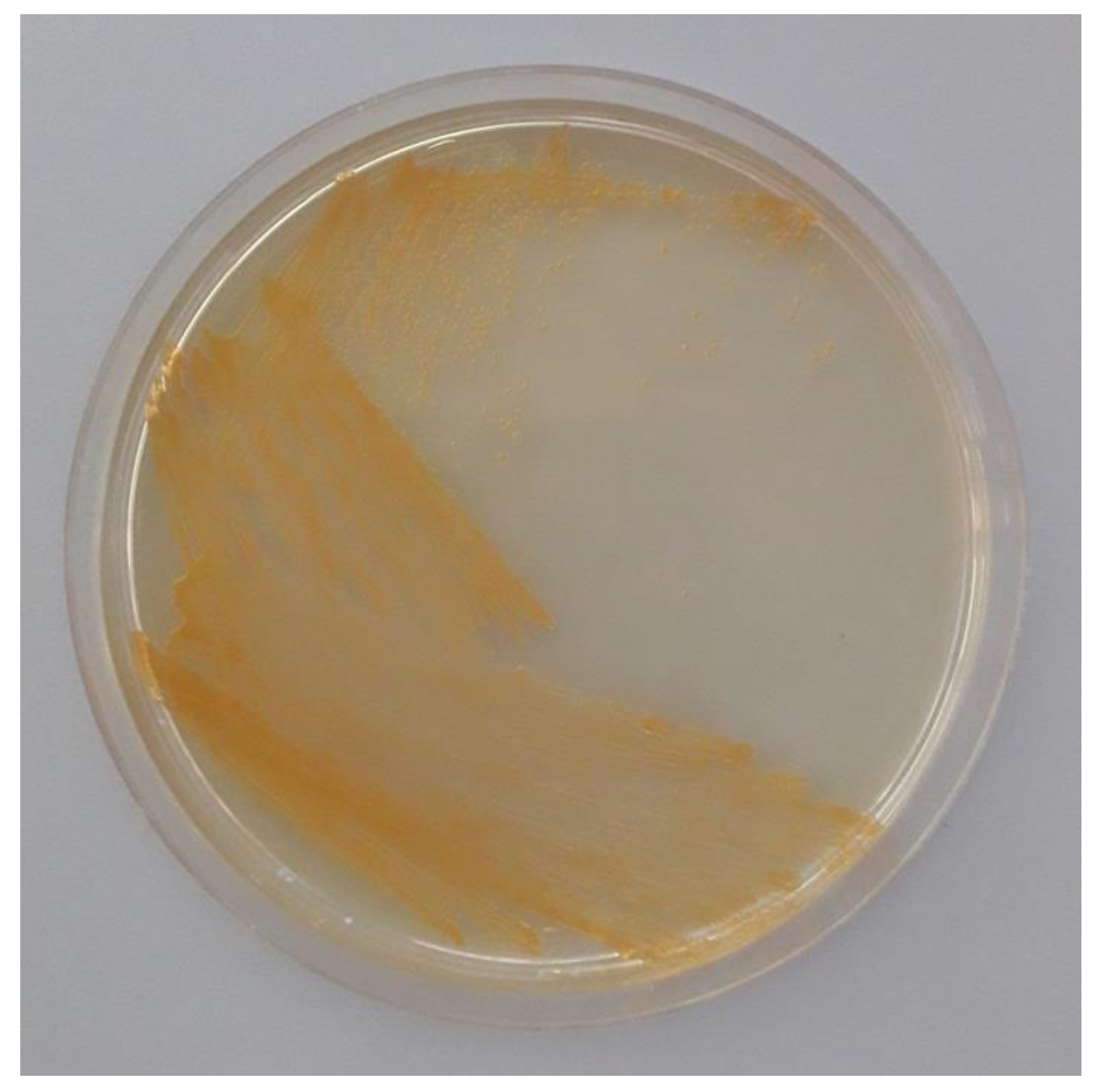
After six days incubation, about 50 round, smooth, convex and apparently bright red colonies were recorded on MF plates. Ten of them were transferred onto new MF plates, and then subcultured on LBA where they produced orange-pigmented growth (Figure 2).
All ten isolates resulted in Gram-negative rod-shaped bacteria able to produce the enzymes catalase and cytochrome oxidase. The 16S rRNA gene sequence is by far the most common housekeeping genetic marker used to study bacterial phylogeny and taxonomy [23]. In order to ascertain the phylogenetic position of the isolates, a near complete sequence of 16S rRNA gene (1413 bp) was determined for one of the ten isolates with identical morphological and growth characteristics. A BLAST search with the 16S rDNA sequence against EMBL nucleotide database indicated that it shared 100% identity with Brevundimonas aurantiaca strain CB-R 16S ribosomal RNA (GenBank: NR_028889.1) [24]. The newly isolated strain was thus designated as B. aurantiaca M3d10. The finding in insects of pigmented extracellular bacteria has been reported over the years, and a possible role of pigmented bacteria in the protection against pathogens or predators of the insect host has been proposed [25]. The species B. aurantiaca was initially isolated from a contaminated Chlorella culture [24], while the first report in insects dates back to 2011 when bacteria showing 99% identity with the 16S rRNA gene of B. aurantiaca strain CB-R [23] were recovered, both by culturable and molecular methods, from field-caught specimens of Anopheles stephensi (Diptera, Culicidae) in south west and northern Iran [26]. One year later, B. aurantiaca strains were cultivated from larval tissues of Ostrinia nubulalis (Lepidoptera, Pyralidae) collected from different maize fields in the eastern Black Sea region of Turkey [27]. Nonetheless, the genus Brevundimonas had earlier been associated with B. oleae, when Belcari et al. [28] reported the isolation of B. vesicularis from the oesophageal bulbs of wild olive flies collected in the Tuscany Region (west-central Italy).

3.2. Identification of Carotenoids

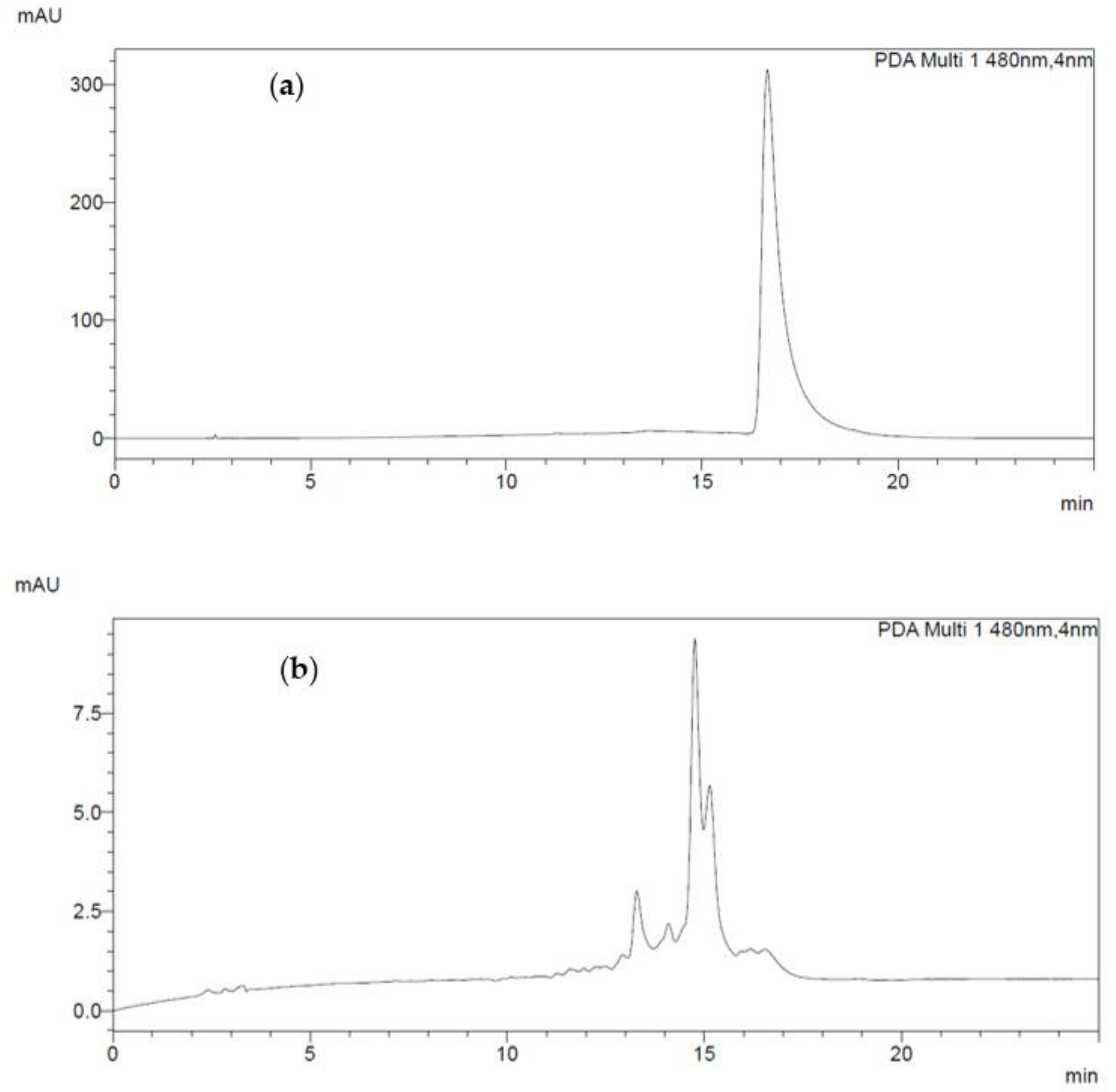
HPLC analysis indicated that B. aurantiaca M3d10 synthesizes at least four compounds with UV absorption spectra very similar to those of ASX used as reference (Figure 3). All of them were likely more polar than ASX since they eluted earlier.
The total carotenoid expressed as ASX was 11.12 mg/L. The component with retention time (Rt) 16.2 min is present at the level of 1.31 mg/L. These results show that B. aurantiaca M3d10 does not produce ASX itself, but its derivatives.
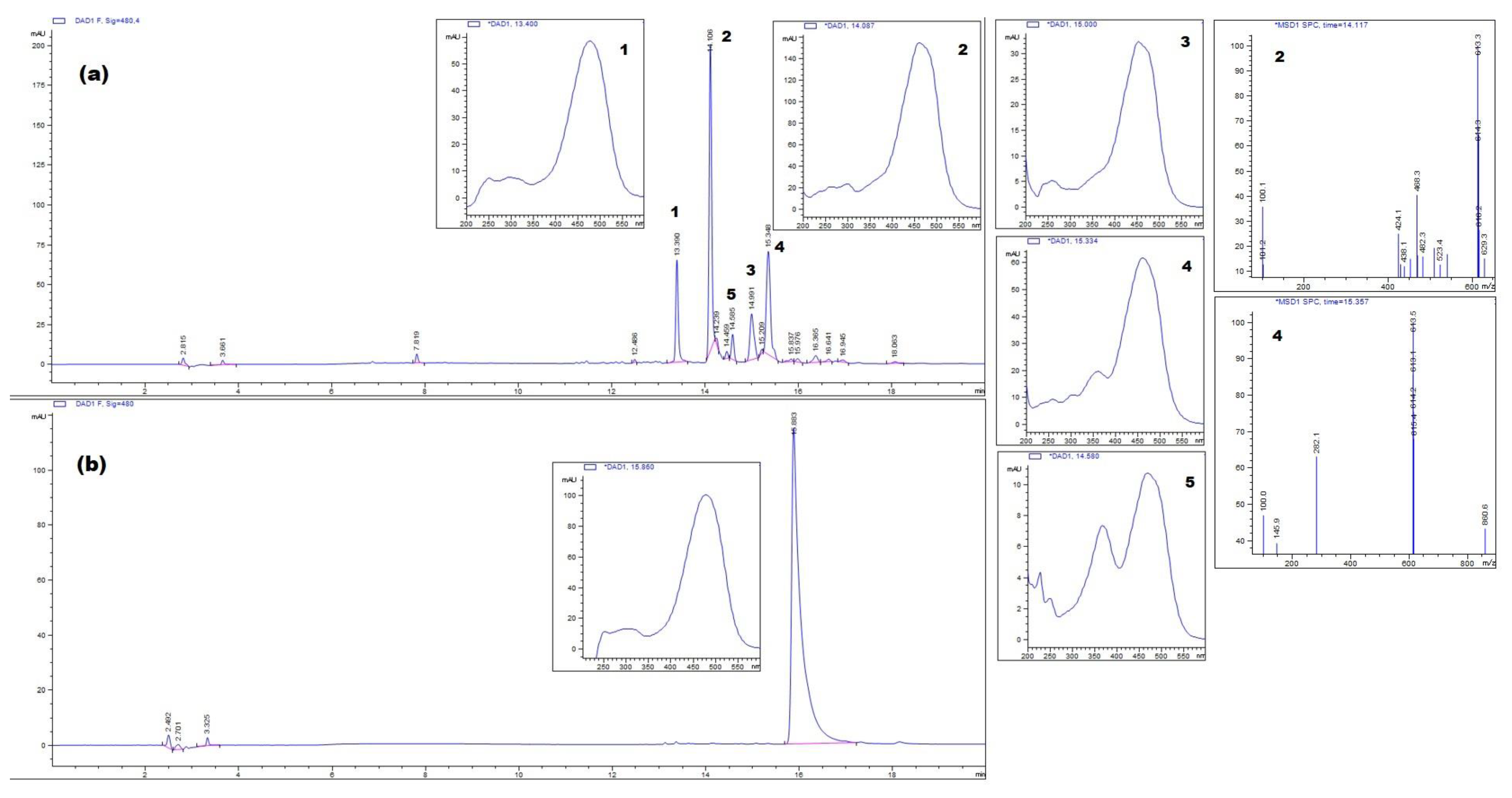
For a better characterization, the extract was further analyzed by HPLC-MS. This analysis detected four major peaks (Figure 4a).
The identified profile was characterized by two main peaks at Rt 14.1 and 15.3 min and two minor peaks at Rt 13.4 and 15.00 min, respectively. ASX, analyzed under the same conditions, showed a Rt 15.8 min (Figure 4b). The four compounds had the absorption spectra identical to that ASX (λmax 480 nm). A second absorption at 360 nm was present for the peak with Rt 15.3 min. This was even more evident in another minor peak at 14.5 min. MS spectra of the peaks at 14.1 and 15.3 min gave a signal of 613 m/z, corresponding to the pseudomolecular ion of an hydroxyl derivative of ASX (Figure 4a).
HPLC elution times, absorption spectra and molecular weights reveal that the two major carotenoids produced by M3d10 are likely hydroxyl-astaxanthins. Moreover, the occurrence of a peak at Rt 13.4 indicates the presence of a more polar compound which might be a dihydroxy-ASX. Similar results were previously reported for other Brevundimonas spp. of different origins [29,30,31]. The marine isolate Brevundimonas sp. strain SD212 was reported to produce seven carotenoid compounds: (2R,3S,32S)-2-hydroxyastaxanthin, (2R,3S,32R)-2-hydroxyadonixanthin, (3S,22R,32R)-erythroxanthin, (2R,3S,22R,32S)-2,3,22,32-tetrahydroxy-β,β-carotene-4,42-dione), (2R,3S,22R,32R)-2,3,22,32-tetrahydroxy-β,β-caroten-4-one, (3S,32S)-astaxanthin and (3S,32R)-adonixanthin [29]. The soil bacterium B. vescicularis strain DC263 was also reported to produce 2,2′-dihydroxyastaxanthin and 2,2′-dihydroxyadonixanthin as its major carotenoids [30]; in this strain, the carotenoid synthesis gene cluster was found to share the same organization as that reported from strain SD212 [11,30]. The 2,2′-dihydroxy-astaxanthin was also the major carotenoid produced by B. scallop isolated from the gut content of a marine bivalve Chlamys nobilis [31]. In addition, the marine bacterium designated as Brevundimonas sp. strain N-5 was reported to produce 2-hydroxyastaxanthin, 2,2′-dihydroxyastaxanthin, and a remarkable amount of optically pure ASX (3S,3S) isomer [22]. In this case, results indicate that culture conditions had great effects on cell growth, carotenoid production and the ratio of ASX to its hydroxylated derivatives [22].

4. Conclusions

This study reports the isolation of the orange-pigmented B. aurantiaca M3d10 strain from adult olive fruit flies. Through analysis by high-performance liquid chromatography mass spectrometry, the orange pigment produced by M3d10 was predicted to be a hydroxylated ASX derivative. The observation that B. aurantiaca M3d10, as well as other Brevundimonas spp. investigated so far, produces mainly ASX derivatives has to be considered the result of the high transformation rate of ASX by the carotenoid β-ring 2(2′)-hydroxylase (CrtG) enzyme [11]. It is worth noticing that hydroxylated ASXs are believed to have superior antioxidant properties compared to regular ASX, and they were shown to protect human dermal fibroblasts in culture from UV-induced damage and oxidative stress [11,32]. Moreover, the presence of hydroxyl groups in the molecular structure enhances their water solubility. Altogether, these characteristics make hydroxylated intermediates of ASX highly valuable for use in cosmetics. In light of these observations, further research should be carried out in order to characterize and to define the significance for cosmetic applications of the carotenoid produced by B. aurantiaca strain M3d10. Nevertheless, this novel bacterial isolate expands the heterogeneity of the group of cultured bacteria involved in ASX biosynthesis.

Author Contributions

Conceptualization, La.M. and M.C.; methodology, I.M.-S., La.M., M.C., Lu.M., M.B. and E.D.; data curation, La.M.; investigation, I.M.-S., La.M., M.C., Lu.M., M.B. and E.D.; writing—original draft preparation, La.M. and M.C.; writing—review and editing, M.C., La.M., I.M.-S., Lu.M., M.B. and E.D.; visualization, M.C., M.B. and E.D.; supervision, La.M. All authors have read and agreed to the published version of the manuscript.

Funding

This research received no external funding.

Institutional Review Board Statement

Not applicable.

Informed Consent Statement

Not applicable.

Data Availability Statement

Data used in this paper is available through GenBank accession number MZ391833.

Conflicts of Interest

The authors declare no conflict of interest.

References

  1. Aruldass, C.A.; Dufossé, L.; Ahmad, W.A. Current perspective of yellowish-orange pigments from microorganisms—A review. J. Clean. Prod. 2018, 180, 168–182. [Google Scholar] [CrossRef]
  2. Jannel, S.; Caro, Y.; Bermudes, M.; Petit, T. Novel insights into the biotechnological production of Haematococcus pluvialis -derived astaxanthin advances and key challenges to allow its industrial use as novel food ingredient. J. Mar. Sci. Eng. 2020, 8, 789. [Google Scholar] [CrossRef]
  3. Davinelli, S.; Nielsen, M.E.; Scapagnini, G. Astaxanthin in skin health, repair, and disease: A comprehensive review. Nutrients 2018, 10, 522. [Google Scholar] [CrossRef] [PubMed] [Green Version]
  4. Si, P.; Zhu, C. Biological and neurological activities of astaxanthin (Review). Mol. Med. Rep. 2022, 26, 300. [Google Scholar] [CrossRef] [PubMed]
  5. Nishshanka, G.K.S.H.; Liyanaarachchi, V.C.; Premaratne, M.; Nimarshana, P.H.V.; Ariyadasa, T.U.; Kornaros, M. Wastwater-based microalgal biorefineries for the production of astaxanthin and co-products: Current status, challenges and future perspectives. Biores. Technol. 2021, 342, 126018. [Google Scholar] [CrossRef] [PubMed]
  6. Zhang, C.; Chen, X.; Too, H.P. Microbial astaxanthin biosynthesis: Recent achievements, challenges, and commercialization outlook. Appl. Microbiol. Biotechnol. 2020, 104, 5725–5737. [Google Scholar] [CrossRef] [PubMed]
  7. Wan, X.; Zhou, X.R.; Moncalian, G.; Su, L.; Chen, W.C.; Zhu, H.Z.; Chen, D.; Gong, Y.M.; Huang, F.H.; Deng, Q.C. Reprogramming microorganisms for the biosynthesis of astaxanthin via metabolic engineering. Prog. Lipid Res. 2021, 81, 101083. [Google Scholar] [CrossRef]
  8. Menin, B.; Lami, A.; Musazzi, S.; Petrova, A.A.; Santabarbara, S.; Casazza, A.P. A Comparison of constitutive and inducible non-endogenous keto-carotenoids biosynthesis in Synechocystis sp. PCC 6803. Microorganisms 2019, 7, 501. [Google Scholar] [CrossRef] [Green Version]
  9. Fang, N.; Wang, C.; Liu, X.; Zhao, X.; Liu, Y.; Liu, X.; Du, Y.; Zhang, Z.; Zhang, H. De novo synthesis of astaxanthin: From organisms to genes. Trends Food Sci. Technol. 2019, 92, 162–171. [Google Scholar] [CrossRef]
  10. Lu, Q.; Bu, Y.F.; Liu, J.Z. Metabolic engineering of Escherichia coli for producing astaxanthin as the predominant carotenoid. Mar. Drugs 2017, 15, 296. [Google Scholar] [CrossRef] [Green Version]
  11. Nishida, Y.; Adachi, K.; Kasai, H.; Shizuri, Y.; Shindo, K.; Sawabe, A.; Komemushi, S.; Miki, W.; Misawa, N. Elucidation of a carotenoid biosynthesis gene cluster encoding a novel enzyme, 2,2′-β-hydroxylase, from Brevundimonas sp. Strain SD212 and combinatorial biosynthesis of new or rare xanthophylls. Appl. Environ. Microbiol. 2005, 71, 4286–4296. [Google Scholar] [CrossRef] [PubMed] [Green Version]
  12. Kanwugu, O.N.; Glukhareva, T.V.; Danilova, I.G.; Kovaleva, E.G. Natural antioxidants in diabetes treatment and management: Prospects of astaxanthin. Crit. Rev. Food Sci. Nutr. 2022, 62, 5005–5028. [Google Scholar] [CrossRef] [PubMed]
  13. Misawa, N. Carotenoid β-Ring hydroxylase and ketolase from marine bacteria-promiscuous enzymes for synthesizing functional xanthophylls. Mar. Drugs 2011, 9, 757–771. [Google Scholar] [CrossRef] [Green Version]
  14. Daane, K.M.; Johnson, M.W. Olive fruit fly: Managing an ancient pest in modern times. Annu. Rev. Entomol. 2010, 55, 151–169. [Google Scholar] [CrossRef]
  15. Marsilio, V.; Campestre, C.; Lanza, B.; De Angelis, M. Sugar and polyol compositions of some European olive fruit varieties (Olea europaea L.) suitable for table olive purposes. Food Chem. 2001, 72, 485–490. [Google Scholar] [CrossRef]
  16. Capuzzo, C.; Firrao, G.; Mazzon, L.; Squartini, A.; Girolami, V. ‘Candidatus Erwinia dacicola’ a coevolved symbiotic bacterium of the olive fly Bactrocera oleae (Gmelin). Int. J. Syst. Evol. Microbiol. 2005, 55, 1641–1647. [Google Scholar] [CrossRef]
  17. Ben-Yosef, M.; Aharon, Y.; Jurkevitch, E.; Yuval, B. Give us the tools and we will do the job: Symbiotic bacteria affect olive fly fitness in a diet-dependent fashion. Proc. Royal Soc. B 2010, 277, 1545–1552. [Google Scholar] [CrossRef]
  18. Powers, E.M. Efficacy of the Ryu nonstaining KOH technique for rapidly determining gram reactions of food-borne and waterborne bacteria and yeasts. Appl. Environ. Microbiol. 1995, 61, 3756–3758. [Google Scholar] [CrossRef] [Green Version]
  19. Smibert, R.M.; Krieg, N.R. General characterization. In Manual of Methods for General Bacteriology; Gerhardt, P., Murray, R.G.E., Costilow, R.N., Nester, E.W., Wood, W.A., Krieg, N.R., Philips, G.B., Eds.; American Society for Microbiology: Washington, DC, USA, 1981; pp. 411–442. [Google Scholar]
  20. Han, X.Y. Bacterial identification based on 16S ribosomal RNA gene sequence analysis. In Advanced Techniques in Diagnostic Microbiology; Springer: Boston, MA, USA, 2006. [Google Scholar] [CrossRef]
  21. Altschul, S.F.; Gish, W.; Miller, W.; Myers, E.W.; Lipman, D.J. Basic local alignment search tool. J. Mol. Biol. 1990, 215, 403–410. [Google Scholar] [CrossRef]
  22. Asker, D. Isolation and characterization of a novel, highly selective astaxanthin-producing marine bacterium. J. Agric. Food. Chem. 2017, 65, 9101–9109. [Google Scholar] [CrossRef] [PubMed]
  23. Janda, J.M.; Abbott, S.L. 16S rRNA gene sequencing for bacterial identification in the diagnostic laboratory: Pluses, perils, and pitfalls. J. Clin. Microbiol. 2007, 45, 2761–2764. [Google Scholar] [CrossRef] [Green Version]
  24. Abraham, W.R.; Stroempl, C.; Meyer, H.; Lindholst, S.; Moore, E.R.B.; Christ, R.; Vancanneyt, M.; Tindall, B.J.; Bennasar, A.; Smit, J.; et al. Phylogeny and polyphasic taxonomy of Caulobacter species. Proposal of Maricaulis gen. nov. with Maricaulis maris (Poindexter) comb. nov. as the type species, and emended description of the genera Brevundimonas and Caulobacter. Int. J. Syst. Bacteriol. 1999, 49, 1053–1073. [Google Scholar] [CrossRef] [Green Version]
  25. Brownlie, J.C.; Karyn, N.J. Symbiont-mediated protection in insect hosts. Trends Microbiol. 2009, 17, 348–354. [Google Scholar] [CrossRef] [PubMed]
  26. Dinparast Djadid, N.; Jazayeri, H.; Raz, A.; Favia, G.; Ricci, I.; Zakeri, S. Identification of the midgut microbiota of An. stephensi and An. maculipennis for their application as a paratransgenic tool against malaria. PLoS ONE 2011, 6, e28484. [Google Scholar] [CrossRef] [PubMed]
  27. Secil, E.S.; Sevim, A.; Demirbag, Z.; Demir, I. Isolation, characterization and virulence of bacteria from Ostrinia nubilalis (Lepidoptera: Pyralidae). Biologia 2012, 67, 767–776. [Google Scholar] [CrossRef]
  28. Belcari, A.; Sacchetti, P.; Marchi, G.; Surico, G. La mosca delle olive e la simbiosi batterica. Inf. Fitopatol. 2003, 53, 55–59. [Google Scholar]
  29. Yokoyama, A.; Miki, W.; Izumida, H.; Shizuri, Y. New trihydroxy-keto-carotenoids isolated from an astaxanthin-producing marine bacterium. Biosci. Biotech. Biochem. 1996, 60, 200–203. [Google Scholar] [CrossRef] [PubMed] [Green Version]
  30. Tao, L.; Rouvière, P.E.; Cheng, Q. A carotenoid synthesis gene cluster from a non-marine Brevundimonas that synthesizes hydroxylated astaxanthin. Gene 2006, 379, 101–108. [Google Scholar] [CrossRef]
  31. Liu, H.; Zhang, C.; Zhang, X.; Tan, K.; Zhang, H.; Cheng, D.; Ye, T.; Li, S.; Ma, H.; Zheng, H. A novel carotenoids-producing marine bacterium from noble scallop Chlamys nobilis and antioxidant activities of its carotenoid compositions. Food Chem. 2020, 320, 126629. [Google Scholar] [CrossRef]
  32. Camera, E.; Mastrofrancesco, A.; Fabbri, C.; Daubrawa, F.; Picardo, M.; Sies, H.; Stahl, W. Astaxanthin, canthaxanthin and beta-carotene differently affect UVA-induced oxidative damage and expression of oxidative stress-responsive enzymes. Exp. Dermatol. 2009, 18, 222–231. [Google Scholar] [CrossRef]
Figure 1. Structure of astaxanthin.
Cosmetics 10 00103 g001
Figure 2. Orange-pigmented growth of B. aurantiaca M3d10 on LBA after three days at room temperature (18–25 °C).
Cosmetics 10 00103 g002
Figure 3. HPLC chromatograms of ASX standard (a) and carotenoid extracted from B. aurantiaca M3d10 (b).
Cosmetics 10 00103 g003
Figure 4. HPLC elution profiles of carotenoid extracted from B. aurantiaca M3d10 (a) and ASX standard (b) with their UV and MS spectra recorded.
Cosmetics 10 00103 g004
Disclaimer/Publisher’s Note: The statements, opinions and data contained in all publications are solely those of the individual author(s) and contributor(s) and not of MDPI and/or the editor(s). MDPI and/or the editor(s) disclaim responsibility for any injury to people or property resulting from any ideas, methods, instructions or products referred to in the content.