Article

Post-Instrumentation Dentinal Microcracks Induced by Two NiTi Rotary Systems with Increased Super Elasticity and Shape Memory: A MicroCT Comparative and Methodological Ex Vivo Study

by
Firas Elmsmari
1,2,
João Nuno Prina
3,
Maria de las Nieves Perez Morales
3,
Juan Gonzalo Olivieri
3,
Fernando Durán-Sindreu
3,
José Antonio González Sánchez
3,* and
Kelvin I. Afrashtehfar
1,4,*
1
Evidence-Based Practice Unit, Clinical Sciences Department, College of Dentistry, Ajman University, Ajman P.O. Box 346, United Arab Emirates
2
Center of Medical and Bio-Allied Health Sciences Research, Ajman University, Ajman P.O. Box 346, United Arab Emirates
3
Department of Endodontics, Faculty of Dentistry, Universitat Internacional de Catalunya (UIC), 08195 Barcelona, Spain
4
Department of Reconstructive Dentistry and Gerodontology, School of Dental Medicine, University of Bern, 3010 Berne, Switzerland
*
Authors to whom correspondence should be addressed.
Cosmetics 2023, 10(1), 37; https://doi.org/10.3390/cosmetics10010037
Submission received: 18 January 2023 / Revised: 13 February 2023 / Accepted: 14 February 2023 / Published: 20 February 2023

Abstract

:
This study aimed to determine the incidence of dentinal microcracks after instrumenting the root canals of maxillary premolars using two novel rotary instrumentation systems. Micro-computed tomography (microCT) scans and images generated by sectioning and observation with a stereomicroscope were used to assess the specimens. Twenty-two freshly extracted maxillary premolars were collected and the specimens were divided into two groups of eleven. The tested radicular shaping systems were XP-Endo shaper and TRUShape (i.e., single file). The specimens were scanned with microCT pre- and post-instrumentation, and the newly formed microcracks were detected. The post-instrumentation scans were also compared with images obtained by sectioning method and stereomicroscope inspection, comparing the incidence of microcracks in either microCT scans or images. The results identified an overall incidence of 0.49% of newly formed microcracks, with no statistically significant differences (p = 0.689) between the shaping systems (0.11% for TRUShape and 0.87% for XP-Endo shaper). There were statistically significant differences (p < 0.001) between the microcracks incidence in microCT scans and the sectioning method (16.6% more for the latter). In conclusion, the results show that neither TRUShape nor XP-Endo shaper created dentinal microcracks during root canal instrumentation. The sectioning method with stereomicroscope evaluation overestimates the presence of microcracks with a statistically significant difference compared to microCT scans.

1. Introduction

The American Association of Endodontics (AAE) defines a vertical root fracture (VRF) as a complete or incomplete fracture initiated from the root at any level, usually directed buccolingually [1]. This complication presents an unfavorable prognosis which leads, in most cases, to tooth extraction; thus, preventive measures are vital [1]. Dentinal preservation is of utmost importance during root canal treatment (RCT). This includes minimizing the removal of interradicular dentin and reducing internal wedging forces during root canal instrumentation and obturation procedures [2,3,4]. These procedures can produce dentinal microcracks, the precursor to VRFs. Cracks can behave as stress concentrator points, leading to plastic deformation at the crack tip when subjected to high strain levels, leading to crack propagation as it releases strain energy. It can leap catastrophically into complete fracture without a solution other than tooth extraction [5,6].
Numerous studies have examined the occurrence of dentinal microcracks because of root canal instrumentation procedures [7,8,9,10,11,12,13,14]. However, there is no consensus among the studies, with some reporting that root canal instrumentation procedures can produce dentinal microcracks [7,8,9,14], while others report that RCT does not result in the creation or propagation of dentinal defects and that any observed defects were pre-existing cracks [10,11,12,13]. Nevertheless, those studies had diverse study designs that utilized different examination models and different instrumentation systems with distinct motions. Interestingly, De Deus et al. [10] performed a study on cadavers concluding that the observed dentinal microcracks were pre-existing as the result of the extraction procedure or the conditions in which the teeth were stored, terming them as experimental dentinal microcracks.
Recently, a new generation of root canal instrumentation systems has been introduced, advocating “3D instrumentation” or “anatomical cleaning and shaping.” The file may transition between the martensite and austenite phases using heat treatment, conforming to the root canal [15], increasing their super-elasticity and shape memory under clinical situations and reducing excessive dentine removal, extrusion of debris, and microcracks formation [15,16,17]. Two of the most popular instrumentation systems of the new-generation are TRUShape® (TS; Dentsply Tulsa Dental Specialties, Tulsa, OK, USA) [18] and XP-Endo® Shaper (XP; FKG Dentaire, La Chaux-de-Fonds, Switzerland) [19]. To the authors’ knowledge, few studies examined the occurrence of dentinal microcracks after using one of these systems [20,21,22], but none directly compared the two systems (TS and XP).
Several ex vivo methods have been used to assess the occurrence of dentinal microcracks post-instrumentation, Micro-computed tomography (MicroCT), sectioning, and subsequent evaluation under a stereomicroscope, scanning electron microscope (SEM) or light-emitting diode (LED) transillumination [23,24,25]. However, MicroCT is currently considered the gold standard in evaluating the occurrence of dentinal microcracks [26,27]. Most importantly, MicroCT offers a non-destructive volumetric quantitative and qualitative imaging method to assess the presence or absence of dentinal microcracks accurately. At the same time, it allows for the observation of opaque objects such as dentine and cementum and the correct mapping of the cracks [28,29]. Moreover, it offers the possibility of assessing the tooth pre- and post-instrumentation, enabling the differentiation between pre-existing and newly formed microcracks. Nevertheless, despite the current evidence, many published studies still utilize the destructive way of sectioning and examining under Stereomicroscope, SEM, or LED [8,9,14,24,25,30].
The main aim of this ex vivo study was to evaluate and compare the occurrence of newly formed dentinal microcracks in extracted maxillary premolars after instrumentation with two novel systems (TS and XP) using MicroCT scans. Secondly, observe the rate of detectable newly formed dentinal microcracks by sectioning the root at 2, 4, and 6 mm from the apex and examination under stereomicroscope compared to their respective counterpart of cross-sectional image stacks from the MicroCT scans after instrumentation.

2. Materials and Methods

2.1. Sample Selection and Root Canal Preparation

This study was conducted under the Declaration of Helsinki [31]. The sample in this study was based on a previous study [32], in which maxillary premolars extracted for reasons unrelated to this study were collected from the Universitat Internacional de Catalunya, University Dental Clinic, with an institutional Ethical Committee approval (REC-ELM-2017-01). A sample size calculation statistic was conducted, utilizing the formula of 8 times the standard divided by the expected minimum difference, in which 0.015 was the standard deviation and 0.02 was the expected minimum average difference, which resulted in each group containing 11 maxillary premolars, with mature root formation and two separate canals. All the selected canals were consistent as they had a curvature ranging from 10° to 20°, as classified by Schneider [33]. As explained in a previous publication [32], a 0.1% Thymol solution was used to preserve the samples kept at 4 °C. Subsequently, to guarantee the standardization of all the included teeth to 20 mm length, a diamond bur multi-use 852 (Jota AG, Rüthi, Switzerland) was employed to excise the occlusal surface. Consequently, the following sequence was used for each sample: access cavity was performed with the help of Start-X ultrasonic tips (DENTSPLY International, Tulsa, OK, USA) with a Suprasson® P5 (Acteon, NJ, USA) for the refinement of the cavity that was previously performed by 2G and 4G burs (Jota AG, Rüthi, Switzerland). A #10 K Flexofile was used to achieve patency of all root canals (DENTSPLY Maillefer, Ballaigues, Switzerland) and confirmed visually once the file end passes the apical foramen. Subsequently, 19.5 mm was established as the working length, which is 0.5 mm shorter than the apical foramen. All these procedures were performed under magnification using a G6 Class A Microscope (global Surgical Corporation, St. Louis, MO, USA). Additionally, a 0.5% chloramine solution was used to disinfect the specimens. Then, they were rinsed with distilled water and stored in a 4 °C 0.1% Thymol solution, wrapped individually in a paraffin film (Sigma-Aldrich, Neenah, WI, USA).

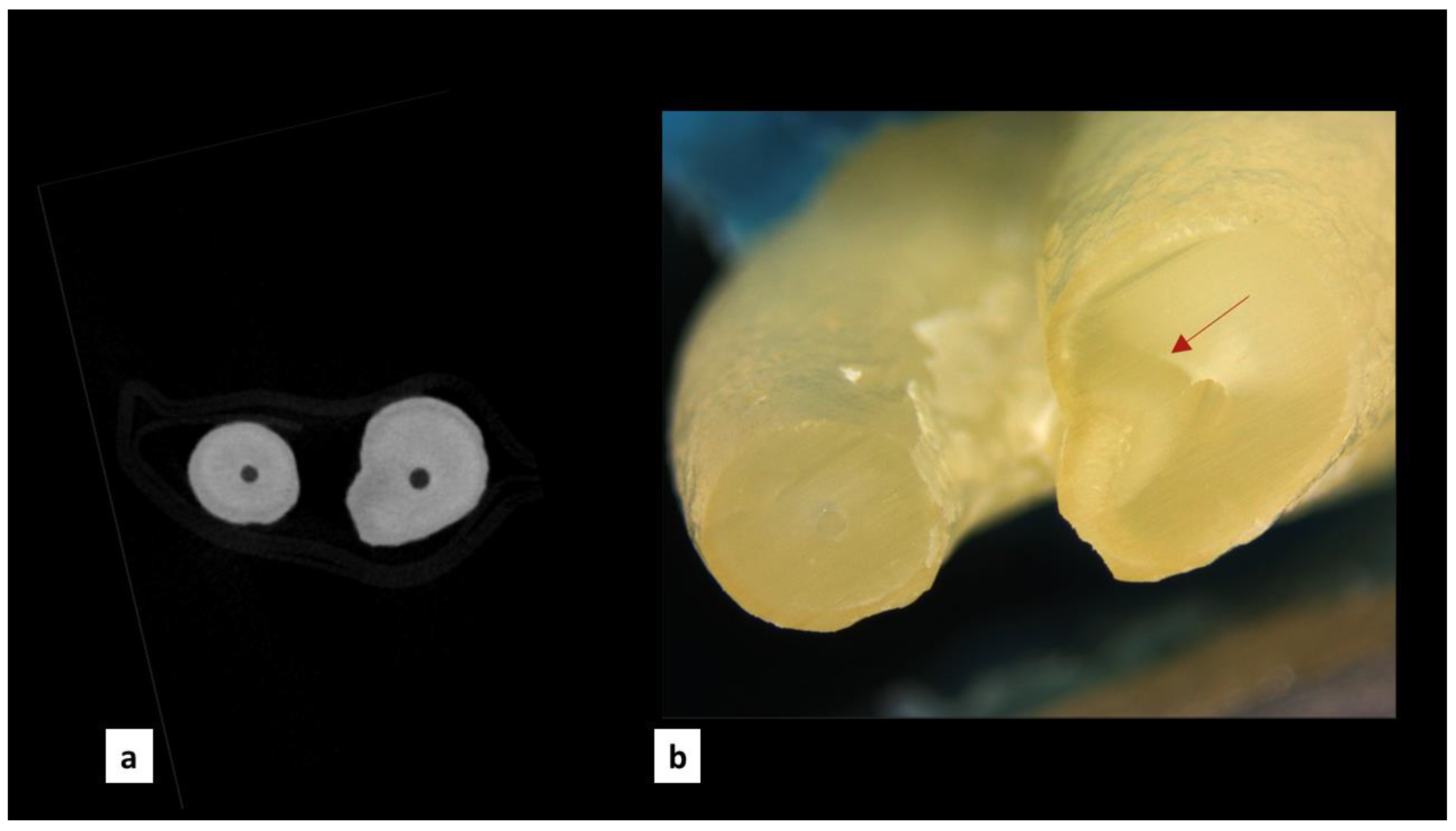
2.2. MicroCT Scanning

All the specimens were scanned before and after the root canal preparation using a MicroCT scanner (Skyscan1275; Bruker micro-CT, Kontich, Belgium) at 142 μA, 70 kV, and 180° rotations with a 0.5° rotation step using a 1 mm aluminum filter, resulting in mean scan duration of 10 min. To standardize the specimens, they were stabilized in a plastic cylinder with their access cavities facing upwards. NRecon GPU-Accelerated 3D reconstruction was used to reconstruct the images using a filtered back-projection algorithm and a graphics card processor (GPU) (Bruker micro-CT, Kontich, Belgium), applying a beam hardening correction of 25% and a ring artifact correction of 4, and limiting the range of images from the root apex till the cementoenamel junction coronally, resulting in around 1200 cross sections per specimen.

2.3. Root Canal Preparation

Two experimental groups were created, in which they obtained 11 premolars containing two canals which were randomly distributed, resulting in 22 root canals per group. The same operator performed all the procedures. The XSmart electric endodontic motor was used for all the instrumentation procedures (DENTSPLY Maillefer, Ballaigues, Switzerland). A Precision Water Bath GP2 was used to keep all the specimens inside at 37 °C (Thermo Fisher Scientific, MA, USA) for 30 min prior to the instrumentation of the canals. Both groups used warm irrigants using the system CanalPro Syringe Warmer (Coltène/Whaledent, Langenau, Germany). During the root canal preparation, irrigation was added with 2.5% sodium hypochlorite (NaOCl) with ProRinse Endo irrigation needles (Dentsply Maillefer, USA), which were kept at 2 to 3 mm from the apical foramen.
In Group 1, the specimens were instrumented with XP-endo® shaper (FKG, La Chaux-de- Fonds, Switzerland), according to the manufacturer’s instructions. Glidepath was created with a #15 k file (DENTSPLY Maillefer, Ballaigues, Switzerland), and the root canal was instrumented with a new single file per tooth. The file went to the working length until the preparation of size #30 with a 0.04 taper was achieved. In Group 2, the specimens were instrumented with TRUshape® (DENTSPLY-Tulsa Dental Specialties, Tulsa, OK, USA), according to the manufacturer’s instructions. For this, a new set of files was used per tooth as follows: the sequence started with the Orifice Modifier (length of 17 mm with a tip/taper 20/0.08), then three files sequence of 20/0.06, 25/0.06, and 30/0.06 were used until full working length. Between each preparation step of the root canal, the instrument was cleaned with an Isopropyl Alcohol 70% impregnated gauze. When the instrumentation was completed, a final rinse with 5 mL 17% EDTA followed by 5 mL of NaOCl 2.5% was performed. Then, absorbent paper points (DENTSPLY Maillefer, Ballaigues, Switzerland) were used to dry the root canals. Ultimately, the specimens were placed again in the same plastic cylinder used previously, which was positioned in the holder for the postoperative MicroCT scan utilizing the same parameters used for the pre-operatory scan.

2.4. Sectioning and Microscope Analysis

Sectioning of teeth was performed using a precision Diamond Section machine (Buehler, Waukegan Road Lake Bluff, IL, USA) with a diamond blade with a radius of 17.9 mm and a thickness of 1.3 mm, at a speed of 350–400 RPM, resulting in a clean and consistent cut in all samples. Each sample was programmed to be sliced at 0.7 mm, 2 mm, and 4.7 mm from the anatomical apex, with the visible section being created at precisely 2 mm, 4 mm, and 6 mm. Each specimen was washed and polished to remove any remaining material. This technique was repeated for the remaining 22 premolars.
After each section, the specimens were positioned in a customized holder made of paraffin wax. Allowing the tooth to be maintained in position while permitting some degree of movement to align the root in the correct orientation, in which the root apex had to be perpendicular to the lens of the microscope so that the images were taken correctly. Proper magnification and lightning were chosen, and the images were taken with a SteREO Discovery.V8 microscope (Zeiss, Oberkochen, Germany). Subsequently, images were adjusted in terms of brightness, contrast, and illumination using the ZEN software (Zeiss, Oberkochen, Germany). This process was repeated for all 22 premolars at three different lengths (2 mm, 4 mm, and 6 mm), resulting in 66 microscopic images.

2.5. Evaluation of the Microcracks

The evaluation of the microcracks consisted of two parts: the first part was performed by evaluating the MicroCT scans before and after instrumentation and comparing the two instrumentation systems, while the second part was conducted by comparing images obtained by the microscope at 2, 4 and 6 mm and the corresponding MicroCT scans after instrumentation. Two independent calibrated evaluators analyzed both the scans and the images, and in case of disagreement, a third independent evaluator was consulted. Firstly, using DataViewer software version 1.5.6.2 (Bruker microCT, Kontich, Belgium) to view the scans, 54.122 cross-sectional images obtained from MicroCT scans before and after instrumentation were evaluated. Furthermore, to detect the difference in the rate of newly formed microcracks according to the detection method, two keynote presentations (macOS, Apple Inc., Cupertino, CA, USA) were made and sent to each evaluator independently. In the first presentation, each slide contained one image obtained using the microscope (n = 66), as well as the second presentation, where each slide contained a counterpart of cross-sectional image stacks from the MicroCT scans after.
Moreover, screening of the presentations was performed under optimal viewing conditions in a darkened room using a 13-inch MacBook Pro (macOS, Apple Inc., Cupertino, CA, USA) with a retina display and a native resolution of 3072 × 1920 at 226 pixels per inch. Microcrack development was binomially classified by both evaluation methods as “absent” or “present.” The slice or microscopic image was characterized as “absent” if there were no microcracks or fractures on both the exterior surface of the root and the interior root canal wall, and was characterized as “present” if any microcracks, or fractures in the root dentine were identified. A microcrack was defined as a break or disruption in the tooth structure that did not result in the separation of pieces [34].

2.6. Statistical Analysis

Statistical analysis was performed using IBM SPSS Statistics software (version 21.0; IBM Corp, Armonk, NY, USA). Descriptive statistics were performed to determine the incidence of newly formed dentinal microcracks in the prepared samples. Pearson’s chi-squared was used to assess and compare the results obtained by the two different instrumentation systems, along with the difference between MicroCT scans and images under the microscope. Alpha was set at 0.05. Thus, p < 0.05 significance level was selected for all the statistical tests. Inter- and intra-examiner agreements were calculated using the Cohen kappa coefficient.

3. Results

After examining 54,122 cross sectional images obtained by the MicroCT scans, pre-existing microcracks were present in 8.5% of the total scans, while newly formed dentinal microcracks were observed in 0.49% of the cross-sectional images after instrumentation (Table 1). In the groups instrumented by TS (27,061 cross sectional images), newly formed dentinal microcracks were observed in 0.11% of the cross-sectional images after instrumentation (Figure 1), whereas in the groups instrumented by XP (27,061 cross-sectional images), newly formed dentinal microcracks were observed in 0.87% of the cross-sectional images after instrumentation (Figure 2). No statistically significant difference between the two instrumentation groups (p = 0.689) as indicated in Table 1.
Additionally, the comparison between the two detection methods showed that the sectioning method and examination under a stereomicroscope led to a higher detection rate of newly formed dentinal microcracks. Among the 66 images obtained by the stereomicroscope, newly formed dentinal microcracks that were not present in their respective cross-sectional images from the MicroCT scans after instrumentation were observed in 16.6% of the microscopic images, as shown in Figure 3. This resulted in a statistically significant difference between the two detection methods (p < 0.0001), as summarized in Table 2. In the instrumentation groups, 15.1% of the images obtained by the TF group showed microcracks that were not detectable in their corresponding MicroCT images, while 18.1% of the images in the XP group displayed microcracks (Table 2). Assessing the Cohen’s Kappa results, an inter-rater agreement value of 0.9 was obtained when evaluating the presence or absence of newly formed microcracks in MicroCT scans, and a value of 0.5 when evaluating the presence of cracks comparing the two detection methods.

4. Discussion

This ex vivo study aimed to evaluate the formation of newly formed dentinal microcracks after instrumentation with two different innovative systems, XP-Endo® Shaper and TruShape® systems, in maxillary premolars. Until this date, no study has been conducted to compare the creation of new dentinal microcracks between both systems. Of the 54,122 MicroCT scans examined, newly formed dentinal microcracks were detected in only 0.49%. These results are consistent with previous findings, as similarly low percentages of newly formed dentinal microcracks were reported in [35,36,37]. Moreover, these studies reported a comparable pattern observed in our study. There is an increased percentage of pre-existing microcracks in extracted human teeth [35,36,37], which could be explained by the extraction method. De Deus et al. [10] found that the dentinal microcracks detected were caused by the extraction techniques or the post-extraction storage conditions, rather than the instrumentation procedures. Additionally, Arashiro et al. [38] examined the effect the extraction method had on dental microcrack formation by comparing atraumatic with traumatic extraction. While no statistically significant difference was found, atraumatic extraction resulted in fewer dentinal microcrack formations, indicating the importance of the extraction techniques and supporting the argument that it should be considered an essential factor in future studies, as microcracks were observed in 8.5% of the pre-instrumentation scans.
Furthermore, in this ex vivo study, two methods for evaluating the presence of dentinal microcracks were used, MicroCT scanning and sectioning followed by examination under a SEM. The sectioning method reported a statistically significant increase in the incidence of dentinal microcracks compared to MicroCT scanning. These findings are partly congruent with the only study comparing various evaluation methods [39], which reported no statistically significant differences between the two methods, despite the sectioning method detecting a larger percentage of dentinal microcracks. Based on a review of the current literature, it appears that evaluation with the sectioning method may overestimate and increase the incidence of observed dentinal microcracks. This might be because these cracks may be created during the tooth sectioning process or because, in the studies that use this technique, only post-instrumentation assessment can be performed. Therefore, it is unknown if the microcrack was pre-existing or newly formed by the instrumentation procedure.
In studies where MicroCT was used, a tooth scan is usually performed before the instrumentation, diminishing possible errors in identifying pre-existing defects [12,23,27,35]. Another possible explanation is that in certain clinical scenarios, the direction of dentinal tubules changes, which may be mistaken for fissure lines when observed visually with a stereomicroscope. Hunter–Schreger bands, which are artifacts produced by mineral component deposition and demineralization spots, creating visual defects that can be misinterpreted as fissure lines [40], have been reported in the literature. This also explains why our evaluators’ Cohen kappa values decreased when comparing the two detection methods, as recognizing the presence of cracks can sometimes be challenging. Hence, the sectioning method should not be used in future studies as a means of detecting newly formed dentinal microcracks and should be replaced with the more accurate and non-invasive MicroCT scanning.
One limitation of the current study is that although the thickness of the blade was considered when the tooth was sectioned, the images do not correspond precisely to the same point of the MicroCT cross-sections due to the angulation of the cut and the aggressive nature of the sectioning method, which can cause damage the tooth structure and produce rough inaccurate sections. Thus, this may not be the most reliable method to detect newly formed microcracks. Another possible limitation is that methylene blue was not used on the sectioned teeth to help identify possible microcracks. Although some studies suggest that 2% methylene blue can aid in detecting defects in apical microsurgery after root resection [41], other studies, such as Ghorbanzadeh et al. [42], have concluded that 2% methylene blue alone is significantly less efficient in diagnosing dentinal cracks formed in the apical third after root resection compared to other methods.

5. Conclusions

Within the limitations of the study, both XP-Endo shaper and TRUShape produced fewer than 1% new dentinal microcracks in all the MicroCT scans examined. Additionally, when comparing the two evaluation methods, significantly more dentinal microcracks were detected using the sectioning method compared to the MicroCT scans, indicating the aggressive nature of this method and the tendency to overestimate the actual presence of dentinal microcracks.

Author Contributions

F.E. and M.d.l.N.P.M. were involved in all sample arrangement and preparation with the stereomicroscope and MicroCT, under the direct supervision and help of J.G.O. and J.A.G.S. Additionally, F.E. and J.N.P. involved in preparing and analyzing MicroCT scans and images obtained under microscope. Finally, K.I.A., J.A.G.S. and F.D.-S. were involved in supervision of the writing process plus, article correction and editing. All authors have read and agreed to the published version of the manuscript.

Funding

This research received no external funding. The APC was partially funded by Universität Bern.

Institutional Review Board Statement

The study protocol was approved by the Institutional Review Ethics Committee of Universitat Internacional de Catalunya (protocol code REC-ELM-2017-01 v.1.0 and approved on 24 December 2017).

Informed Consent Statement

Not applicable.

Data Availability Statement

Data described in this paper are available on request.

Acknowledgments

The authors thank the Universität Bern for partially supporting the open access publication modality of this work.

Conflicts of Interest

The authors declare no competing interest with respect to the authorship and/or publication of this article.

References

  1. Endodontists, A.A. Endodontics: Colleagues for Excellence-Cracking the Cracked Tooth Code; Fall-Winter: Chicago, IL, USA, 2008. [Google Scholar]
  2. Chai, H.; Tamse, A. Vertical Root Fracture in Buccal Roots of Bifurcated Maxillary Premolars from Condensation of Gutta-Percha. J. Endod. 2018, 44, 1159–1163. [Google Scholar] [CrossRef] [PubMed]
  3. Afrashtehfar, K.I.; MacDonald, D. Vertical tooth root fracture detection through cone-beam computed tomography: An umbrella review protocol testing four hypotheses. Open Dent. J. 2019, 13, 449–453. [Google Scholar] [CrossRef] [Green Version]
  4. Al-Jadaa, A.; Alsmadi, R.F.; Salem, W.M.; Abdulridha, A.A.; Afrashtehfar, K.I. Radicular Intracanal Splitting Forces and Cutting Efficiency of NiTi Rotary versus Reciprocating Systems: A Comparative In Vitro Study. Cosmetics 2023, 10, 23. [Google Scholar] [CrossRef]
  5. Roylance, D. Introduction to Fracture Mechanics; Elsevier: Cambridge, MA, USA, 2001. [Google Scholar]
  6. Kishen, A. Biomechanics of Fractures in Endodontically Treated Teeth. Endod. Topics 2015, 33, 3–13. [Google Scholar] [CrossRef]
  7. Yoldas, O.; Yilmaz, S.; Atakan, G.; Kuden, C.; Kasan, Z. Dentinal Microcrack Formation during Root Canal Preparations by Different NiTi Rotary Instruments and the Self-Adjusting File. J. Endod. 2012, 38, 232–235. [Google Scholar] [CrossRef]
  8. Adorno, C.G.; Yoshioka, T.; Suda, H. The Effect of Root Preparation Technique and Instrumentation Length on the Development of Apical Root Cracks. J. Endod. 2009, 35, 389–392. [Google Scholar] [CrossRef]
  9. Del Fabbro, M.; Afrashtehfar, K.I.; Corbella, S.; El-Kabbaney, A.; Perondi, I.; Taschieri, S. In Vivo and in vitro effectiveness of ro-tary nickel-titanium vs manual stainless steel instruments for root canal therapy: Systematic review and meta-analysis. J. Evid. Based Dent. Pract. 2018, 18, 59–69. [Google Scholar] [CrossRef]
  10. De-Deus, G.; Cavalcante, D.M.; Belladonna, F.G.; Carvalhal, J.; Souza, E.M.; Lopes, R.T.; Versiani, M.A.; Silva, E.J.N.L.; Dummer, P.M.H. Root Dentinal Microcracks: A Post-Extraction Experimental Phenomenon? Int. Endod. J. 2019, 52, 857–865. [Google Scholar] [CrossRef]
  11. De-Deus, G.; César de Azevedo Carvalhal, J.; Belladonna, F.G.; Silva, E.J.N.L.; Lopes, R.T.; Moreira Filho, R.E.; Souza, E.M.; Provenzano, J.C.; Versiani, M.A. Dentinal Microcrack Development after Canal Preparation: A Longitudinal In Situ Micro–Computed Tomography Study Using a Cadaver Model. J. Endod. 2017, 43, 1553–1558. [Google Scholar] [CrossRef]
  12. PradeepKumar, A.R.; Shemesh, H.; Archana, D.; Versiani, M.A.; Sousa-Neto, M.D.; Leoni, G.B.; Silva-Sousa, Y.T.C.; Kishen, A. Root Canal Preparation Does Not Induce Dentinal Microcracks In Vivo. J. Endod. 2019, 45, 1258–1264. [Google Scholar] [CrossRef]
  13. de Oliveira, B.P.; Câmara, A.C.; Duarte, D.A.; Heck, R.J.; Antonino, A.C.D.; Aguiar, C.M. Micro–Computed Tomographic Analysis of Apical Microcracks before and after Root Canal Preparation by Hand, Rotary, and Reciprocating Instruments at Different Working Lengths. J. Endod. 2017, 43, 1143–1147. [Google Scholar] [CrossRef] [PubMed]
  14. Kansal, R.; Rajput, A.; Talwar, S.; Roongta, R.; Verma, M. Assessment of Dentinal Damage during Canal Preparation Using Reciprocating and Rotary Files. J. Endod. 2014, 40, 1443–1446. [Google Scholar] [CrossRef] [PubMed]
  15. Peters, O.A.; Arias, A.; Paqué, F. A Micro-Computed Tomographic Assessment of Root Canal Preparation with a Novel Instrument, TRUShape, in Mesial Roots of Mandibular Molars. J. Endod. 2015, 41, 1545–1550. [Google Scholar] [CrossRef] [PubMed] [Green Version]
  16. Arias, A.; Paqué, F.; Shyn, S.; Murphy, S.; Peters, O.A. Effect of Canal Preparation with TRUShape and Vortex Rotary Instruments on Three-Dimensional Geometry of Oval Root Canals. Aust. Endod. J. 2018, 44, 32–39. [Google Scholar] [CrossRef] [PubMed] [Green Version]
  17. Uslu, G.; Özyürek, T.; Yılmaz, K.; Gündoğar, M.; Plotino, G. Apically Extruded Debris during Root Canal Instrumentation with Reciproc Blue, HyFlex EDM, and XP-Endo Shaper Nickel-Titanium Files. J. Endod. 2018, 44, 856–859. [Google Scholar] [CrossRef] [PubMed]
  18. TruShape|Dentsply Sirona. Available online: https://www.dentsplysirona.com/en-us/categories/endodontics/trushape.html (accessed on 12 July 2022).
  19. XP-Endo Shaper|XP-Endo Solutions. Available online: https://www.fkg.ch/xpendo/shaper (accessed on 12 July 2022).
  20. Aksoy, Ç.; Keriş, E.Y.; Yaman, S.D.; Ocak, M.; Geneci, F.; Çelik, H.H. Evaluation of XP-Endo Shaper, Reciproc Blue, and ProTaper Universal NiTi Systems on Dentinal Microcrack Formation Using Micro–Computed Tomography. J. Endod. 2019, 45, 338–342. [Google Scholar] [CrossRef]
  21. Zuolo, M.L.; De-Deus, G.; Belladonna, F.G.; da Silva, E.J.N.L.; Lopes, R.T.; Souza, E.M.; Versiani, M.A.; Zaia, A.A. Micro–Computed Tomography Assessment of Dentinal Micro-Cracks after Root Canal Preparation with TRUShape and Self-Adjusting File Systems. J. Endod. 2017, 43, 619–622. [Google Scholar] [CrossRef]
  22. Bayram, H.M.; Bayram, E.; Ocak, M.; Uzuner, M.B.; Geneci, F.; Celik, H.H. Micro–Computed Tomographic Evaluation of Dentinal Microcrack Formation after Using New Heat-Treated Nickel-Titanium Systems. J. Endod. 2017, 43, 1736–1739. [Google Scholar] [CrossRef]
  23. Alkahtany, S.M.; Al-Madi, E.M. Dentinal Microcrack Formation after Root Canal Instrumentation by XP-Endo Shaper and ProTaper Universal: A Microcomputed Tomography Evaluation. Int. J. Dent. 2020, 2020, 4030194. [Google Scholar] [CrossRef] [Green Version]
  24. Coelho, M.S.; Card, S.J.; Tawil, P.Z. Light-Emitting Diode Assessment of Dentinal Defects after Root Canal Preparation with Profile, TRUShape, and WaveOne Gold Systems. J. Endod. 2016, 42, 1393–1396. [Google Scholar] [CrossRef]
  25. Çiçek, E.; Koçak, M.M.; Saʇlam, B.C.; Koçak, S. Evaluation of Microcrack Formation in Root Canals after Instrumentation with Different NiTi Rotary File Systems: A Scanning Electron Microscopy Study. Scanning 2015, 37, 49–53. [Google Scholar] [CrossRef] [PubMed]
  26. Dowker, S.E.P.; Davis, G.R.; Elliott, J.C. X-ray Microtomography Nondestructive Three-Dimensional Imaging for In Vitro Endodontic Studies. Oral. Surg. Oral. Med. Oral. Pathol. Oral. Radiol. Endod. 1997, 83, 510–516. [Google Scholar] [CrossRef] [PubMed]
  27. De-Deus, G.; Belladonna, F.G.; Souza, E.M.; Silva, E.J.N.L.; Neves, A.D.A.; Alves, H.; Lopes, R.T.; Versiani, M.A. Micro-Computed Tomographic Assessment on the Effect of ProTaper Next and Twisted File Adaptive Systems on Dentinal Cracks. J. Endod. 2015, 41, 1116–1119. [Google Scholar] [CrossRef] [PubMed]
  28. Rhodes, J.S.; Pitt Ford, T.R.; Lynch, J.A.; Liepins, P.J.; Curtis, R.V. Micro-Computed Tomography: A New Tool for Experimental Endodontology. Int. Endod. J. 1999, 32, 165–170. [Google Scholar] [CrossRef]
  29. Siqueira, J.F.; Alves, F.R.F.; Versiani, M.A.; Rôças, I.N.; Almeida, B.M.; Neves, M.A.S.; Sousa-Neto, M.D. Correlative Bacteriologic and Micro-Computed Tomographic Analysis of Mandibular Molar Mesial Canals Prepared by Self-Adjusting File, Reciproc, and Twisted File Systems. J. Endod. 2013, 39, 1044–1050. [Google Scholar] [CrossRef]
  30. Ashwinkumar, V.; Krithikadatta, J.; Surendran, S.; Velmurugan, N. Effect of reciprocating file motion on microcrack formation in root canals: An SEM study. Int. Endod. J. 2014, 47, 622–627. [Google Scholar] [CrossRef]
  31. World Medical Association Declaration of Helsinki: Ethical Principles for Medical Research Involving Human Subjects. JAMA 2013, 310, 2191–2194. [CrossRef] [Green Version]
  32. de las Perez Morales, M.N.; González Sánchez, J.A.; Olivieri Fernández, J.G.; Laperre, K.; Abella Sans, F.; Jaramillo, D.E.; Terol, F.D.S. TRUShape Versus XP-Endo Shaper: A Micro–Computed Tomographic Assessment and Comparative Study of the Shaping Ability—An In Vitro Study. J. Endod. 2020, 46, 271–276. [Google Scholar] [CrossRef]
  33. Schneider, S.W.; Austin, D.D.S. A Comparison of Canal Preparations in Straight and Curved Root Canals. Oral Surg. Oral Med. Oral Pathol. 1971, 32, 271–275. [Google Scholar] [CrossRef]
  34. Ratcliff, S.; Becker, I.M.; Quinn, L. Type and Incidence of Cracks in Posterior Teeth. J. Prosthet. Dent. 2001, 86, 168–172. [Google Scholar] [CrossRef]
  35. PradeepKumar, A.R.; Shemesh, H.; Chang, J.W.W.; Bhowmik, A.; Sibi, S.; Gopikrishna, V.; Lakshmi-Narayanan, L.; Kishen, A. Preexisting Dentinal Microcracks in Nonendodontically Treated Teeth: An Ex Vivo Micro–Computed Tomographic Analysis. J. Endod. 2017, 43, 896–900. [Google Scholar] [CrossRef] [PubMed]
  36. Miguéns-Vila, R.; Martín-Biedma, B.; De-Deus, G.; Belladonna, F.G.; Peña-López, A.; Castelo-Baz, P. Micro-computed Tomographic Evaluation of Dentinal Microcracks after Preparation of Curved Root Canals with ProTaper Gold, WaveOne Gold, and ProTaper Next Instruments. J. Endod. 2021, 47, 309–314. [Google Scholar] [CrossRef] [PubMed]
  37. Uğur Aydın, Z.; Keskin, N.B.; Özyürek, T. Effect of Reciproc Blue, XP-Endo Shaper, and WaveOne Gold Instruments on Dentinal Microcrack Formation: A Micro-Computed Tomographic Evaluation. Microsc. Res. Tech. 2019, 82, 856–860. [Google Scholar] [CrossRef] [PubMed]
  38. Arashiro, F.N.; De-Deus, G.; Belladonna, F.G.; Cavalcante, D.M.; Coelho, M.S.; Silva, E.J.N.L.; Pereira, K.F.S.; da Silva, P.G.; Lopes, R.T.; Souza, E.M. Dentinal Microcracks on Freshly Extracted Teeth: The Impact of the Extraction Technique. Int. Endod. J. 2020, 53, 440–446. [Google Scholar] [CrossRef]
  39. Çapar, İ.D.; Gök, T.; Uysal, B.; Keleş, A. Comparison of Microcomputed Tomography, Cone Beam Tomography, Stereomicroscopy, and Scanning Electron Microscopy Techniques for Detection of Microcracks on Root Dentin and Effect of Different Apical Sizes on Microcrack Formation. Microsc. Res. Tech. 2019, 82, 1748–1755. [Google Scholar] [CrossRef]
  40. Harper, R.A.; Shelton, R.M.; James, J.D.; Salvati, E.; Besnard, C.; Korsunsky, A.M.; Landini, G. Acid-Induced Demineralisation of Human Enamel as a Function of Time and PH Observed Using X-ray and Polarised Light Imaging. Acta Biomater. 2021, 120, 240–248. [Google Scholar] [CrossRef]
  41. Floratos, S.; Kim, S. Modern Endodontic Microsurgery Concepts: A Clinical Update. Dent. Clin. N. Am. 2017, 61, 81–91. [Google Scholar] [CrossRef]
  42. Ghorbanzadeh, A.; Aminifar, S.; Shadan, L.; Ghanati, H.; Aminifar, S. Evaluation of Three Methods in the Diagnosis of Dentin Cracks Caused by Apical Resection. J. Dent. 2013, 10, 175. [Google Scholar]
Figure 1. Three-dimensional representation of the Micro-computed tomography (MicroCT) scan of the maxillary premolar instrumented using XP-endo shaper. (a) Lateral view of the 3D model of the maxillary premolar. (b) Coronal view of the 3D model of the maxillary premolar. (c) Cross section of radicular canals before instrumentation. (d) Cross section of the canals after instrumentation with the arrow pointing at the newly formed microcrack.
Cosmetics 10 00037 g001
Figure 2. Three-dimensional representation of the Micro-computed tomography (MicroCT) scan of the maxillary premolar instrumented using TruShape. (a) Lateral view of the 3D model of the maxillary premolar. (b) Coronal view of the 3D model of the maxillary premolar. (c) Cross section of the canals before instrumentation. (d) Cross section of radicular canals after instrumentation with the arrow pointing at the newly formed microcrack.
Cosmetics 10 00037 g002
Figure 3. Comparison between the two detection methods. (a) Dentinal microcrack observed in the Stereomicroscope image. (b) The counterpart Micro-computed tomography (MicroCT) scan, in which the dentinal microcrack is not detectable.
Cosmetics 10 00037 g003
Table 1. Frequency of microcrack formation with TRUshape and XP-Endo Shaper.
GroupMicroCT Scans Examined (n)Pre-Existing Dentinal MicrocracksNewly Formed Dentinal Microcracks
(After Instrumentation)
Scans (n)Percentage (%)Scans (n)Percentage (%)
TRUshape27,06117096.3300.11
XP-Endo shaper27,061291110.72350.87
Total54,12246208.52650.49
Table 2. Frequency of dentinal microcracks only detected after sectioning and examination under stereomicroscope.
GroupImages Obtained with Stereomicroscope after Sectioning (n)Dentinal Microcracks Detected Only under Stereomicroscope and Not in Their Counterpart MicroCT Scans
Images (n)Percentage (%)
TRUshape33515.1
XP-Endo shaper33618.1
Total661116.6
Disclaimer/Publisher’s Note: The statements, opinions and data contained in all publications are solely those of the individual author(s) and contributor(s) and not of MDPI and/or the editor(s). MDPI and/or the editor(s) disclaim responsibility for any injury to people or property resulting from any ideas, methods, instructions or products referred to in the content.

