Article

Radicular Intracanal Splitting Forces and Cutting Efficiency of NiTi Rotary Versus Reciprocating Systems: A Comparative In Vitro Study

by
Anas Al-Jadaa
1,*,
Ranya F. Alsmadi
1,
Wesal M. Salem
1,
Aya A. Abdulridha
1 and
Kelvin I. Afrashtehfar
1,2,*
1
Clinical Sciences Department, Ajman University College of Dentistry, Ajman City P.O. Box 346, United Arab Emirates
2
Department of Reconstructive Dentistry & Gerodontology, School of Dental Medicine, University of Bern, 3010 Bern, Switzerland
*
Authors to whom correspondence should be addressed.
Cosmetics 2023, 10(1), 23; https://doi.org/10.3390/cosmetics10010023
Submission received: 2 December 2022 / Revised: 11 January 2023 / Accepted: 20 January 2023 / Published: 24 January 2023

Abstract

:
This study aimed to compare the intracanal lateral force and cutting efficiency associated with two engine-driven nickel-titanium (NiTi) systems during root canal shaping. Bovine single-rooted teeth models were assembled to a custom-made splitting force measuring platform while being endodontically treated with rotary (ProTaper Gold [PTG]) or reciprocal systems (WaveOne Gold [WOG]) by two clinicians. The cutting efficiency test was run for each group by a free-falling endomotor on dentin discs for 3 min. The resulting force, files, and stroke force peaks were recorded. Data were analyzed by the Shapiro-Wilk test and ANOVA. Statistical significance was set at alpha = 0.05. Intracanal lateral forces (p < 0.01), cutting efficiency (p < 0.01), and time efficiency (p < 0.01) were significantly different between the NiTi groups. The maximum lateral splitting force was detected using the PTG shaping full-length file (S1) and during the WOG final stroke. Thus, caution is advised when reciprocating single-file systems approach the apical third or when using a large taper and wide-diameter rotatory instruments. The WOG had significantly less maximum horizontal splitting force and significantly greater cutting efficiency but less time efficiency than the PTG system.

1. Introduction

The preparation of the root canal system after pathological or physical stress comprises the removal of essential and necrotic tissues and contaminated root dentine using endodontic files combined with irrigants and medicaments to clean the canal space chemically [1,2]. A shift in endodontic instrumentation toward using nickel-titanium (NiTi) files was observed in recent decades. This can be referred to as their safe use, as they can overcome the predicament of both rigidity and low resistance to cyclic fatigue associated with stainless steel instruments [3,4]. The ability of ‘engine-driven’ NiTi instruments to prepare root canals, such as hand instruments, granted them further attractiveness [5,6]. Rotation and reciprocation are the popular modes of NiTi files in engine-driven action of root canal instrumentation [7]. There is a wide variety of rotary systems, which includes one file with continuous motion, one file with reciprocating motion, multiple files with continuous motion, and multiple files with a reverse motion option. In general, reciprocation uses a single-file instrumentation system to prepare the canal to the desired size and shape, which shortens the time to complete the canal cleaning and shaping compared to rotary multiple-file systems [8]. However, the antimicrobial agent volume and its contact time in the canal are reduced.
Regardless of the technique used in preparing the canals and the irrigant type used for disinfection, there is a risk of treatment failure. Although insufficient disinfection and obturation of the root canal system by the conclusion of treatment are said to be the main reasons for root canal therapy failure, vertical root fracture (VRF) is considered a major cause of treatment failure [9,10]. The location, direction, and type of root fractures are influenced by instrumentation, post presence, compaction/obturation, root canal geometry, external root morphology, and dentin wall thickness [11,12]. As NiTi files can better maintain the canal center, leaving thicker dentine walls in curved root canals [13], it was suggested to improve the fracture resistance and stability of treated roots [14].
A major concern in endodontic therapy is the induction of microcracks on the radicular canal walls during root canal preparation. Microcracks and craze lines on the root surface, which do not extend through the root canal walls, are supposedly the result of root canal preparation procedures. These dentinal defects may be aggravated in time into VRF [15,16]. A positive relationship between dentinal cracks and biomechanical preparation was suggested [17]. The possible suggested factors influencing the initiation of dentinal defects are file size, taper, cross-section, manufacturing method, materials or alloys used in instruments, and preparation technique [18].
Some studies reported no difference between the reciprocating and rotary types of motion on dentinal defect induction [19]. Nevertheless, the advantage of the reciprocating motion may reduce the file torsional stress by disengaging dentine, thus reducing the magnitude of generated forces [20]. In contrast, a single reciprocating file may induce more dentinal defects than full-sequence rotary files [21]. A larger taper could increase the initiation of dentinal defects [22] since it results in less remaining dentine thickness, which may increase the likelihood of root fracture [23]. Other researchers disagreed with both opinions and reported that full-sequence rotary files produce significantly more dentinal defects than reciprocating single-file systems [24].
Some research relates microcrack initiation to other factors, such as file design [22] and instrument alloy [25]. Regardless of the debate associated with factors affecting microcrack initiation, all factors can be linked to the file performance through cutting efficiency and resulting in intracanal stresses. Thus, a good strategy is to enhance the force distribution during the preparation and obturation of the root canals to reduce the incidence of root fracture initiation [26].
This comparative in vitro study aimed to compare the horizontal intracanal force with two engine-driven NiTi systems during root canal shaping and instrumentation. Additionally, identifying the root canal instrument and instrumentation stage with the highest radicular splitting force with each system could inform clinicians and researchers of safety precautions. A secondary aim was to test the cutting efficiency of both systems. The tested null hypotheses were that both engine-driven instrumentation systems have similar (1) lateral splinting forces and (2) cutting efficiency.

2. Materials and Methods

2.1. Ethical Approval and Registration

This comparative in vitro study was conducted after its study protocol obtained approval from the Review and Ethical Committee (REC) of the educational institution. The two voluntary participant operators were aware of the study’s objectives, and informed consent was obtained before enrolment. None of the operators received any compensation for their time or participation.

2.2. Power Calculation

Calculation of the sample size was carried out after a triplicate trial. It was based on an effect size of 12.74, an alpha error probability of 0.05, and 95% power (G*Power 3.1, Heinrich Heine Universität Düseldorf, Germany). Accordingly, the sample size was set to 7 per operator for each applied system (n = 14).

2.3. Part 1: Sample Preparation for Lateral Splinting Force Testing

Twenty-eight mandibular anterior bovine teeth were obtained. The roots were cleaned with scalers and disinfected following the college’s policy to be stored in 1% chloramine trihydrate solution (Figure 1A).

2.3.1. Sample Sectioning and Mounting for the Testing Platform

The samples were prepared according to a previously published methodology [27]. In short, the teeth were decoronated with a slow-speed round diamond disk at the cementoenamel junction (CEJ) level on the interproximal aspect, leaving approximately 2 mm of coronal tooth structure on the buccal and lingual aspects (Figure 1B). They were then aligned and mounted with acryl in a perpendicular position to a casted acrylic stump (Veracril, New Stetic, Antioquia, Colombia) with the aid of a centering device. The acrylic stump allowed the roots to be held in a parallel alignment against a vibrating blade microtome (Leica SP1600, Nussloch, Germany) (Figure 2). Under cooling water, each root received two parallel cuts along its long axis (right and left to the canal). This allowed the middle part (canal) to be excluded while obtaining the outer two dentinal walls. The obtained dentinal walls were aligned against each other in a custom-made Teflon cylindrical mold. They were cast with pink self-cured acrylic (Veracril, New Stetic, Antioquia, Colombia) to fix the roots firmly with acrylic holding parts.

2.3.2. Creation of the Artificial Canal

A straight artificial canal was created parallel to the long axis on one side of the root intaglio portion with a 0.2 mm thick diamond disc mounted to a precision milling machine (Figure 3a). The resulting artificial canal simulated an unprepared canal (Figure 3b).

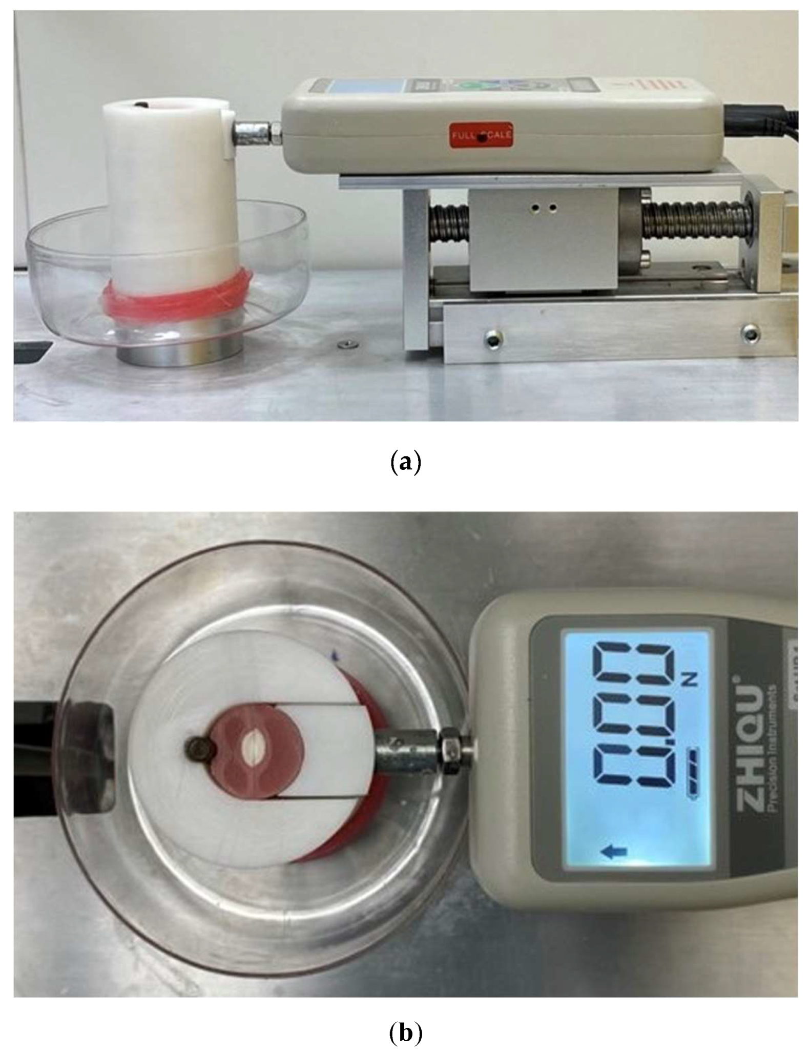
2.3.3. The Splitting Force Platform

A custom-made splitting force measuring device was used to measure the intracanal lateral forces [27]. The device used in the present in vitro study consisted of a high-precision digital force gauge (DS2-50N, Zhique Precision Instruments, Shanghai, China) mounted on a linear sliding table (Figure 4a). The measuring end of the device was connected to the cylindrical Teflon holder that contained the two-piece sample in intimate contact during the testing procedures. The resulting assembly registered any lateral force applied between the two pieces of the specimen with an accuracy of 0.01 N. The sliding table permitted precisely reaching a 0 N force reading before initiating the instrumentation (Figure 4b). The principle of the splitting force platform is based on detecting horizontal (lateral) splitting forces resulting from a given procedure within the radicular canal. This was performed through force transmission from the terminal probe to the force-measuring device.

2.4. Instrumentation

Two operators were assigned to instrument the samples following the manufacturers’ recommendations. One operator was a senior resident, and the other was a junior resident of the Endodontology Postgraduate Program of a Dental Institution. The 28 samples were divided into four groups (n = 7). Each operator had 7 specimens randomly assigned from each engine-driven instrumentation group. Thus, each group consisted of 14 tested specimens. The NiTi rotary group (PTG) comprised the ProTaper Gold (Dentsply Sirona, Ballaigues, Switzerland), whereas the NiTi reciprocating group (WOG) comprised the WaveOne Gold (Dentsply Sirona, Ballaigues, Switzerland).
The initial canal preparation used the TruNatomy glide path file (Dentsply Sirona, Maillefer, Ballaigues, Switzerland) and 1 mL of sodium hypochlorite (NaOCl) 1.3% concentration irrigation through a side vented needle over 6 s utilizing a custom-made irrigation device at a rate of 10 mL/min (Figure 5). This step aimed to create a standardized canal in all samples before the preparation process. An endodontic motor (X-Smart Plus, Dentsply Maillefer, Switzerland) was set to the manufacturer’s pre-settings. After each file/stroke in both groups, NaOCl irrigation was delivered to the canal at a rate of 10 mL/min.
During instrumentation, the testing device was connected to a computer through graph software (Zhique Precision Instruments, China) that received and registered the measured force data in Newtons at a rate of 5 measurements per second. The data were captured in an Excel worksheet (MS Excel, Microsoft, Redmond, WA, USA).

2.4.1. Instrumentation Protocol

The entire PTG file sequence (SX–F2) had to be followed, and a new set of files was used for each specimen. The SX file was used to prepare 8 mm of the 16 mm total full length, which was subsequently fully prepared by the full-sequence S1-F2 reaching the total working length in a maximum of three strokes.
In the WOG system, the primary file size was selected for root canal preparation because it has dimensions comparable to the final preparation file (F2) of the PTG system. The total working length was reached in 5 strokes, each of 3 picking motions of 1 mm, except for the last stroke, which completed the entire working length of 16 mm by preparing the last 1 mm. A new file set in PTG and a new file in the WOG were used to prepare each sample.
Each file use or stroke was followed by irrigation with 1 mL of sodium hypochlorite (NaOCl) 1.3% concentration irrigation through a side vented needle over 6 s utilizing the custom-made irrigation device at a rate of 10 mL/min.

2.4.2. Data Extraction

Five intracanal lateral force peaks could be detected in the data set. These peaks presented the splitting force associated with the file as a full-length file, as in the ProTaper Gold system, or as a stroke in the WOG system. Furthermore, the timing of the first detected force raise was reported up to the force drop to 0 N by the last file stroke. The time required for the root canal preparation was calculated by dividing the number of detected force measurements by 5 (measuring rate is 5 per 1 sec).

2.5. Part 2: Cutting Efficiency Testing Sample Preparation and Instrumentation

Dentine discs of 1 mm thickness were obtained from the lingual side of 10 bovine teeth crowns by cutting two parallel cuts extending from the incisal edge to the cingulum of the crown. The 20 paired half discs were assigned to one of the tested systems (10 specimens per group). In other words, each disc was cut into 2 parts for further testing using each system. The dentine side was then mounted perpendicular to the tested system holder. The tested file (F2 or Primary) was inserted in the handpiece while left free-falling on a 350 g ball bearing slide in the Z-axis direction (Figure 6).
For each sample and file combination, the handpiece of the X-Smart Endomotor was automatically set at the preset settings of the manufacturer. The engine-driven NiTi files were active at D16 in contact with the dentine disc for 3 min under a continuous flow of NaOCl 1.3%. Photos were obtained with a metal calibration scale attached to the same dentine disc plane. The photos were analyzed utilizing ImageJ analysis software (V. 1.44p; National Institutes of Health, Bethesda, MD, USA). The effective depth of the notch in mm represented the cutting efficiency of the file on each evaluated sample. The definition of ‘better efficacy’ was: the higher the notch depth, the higher the efficacy.

2.6. Statistical Analysis

The sample size was calculated based on the first triplicates’ mean values and standard deviations, with a power of 80% and a Type I error rate of 5%. The maximum exerted force values for each file/stroke and maximum force values per system are presented as the means and standard deviations (SDs). The normal distribution within the same instrumentation step/stage was tested using the Shapiro-Wilk test with α set at 0.001. The data were normally distributed, as all data sets were not statically significant. Comparisons within the systems and operators were conducted by setting p < 0.001 for analysis of variance (ANOVA). The different values obtained compared the forces and timing between the two groups of engine-driven systems through ANOVA. All statistical analyses were performed using statistical software (SPSS ver. 25, IBM Corp., Armonk, NY, USA). The level of significance was set to α = 0.05.

3. Results

The WOG had significantly less maximum horizontal intracanal lateral force (p < 0.01; Table 1) as well as significantly greater cutting efficiency (p < 0.01; Table 2) and time efficiency (p < 0.01; Table 3). In the PTG group, the maximum force detected with significance was associated with using the S1 files for both operators (p ≤ 0.05). The least exerted force was associated with the SX file (Table 3). The WOG system revealed a significant difference between the fourth and fifth strokes compared to the first stroke for the first operator (p < 0.05). All strokes showed significant differences between operators (p < 0.05; Table A1). Additionally, the fifth stroke was significantly different from the other strokes (p < 0.05; Table 3) compared to the PTG system.
No inter-operator differences were detected regardless of the system preparation mode (PTG p = 0.37; WOG p = 0.06). After comparing the operators’ time required during radicular preparation using the same system (Figure A1), no significant inter-operator difference could be detected when comparing the same system (PTG, p = 0.37, WOG, p = 0.06).
There were no significant differences in splitting forces or instrumentation time between the operators in the PTG (p = 0.11 and p = 0.13, respectively) and WOG groups (p = 0.12 and p = 0.24, respectively) (Table A1). However, both operators had significantly different performances when comparing both systems (p = 0.01 for operator 1 and p = 0.01 for operator 2).

4. Discussion

To the authors’ knowledge, this is the first study that compares the radicular horizontal splitting forces of rotating and reciprocating systems. The current study provides safety recommendations as engine-driven instrumentation systems are applied in dental practice. Since the radicular lateral splinting forces during instrumentation and the cutting efficiency significantly differed between the engine-driven NiTi instrumentation systems, the null hypotheses were rejected. These findings align with previous studies that reported that full-sequence rotary files significantly produce more dentinal defects than reciprocating single-file systems [18,24,28,29]. Since the highest forces were provoked once the file was inserted to the full working length due to the maximum surface contact (Table 2 and Table 3), caution is advised when preparing the apical third with thin dentinal walls when large taper and wide-diameter instruments are applied. The rotary instrumentation findings align with previous studies [27]. Another in vitro study concluded that the low fracture threshold of mandibular incisor roots during an obturation procedure was 1.5 kg or 14.7 N [11]. Within this context, the reciprocating mode of action showed more safety using the exerted intracanal forces than the rotatory mode.
The PTG’s S1 file and the WOG’s 5th stroke exerted the highest horizontal force within the systems’ performance. This might be attributed to the S1 file design, which is intended to flare the coronal third of the canal. The following files have a narrower diameter as they taper to prepare the canal’s middle and apical thirds. Generally, the file with the widest diameter is expected to exert the maximum force in a standardized canal [27]. In the case of the reciprocating system’s primary file, its full contact with the canal walls is reached at the entire working length. This file exerts a lower force because the unlocking mechanism is associated with reciprocating motion [20]. An in vitro study testing two alloys with the PT rotatory systems’ lateral forces adapted for a standardized insertion reported that the S1 file was the file exerting the maximum force (i.e., PT Universal = 13.4 ± 3.7 N; PTG = 15.8 ± 4.5 N) [27]. This coincides with the current study’s findings.
Regarding the instrumentation duration when applying the continuous rotation system and the reciprocating system, the latter required significantly less time by both operators. This might be attributed to the five files to be used in the continuous rotation system compared to the single file used with the reciprocating system. The strokes in the reciprocating system simulated the number of files applied in the continuous rotation system. However, the file exchange in the rotation system adds to the instrumentation time. Less time required for preparation is not equivalent to a better outcome since it means less irrigation contact time.
Consequently, the time difference between systems must be compensated for with a final irrigation protocol ensuring proper canal disinfection. Moreover, there was no significant difference between operators for the time required to complete the canal instrumentation using the same system despite their different levels of expertise. This finding contradicts a previous study that reported that the operator experience influences the instrumentation duration [30]. The operator’s physical factors, mishaps, and psychological status at the time of the experiment significantly contribute to the procedural and treatment outcomes [31]. Operators’ academic performance and gender also account for the variability among forces [32]. Thus, operators have unidentical endodontic treatment outcomes. No differences were detected between the two operators included in this study regardless of the system preparation mode (rotary p = 0.37; reciprocating p = 0.06). However, only two operators were included in this study.
A strength of this study is that it reported the forces that root canal instrumentation generates towards the radicular tooth portion rather than on the file itself. The setup by Daher et al. was emulated by where a device permits simulated clinical preparation using a torque-controlled endodontic handpiece. Standardized bovine tooth samples were generated for precise data comparison, simulating the mechanical testing of premolars for root canal instrumentation. Despite this effort, it is acknowledged that the laboratory outcome might not be extrapolated directly to clinical practice. Nonetheless, older bovine teeth were used, as they have greater similarity with human teeth, as reported elsewhere [33].
Not all teeth prepared with the tested systems will present VRF. Several risk factors, such as dentin thickness during preparation, influence VRF incidence. For example, thicker canal walls can withstand the exerted splitting forces better. Thus, dentine structure preservation is recommended, less-tapered instruments are preferred, and the file design has a possible effect on the propagation of VRF [32]. Additionally, gradual slow file insertion, interval irrigation between file insertion, and proper instrument size for each canal might reduce tooth weakening or microcracks leading to VRFs. Lastly, this in vitro study compared two very different shaping systems and it is acknowledged that there a plenty of systems available in the market.
Future in vitro studies may compare the intracanal stress generated from other root canal instrumentation systems and include more operators with a broader range of experience. The correlation between the propagation of preexisting radicular microcracks and the instrumentation forces could also be assessed.

5. Conclusions

Within the limitations of this in vitro study, the intracanal lateral forces during root canal preparation and dentine cutting efficiency were system dependent as follows:
  • The maximum lateral splinting forces of the rotary system were significantly higher than those of the reciprocating system.
  • The dentine cutting efficiency was significantly greater using the reciprocating system than using the rotating system.
Moreover, caution should be considered when using a large taper and wide-diameter instruments rather than canal geometry. The reciprocating single-file approach was significantly more time efficient than the full-sequence rotary system. Further laboratory tests are needed to build a solid foundation for understanding the various forces exerted by the vast range of preparation procedures and systems utilized in clinical dentistry.

Author Contributions

A.A.-J. conceived, designed, and supervised the study and the experiments, contributed to data analysis and interpretation, wrote, and revised the manuscript; R.F.A. performed the experiments, wrote the manuscript, data analysis, and interpretation; W.M.S. performed the experiments, wrote the manuscript, data analysis, and interpretation; A.A.A. performed the experiments, wrote the manuscript, data analysis, and interpretation; K.I.A. contributed to study design and conception, data analysis and interpretation, and revised the manuscript. All authors have read and agreed to the published version of the manuscript.

Funding

This study received no external funding.

Institutional Review Board Statement

Approved by the Ajman University Research Ethics Committee on 9 October 2021 (Ethic Code #: AU-COD-D-A-S-2021-OCT-9-1).

Informed Consent Statement

Informed consent was obtained from the subjects involved in the study.

Data Availability Statement

The data presented in this study are available on request from the corresponding authors (A.A.-J. and K.I.A.). The data are not publicly available due to privacy restrictions.

Acknowledgments

The authors are grateful to M. Hassan (Ajman University) for the statistical analysis. The authors thank D. Sharaf for assisting with the equipment and access to the AU Health Sciences research lab facility. The authors would like to thank the operators who performed the root canal instrumentation of the specimens used in the study. KIA thanks the Universität Bern for partially supporting the open-access publication modality of this work.

Conflicts of Interest

The authors declare no conflict of interest.

Appendix A

Table A1. Comparison of operator performance through maximum exerted forces and time required for system instrumentation. Common superscript letters indicate significance (Read vertically).
System of InstrumentationMaximum Force Exerted by System (N)Time Required for Instrumentation (sec)
Operator 1, ProTaper Gold21.89 ± 4.72 A242.91 ± 39.56 A
Operator 1, WaveOne Gold13.50 ± 3.04 A157.14 ± 17.30 A
Operator 2, ProTaper Gold24.11 ± 4.17 B143.86 ± 89.27 B
Operator 2, WaveOne Gold16.92 ± 3.15 B150.50 ± 20.33 B
Figure A1. Graphical representation of the time (unit: seconds) needed for radicular preparation plotted for operators with different systems.
Cosmetics 10 00023 g0a1

References

  1. Hulsmann, M.; Peters, O.A.; Dummer, P.M. Mechanical preparation of root canals: Shaping goals, techniques and means. Endod. Top. 2005, 10, 30–76. [Google Scholar] [CrossRef]
  2. Fidan, M.E.; Erdemir, A. Effect of different irrigation activation techniques on irrigation penetration into the simulated lateral canals. Odontology 2022, 111, 132–141. [Google Scholar] [CrossRef] [PubMed]
  3. Tabassum, S.; Zafar, K.; Umer, F. Nickel-Titanium Rotary File Systems: What’s New? Eur. Endod. J. 2019, 4, 111–117. [Google Scholar] [CrossRef]
  4. Del Fabbro, M.; Afrashtehfar, K.I.; Corbella, S.; El-Kabbaney, A.; Perondi, I.; Taschieri, S. In Vivo and In Vitro Effectiveness of Rotary Nickel-Titanium vs Manual Stainless Steel Instruments for Root Canal Therapy: Systematic Review and Meta-analysis. J. Évid. Based Dent. Pr. 2018, 18, 59–69. [Google Scholar] [CrossRef]
  5. Amorim, A.C.; Caldeira, A.V.; Sampaio, S.C.; Neto, N.L.; Oliveira, T.M.; Nogueira, D.A.; Moretti, A.B.D.S.; Sakai, V.T. Comparison between the rotary (Hyflex EDM®) and manual (k-file) technique for instrumentation of primary molars: A 12-month randomized clinical follow-up study. J. Appl. Oral Sci. 2022, 30, e20210527. [Google Scholar] [CrossRef]
  6. Palma, P.J.; Messias, A.; Cerqueira, A.R.; Tavares, L.D.; Caramelo, F.; Roseiro, L.; Santos, J.M. Cyclic fatigue resistance of three rotary file systems in a dynamic model after immersion in sodium hypochlorite. Odontology 2018, 107, 324–332. [Google Scholar] [CrossRef] [PubMed]
  7. Wei, X.; Hu, B.; Peng, H.; Tang, M.; Song, J. The incidence of dentinal cracks during root canal preparations with reciprocating single-file and rotary-file systems: A meta-analysis. Dent. Mater. J. 2017, 36, 243–252. [Google Scholar] [CrossRef] [Green Version]
  8. Boonchoo, K.; Leelataweewud, P.; Yanpiset, K.; Jirarattanasopha, V. Simplify pulpectomy in primary molars with a single-file reciprocating system: A randomized controlled clinical trial. Clin. Oral Investig. 2019, 24, 2683–2689. [Google Scholar] [CrossRef]
  9. Moule, A.J.; Kahler, B. Diagnosis and management of teeth with vertical root fractures. Aust. Dent. J. 1999, 44, 75–87. [Google Scholar] [CrossRef] [Green Version]
  10. Afrashtehfar, K.I.; Macdonald, D. Vertical Tooth Root Fracture Detection through Cone-beam Computed Tomography: An Umbrella Review Protocol Testing Four Hypotheses. Open Dent. J. 2019, 13, 449–453. [Google Scholar] [CrossRef] [Green Version]
  11. Holcomb, J.Q.; Pitts, D.L.; Nicholls, J.I. Further investigation of spreader loads required to cause vertical root fracture during lateral condensation. J. Endod. 1987, 13, 277–284. [Google Scholar] [CrossRef]
  12. Silva, L.R.; de Lima, K.L.; Santos, A.A.; Leles, C.R.; Estrela, C.; Silva, B.S.D.F.; Yamamoto-Silva, F.P. Dentin thickness as a risk factor for vertical root fracture in endodontically treated teeth: A case-control study. Clin. Oral Investig. 2020, 25, 1099–1105. [Google Scholar] [CrossRef] [PubMed]
  13. Chan, A.W.K.; Cheung, G.S.P. A comparison of stainless steel and nickel-titanium K-files in curved root canals. Int. Endod. J. 1996, 29, 370–375. [Google Scholar] [CrossRef] [PubMed]
  14. Rivera, E.M.; Williamson, A. Diagnosis and treatment planning: Cracked tooth. J. Tenn. Dent. Assoc. 2003, 83, 38–41. [Google Scholar] [PubMed]
  15. Yoldas, O.; Yilmaz, S.; Atakan, G.; Kuden, C.; Kasan, Z. Dentinal Microcrack Formation during Root Canal Preparations by Different NiTi Rotary Instruments and the Self-Adjusting File. J. Endod. 2012, 38, 232–235. [Google Scholar] [CrossRef] [PubMed]
  16. Lin, G.S.S.; Ghani, N.R.N.A.; Noorani, T.Y. The existence of butterfly effect and its impact on the dentinal microhardness and crack formation after root canal instrumentation. Odontology 2021, 109, 672–678. [Google Scholar] [CrossRef] [PubMed]
  17. Plotino, G.; Nagendrababu, V.; Bukiet, F.; Grande, N.M.; Veettil, S.K.; De-Deus, G.; Ahmed, H.M.A. Influence of Negotiation, Glide Path, and Preflaring Procedures on Root Canal Shaping—Terminology, Basic Concepts, and a Systematic Review. J. Endod. 2020, 46, 707–729. [Google Scholar] [CrossRef] [PubMed]
  18. Jamleh, A.; Komabayashi, T.; Ebihara, A.; Nassar, M.; Watanabe, S.; Yoshioka, T.; Miyara, K.; Suda, H. Root surface strain during canal shaping and its influence on apical microcrack development: A preliminary investigation. Int. Endod. J. 2014, 48, 1103–1111. [Google Scholar] [CrossRef] [PubMed]
  19. Ustun, Y.; Aslan, T.; Sagsen, B.; Kesim, B. The effects of different nickel-titanium instruments on dentinal microcrack formations during root canal preparation. Eur. J. Dent. 2015, 09, 041–046. [Google Scholar] [CrossRef] [PubMed]
  20. Ahn, S.-Y.; Kim, H.-C.; Kim, E. Kinematic Effects of Nickel-Titanium Instruments with Reciprocating or Continuous Rotation Motion: A Systematic Review of In Vitro Studies. J. Endod. 2016, 42, 1009–1017. [Google Scholar] [CrossRef] [PubMed]
  21. Li, S.-H.; Lu, Y.; Song, D.; Zhou, X.; Zheng, Q.-H.; Gao, Y.; Huang, D.-M. Occurrence of Dentinal Microcracks in Severely Curved Root Canals with ProTaper Universal, WaveOne, and ProTaper Next File Systems. J. Endod. 2015, 41, 1875–1879. [Google Scholar] [CrossRef] [PubMed]
  22. Kim, H.-C.; Lee, M.-H.; Yum, J.; Versluis, A.; Lee, C.-J.; Kim, B.-M. Potential Relationship between Design of Nickel-Titanium Rotary Instruments and Vertical Root Fracture. J. Endod. 2010, 36, 1195–1199. [Google Scholar] [CrossRef]
  23. Wilcox, L.R.; Roskelley, C.; Sutton, T. The relationship of root canal enlargement to finger-spreader induced vertical root fracture. J. Endod. 1997, 23, 533–534. [Google Scholar] [CrossRef]
  24. Monga, P.; Bajaj, N.; Mahajan, P.; Garg, S. Comparison of incidence of dentinal defects after root canal preparation with continuous rotation and reciprocating instrumentation. Singap. Dent. J. 2015, 36, 29–33. [Google Scholar] [CrossRef] [PubMed] [Green Version]
  25. PradeepKumar, A.R.; Shemesh, H.; Archana, D.; Versiani, M.A.; Sousa-Neto, M.D.; Leoni, G.B.; Silva-Sousa, Y.T.; Kishen, A. Root Canal Preparation Does Not Induce Dentinal Microcracks In Vivo. J. Endod. 2019, 45, 1258–1264. [Google Scholar] [CrossRef] [PubMed]
  26. Hatton, J.F.; Ferrillo, P.J.; Wagner, G.; Stewart, G.P. The effect of condensation pressure on the apical seal. J. Endod. 1988, 14, 305–308. [Google Scholar] [CrossRef] [PubMed]
  27. Deari, S.; Zehnder, M.; Al-Jadaa, A. Effect of dentine cutting efficiency on the lateral force created by torque-controlled rotary instruments. Int. Endod. J. 2020, 53, 1153–1161. [Google Scholar] [CrossRef]
  28. Karatas, E.; Gunduz, H.A.; Kirici, D.O.; Arslan, H. Incidence of dentinal cracks after root canal preparation with ProTaper Gold, Profile Vortex, F360, Reciproc and ProTaper Universal instruments. Int. Endod. J. 2015, 49, 905–910. [Google Scholar] [CrossRef] [PubMed]
  29. Ashwinkumar, V.; Krithikadatta, J.; Surendran, S.; Velmurugan, N. Effect of reciprocating file motion on microcrack formation in root canals: An SEM study. Int. Endod. J. 2013, 47, 622–627. [Google Scholar] [CrossRef] [PubMed]
  30. Al-Omari, M.; Aurich, T.; Wirtti, S. Shaping canals with ProFiles and K3 instruments: Does operator experience matter? Oral Surgery, Oral Med. Oral Pathol. Oral Radiol. Endodontology 2010, 110, e50–e55. [Google Scholar] [CrossRef] [PubMed]
  31. Bailey, E.; Tickle, M.; Campbell, S. Patient safety in primary care dentistry: Where are we now? Br. Dent. J. 2014, 217, 339–344. [Google Scholar] [CrossRef] [PubMed] [Green Version]
  32. Jamleh, A.; Alfouzan, K.; Awawdeh, L.; Alfadley, A.; Ibrahim, N.; Alhijji, S.; Masuadi, E. Successive spreader insertion forces induced by undergraduate students during canal obturation. Saudi Endod. J. 2017, 7, 110–114. [Google Scholar]
  33. Fonseca, R.; Haiter-Neto, F.; Carlo, H.; Soares, C.; Sinhoreti, M.; Puppin-Rontani, R.; Correr-Sobrinho, L. Radiodensity and hardness of enamel and dentin of human and bovine teeth, varying bovine teeth age. Arch. Oral Biol. 2008, 53, 1023–1029. [Google Scholar] [CrossRef] [PubMed]
Figure 1. Cleaned bovine tooth (A) and decoronated tooth (B).
Cosmetics 10 00023 g001
Figure 2. Sample sectioning: root centred on acrylic stump (a) and vertical root sectioning (b).
Cosmetics 10 00023 g002
Figure 3. Creation of artificial canal on a precision milling machine (a) and aspect of the artificial canal (b).
Cosmetics 10 00023 g003
Figure 4. Lateral intracanal force measuring system. Display of the system setup (a) and force reading 0 N (b).
Cosmetics 10 00023 g004
Figure 5. Custom-made device for standardized irrigation.
Cosmetics 10 00023 g005
Figure 6. Cutting efficiency testing: engine-driven system placed in a perpendicular position to the tested specimen.
Cosmetics 10 00023 g006
Table 1. Maximum force (N) per system excreted during radicular preparation.
Tested Group InstrumentsMean (SD)p-Value
PTG (F2)23.00 (4.43)
<0.001
WOG (Primary)15.21 (3.46)
PTG, ProTaper Gold; WOG, WaveOne Gold; SD, standard deviation.
Table 2. Cutting efficiency (mm) by engine-driven endodontic instrumentation system.
Tested Group InstrumentsMean (SD)p-Value
PTG (F2)0.89 (0.22)
<0.001
WOG (Primary)1.34 (0.26)
PTG, ProTaper Gold; WOG, WaveOne Gold; SD, standard deviation.
Table 3. Maximum exerted forces and instrumentation duration by engine-driven endodontic instrumentation system.
Maximum Force Exerted by File and Stroke, (N)Total Instrumentation Time (s)
System Instrumentation Sequence
Sequence12345
Instrument/StrokeSXStroke 1S1Stroke 2S2Stroke 3F1Stroke 4F2Stroke 5
WOG 3.38 ± 2.42
abcde
7.17 ± 2.02
abe
8.75 ±1.74
ace
10.03 ± 3.88
ade
15.16 ± 3.50
abcde
150.50 ± 20.33
PTG3.94 ± 2.17
abcde
23.00 ± 4.46
abcde
13.14 ± 6.39
abc
11.97 ± 4.84
abd
11.16 ± 5.85
abe
272.81 ± 73.24
Common superscript letters indicate significant difference (vertical = intergroup; horizontal = intergroup). Intergroup time instrumentation (Read vertical, p = 0.001).
Disclaimer/Publisher’s Note: The statements, opinions and data contained in all publications are solely those of the individual author(s) and contributor(s) and not of MDPI and/or the editor(s). MDPI and/or the editor(s) disclaim responsibility for any injury to people or property resulting from any ideas, methods, instructions or products referred to in the content.

