


Osteonal Microcracking Pattern: A Potential Vitality Marker in Human Bone Trauma
 , , ,
, , ,
Simple Summary
Abstract
1. Introduction
2. Materials and Methods
2.1. Samples
2.2. Fracture Reproduction
2.3. Bone Preparation and Histological Analysis
2.4. Statistical Analysis
3. Results
4. Discussion
Limitations of the Study
5. Conclusions
Author Contributions
Funding
Institutional Review Board Statement
Informed Consent Statement
Data Availability Statement
Acknowledgments
Conflicts of Interest
References
- Galloway, A.; Zephro, L.; Wedel, V.L. Diagnostic criteria for the determination of timing and fracture mechanism. In Broken Bones: Anthropological Analysis of Blunt Force Trauma; Charles C Thomas Publisher LTD.: Springfield, IL, USA, 2014; pp. 47–58. [Google Scholar]
- Christensen, A.M.; Passalacqua, N.V.; Bartelink, E.J. Forensic Anthropology: Current Methods and Practice; Academic Press: Cambridge, MA, USA, 2019; ISBN 0-12-815735-6. [Google Scholar]
- Cappella, A.; Amadasi, A.; Castoldi, E.; Mazzarelli, D.; Gaudio, D.; Cattaneo, C. The Difficult Task of Assessing Perimortem and Postmortem Fractures on the Skeleton: A Blind Text on 210 Fractures of Known Origin. J. Forensic Sci. 2014, 59, 1598–1601. [Google Scholar] [CrossRef] [Scilit]
- Symes, S.A.; L’Abbé, E.N.; Stull, K.E.; LaCroix, M.; Pokines, J.T. Taphonomy and the timing of bone fractures in trauma analysis. In Manual of Forensic Taphonomy; CRC Press: Boca Raton, FL, USA, 2014; pp. 341–365. [Google Scholar]
- Symes, S.A.; Ericka, N.; L’Abbé, E.N.C.; Wolff, I.; Dirkmaat, D.C. Chapter 17—Interpreting traumatic injury to bone in medicolegal investigations. In A Companion Forensic Anthropology; John Wiley & Sons: Hoboken, NJ, USA, 2012; Volume 39, p. 340. [Google Scholar]
- Wieberg, D.A.M.; Wescott, D.J. Estimating the Timing of Long Bone Fractures: Correlation Between the Postmortem Interval, Bone Moisture Content, and Blunt Force Trauma Fracture Characteristics. J. Forensic Sci. 2008, 53, 1028–1034. [Google Scholar] [CrossRef] [Scilit]
- Ortner, D. Differential diagnosis of skeletal injuries. In Skeletal Trauma: Identification of Injuries Resulting from Human Rights Abuse and Armed Conflict; CRC Press: Boca Raton, FL, USA, 2008; pp. 21–86. [Google Scholar]
- Pechníková, M.; Porta, D.; Cattaneo, C. Distinguishing between perimortem and postmortem fractures: Are osteons of any help? Int. J. Leg. Med. 2011, 125, 591–595. [Google Scholar] [CrossRef] [Scilit]
- Winter-Buchwalder, M.; Schwab, N.; Galtés, I.; Ortega-Sánchez, M.; Scheirs, S.; Jordana, X. Microcracking pattern in fractured bones: New approach for distinguishing between peri- and postmortem fractures. Int. J. Leg. Med. 2022, 1–8. [Google Scholar] [CrossRef] [Scilit]
- Kieser, J.; Taylor, M.; Carr, D. Forensic Biomechanics; John Wiley & Sons: Hoboken, NJ, USA, 2012; ISBN 978-1-119-99011-6. [Google Scholar]
- Cohen, H.; Kugel, C.; May, H.; Medlej, B.; Stein, D.; Slon, V.; Brosh, T.; Hershkovitz, I. The influence of impact direction and axial loading on the bone fracture pattern. Forensic Sci. Int. 2017, 277, 197–206. [Google Scholar] [CrossRef] [Scilit] [PubMed]
- Scheirs, S.; Hevink, B.; Ortega-Sánchez, M.; Jordana, X.; McGlynn, H.; Rodriguez-Baeza, A.; Malgosa, A.; Galtés, I. Intra vitam trauma pattern: Changing the paradigm of forensic anthropology? Int. J. Leg. Med. 2019, 133, 661–668. [Google Scholar] [CrossRef] [Scilit]
- Scheirs, S.; Malgosa, A.; Sanchez-Molina, D.; Ortega-Sánchez, M.; Velázquez-Ameijide, J.; Arregui-Dalmases, C.; Medallo-Muñiz, J.; Galtés, I. New insights in the analysis of blunt force trauma in human bones. Preliminary results. Int. J. Leg. Med. 2017, 131, 867–875. [Google Scholar] [CrossRef] [Scilit]
- Sen, S.; Ando, T.; Kobayashi, E.; Miyamoto, H.; Ohashi, S.; Tanaka, S.; Joung, S.; Park, I.-H.; Sakuma, I. Development of femoral bone fracture model simulating muscular contraction force by pneumatic rubber actuator. In Proceedings of the 2014 36th Annual International Conference of the IEEE Engineering in Medicine and Biology Society, Chicago, IL, USA, 26–30 August 2014; IEEE: New York, NY, USA, 2014; Volume 2014, pp. 6872–6875. [Google Scholar]
- Ebacher, V.; Guy, P.; Oxland, T.R.; Wang, R. Sub-lamellar microcracking and roles of canaliculi in human cortical bone. Acta Biomater. 2012, 8, 1093–1100. [Google Scholar] [CrossRef] [Scilit]
- De Boer, H.H.; Wood, M.B. Bone changes in the vascularised fibular graft. J. Bone Jt. Surgery. Br. Vol. 1989, 71, 374–378. [Google Scholar] [CrossRef] [Scilit] [PubMed]
- O’Brien, F.J.; Taylor, D.; Lee, T.C. The effect of bone microstructure on the initiation and growth of microcracks. J. Orthop. Res. 2005, 23, 475–480. [Google Scholar] [CrossRef] [Scilit] [PubMed]
- Skedros, J.G.; Holmes, J.L.; Vajda, E.G.; Bloebaum, R.D. Cement lines of secondary osteons in human bone are not mineral-deficient: New data in a historical perspective. Anat. Rec. Part A Discov. Mol. Cell. Evol. Biol. Off. Publ. Am. Assoc. Anat. 2005, 286, 781–803. [Google Scholar] [CrossRef] [Scilit]
- Milovanovic, P.; vom Scheidt, A.; Mletzko, K.; Sarau, G.; Püschel, K.; Djuric, M.; Amling, M.; Christiansen, S.; Busse, B. Bone tissue aging affects mineralization of cement lines. Bone 2018, 110, 187–193. [Google Scholar] [CrossRef] [Scilit]
- Zimmermann, E.A.; Gludovatz, B.; Schaible, E.; Busse, B.; Ritchie, R.O. Fracture resistance of human cortical bone across multiple length-scales at physiological strain rates. Biomaterials 2014, 35, 5472–5481. [Google Scholar] [CrossRef] [Scilit] [PubMed]
- Wang, M.; Gao, X.; Abdel-Wahab, A.; Li, S.; Zimmermann, E.A.; Riedel, C.; Busse, B.; Silberschmidt, V.V. Effect of micromorphology of cortical bone tissue on crack propagation under dynamic loading. EPJ Web Conf. 2015, 94, 03005. [Google Scholar] [CrossRef] [Scilit]
- Hazenberg, J.G.; Taylor, D.; Lee, T.C. The role of osteocytes and bone microstructure in preventing osteoporotic fractures. Osteoporos. Int. 2007, 18, 1–8. [Google Scholar] [CrossRef] [Scilit] [PubMed]
- Boyce, T.M.; Fyhrie, D.P.; Glotkowski, M.C.; Radin, E.L.; Schaffler, M.B. Damage type and strain mode associations in human compact bone bending fatigue. J. Orthop. Res. 1998, 16, 322–329. [Google Scholar] [CrossRef] [Scilit] [PubMed]
- Jepsen, K.J.; Akkus, O.J.; Majeska, R.J.; Nadeau, J.H. Hierarchical relationship between bone traits and mechanical properties in inbred mice. Mamm. Genome 2003, 14, 97–104. [Google Scholar] [CrossRef] [Scilit]
- Smit, T.H. Closing the osteon: Do osteocytes sense strain rate rather than fluid flow? BioEssays 2021, 43, 2000327. [Google Scholar] [CrossRef] [Scilit]
- Burr, D.B.; Martin, R.B.; Schaffler, M.B.; Radin, E.L. Bone remodeling in response to in vivo fatigue microdamage. J. Biomech. 1985, 18, 189–200. [Google Scholar] [CrossRef] [Scilit]
- Frost, H.L. Presence of microscopic cracks in vivo in bone. Henry Hosp. Med. J. 1960, 8, 25–35. [Google Scholar]
- Robling, A.; Castillo, A.; Turner, C. Biomechanical and Molecular Regulation of Bone Remodeling. Annu. Rev. Biomed. Eng. 2006, 8, 455–498. [Google Scholar] [CrossRef] [Scilit] [PubMed]
- Aarden, E.M.; Nijweide, P.J.; Burger, E.H. Function of osteocytes in bone. J. Cell. Biochem. 1994, 55, 287–299. [Google Scholar] [CrossRef] [Scilit]
- Burger, E.H.; Klein-Nulend, J.; Van Der Plas, A.; Nijweide, P.J. Function of Osteocytes in Bone—Their Role in Mechanotransduction. J. Nutr. 1995, 125, 2020S–2023S. [Google Scholar] [CrossRef] [Scilit] [PubMed]
- Turner, C.H.; Pavalko, F.M. Mechanotransduction and functional response of the skeleton to physical stress: The mechanisms and mechanics of bone adaptation. J. Orthop. Sci. 1998, 3, 346–355. [Google Scholar] [CrossRef] [Scilit]
- You, L.-D.; Weinbaum, S.; Cowin, S.C.; Schaffler, M.B. Ultrastructure of the osteocyte process and its pericellular matrix. Anat. Rec. Part A Discov. Mol. Cell. Evol. Biol. Off. Publ. Am. Assoc. Anat. 2004, 278, 505–513. [Google Scholar] [CrossRef] [Scilit]
- Currey, J.D. Bones: Structure and Mechanics; Princeton University Press: Princeton, NJ, USA, 2006; ISBN 978-0-691-12804-7. [Google Scholar]
- Ritchie, R.O. Mechanisms of fatigue-crack propagation in ductile and brittle solids. Int. J. Fract. 1999, 100, 55–83. [Google Scholar] [CrossRef] [Scilit]
- Parfitt, A.M. New concepts of bone remodeling: A unified spatial and temporal model with physiologic and pathophysiologic implications. In Bone Loss and Osteoporosis: An Anthropological Perspective; Agarwal, S.C., Stout, S.D., Eds.; Springer: Boston, MA, USA, 2003; pp. 3–17. ISBN 978-1-4419-8891-1. [Google Scholar]
- Schaffler, M.B.; Choi, K.; Milgrom, C. Aging and matrix microdamage accumulation in human compact bone. Bone 1995, 17, 521–525. [Google Scholar] [CrossRef] [Scilit]
- Ebacher, V.; Tang, C.; McKay, H.; Oxland, T.R.; Guy, P.; Wang, R. Strain redistribution and cracking behavior of human bone during bending. Bone 2007, 40, 1265–1275. [Google Scholar] [CrossRef] [Scilit]
- Carter, D.R.; Hayes, W.C. Compact Bone Fatigue Damage: A Microscopic Examination. Clin. Orthop. Relat. Res. 1977, 127, 265. [Google Scholar] [CrossRef] [Scilit]
- Heřt, J.; Fiala, P.; Petrtỳl, M. Osteon orientation of the diaphysis of the long bones in man. Bone 1994, 15, 269–277. [Google Scholar] [CrossRef] [Scilit]
- Petrtýl, M.; Heřt, J.; Fiala, P. Spatial organization of the haversian bone in man. J. Biomech. 1996, 29, 161–169. [Google Scholar] [CrossRef] [Scilit]
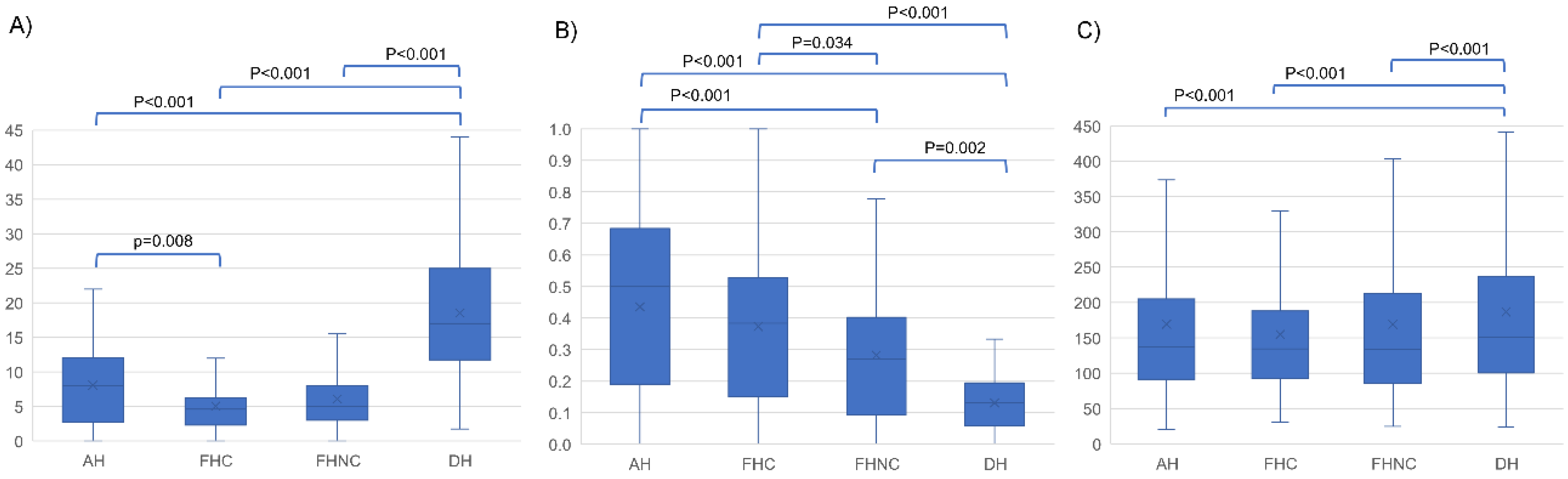

| Sample Groups | n | Mean | Median | SD | IQR | |
|---|---|---|---|---|---|---|
| MCK count | AH | 64 | 8.10 | 8.00 | 6.01 | 9.31 |
| FHC | 74 | 5.07 | 4.63 | 3.48 | 3.94 | |
| FHNC | 82 | 6.09 | 5.00 | 4.30 | 5.00 | |
| DH | 29 | 18.53 | 17.00 | 10.76 | 13.25 | |
| Proportion of osteonal MCKs | AH | 64 | 0.44 | 0.50 | 0.28 | 0.47 |
| FHC | 74 | 0.37 | 0.39 | 0.28 | 0.35 | |
| FHNC | 82 | 0.28 | 0.27 | 0.23 | 0.31 | |
| DH | 29 | 0.13 | 0.13 | 0.09 | 0.11 | |
| Interstitial MCK lenght (µm) | AH | 922 | 169.00 | 137.00 | 118.60 | 113.30 |
| FHC | 408 | 155.00 | 135.00 | 90.70 | 95.60 | |
| FHNC | 945 | 169.00 | 134.00 | 125.10 | 126.90 | |
| DH | 1304 | 187.00 | 151.00 | 124.50 | 136.20 |
Disclaimer/Publisher’s Note: The statements, opinions and data contained in all publications are solely those of the individual author(s) and contributor(s) and not of MDPI and/or the editor(s). MDPI and/or the editor(s) disclaim responsibility for any injury to people or property resulting from any ideas, methods, instructions or products referred to in the content. |
© 2023 by the authors. Licensee MDPI, Basel, Switzerland. This article is an open access article distributed under the terms and conditions of the Creative Commons Attribution (CC BY) license (https://creativecommons.org/licenses/by/4.0/).
Share and Cite
Schwab, N.; Galtés, I.; Winter-Buchwalder, M.; Ortega-Sánchez, M.; Jordana, X. Osteonal Microcracking Pattern: A Potential Vitality Marker in Human Bone Trauma. Biology 2023, 12, 399. https://doi.org/10.3390/biology12030399
Schwab N, Galtés I, Winter-Buchwalder M, Ortega-Sánchez M, Jordana X. Osteonal Microcracking Pattern: A Potential Vitality Marker in Human Bone Trauma. Biology. 2023; 12(3):399. https://doi.org/10.3390/biology12030399
Chicago/Turabian StyleSchwab, Nathalie, Ignasi Galtés, Michelle Winter-Buchwalder, Marisa Ortega-Sánchez, and Xavier Jordana. 2023. "Osteonal Microcracking Pattern: A Potential Vitality Marker in Human Bone Trauma" Biology 12, no. 3: 399. https://doi.org/10.3390/biology12030399
APA StyleSchwab, N., Galtés, I., Winter-Buchwalder, M., Ortega-Sánchez, M., & Jordana, X. (2023). Osteonal Microcracking Pattern: A Potential Vitality Marker in Human Bone Trauma. Biology, 12(3), 399. https://doi.org/10.3390/biology12030399

