The Role of Selenoproteins SELENOM and SELENOT in the Regulation of Apoptosis, ER Stress, and Calcium Homeostasis in the A-172 Human Glioblastoma Cell Line

Simple Summary
Abstract
1. Introduction
2. Materials and Methods
2.1. Isolation of RNA
2.2. Reverse Transcription and Real-Time PCR
2.3. Western Blotting
2.4. Gene Knockdown by RNA Interference, Transduction with Lentiviral Particles
2.5. MTT Analysis
2.6. Ability of Cells to Proliferate on Soft Agar
2.7. Wound Healing Assay
2.8. Registration of Changes in Cytosolic Ca2+
2.9. Statistical Analysis
3. Results
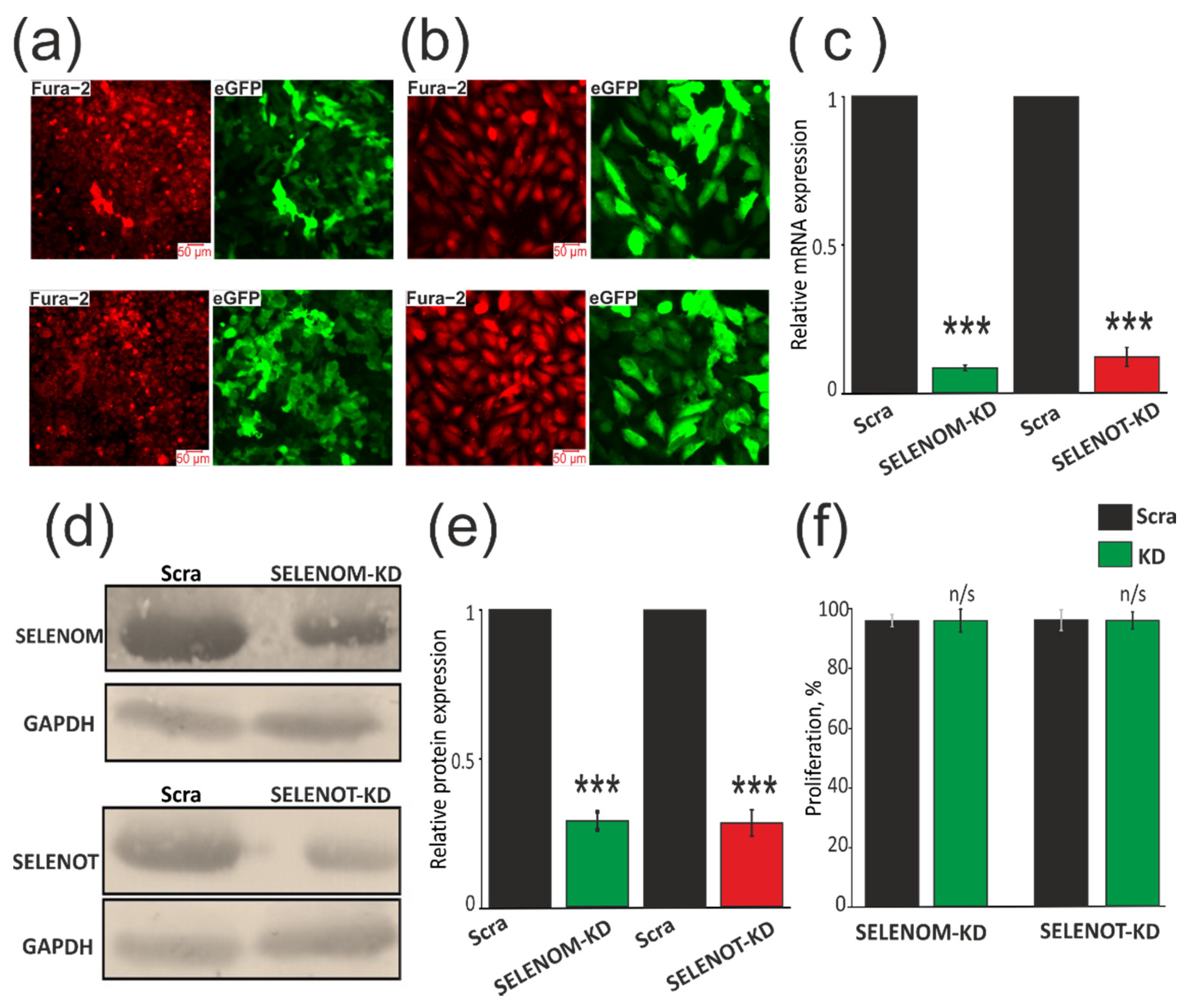
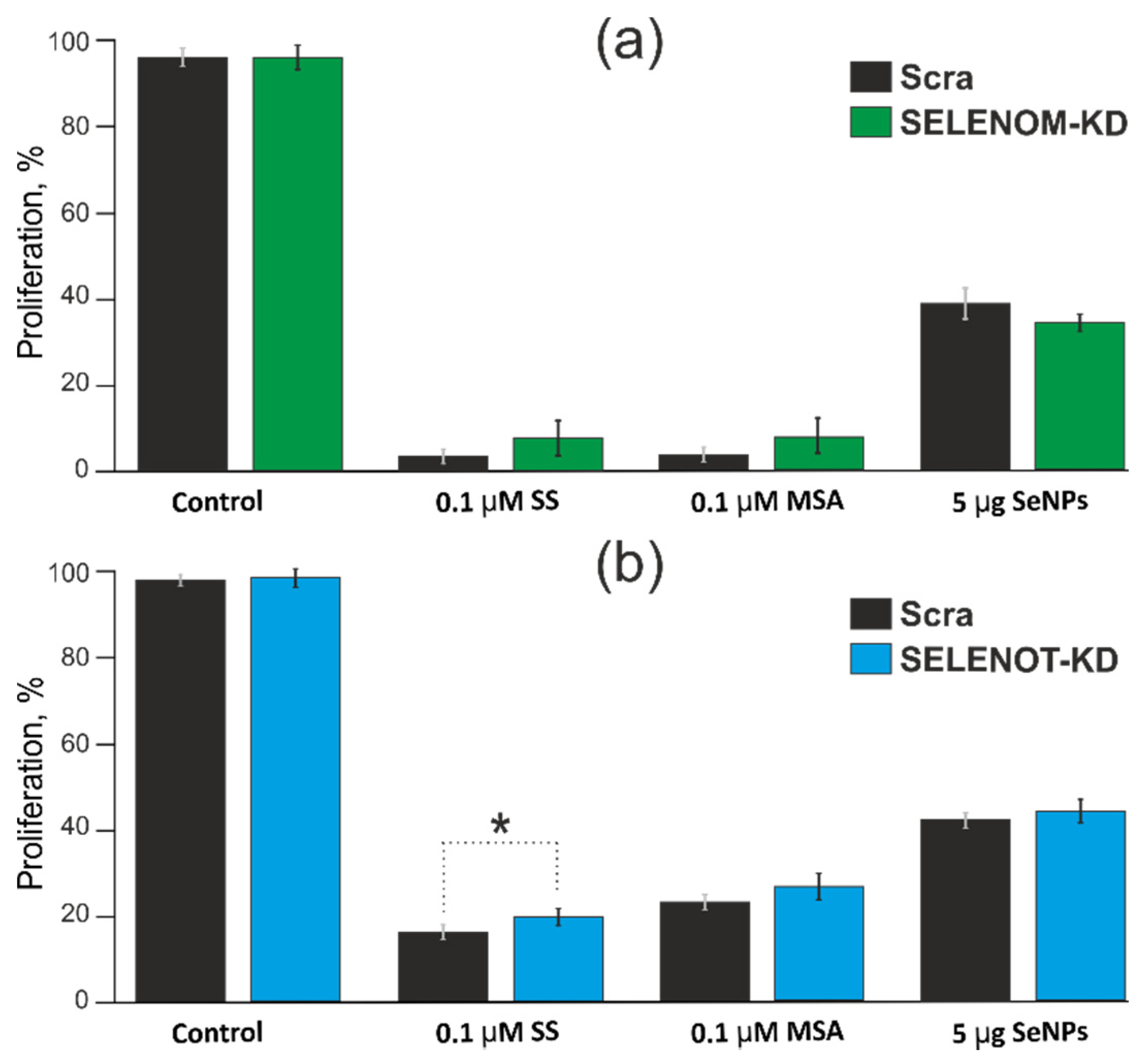
3.1. SELENOM-KD and SELENOT-KD Do Not Significantly Affect the Proliferative Properties of A-172 Cells
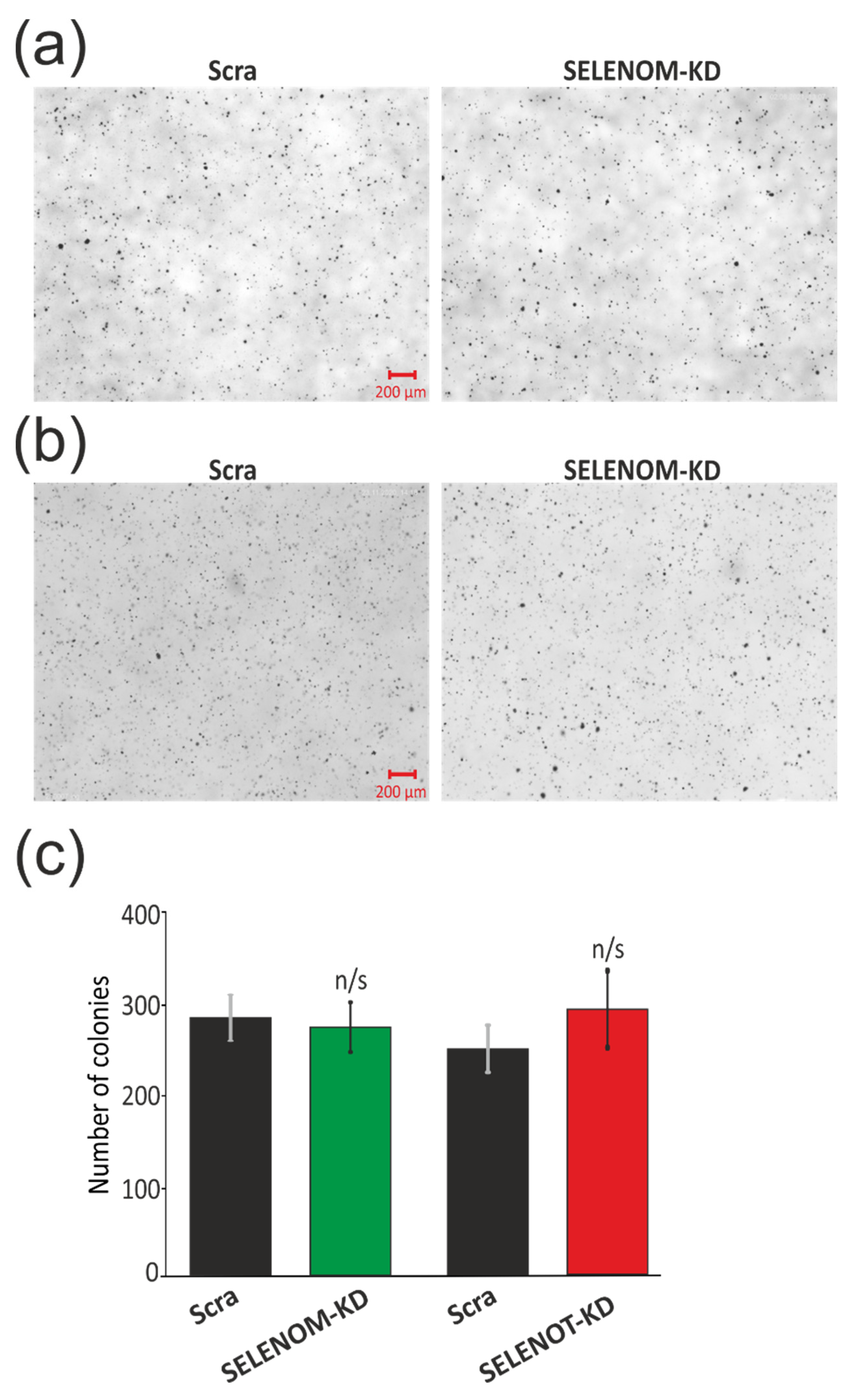
3.2. SELENOM-KD and SELENOT-KD Do Not Contribute to the Acquisition of Signs of Normal Cells by A-172 Cancer Cells
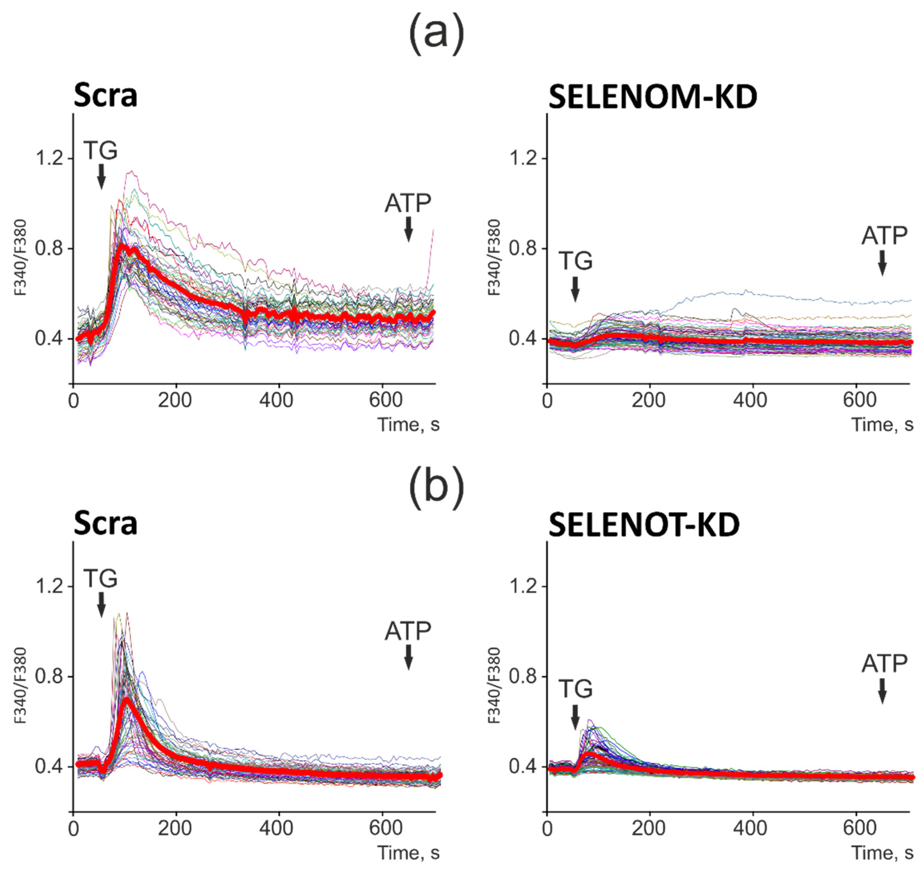
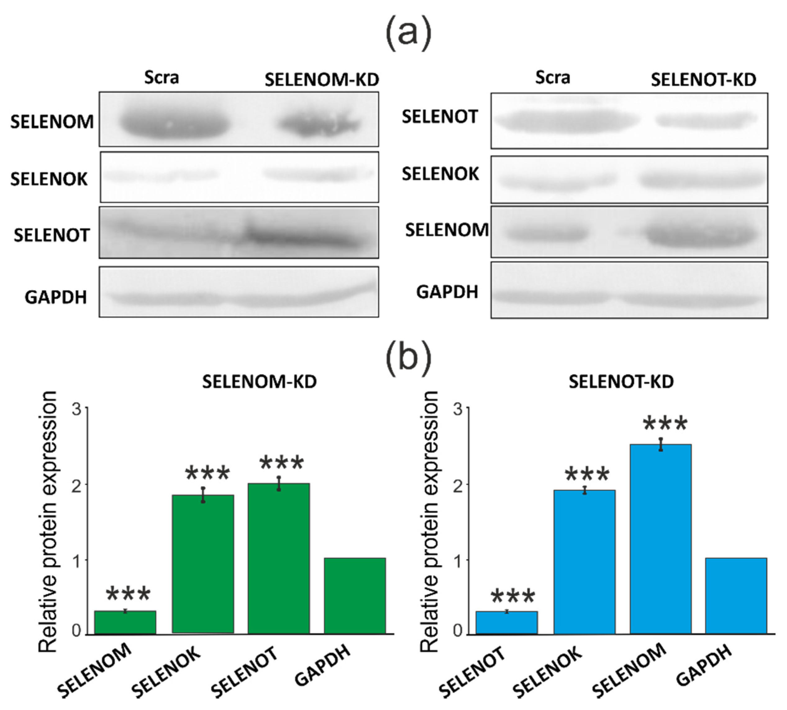
3.3. SELENOM-KD and SELENOT-KD Lead to Impaired Activity and Ca2+ Capacity in the ER
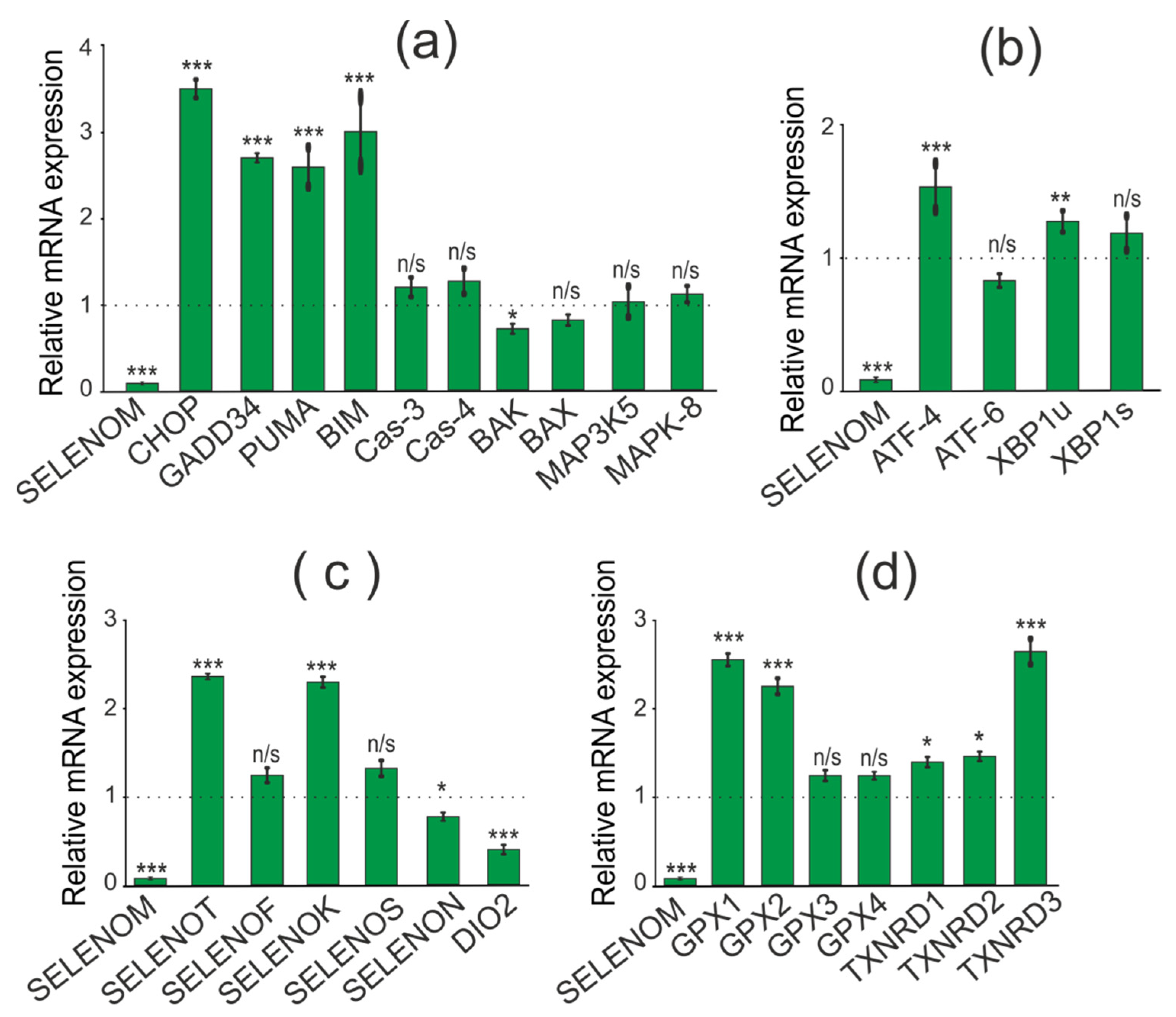
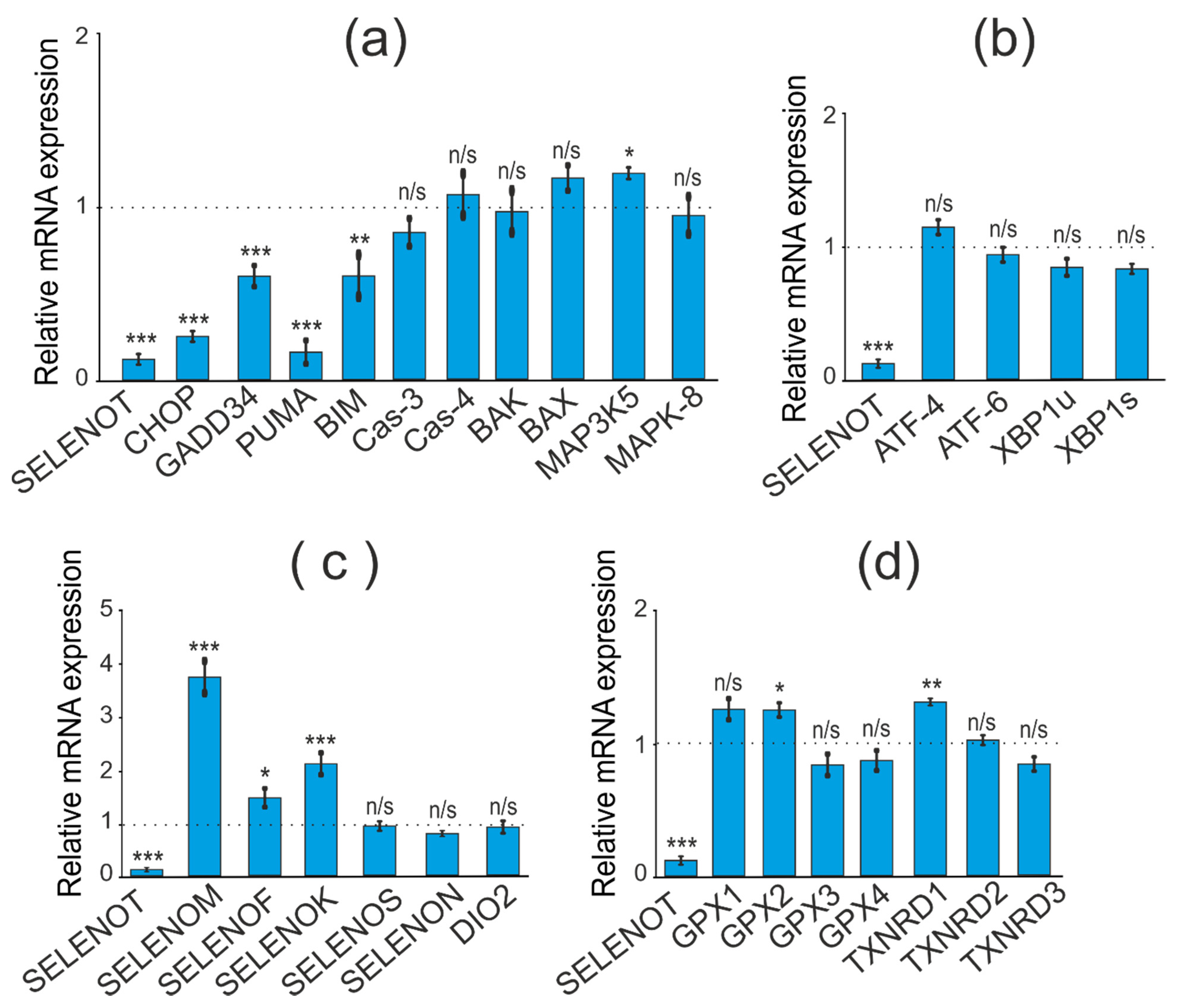
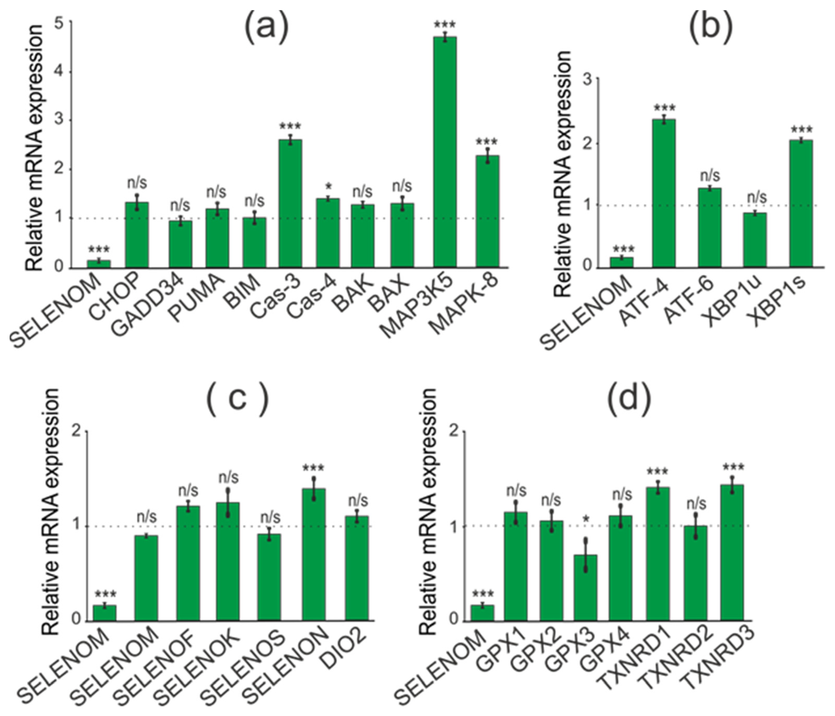
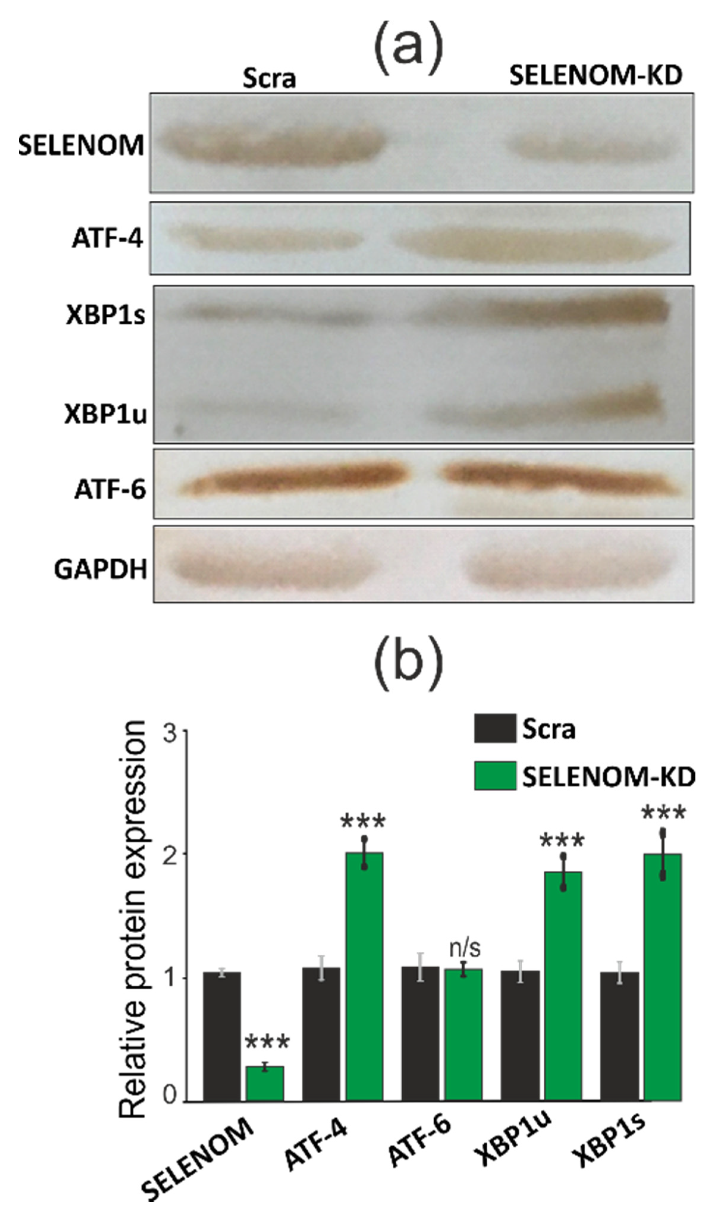
3.4. SELENOM-KD and SELENOT-KD Affect Expression Patterns of Pro-Apoptotic Genes, ER Stress Markers, and Selenoproteins in Different Ways
3.5. SELENOM-KD and SELENOT-KD Do Not Significantly Affect the Proliferative Properties of A-172 Cells under ER Stress
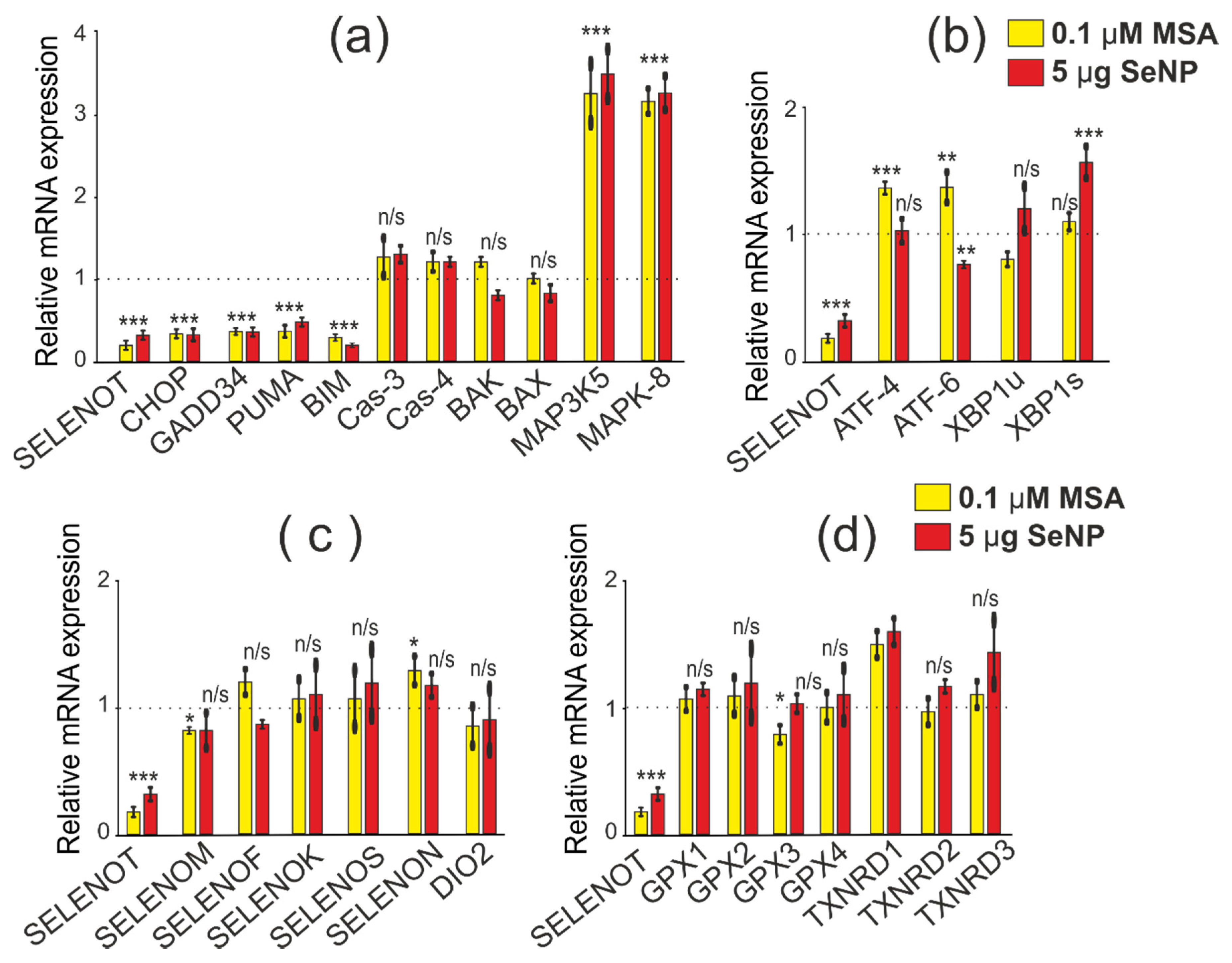
3.6. SELENOM-KD and SELENOT-KD Induce Changes in the Expression Patterns of Pro-Apoptotic Genes, ER Stress Markers, and Selenoproteins under Conditions of ER Stress Caused by Various Selenium-Containing Inducers
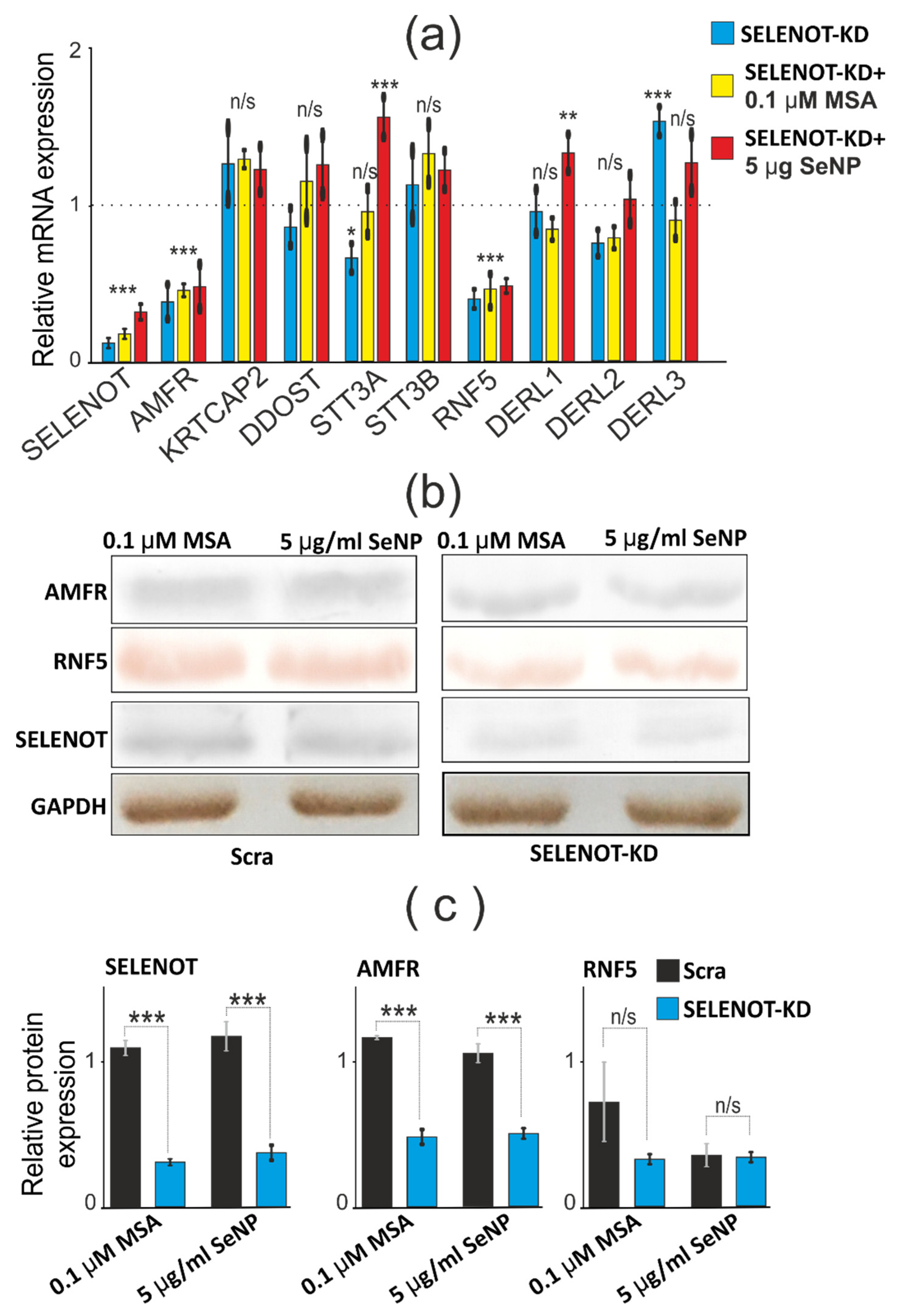
3.7. SELENOT-KD Causes a Decrease in the Expression of AMFR and RNF5 mRNA in A-172 Cells Both under Conditions of ER Stress and without It
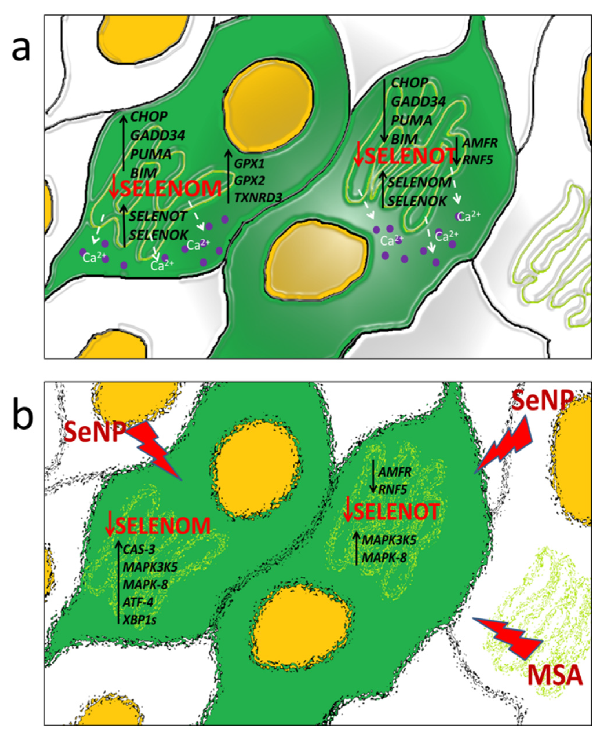
4. Discussion
5. Conclusions
Supplementary Materials
Author Contributions
Funding
Informed Consent Statement
Data Availability Statement
Conflicts of Interest
Abbreviations
References
- Arnér, E.S.J.; Holmgren, A. Physiological functions of Trx and Trx reductase. Eur. J. Biochem. 2000, 267, 6102–6109. [Google Scholar] [CrossRef] [Scilit] [PubMed]
- Korotkov, K.V.; Novoselov, S.V.; Hatfield, D.L.; Gladyshev, V.N. Mammalian selenoprotein in which selenocysteine (Sec) incorporation is supported by a new form of Sec insertion sequence element. J. Mol. Cell Biol. 2002, 22, 1402–1411. [Google Scholar] [CrossRef] [Scilit] [PubMed]
- Dikiy, A.; Novoselov, S.V.; Fomenko, D.E.; Sengupta, A.; Carlson, B.A.; Cerny, R.L.; Ginalski, K.; Grishin, N.V.; Hatfield, D.L.; Gladyshev, V.N. SelT, SelW, SelH, and Rdx12: Genomics and molecular insights into the functions of selenoproteins of a novel Trx–like family. J. Biochem. 2007, 46, 6871–6882. [Google Scholar] [CrossRef] [Scilit] [PubMed]
- Varlamova, E.G. Protein-protein interactions of ER-resident selenoproteins with their physiological partners. Biochimie 2020, 171–172, 197–204. [Google Scholar] [CrossRef] [Scilit] [PubMed]
- Zhou, J.C.; Zhao, H.; Tang, J.-Y.; Li, J.G.; Liu, X.L.; Zhu, Y.M. Molecular cloning, chromosomal localization and expression profiling of porcine selenoprotein M gene. Genes Genom. 2011, 33, 529–534. [Google Scholar] [CrossRef] [Scilit]
- Huang, J.Q.; Ren, F.Z.; Jiang, Y.Y.; Lei, X. Characterization of Selenoprotein M andIts Response to Selenium Deficiency in Chicken Brain. Biol. Trace Elem. Res. 2016, 170, 449–458. [Google Scholar] [CrossRef] [Scilit]
- Reeves, M.A.; Bellinger, F.P.; Berry, M.J. The neuroprotective functions of selenoprotein M and its role in cytosolic calcium regulation. Antioxid. Redox Signal. 2010, 12, 809–818. [Google Scholar] [CrossRef] [Scilit]
- Hwang, D.Y.; Sin, J.S.; Kim, M.S.; Yim, S.Y.; Kim, Y.K.; Kim, C.K.; Kim, B.G.; Shim, S.B.; Jee, S.W.; Lee, S.H.; et al. Overexpression of human selenoprotein M differentially regulates the concentrations of antioxidants and H2O2, the activity of antioxidant enzymes, and the composition of white blood cells in a transgenic rat. Int. J. Mol. Med. 2008, 21, 169–179. [Google Scholar] [CrossRef] [Scilit]
- Moustafa, M.E.; Antar, H.A. A bioinformatics approach to characterize mammalian selenoprotein T. Biochem. Genet. 2012, 50, 736–747. [Google Scholar] [CrossRef] [Scilit]
- Grumolato, L.; Ghzili, H.; Montero–Hadjadje, M.; Gasman, S.; Lesage, J.; Tanguy, Y.; Galas, L.; Ait–Ali, D.; Leprince, J.; Guérineau, N.C.; et al. Selenoprotein T is a PACAP–regulated gene involved in intracellular Ca2+ mobilization and neuroendocrine secretion. FASEB J. 2008, 22, 1756–1768. [Google Scholar] [CrossRef] [Scilit]
- Shchedrina, V.A.; Zhang, Y.; Labunskyy, V.M.; Hatfield, D.L.; Gladyshev, V.N. Structure–Function Relations, Physiological Roles, and Evolution of Mammalian ER– Resident Selenoproteins. Antioxid. Redox Signal. 2010, 12, 839–849. [Google Scholar] [CrossRef] [Scilit]
- Liu, J.; Rozovsky, S. Membrane–bound selenoproteins. Antioxid. Redox Signal. 2015, 23, 795–813. [Google Scholar] [CrossRef] [Scilit]
- Boukhzar, L.; Hamieh, A.; Cartier, D.; Tanguy, Y.; Alsharif, I.; Castex, M.; Arabo, A.; El Hajji, S.; Bonnet, J.J.; Errami, M.; et al. Selenoprotein T Exerts an Essential Oxidoreductase Activity that Protects Dopaminergic Neurons in Mouse Models of Parkinson’s Disease. Antioxid. Redox Signal. 2016, 24, 557–574. [Google Scholar] [CrossRef] [Scilit]
- Tanguy, Y.; Falluel–Morel, A.; Arthaud, S.; Boukhzar, L.; Manecka, D.L.; Prevost, G.; Elias, S.; Dorval–Coiffec, I.; Lesage, J.; Vieau, D.; et al. The PACAP–regulated gene selenoprotein T is highly induced in nervous, endocrine, and metabolic tissues during ontogenetic and regenerative processes. Endocrinology 2011, 152, 4322–4335. [Google Scholar] [CrossRef] [Scilit]
- Hamieh, A.; Cartier, D.; Abid, H.; Calas, A.; Burel, C.; Bucharles, C.; Jehan, C.; Grumolato, L.; Landry, M.; Lerouge, P.; et al. Selenoprotein T is a novel OST subunit that regulates UPR signaling and hormone secretion. EMBO 2017, 18, 1935–1946. [Google Scholar] [CrossRef] [Scilit]
- Roboti, P.; High, S. The oligosaccharyltransferase subunits OST48, DAD1 and KCP2 function as ubiquitous and selective modulators of mammalian N–glycosylation. J. Cell Sci. 2012, 125, 3474–3484. [Google Scholar] [CrossRef] [Scilit]
- Castex, M.T.; Arabo, A.; Bénard, M.; Roy, V.; Le Joncour, V.; Prévost, G.; Bonnet, J.J.; Anouar, Y.; Falluel–Morel, A. Selenoprotein T deficiency leads to neurodevelopmental abnormalities and hyperactive behavior in mice. Mol. Neurobiol. 2016, 53, 5818–5832. [Google Scholar] [CrossRef] [Scilit]
- Varlamova, E.G.; Turovsky, E.A. The main cytotoxic effects of methylseleninic acid on various cancer cells. Int. J. Mol. Sci. 2021, 22, 6614. [Google Scholar] [CrossRef] [Scilit]
- Goltyaev, M.V.; Mal’tseva, V.N.; Varlamova, E.G. Expression of ER-resident selenoproteins and activation of cancer cells apoptosis mechanisms under ER-stress conditions caused by methylseleninic acid. Gene 2020, 755, 144884. [Google Scholar] [CrossRef] [Scilit]
- Goltyaev, M.V.; Varlamova, E.G.; Novoselov, S.V.; Fesenko, E.E. Activation of Signal Pathways of Apoptosis under Conditions of Prolonged ER-Stress Caused by Exposure of Mouse Testicular Teratoma Cells to Selenium-Containing Compounds. Dokl. Biochem. Biophys. 2020, 490, 9–11. [Google Scholar] [CrossRef] [Scilit]
- Varlamova, E.G.; Turovsky, E.A.; Blinova, E.V. Therapeutic Potential and Main Methods of Obtaining Selenium Nanoparticles. Int. J. Mol. Sci. 2021, 22, 10808. [Google Scholar] [CrossRef] [Scilit]
- Turovsky, E.A.; Varlamova, E.G. Mechanism of Ca2+-Dependent Pro-Apoptotic Action of Selenium Nanoparticles, Mediated by Activation of Cx43 Hemichannels. Biology 2021, 10, 743. [Google Scholar] [CrossRef] [Scilit]
- Varlamova, E.G.; Goltyaev, M.V.; Mal’tseva, V.N.; Turovsky, E.A.; Sarimov, R.M.; Simakin, A.V.; Gudkov, S.V. Mechanisms of the Cytotoxic Effect of Selenium Nanoparticles in Different Human Cancer Cell Lines. Int. J. Mol. Sci. 2021, 22, 7798. [Google Scholar] [CrossRef] [Scilit]
- Kuznetsova, Y.P.; Goltyaev, M.V.; Gorbacheva, O.S.; Novoselov, S.V.; Varlamova, E.G.; Fesenko, E.E. Influence of Sodium Selenite on the mRNA Expression of the Mammalian Selenocysteine–Containing Protein Genes in Testicle and Prostate Cancer Cells. Dokl. Biochem. Biophys. 2018, 480, 131–134. [Google Scholar] [CrossRef] [Scilit]
- Shigemi, Z.; Manabe, K.; Hara, N.; Baba, Y.; Hosokawa, K.; Kagawa, H.; Watanabe, T.; Fujimuro, M. Methylseleninic acid and sodium selenite induce severe ER stress and subsequent apoptosis through UPR activation in PEL cells. Chem. Biol. Interact. 2017, 266, 28–37. [Google Scholar] [CrossRef] [Scilit]
- Varlamova, E.G. Participation of selenoproteins localized in the ER in the processes occurring in this organelle and in the regulation of carcinogenesis-associated processes. J. Trace Elem. Med. Biol. 2018, 48, 172–180. [Google Scholar] [CrossRef] [Scilit]
- Varlamova, E.G.; Cheremushkina, I.V. Contribution of mammalian selenocysteine-containing proteins to carcinogenesis. J. Trace Elem. Med. Biol. 2017, 39, 76–85. [Google Scholar] [CrossRef] [Scilit]
- Varlamova, E.G.; Goltyaev, M.V.; Novoselov, V.I.; Fesenko, E.E. Cloning, intracellular localization, and expression of the mammalian selenocysteine-containing protein SELENOI (SelI) in tumor cell lines. Dokl. Biochem. Biophys. 2017, 476, 320–322. [Google Scholar] [CrossRef] [Scilit]
- Oyadomari, S.; Mori, M. Roles of CHOP/GADD153 in endoplasmic reticulum stress. Cell Death Differ. 2004, 11, 381–389. [Google Scholar] [CrossRef] [Scilit]
- Marciniak, S.J.; Yun, C.Y.; Oyadomari, S.; Novoa, I.; Zhang, Y.; Jungreis, R. CHOP induces death by promoting protein synthesis and oxidation in the stressed endoplasmic reticulum. Genes Dev. 2004, 18, 3066–3077. [Google Scholar] [CrossRef] [Scilit]
- Chu, F.F.; Doroshow, J.H.; Esworthy, R.S. Expression, characterization and tissue distribution of anew cellular seleniumdependent glutathione peroxidase, GSHPx. GI J. Biol. Chem. 1993, 268, 2571–2576. [Google Scholar] [CrossRef] [Scilit]
- Su, D.; Novoselov, S.V.; Sun, Q.-A.; Moustafa, M.E.; Zhou, Y.; Oko, R.; Hatfield, D.L.; Gladyshev, V.N. Mammalian selenoprotein thioredoxin-glutathione reductase. Roles in disulfide bond formation and sperm maturation. J Biol. Chem. 2005, 280, 26491–26498. [Google Scholar] [CrossRef] [Scilit] [PubMed]
- Shchedrina, V.A.; Everley, R.A.; Zhang, Y.; Gygi, S.P.; Hatfield, D.L.; Gladyshev, V.N. Selenoprotein K binds multiprotein complexes and is involved in the regulation of endoplasmic reticulum homeostasis. J. Biol. Chem. 2011, 286, 42937–42948. [Google Scholar] [CrossRef] [Scilit] [PubMed]
- Ye, Y.; Shibata, Y.; Kikkert, M.; van Voorden, S.; Wiertz, E.; Rapoport, T.A. Recruitment of the p97 ATPase and ubiquitin ligases to the site of retrotranslocation at the endoplasmic reticulum membrane. Proc. Natl. Acad. Sci. USA 2005, 102, 14132–14138. [Google Scholar] [CrossRef] [Scilit] [PubMed]
- Lee, J.H.; Park, K.J.; Jang, J.K.; Jeon, Y.H.; Ko, K.Y.; Kwon, J.H.; Lee, S.R.; Kim, I.Y. Selenoprotein S–dependent Selenoprotein K Binding to p97(VCP) Protein Is Essential for Endoplasmic Reticulum–associated Degradation. J. Biol. Chem. 2015, 290, 29941–29952. [Google Scholar] [CrossRef] [Scilit]
- Lee, J.H.; Kwon, J.H.; Jeon, Y.H.; Ko, K.Y.; Lee, S.-R.; Kim, I.Y. Pro–178 and Pro–183 of selenoprotein S Are essential residues for interaction with p97 (VCP) during endoplasmic reticulum–associated degradation. J. Biol. Chem. 2014, 289, 13758–13768. [Google Scholar] [CrossRef] [Scilit]
- Fredericks, G.J.; Hoffmann, F.K.W.; Rose, A.H.; Osterheld, H.J.; Hess, F.M.; Mercier, F.; Hoffmann, P.R. Stable expression and function of the inositol 1,4,5–triphosphate receptor requires palmitoylation by a DHHC6/selenoprotein K complex. Proc. Natl. Acad. Sci. USA 2014, 111, 16478–16483. [Google Scholar] [CrossRef] [Scilit]
- Abid, H.; Cartier, D.; Hamieh, A.; Francois-Bellan, A.M.; Bucharles, C.; Pothion, H.l. AMPK Activation of PGC-1alpha/NRF-1-Dependent SELENOT Gene transcription promotes PACAP-induced neuroendocrine cell differentiation through tolerance to oxidative stress. Mol. Neurobiol. 2019, 56, 4086–4101. [Google Scholar] [CrossRef] [Scilit]
- Brevik, E.J.; van Donkelaar, M.M.; Weber, H.; Sanchez-Mora, C.; Jacob, C.; Rivero, O. Genome-wide analyses of aggressiveness in attention-deficit hyperactivity disorder. Am. J. Med. Genet. B Neuropsychiatr. Genet. 2016, 171, 733–747. [Google Scholar] [CrossRef] [Scilit]
- Shao, Z.Q.; Zhang, X.; Fan, H.H.; Wang, X.S.; Wu, H.M.; Zhang, L.; Cheng, W.H.; Zhu, J.H. Selenoprotein T Promotes Proliferation and G1-to-S Transition in SK-N-SH Cells: Implica-tions in Parkinson’s Disease. J. Nutr. 2019, 149, 2110–2119. [Google Scholar] [CrossRef] [Scilit]
- Sengupta, A.; Carlson, B.A.; Labunskyy, V.M.; Gladyshev, V.N.; Hatfield, D.L. Selenoprotein T deficiency alters cell adhesion and elevates selenoprotein W expression in murine fibroblast cells. Biochem. Cell Biol. 2009, 87, 953–961. [Google Scholar] [CrossRef] [Scilit]
- Rocca, C.; Boukhzar, L.; Granieri, M.C.; Alsharif, I.; Mazza, R.; Lefranc, B.; Tota, B.; Leprince, J.; Cerra, M.C.; Anouar, Y.; et al. A selenoprotein T-derived peptide protects the heart against ischaemia/reperfusion injury through inhibition of apoptosis and oxidative stress. Acta Physiol. 2018, 223, 13067. [Google Scholar] [CrossRef] [Scilit]
- Zhang, Z.H.; Song, G.L. Roles of Selenoproteins in Brain Function and the Potential Mechanism of Selenium in Alzheimer’s Disease. Front. Neurosci. 2021, 15, 646518. [Google Scholar] [CrossRef] [Scilit]
- Lockshin, R.A.; Zakeri, Z. Apoptosis, autophagy, and more. Int. J. Biochem. Cell Biol. 2004, 36, 2405–2419. [Google Scholar] [CrossRef] [Scilit]
- Nakamura, K.; Bossy-Wetzel, E.; Burns, K.; Fadel, M.P.; Lozyk, M.; Goping, I.S.; Opas, M.; Bleackley, R.C.; Green, D.R.; Michalak, M. Changes in endoplasmic reticulum luminal environment affect cell sensitivity to apoptosis. J. Cell Biol. 2000, 150, 731–740. [Google Scholar] [CrossRef] [Scilit]



| Gene Name | Forward Primer 5′→3′ | Reverse Primer 5′→3′ |
|---|---|---|
| GAPDH | ACATCGCTCAGACACCATG | GCCAGTGAGCTTCCCGTT |
| SELENOT | TCTCCTAGTGGCGGCGTC | GTCTATATATTGGTTGAGGGAGG |
| SELENOM | AGCCTCCTGTTGCCTCCGC | AGGTCAGCGTGGTCCGAAG |
| SELENOF | TACGGTTGTTGTTGGCGAC | CAAATTGTGCTTCCTCCTGAC |
| SELENOK | TTTACATCTCGAACGGACAAG | CAGCCTTCCACTTCTTGATG |
| SELENOS | TGGGACAGCATGCAAGAAG | GCGTCCAGGTCTCCAGG |
| SELENON | TGATCTGCCTGCCCAATG | TCAGGAACTGCATGTAGGTGG |
| DIO2 | AGCTTCCTCCTCGATGCC | AAAGGAGGTCAAGTGGCTG |
| AMFR | TCCTGGCTAGAACAAGACACC | CTGCGTAATGCCAAGAATG |
| KRTCAP2 | GCAAAGGATTCCAAGCAAAG | TTCTTGCTCTTGCCTGTGAC |
| DDOST | CGCATTGATCCTTTTGTGAG | GAAGACGATGCTGAAGATGAAG |
| STT3A | TACAGGCAAACATATCAAGGAG | TCCAGGACATCAAGCTCAAAG |
| STT3B | CTACTTTGTTGAATTGCCTTATG | TTCCTTTTGGTAGTCTTCTTTG |
| RNF5 | AAGGGCCAAATCGCGAGCG | GCGGGACAACCTTCTCTCTG |
| DERL1 | ATTCAACTATATCATCGGAGGC | CAAGTCGAAAGCCCTGGCC |
| DERL2 | TTCGGCCTTCTCAACTTCCAG | TGGGTCTCATTATTGGCACTG |
| DERL3 | CTTCTCGCTGCTGCTGGGC | CCCAGGAAGCCAGGGGTC |
| CHOP | GCTCTGATTGACCGAATGG | TCTGGGAAAGGTGGGTAGTG |
| GADD34 | CTCCGAGAAGGTCACTGTCC | GACGAGCGGGAAGGTGTGG |
| PUMA | CAGATATGCGCCCAGAGAT | CCATTCGTGGGTGGTCTTC |
| BIM | GGACGACCTCAACGCACAGTACGAG | GTAAGGGCAGGAGTCCCA |
| ATF-4 | GTGTTCTCTGTGGGTCTGCC | GACCCTTTTCTTCCCCCTTG |
| ATF-6 | AACCCTAGTGTGAGCCCTGC | GTTCAGAGCACCCTGAAGA |
| XBPu | ACTCAGACTACGTGCACCTC | GTCAATACCGCCAGAATCC |
| XBPs | CTGAGTCCGCAGCGGTGCAGG | GGTCCAAGTTGTCCAGAATG |
| CAS-3 | GCATTGAGACAGACAGTGGTG | AATAGAGTTCTTTTGTGAGCATG |
| CAS-4 | CACGCCTGGCTCTCATCATA | TAGCAAATGCCCTCAGCG |
| MAP3K5 | AACACCTGAAGCTTAAGTCCC | TCAATGATAGCCTTCCACAGTG |
| MAPK-8 | AAAGGGAACACACAATAGAAGAG | GCTGCTGCTTCTAGACTG |
| BAX | GGGCTGGACATTGGACTTC | AACACAGTCCAAGGCAGCTG |
| BAK | GAGAGTGGCATCAATTGGGG | CAGCCACCCCTCTGTGCAATCCA |
| GPX1 | CTACTTATCGAGAATGTGGCG | CGAAGAGCATGAAGTTGGG |
| GPX2 | CCCTTCCGACGCTACAGCCG | GGAGCCCAAGTTGAATCACC |
| GPX3 | CCCCCACTCCTACTTCCTG | CCGAAGGAGCAGGGGTGG |
| GPX4 | CCATGCACGAGTTTTCCG | AATTTGACGTTGTAGCCCG |
| TXNRD1 | GGTCTGGCAGCTGCTAAGG | TAGCCCCAATTCAAAGAGC |
| TXNRD2 | TGGGTGTGGCAGTGGGAGAC | TCCCCTGAGCCATCCCTGTG |
| TXNRD3 | CCTTTGCTTTGTTGTTTCTGTG | TAGTGAGTGTGAGGGTGAAGC |
Publisher’s Note: MDPI stays neutral with regard to jurisdictional claims in published maps and institutional affiliations. |
© 2022 by the authors. Licensee MDPI, Basel, Switzerland. This article is an open access article distributed under the terms and conditions of the Creative Commons Attribution (CC BY) license (https://creativecommons.org/licenses/by/4.0/).
Share and Cite
Varlamova, E.G.; Goltyaev, M.V.; Turovsky, E.A. The Role of Selenoproteins SELENOM and SELENOT in the Regulation of Apoptosis, ER Stress, and Calcium Homeostasis in the A-172 Human Glioblastoma Cell Line. Biology 2022, 11, 811. https://doi.org/10.3390/biology11060811
Varlamova EG, Goltyaev MV, Turovsky EA. The Role of Selenoproteins SELENOM and SELENOT in the Regulation of Apoptosis, ER Stress, and Calcium Homeostasis in the A-172 Human Glioblastoma Cell Line. Biology. 2022; 11(6):811. https://doi.org/10.3390/biology11060811
Chicago/Turabian StyleVarlamova, Elena G., Michael V. Goltyaev, and Egor A. Turovsky. 2022. "The Role of Selenoproteins SELENOM and SELENOT in the Regulation of Apoptosis, ER Stress, and Calcium Homeostasis in the A-172 Human Glioblastoma Cell Line" Biology 11, no. 6: 811. https://doi.org/10.3390/biology11060811
APA StyleVarlamova, E. G., Goltyaev, M. V., & Turovsky, E. A. (2022). The Role of Selenoproteins SELENOM and SELENOT in the Regulation of Apoptosis, ER Stress, and Calcium Homeostasis in the A-172 Human Glioblastoma Cell Line. Biology, 11(6), 811. https://doi.org/10.3390/biology11060811