Abstract
In this study, hybrid materials were synthesized incorporating curcumin, Cu2+ or Fe3+, and Kappa-carrageenan as a reducing agent to improve stability, considering that curcumin has low thermal and solution stability, which limits its applications. Colorimetric analysis showed color changes in the hybrids, ultraviolet–visible spectroscopy revealed band shifts in the hybrids, and infrared analysis indicated shifts in wavenumbers, suggesting changes in the vibrational state of curcumin after bonding with metal ions. These techniques confirmed the formation of hybrid materials. Thermogravimetric and chromatographic analyses demonstrated greater thermal and solution stability for the hybrids compared to curcumin. Additionally, the hybrid composites effectively developed natural and sebaceous latent fingerprints with good clarity and contrast on glass surfaces. Both composites performed similarly to commercial Gold® powder. When applied to surfaces representative of forensic scenarios, the composites were versatile, revealing sufficient fingerprint details for human identification on both porous and non-porous surfaces. Scanning electron microscopy images showed greater clarity in sebaceous and natural fingerprints developed with the Fe composite compared to the Cu composite.
1. Introduction
Papilloscopy refers to the science dedicated to studying fingerprints, which vary between individuals and even in the same person. This uniqueness has made fingerprints a reliable method of human identification for over a century [1]. In forensic sciences, fingerprint analysis is based on Locard’s exchange principle, which establishes that every contact between two objects results in a transfer of traces between them. Consequently, the act of touching a surface transfers material from the ridges of the fingers onto a surface [2]. In criminal investigations, fingerprints are considered physical evidence, as they not only capture the pattern ridges but can also retain exogenous compounds such as drugs, explosives, and chemicals [3]. Latent fingerprints (LFPs) found at crime scenes are invisible to the naked eye and require specific methods to be visualized, which include optical (ultraviolet laser), chemical (cyanoacrylate fuming), and physical (powder dusting and deposition) methods [4,5]. One of the oldest techniques, still widely used by forensic experts, is the powder technique, especially when LFPs are deposited on smooth surfaces such as glass, metal, and plastic. The technique is simple, involving the application of powder with a specific brush onto the surface where the LFP is found, making the ridge patterns and their minutiae visible to the naked eye [6]. An ideal compound for developing fingerprint should provide high contrast, good adhesion properties, and desirable selectivity [1]. In this context, the search for a selective, high-contrast, low-cost, and reliable powder for developing LFPs has been relentless. Most available LFP powders use hazardous chemicals in their synthesis, and continuous use can cause side effects. Therefore, the use of natural products as a raw material source for the development of new LFPs is of great interest, as their use may result in a final product with little to no toxicity [7].
Based on this, curcumin, a natural dye extracted from the rhizomes of Curcuma longa, has emerged as a promising candidate [8]. Its structure comprises a β-diketone fraction, methoxy groups, phenolic hydroxyls, doubly conjugated bonds, and keto-enol tautomerism [9]. Studies report that the properties found in turmeric are mainly attributed to curcumin, making the isolated compound of great interest [10]. Interesting properties have been reported in isolated curcumin, including high antimicrobial activity [11], antibacterial properties [12], and antioxidant features [13], among others. However, isolated curcumin exhibits rapid photodegradation when exposed to light [14] and air [15], which limits its stability and effectiveness. Furthermore, the curcumin molecule is not stable in solution due to the β-diketone fraction of curcumin, which is a hydrolytic center. This hydrolytic center is involved in the elimination of the hydroxyl radical and in redox reactions, leading to the formation of degradation products, that is, minor phenolics [14], such as ferulic acid, vanillic acid, and vanillin [16]. To overcome these limitations, hybrid materials can be designed from various components in order to achieve original properties in a single material [17]. The β-diketone groups enable curcumin to chelate metal ions, leading to the formation of hybrid materials, which comprise a combination of two or more phases, whether organic or inorganic, with unique properties [18]. However, the properties of a hybrid material are not merely a sum of the individual contributions of its constituents but depend on the chemical nature of the organic and inorganic compounds. Due to their complexity, they can be classified into different categories. Among them, the usual classification is performed according to the type of interaction or the nature of the chemical bonds between the organic and inorganic components. As mentioned earlier, curcumin contains a fraction that acts as a strong chelating agent, and thus, hybrid materials synthesized from curcumin belong to class II, which consists of materials formed through covalent or ionic bonds [19]. For the synthesis of a hybrid material containing curcumin and a metal as organic and inorganic phases, respectively, a third phase with metal-reducing properties is needed. In this scenario, the use of Kappa-carrageenan (κ-car) as a reducer agent has attracted interest [20]. κ-car is a polysaccharide extracted from red macroalgae, featuring non-toxic, low-cost, edible, biodegradable, and biocompatible properties [21]. The negatively charged surface of κ-car enables easy interaction with positively charged metal ions through electrostatic attraction. Moreover, the presence of hydroxyl (OH) groups facilitates the reduction of these metals [22].
According to what has previously been reported, in this work, we continue our efforts to develop derivatives of organic compounds with applications in papilloscopy [7,23,24,25,26,27,28,29,30,31,32]. Therefore, this study reports the development of a new hybrid material of curcumin and Kappa-carrageenan combined with two low-cost copper and iron metals. The hybrid materials were evaluated for thermal stability and solution behavior. Finally, these hybrid composites were tested as potential LFP developers, offering a novel, low-toxicity alternative for fingerprint analysis.
2. Materials and Methods
The curcumin (diferuloylmethane, 65% purity) and Kappa-carrageenan were purchased from Sigma-Aldrich (St. Louis, MO, USA). Acetonitrile was purchased from Biograde (Durham, USA, USA–HPLC ≥ 99.99%). Tetrahydrofuran (THF) was purchased from Honeywell (Charlotte, USA, USA–HPLC ≥ 99.99%). Fingerprint powder (developer, Gold®) was purchased from Sirchie® (Youngsville, LA, USA). The other chemicals were of analytical grade and from commercial sources.
2.1. Synthesis of the Hybrid Materials
The hybrid materials were synthesized following a one-pot methodology adapted from Li et al. (2021) [33]. Briefly, in a beaker, we added copper acetate monohydrate (Cu(CO2CH3)2·H2O, Vetec-P.A) (0.1359 g, 0.0012 mmol) or iron (III) chloride hexahydrate (FeCl3·6H2O, Vetec-P.A) (0.1834 g, 0.0008 mmol) into curcumin (0.5000 g, 0.0014 mmol) previously dissolved in a solution of ethanol (8 mL) and deionized water (4 mL). Then, Kappa-carrageenan (1.0000 g, 0.0023 mmol) was added to the mixture in the beaker, which reacted for 30 min at room temperature. The mixture was rinsed with deionized water (200 mL) and absolute ethanol (200 mL). Finally, the material was centrifuged (10 min, 4000 rpm) to separate the solid–liquid phases. The solid fraction was dried in an oven at 60 °C for 2 h. Scheme 1 presents the synthesis of new hybrid materials.
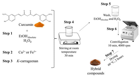
Scheme 1.
Scheme for the synthesis of new hybrid materials. Step 1. Dissolution of curcumin in absolute ethanol and distilled water. Step 2. Addition of Cu2+ acetate or Fe3+ chloride. Step 3. Addition of K-carrageenan. Step 4. Stirring for 30 min. Step 5. Addition of distilled water and absolute ethanol for washing. Step 6. Centrifugation to separate the solid–liquid phases. Step 7. Drying of the hybrid materials in an oven at 60 °C for 2 h.
2.2. Characterization
The ultraviolet–visible spectroscopy (UV-Vis) of pure curcumin and both Fe and Cu hybrid materials was performed using a Bel LGS53 instrument, with the samples diluted in methanol solvent. The samples were also investigated by Fourier transform infrared spectroscopy (FT-IR), model SPIRIT (Shimadzu, Tokyo, Japan), using potassium bromide (KBr—[0.5%]). X-ray diffraction (XRD) patterns were recorded in the 2θ range from 10° to 60° using a Shimadzu XRD-6000 instrument. Thermogravimetric analysis (TGA) was performed using a Shimadzu DTG-60 instrument. The samples were evaluated in N2 atmosphere, from 30 to 800 °C and with a heating rate of 10 °C/min. To determine the color of the samples, Kappa-carrageenan, curcumin, copper acetate, iron chloride, copper hybrid, and iron hybrid samples were analyzed in a CR-400 colorimeter (Konica Minolta), with an observation angle of 10°, calibrated with porcelain, configured with D65 light. All analyses were performed in triplicate. The CIELab system (L*, a*, and b*) was used to determine the colorimetric parameters. In this system, L* represents lightness (from 0 (black) to 100 (white)), a* corresponds to the green–red axis (negative values indicate shades of green, while positive values indicate shades of red), and b* corresponds to the blue–yellow axis (negative values indicate shades of blue, while positive values indicate shades of yellow). To identify the variation between colors, ΔE was used, according to the following equation:
where ΔE is the variation in all colors; ΔL is the variation in lightness; Δa is the variation in the red–green coordinates; and Δb is the variation in the blue–yellow coordinatess [33].
2.3. High-Performance Liquid Chromatography with UV-Vis Detector (HPLC-UV-Vis)
The chromatographic analyses were performed using a Thermo Scientific Ultimate 3000, Waltham model, USA, with a standard auto-injector and UV-Vis detector. The separation of analytes was carried out on a C18 column (Ascentis, Bellevue, WA, USA) with a particle size of 5 μm, a pore size of 90 Å, and a column size of 150 mm × 4.6 mm. For the determination of curcuminoids in the commercial curcumin sample, we employed an adapted method based on [34], where the isocratic mode was used with a mobile phase of 0.001% phosphoric acid (in water) and acetonitrile (20:80, v/v). The analysis was performed at a temperature of 45 °C, with an injection volume of 20 µL and a flow rate of 0.8 mL/min, for 5 min. The detection wavelength was 425 nm, and the samples were diluted in acetonitrile.
For the determination of the solution stability of curcumin and the hybrid materials, the method employed was adapted from Kumar et al. (2021) [16]. The isocratic mode was used, and a mobile phase mixture was employed: acetonitrile, tetrahydrofuran, and 2% acetic acid (aqueous) (26.3:63.2:10.5 v/v). The analysis was carried out at room temperature for 7 min. The injection volume was 20 µL, the flow rate was 1 mL/min, detection was achieved at 254 nm, and the samples were dissolved in acetonitrile. The analysis was conducted by program time: time zero (immediately after sample dilution), after 2 h, and after 24 h.
2.4. Preparation of Hybrid Composites
An adapted methodology previously described by Modwi was used to prepare the Fe and Cu hybrid composites [35]. Briefly, copper or iron precursor, silver mica powder (TiO2/Ag2O), carboxymethyl cellulose, and cellulose acetate were combined in the proportions of (20:15:15:50), respectively. The mixture was added to 200 mL of absolute ethyl alcohol. The solution was sonicated in an ultrasonic bath for 30 min. The resulting solution was transferred to a container of a dispersing mill (PE-075), where 150 g of zirconia beads and 50 mL of ethyl alcohol were added. The composites were processed for 3 h at a rotation speed of 18,000 RPM. The final solution was centrifuged for 10 min at 4000 RPM. The supernatant was discarded, and the powder was dried in an oven at 100 °C for 4 h.
2.5. Determination of the Concentration of Cu and Fe in Hybrids
For the determination of Cu and Fe elements, the hybrids were first digested through an acid digestion process in a digestion block (Tecnal, Piracicaba, Brazil), following the methodology described by Fioravante [36]. Thus, 0.25 g of the samples was weighed into a glass tube, and 5 mL of nitric acid was added. The mixture was heated in a digestion block for 2 h at 100 °C. Subsequently, 2 mL of hydrogen peroxide was added, and the solution was returned to the digestion block for another 2 h at 100 °C. After cooling, the content was transferred to polypropylene tubes and brought to a volume of 12 mL with deionized water.
The elements were quantified using a flame atomic absorption spectrometer (FAAS), model AAnalyst 200 (Perkin Elmer, Singapore, Singapore), equipped with a hollow cathode lamp for each analyte to be determined (Lumina, Perkin Elmer) and a deuterium arc lamp as a background corrector. Additionally, acetylene (Linde, Barueri, Brazil) was used as the fuel gas, and compressed air was used as the oxidant gas. The calibration curve was prepared from a 1000 mg L−1 standard containing the elements to be determined.
2.6. Papilloscopy
For fingerprint development, the powder technique was used with the aid of specific brushes (132LBW and CFB100, acquired from Sirchie®). The developed fingerprints were then photographed with a Canon EOS Rebel T6 18MP digital camera, with the close-up + 458 mm lens, a distance of 9 cm, a focus of 5.6, and the automatic ISO speed photo mode. For better visualization, three white LED lamps were used to improve the brightness with a black background under the fingermarks. The fingermark images were processed in Adobe Photoshop. All the surfaces used for the deposition and development of fingerprints were washed with water and neutral soap after being photographed. All the photos taken were deleted after the publication of this work. The method of Pacheco et al. (2021) [28] was used for the deposition of natural and sebaceous fingerprints. Natural fingerprints are those that donors deposit after 30 min of washing their hands. For sebaceous prints, donors were asked to rub their thumbs on sebaceous parts of the face such as the forehead or nose. Natural and sebaceous fingerprints, a depletion series, and a comparison with standard Gold® powder acquired from Sirchie® were evaluated.
The evaluation of latent, natural, and sebaceous fingerprints from four donors, developed with copper and iron hybrid composites, was carried out according to the scale proposed by Sears et al. (2012) [37] (Table S1—Supplementary Information). This assessment was conducted by five independent analysts, all of whom hold Bachelor’s degrees in Forensic Chemistry from the Federal University of Pelotas. To determine the final score, located to the left of the respective image in each corresponding figure, the most frequently assigned evaluation among the five analysts was chosen.
For SEM analysis, a Shimadzu SSX-550 Superscan was used. A glass slide was used as the substrate for the deposition of natural and sebaceous fingerprints, which were further developed with the Fe and Cu hybrid materials. The substrate was fixed with carbon tape, gold-coated, and analyzed at 15 kV. Secondary electron imaging was used, along with the following analytical parameters: AccV of 15.0 kV and a 4.0 probe.
3. Results
The influence of metals on the color of hybrid materials was observed, and the images are shown in Figure 1. It can be observed that curcumin (Figure 1B) and Kappa-carrageenan (Figure 1C) exhibit saffron yellow and whitish colors, respectively. Additionally, after the addition of Kappa-carrageenan in curcumin (Figure 1E), only the hue was altered, while the overall color remained unchanged, which agrees with the L*, a*, and b* values, as indicated in Table S2 (Supplementary Information). In the presence of metals (Figure 1F,G), significant changes in the color of the materials were clearly noticed. As shown in Table S1 (Supplementary Materials), the a* and b* values for the copper hybrid material (a* 9.10 and b* 58.44) and the iron hybrid material (a* 2.41 and b* 11.98) decreased compared to the values obtained for the curcumin/Kappa-carrageenan mixture (a* 16.46 and b* 82.17), suggesting that the red and yellow components were reduced. Among the hybrid materials, the copper hybrid exhibited the highest red and yellow values, indicating that Cu2+ has a more significant role in enhancing the color of curcumin/Kappa-carrageenan. Additionally, the L* values were reduced for the copper (L* 43.92) and iron (L* 14.33) hybrid materials, indicating that they exhibited a darker color compared to the curcumin/Kappa-carrageenan (L* 63.40), which is consistent with reports from the literature [33]. Additionally, when determining the concentration of Cu and Fe in the hybrid materials, values of 13.31 and 13.51 mg g−1, respectively, were obtained, thus confirming the presence of the metals in their respective hybrid materials.
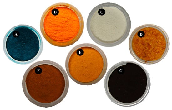
Figure 1.
(A) copper acetate; (B) curcumin; (C) Kappa-carrageenan; (D) iron chloride; (E) curcumin/kappa-carrageenan; (F) copper hybrid; (G) iron hybrid.
Figure 2A shows the UV–Vis spectrum of curcumin and the Cu and Fe hybrid materials. The curcumin exhibited maximum absorption at the wavelength of 425 nm and weak absorption at 262 nm [38]. The samples containing Cu and Fe exhibited more intense absorption bands, with a shift of approximately 6 nm [39]. Furthermore, bands at 419 nm and 255 nm were clearly observed for the Cu hybrid material, while peaks at 428 nm and 257 nm are observed for the Fe hybrid material. The crystal structure of samples was further investigated through XRD, and the results are presented in Figure 2B. The XRD of curcumin exhibits its typical crystal structure with intense peaks, especially at angles below 30 degrees, similar to what has been reported in other similar studies [40]. For the Kappa-carrageenan samples, an amorphous structure was observed, similar to that reported by other authors [41]. The amorphous behavior remained predominant for the hybrid samples, where no peaks corresponding to the crystal planes could be identified. The FT-IR was used to identify the functional groups on the surface of the samples, and the results are displayed in Figure 2C,D. The infrared spectra of curcumin show a typical band at 3461 cm−1 corresponding to phenolic stretching (O-H). Thus, aromatic stretching (C=C) and stretching vibrations (C=C) were observed at 1629 cm−1 and 1509 cm−1, respectively. Stretching vibrations (C=O) were observed at 1599 cm−1, and bending vibrations (C-H) and aromatic stretching (C-O) vibrations were noticed at 1427 cm−1 and 1280 cm−1, respectively. Finally, stretching vibrations (C-O-C) were observed at 1025 cm−1 [42]. The FT-IR of the Fe and Cu hybrid materials exhibit similar bands to those observed in curcumin. Furthermore, the vibration modes underwent shifts in their wavenumber, indicating changes in the vibrational state of curcumin upon binding to the metal ions [43,44].
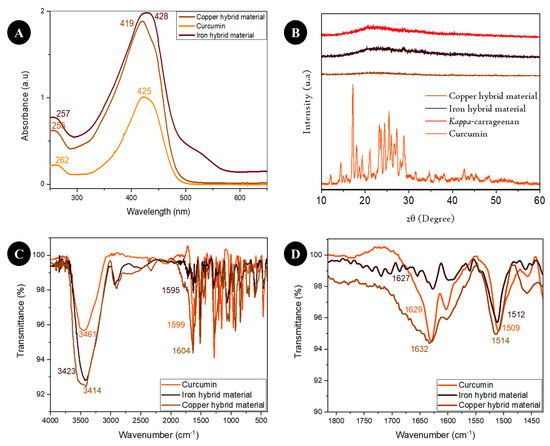
Figure 2.
(A) UV-Vis spectra; (B) X-ray diffractogram; (C) FT-IR spectra; (D) zoomed-in FT-IR spectra.
TGA was performed on the hybrid materials, curcumin, and Kappa-carrageenan, and the results are shown in Figure 3. Kappa-carrageenan (Figure 3A) exhibited three stages of weight loss, which is consistent with previous reports [45]. The first thermal event occurred around 218 °C. The second and third thermal event occurred at approximately 531 °C and 747 °C, respectively. The decomposition of curcumin began around 333 °C, with total degradation occurring around 575 °C, reaching 45.43%, which is in accordance with the literature reports. The TGA curve for Curcumin/Kappa-carrageenan (Figure 3B) shows three stages of mass losses of 12.43%, 14.29%, and 10.46% at temperatures of 207 °C, 342 °C, and 722 °C, respectively. Finally, two and three stages of mass loss were observed for the iron and copper hybrid materials, respectively. Both samples exhibited the greatest mass loss in the first thermal event, 17.25% and 18.12%, at temperatures of 198 °C and 229 °C, respectively [33].
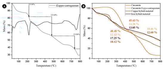
Figure 3.
TGA curves of (A) Kappa-carrageenan, (B) curcumin, curcumin/Kappa-carrageenan, and hybrid materials.
In the analysis of curcuminoids in a commercial sample of natural curcumin, three distinct peaks were observed, as shown in Figure S1 (Supplementary Information). Peak 1 has a retention time of 2.957 min, Peak 2 has a retention time of 3.103 min, and Peak 3 has a retention time of 3.263 min. To study curcumin and hybrid material degradation, the samples were injected into the HPLC at different time intervals starting from the moment of dissolution. During this period, the samples were incubated in clear glass vials and left in an unprotected lit area throughout the analysis period. Figure 4A shows the chromatograms of the curcumin solution at different time points, while Figure 4B presents the chromatogram of the curcumin solution after 24 h, where the presence of more than one peak can be noted. In Figure 4C,D, the chromatograms of copper and iron hybrid materials, respectively, are shown. It can be observed that new peaks were not detected after 24 h.
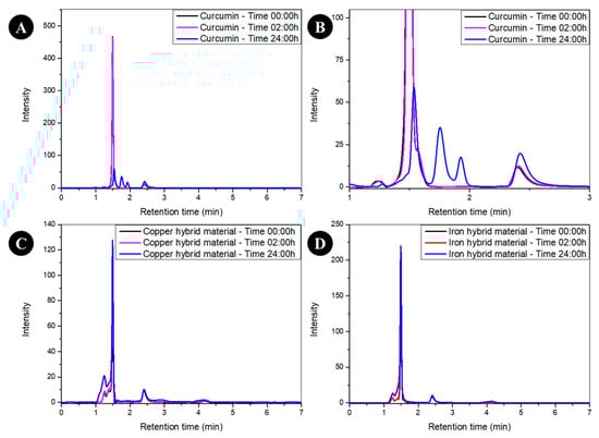
Figure 4.
(A) Chromatogram of the analyzed curcumin solution at different time intervals; (B) enlargement of the curcumin chromatogram at the 24:00 h time point; (C) chromatogram of the analyzed copper hybrid material solution at different time intervals; (D) chromatogram of the analyzed iron hybrid material solution at different time intervals.
In the development of fingerprints using composites generated from hybrid materials, an initial study was conducted with a variety of donors. To convert the visual appearance of the fingerprints developed with powders into numerical values, recognizing that the researchers in this study were not fingerprint identification experts, a scoring system was employed instead of relying solely on the detection and counting of minutiae. This scoring system has been in use for over 35 years. The fingerprint evaluation scheme is presented in Table S1 (Supplementary Information). Cu and Fe composites were then applied to develop fingerprints using the physical powdering method on a glass surface. Figure 5 and Figure 6 show the natural and sebaceous fingerprints from four donors, along with their respective scores.
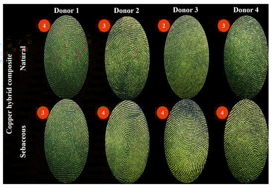
Figure 5.
Natural and sebaceous fingerprints developed with the Cu hybrid composite and their respective scores.
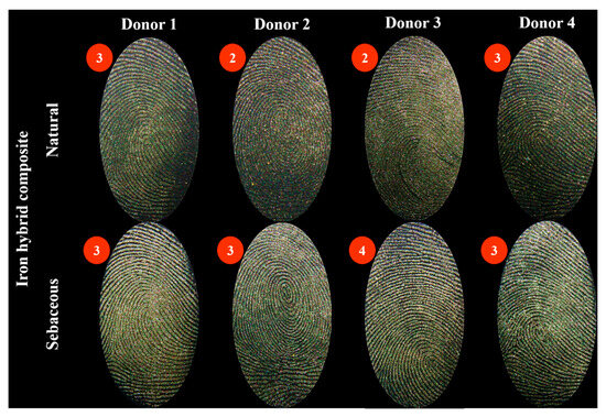
Figure 6.
Natural and sebaceous fingerprints developed with the Fe hybrid composite and their respective scores.
Figure 7 and Figure 8 represent a depletion series from donor 3, developed with copper and iron composites. In Supplementary Figures S2–S7, the depletion series from the other donors developed with both composites can be observed.
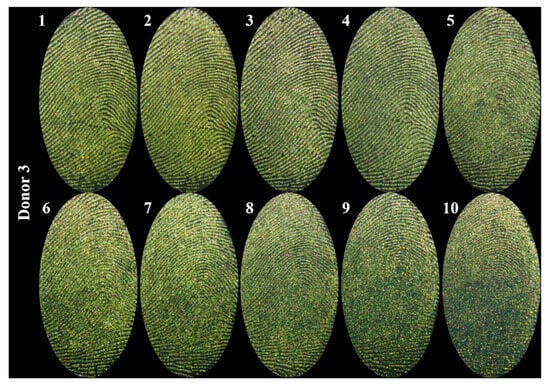
Figure 7.
Depletion series developed with the Cu composite from donor 3.
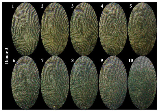
Figure 8.
Depletion series developed with the Fe composite from donor 3.
Figure 9 shows the same fingerprint (natural and sebaceous) developed on the left with composites and on the right with commercial Gold® powder. Supplementary Figures S8 and S9 display the development achieved for the same natural and sebaceous fingerprints from the other donors, using both composites and the commercial powder.
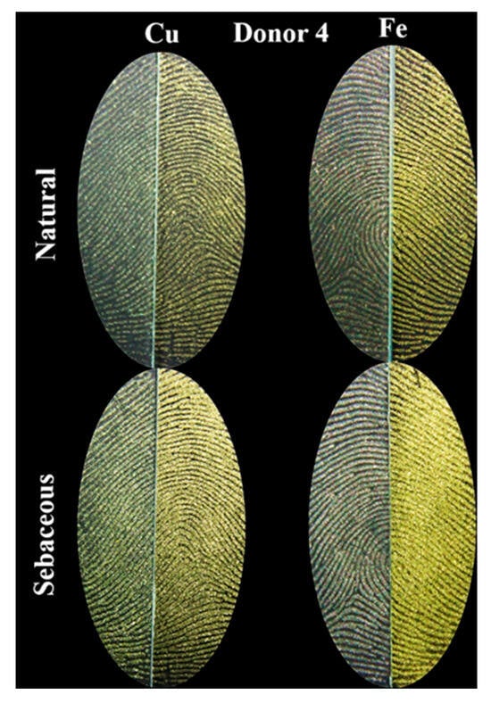
Figure 9.
Half-and-half fingerprint (natural (N) and sebaceous (S)) from donor 4, with the left half developed using composites and the right half developed with Gold® commercial powder purchased from Sirchie®.
The SEM images (Figure 10) provided a detailed analysis of the development of latent fingerprints using both composites. Magnifications of 40×, 600×, and 2000× were applied to investigate the minutiae of the fingerprints, the interaction of the particles with the surface, and the shape and size of the particles. For this analysis, latent fingerprints, both natural and sebaceous, were deposited on glass surfaces and developed with Cu and Fe composites. These magnification levels offered a more precise view of the particle adhesion to the ridges of the fingerprints and the effectiveness of the composites in developing the fingerprints.
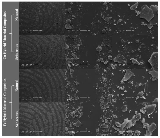
Figure 10.
SEM analysis of natural and sebaceous latent fingerprints developed with Cu and Fe composites on a glass surface.
4. Discussion
According to the literature, it is known that the conjugated β-diketone portion of curcumin is a strong chelating agent, capable of interacting with several metal ions through a coordination bond, which can modify the color of the organic phase of curcumin [46,47]. Thus, the colorimetry results indicated the effective formation of hybrid materials, evidenced by the color change after the addition of these metals and confirmed by their concentration found in the materials.
Thus, the UV-Vis, X-ray, and FT-IR results suggest that the one-pot synthesis was efficient in forming hybrid materials using Kappa-carrageenan as a reducing agent. A strong absorption in the 400–550 nm range of the UV-Vis spectrum (Figure 2A) was observed for all samples, corresponding to the blue and green light region, which agrees with the colorimetric results. The shift in wavelength from curcumin to the hybrid materials suggests an interaction between the carbonyl group of curcumin in metal coordination [39]. The XRD analysis (Figure 2B) of curcumin confirmed its typical crystalline structure [40], while both Kappa-carrageenan and the hybrid materials exhibited amorphous structures. FT-IR was used to identify the functional groups on the surface of the samples, and the results are displayed in Figure 2C,D. The infrared spectra of curcumin show a characteristic band at 3461 cm−1 corresponding to phenolic stretching (O-H). The aromatic stretching of (C=C) and stretching vibrations of (C=C) were observed at 1629 cm−1 and 1509 cm−1, respectively. The stretching vibrations of (C=O) were observed at 1599 cm−1, while the bending corresponding to (C-H) and the aromatic stretching of (C-O) were noticed at 1427 cm−1 and 1280 cm−1, respectively. Finally, stretching vibrations related to (C-O-C) were observed at 1025 cm−1 [42]. The FT-IR of the Fe and Cu hybrid materials, shown in Figure 2C,D, exhibited similar bands to those observed in curcumin, with shifts in wavenumber indicating changes in the vibrational state of curcumin upon binding to the metal ions [43,44].
The thermal stability of hybrid materials is crucial in their practical applications. Kappa-carrageenan exhibited three thermal events (Figure 3A): the first corresponded to moisture loss, while the second and third events were related to the decomposition of the carbohydrate skeleton and complete degradation. Curcumin began with the dehydroxylation process, followed by its total degradation. The curcumin/Kappa-carrageenan composite showed similar behavior to pure Kappa-carrageenan (Figure 3B). On the other hand, the hybrid materials displayed differences due to the varying amounts of curcumin present in the samples. Thus, the TGA curves indicate that the hybrid materials possess superior thermal stability to pure curcumin [34].
The presence of three peaks in the chromatogram (Figure S1—Supplementary Information) can be explained by the composition of commercial curcumin (65%), which contains two other curcuminoids: desmethoxycurcumin and bisdemethoxycurcumin [48], representing the remaining 35% of the sample. Thus, the method adapted proved to be efficient, as the three peaks did not overlap. In addition, it provided a fast analysis time of 5 min [33]. The curcuminoid diferuloylmethane, commonly known as curcumin, is the largest fraction present in the sample, as curcumin was acquired at 65% purity. Therefore, the peak referring to the retention time of 3.263 min, which has the greatest area and intensity, represents curcumin. Desmethoxycurcumin is shown by the second largest fraction of the sample and was identified with a retention time of 3.103 min. Finally, bisdemethoxycurcumin, which denotes the smallest fraction of the sample, was identified with a retention time of 2.957 min. Several reports describe the same order of curcuminoids, with bisdemethoxycurcumin being the first peak and curcumin the last peak [49,50], and no interference in the formation of hybrid materials was found, since all curcuminoids contain the β-diketone fraction responsible for binding the metals. The absence or presence of methoxy groups (the only difference between the curcuminoids) does not influence the process, as they remain intact after the synthesis of the hybrid materials [18].
The instability of curcumin has been investigated in previous studies, where curcumin was dissolved in ethanol [42] or in phosphate-buffered saline (PBS) [16]. Under these conditions, degradation products such as vanillin, vanillic acid, and ferulic acid were identified [18,51]. In particular, curcumin degradation was detected after 24 h of incubation, observed by a reduction in the corresponding peak height, as previously reported [16]. Figure 4A,B clearly illustrate the decrease in curcumin peak intensity after 24 h, as well as the appearance of new peaks corresponding to degradation products with retention times longer than that of curcumin, consistent with the findings of Kumar et al. (2021) [16], as shown in Figure 4B. In contrast, no degradation products were observed in the hybrid materials. Figure 4C,D show the same intensity and peak behavior at all analyzed times, corroborating the results of Kumar et al. (2021) for curcumin-based metal complexes [16].
The use of powders to develop fingerprints is crucial in addressing the limitations of curcumin, especially in terms of stability. The previous results demonstrated that the combination of curcumin with Cu and Fe metals led to a significant increase in the stability of samples, which is one of the key requirements for practical use [52,53]. Based on these results, the potential of these compounds as developers for latent fingerprints was investigated. Composites were applied to fingerprints from various donors, aiming to capture variations related to age, sex, and other characteristics. It is known that factors such as occupation, health (both physical and mental), diet, and medications directly affect the fingerprint composition. Both natural and sebaceous fingerprints were evaluated to assess interactions with the water-soluble and lipid-soluble compounds [24,54]. Through the method of Sears et al. (2012) [37], the visual appearance of the fingerprints was converted to numerical values for classification. The results demonstrated that both composites were effective in developing natural and sebaceous fingerprints from all donors, resulting in clear images on glass surfaces. The composites adhered to the fingerprints without having adhesion to the glass, providing good contrast in the images (Figure 5 and Figure 6). In numerical values, the lowest scores, such as zero (no evidence of mark) and one (evidence of contact without ridge details), were not assigned to any fingerprint developed with the composites.
Only one natural fingerprint from the Cu hybrid composite (donor 3) (Figure 5) and two from the Fe hybrid composite (donors 2 and 3) (Figure 6) showed limited development, with about 1/3 of the ridge details visible (score 2). The Cu hybrid composite showed a high level of development for natural fingerprints, with complete ridge details (score 4) for donor 1 and strong development, showing between 1/3 and 2/3 of ridge details (score 3), for donors 2 and 4 (Figure 5). In the case of the Fe hybrid composite, natural fingerprints exhibited strong detail (score 3) for donors 1 and 4 (Figure 6). The Cu composite (Figure 5) more efficiently developed sebaceous fingerprints than natural ones, with a score of 4 for donors 2, 3, and 4 and a score of 3 for donor 1. Thus, all sebaceous fingerprints developed with this powder showed very strong or strong detail. Similarly, the Fe composite (Figure 6) also showed superior performance in sebaceous fingerprints compared to natural ones, with a score of 3 for donors 1, 2, and 4 and a score of 4 for donor 3, indicating well-defined fingerprints with strong or very strong detail levels. These findings indicate that, although the same deposition and development method was applied consistently, the influence of the donor on the development process was evident. Nevertheless, the results were consistent and satisfactory, with the Cu composite showing higher contrast and better scores compared to the Fe composite.
To evaluate the sensitivity of the composites in developing fingerprints, depletion series were performed. These series consist of successive contacts of the same finger with the surface, resulting in a progressive reduction in the amount of residue left with each new contact. Thus, the effective development of fingerprints left in the final contact indicates higher sensitivity in the developing powder, as there is less material available for adhesion [37]. Figure 7 and Figure 8 present the depletion series for donor 3, developed with Cu and Fe composites, respectively. Although the Cu composite showed better performance with the previously discussed fingerprints, when comparing the depletion series for donor 3, greater clarity was observed in the revelations made with the Fe composite. This composite demonstrated good development, with excellent sharpness and contrast in most deposits, except for the tenth, where there was a significant decrease in quality. On the other hand, the Cu composite showed better clarity in the first four deposits, but from the fifth onward, there was a progressive decline in the quality of development. This decline may be attributed to the higher contrast provided by the Cu composite, which could have hindered the clarity of the fingerprints in deposits with less sebaceous residue. Similar results were observed for the other donors in the depletion series, as shown in Supplementary Figures S2–S7. Although the Fe composite offers lower contrast compared to Cu, it demonstrated superior sharpness in the depletion series for all donors, often effectively revealing fingerprints up to the tenth deposit. In contrast, the Cu composite showed satisfactory development, with good results up to the fourth deposit, followed by a progressive decline in quality from the fifth onward.
It is important to consider that fingerprints exhibit inherent variability in their composition, including factors such as chemical composition, the amount of material deposited, and the pressure applied during deposition. To minimize the impact of these variables on fingerprint development, the use of half-and-half fingerprint tests is an effective approach. In this method, fingerprints are deposited and physically split along the centerline, allowing each half to be developed using different techniques or powders (powdering method) [37]. This “half-and-half” strategy, applied to the comparative analysis between synthesized powders and commercially available powders, is particularly relevant as it allows the evaluation of whether the developed material performs similarly to those currently used by the Federal Police [32]. Figure 9 illustrates the natural and sebaceous fingerprints of donor 4, developed using the half-and-half method. The left halves were developed with Cu or Fe composites, while the right halves were developed with the commercial Gold® powder. Good development of natural fingerprints was observed with both composites, exhibiting a quality comparable to that of the commercial Gold® powder. In the case of sebaceous fingerprints, the Cu composite showed a performance similar to that of the commercial powder, while the Fe composite demonstrated superior sharpness compared to the commercial powder. Figures S8 and S9 (Supplementary Information) show the half-and-half fingerprints of the other donors. A similar behavior was observed in the development of the composites, with a performance equivalent to, or better than, that of the commercial powder. When comparing both composites, the Fe one stood out by providing greater sharpness, resulting in clearer definition of the fingerprint minutiae; this may be related to the weak magnetic cohesion that can favor the uniform distribution of particles in certain contexts, such as when applied to irregular surfaces. In the case of iron (Fe) composites, this property allows the material to be easily dispersed and adhere to other surfaces, which can result in greater-definition details when applied as a fingerprint developer. In contrast, materials with stronger magnetic cohesive forces tend to form agglomerates and may have a lower capacity for adhesion and homogeneous distribution [55,56].
So far, hybrid material composites have been evaluated as developers for latent fingerprints using various methods, all applied to glass surfaces. Considering that the surface on which a fingerprint is deposited can significantly influence the effectiveness of its development, the final analysis of these powders included tests on surfaces representative of those encountered in operational work. For this, the same composites were applied to different surfaces commonly found in forensic investigations [37]. Examples of these surfaces can be seen in Figure 11.
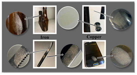
Figure 11.
Development of latent fingerprints using Cu and Fe composites on different objects representing porous and non-porous surfaces: glass bottle, doorknob and door, knife and knife handle, and a black smartphone.
Figure 11 illustrates the development of latent fingerprints with both composites on porous and non-porous surfaces. Objects commonly found in real-world scenarios were selected, including glass bottles, metal knife surfaces, plastic knife handles, wooden door surfaces, metal doorknobs, and the glass screens of smartphones. Both composites were effective in enhancing latent fingerprints on both light and dark surfaces, as well as porous and non-porous ones, demonstrating the versatility of the developed powders. The revealed fingerprints exhibited sufficient detail to be used in human identification processes.
The SEM images (Figure 10) provide a detailed analysis of the minutiae in both natural and sebaceous fingerprints developed with both Fe and Cu composites. At 40× magnification, it can be observed that the Fe composite offers greater clarity compared to the Cu composite, which is directly related to the strong adhesion of the Fe composite to the fingerprint compounds, while interacting minimally with the glass surface, as evidenced in the 600× magnified images. Regarding morphology, both composites exhibit irregular shapes and heterogeneous particle sizes [28], ranging from 50 μm to less than 10 μm. Despite these characteristics, the composites were effective in developing latent fingerprints, providing sufficient detail for human identification.
5. Conclusions
The hybrid copper and iron materials were synthesized via a one-pot methodology using natural curcumin and Kappa-carrageenan as a reducing agent. These materials were characterized by different techniques such as FT-IR, UV-Vis, XRD, and FAAS. The observed color changes and enhanced stability of these materials can be attributed to the coordination of Cu2+ or Fe3+ metal ions with curcumin, as well as potential interactions with Kappa-carrageenan. To verify whether curcumin hybridization effectively improved its stability, thermogravimetric analysis was performed, showing that the hybrid materials had greater thermal stability than curcumin. Solution stability studies using liquid chromatography revealed that curcumin showed degradation product peaks after 24 h of solvent exposure, whereas the hybrid materials remained stable after 24 h. The hybrid composites were further applied as latent fingerprint developers. The results showed that both materials effectively developed natural and sebaceous fingerprints from various donors, with good clarity and contrast on glass surfaces. In successive contact depletion series, the Fe composite demonstrated higher sensitivity, revealing sharp fingerprints up to the tenth contact, while the Cu composite showed lower quality after the fifth contact. In comparative tests with commercial Gold® powder, both hybrid materials performed similarly, with the Fe composite standing out for its fingerprint sharpness. Additionally, when applied to surfaces representative of forensic scenarios (such as glass bottles, metal doorknobs, knife surfaces, and smartphones), the composites were versatile, revealing sufficient fingerprint details for human identification on both porous and non-porous surfaces. Overall, these findings highlight the potential of hybrid materials to enhance forensic applications and contribute to the sustainable use of natural resources.
Supplementary Materials
The following supporting information can be downloaded at: https://www.mdpi.com/article/10.3390/colorants4010003/s1, Table S1: Classification scheme used for the evaluation of developed fingerprints; Table S2: Color parameters of reagents and hybrid materials; Figure S1: Chromatogram of commercial natural curcumin; Figure S2: Depletion series developed with the Cu composite from donor 1; Figure S3: Depletion series developed with the Fe composite from donor 1; Figure S4: Depletion series developed with the Cu composite from donor 2; Figure S5: Depletion series developed with the Fe composite from donor 2; Figure S6: Depletion series developed with the Cu composite from donor 4; Figure S7: Depletion series developed with the Fe composite from donor 4; Figure S8: Half-and-half fingerprint (natural (N) and sebaceous (S)) from donors 1, 2, and 3, with the left half developed using Cu composite and the right half developed with Gold® commercial powder purchased from Sirchie; Figure S9: Half-and-half fingerprint (natural (N) and sebaceous (S)) from donors 1, 2, and 3, with the left half developed using Fe composite and the right half developed with Gold® commercial powder purchased from Sirchie®.
Author Contributions
Conceptualization, D.T.B.; methodology, D.T.B.; validation, A.F.L.; formal analysis, D.H.B., B.V.L., D.A.G. and M.d.P.G.; investigation, D.T.B., A.F.L., J.P.d.S. and G.Q.S.; data curation, T.P. and B.N.d.R.; writing—original draft preparation, D.T.B.; writing—review and editing, A.F.L. and G.K.M.; supervision, C.M.P.d.P., A.L.M. and N.L.V.C.; funding acquisition, C.M.P.d.P.; project administration, C.M.P.d.P. All authors have read and agreed to the published version of the manuscript.
Funding
Financial support for this research was provided by FAPERGS (Research Support Foundation of the Rio Grande do Sul State 22/2551-0000840-2), Coordination for Improvement of Higher-Level Personnel (CAPES), and the Forensic National Institute of Science and Technology Grant number (465450/2014-8).
Informed Consent Statement
Not applicable.
Data Availability Statement
The data can be made available on request.
Acknowledgments
The authors are thankful to FAPERGS, CAPES, Forensic National Institute of Science and Technology, and the Brazilian Federal Police for their assistance.
Conflicts of Interest
The authors declare no conflicts of interest.
References
- Barros, H.L.; Tavares, L.; Stefani, V. Dye-doped starch microparticles as a novel fluorescent agent for the visualization of latent fingermarks on porous and non-porous substrates. Forensic Chem. 2020, 20, 100264. [Google Scholar] [CrossRef]
- Khare, V.; Singla, A. A review on the advancements in chemical examination of composition of latent fingerprint residues. Egypt. J. Forensic Sci. 2022, 12, 6. [Google Scholar] [CrossRef]
- Robson, R.; Ginige, T.; Mansour, S.; Khan, I.; Assi, S. Analysis of fingermark constituents: A systematic review of quantitative studies. Chem. Pap. 2022, 76, 4645–4667. [Google Scholar] [CrossRef]
- Li, L.; Li, Q.; Chu, J.; Xi, P.; Wang, C.; Liu, R.; Wang, X.; Cheng, B. Dual-mode luminescent multilayer core-shell UCNPs@SiO2@TEuTbB nanospheres for high-level anti-counterfeiting and recognition of latent fingerprints. Appl. Surf. Sci. 2022, 581, 152395. [Google Scholar] [CrossRef]
- Ansari, A.A.; Aldajani, K.M.; AlHazaa, A.N.; Albrithen, H.A. Recent progress of fluorescent materials for fingermarks detection in forensic science and anti-counterfeiting. Coord. Chem. Rev. 2022, 462, 214523. [Google Scholar] [CrossRef]
- Poletti, T.; Berneira, L.M.; Bueno, D.T.; da Silva, C.C.; da Silva, R.; Pereira, C.M. Chemical evaluation and application of cinnamaldehyde-derived curcumins as potential fingerprint development agents. Talanta Open 2022, 6, 100133. [Google Scholar] [CrossRef]
- Passos, L.F.; Berneira, L.M.; Poletti, T.; Mariotti, K.D.C.; Carreño, N.L.; Hartwig, C.A.; Pereira, C.M. Evaluation and characterization of algal biomass applied to the development of fingermarks on glass surfaces. Aust. J. Forensic Sci. 2021, 53, 337–346. [Google Scholar] [CrossRef]
- Pereira, C.M.; Pacheco, B.S.; da Silva, C.C. Curcumin and Analogues: Chemical and Biological Aspects; LAP LAMBERT Academic Publishing: Saarbrücken, Germany, 2017. [Google Scholar]
- Arab, C.; El Kurdi, R.; Patra, D. Effect of pH on the removal of anionic and cationic dyes using zinc curcumin oxide nanoparticles as adsorbent. Mater. Chem. Phys. 2022, 277, 125504. [Google Scholar] [CrossRef]
- Verma, R.K.; Kumari, P.; Maurya, R.K.; Kumar, V.; Verma, R.B.; Singh, R.K. Medicinal properties of turmeric (Curcuma longa L.): A review. Int. J. Chem. Stud. 2018, 6, 1354–1357. [Google Scholar]
- Yang, M.Y.; Chang, K.C.; Chen, L.Y.; Hu, A. Low-dose blue light irradiation enhances the antimicrobial activities of curcumin against Propionibacterium acnes. J. Photochem. Photobiol. B Biol. 2018, 189, 21–28. [Google Scholar] [CrossRef]
- Maghsoudi, A.; Yazdian, F.; Shahmoradi, S.; Ghaderi, L.; Hemati, M.; Amoabediny, G. Curcumin-loaded polysaccharide nanoparticles: Optimization and anticariogenic activity against Streptococcus mutans. Mater. Sci. Eng. 2017, 75, 1259–1267. [Google Scholar] [CrossRef] [PubMed]
- Lin, L.; Li, C.; Zhang, D.; Yuan, M.; Chen, C.H.; Li, M. Synergic effects of berberine and curcumin on improving cognitive function in an Alzheimer’s disease mouse model. Neurochem. Res. 2020, 45, 1130–1141. [Google Scholar] [CrossRef]
- Buliga, D.I.; Diacon, A.; Calinescu, I.; Popa, I.; Rusen, E.; Ghebaur, A.; Tutunaru, O.; Boscornea, C.A. Enhancing the light fastness of natural dyes by encapsulation in silica matrix. J. Photochem. Photobiol. A Chem. 2022, 432, 114085. [Google Scholar] [CrossRef]
- Souza, C.R.A.; Osme, S.F.; Glória, M.B.A. Stability of curcuminoid pigments in model systems. J. Food Process. Preserv. 1997, 21, 353–363. [Google Scholar] [CrossRef]
- Kumar, P.; Saha, T.; Behera, S.; Gupta, S.; Das, S.; Mukhopadhyay, K. Enhanced efficacy of a Cu2+ complex of curcumin against Gram-positive and Gram-negative bacteria: Attributes of complex formation. J. Inorg. Biochem. 2021, 222, 111494. [Google Scholar] [CrossRef]
- Hernández, M.; Felipe, C.; Guzmán-Vargas, A.; Rivera, J.L.; Lima, E. Highly Stable Hybrid Pigments Prepared from Organic Chromophores and Fluorinated Hydrotalcites. Colorants 2024, 3, 125–135. [Google Scholar] [CrossRef]
- Prasad, S.; Lall, R. Zinc-Curcumin Based Complexes in Health and Diseases: An Approach in Chemopreventive and Therapeutic Improvement. J. Trace Elem. Med. Biol. 2022, 73, 127023. [Google Scholar] [CrossRef] [PubMed]
- Sanchez, C.; Lebeau, B. Design and properties of hybrid organic–inorganic nanocomposites for photonics. Mrs Bulletin 2001, 26, 377–387. [Google Scholar] [CrossRef]
- Lee, J.Y.; Lee, S.; Choi, J.H.; Na, K. ι-Carrageenan nanocomposites for enhanced stability and oral bioavailability of curcumin. Biomater. Res. 2021, 25, 32. [Google Scholar] [CrossRef]
- Tan, K.X.; Ng, L.L.E.; Loo, S.C.J. Formulation development of a food-graded curcumin-loaded medium chain triglycerides-encapsulated kappa carrageenan (CUR-MCT-KC) gel bead based oral delivery formulation. Materials 2021, 14, 2783. [Google Scholar] [CrossRef]
- Wan, H.; Li, C.; Mahmud, S.; Liu, H. Kappa carrageenan reduced-stabilized colloidal silver nanoparticles for the degradation of toxic azo compounds. Colloids Surf. A Physicochem. Eng. Asp. 2021, 616, 126325. [Google Scholar] [CrossRef]
- Leitzke, A.F.; Berneira, L.M.; Rosa, B.N.D.; Moreira, B.C.; Mariotti, K.D.C.; Venzke, D.; Pereira, C.M. A Química de Produtos Naturais aplicados a reveladores de impressões digitais latentes. Química Nova 2022, 45, 424–434. [Google Scholar] [CrossRef]
- Bueno, D.T.; Leitzke, A.F.; Crizel, R.L.; Jansen-Alves, C.; Bertizzolo, E.G.; da Silva, J.P.; Sejanes, G.Q.; Mariotti, K.C.; de Pereira, C.M.P. Characterization of Bixin by UV-Visible Spectroscopy and HPLC, and Its Application as Latent Fingermark Developer. Analytica 2024, 5, 107–118. [Google Scholar] [CrossRef]
- Balsan, J.D.; Rosa, B.N.; Pereira, C.M.; Santos, C.M. Desenvolvimento de metodologia de revelação de impressão digital latente com chalconas. Química Nova 2019, 42, 845–850. [Google Scholar] [CrossRef]
- da Rosa, B.N.; da Rosa, M.P.; Poletti, T.; de Lima, N.P.K.; Maron, G.K.; Lopes, B.V.; Mariotti, K.C.; Beck, P.H.; Carreno, N.L.V.; de Pereira, C.M.P. Green composites from thiophene chalcones and rice husk lignin: An alternative of powder for latent fingermark. Surfaces 2022, 5, 481–488. [Google Scholar] [CrossRef]
- da Rosa, B.N.; Maron, G.K.; Lopes, B.V.; Rocha, A.C.S.; de Moura Gatti, F.; Machado, J.O.A.; Barichello, J.M.; Mariotti, K.C.; Trossini, G.H.G.; Carreno, N.L.V.; et al. Dimethylaminochalcones with silicon dioxide and zinc oxide as latent fingermark developer powder. Mater. Chem. Phys. 2023, 295, 127033. [Google Scholar] [CrossRef]
- Pacheco, B.S.; Da Silva, C.C.; Da Rosa, B.N.; Mariotti, K.C.; Nicolodi, C.; Poletti, T.; Segatto, N.V.; Collares, T.; Seixas, F.K.; Paniz, O.; et al. Monofunctional curcumin analogues: Evaluation of green and safe developers of latent fingerprints. Chem. Pap. 2021, 75, 3119–3129. [Google Scholar] [CrossRef]
- Poletti, T.; Berneira, L.M.; Passos, L.F.; da Rosa, B.N.; de Pereira, C.M.; Mariotti, K.D.C. Preliminary efficiency evaluation of development methods applied to aged sebaceous latent fingermarks. Sci. Justice 2021, 61, 378–383. [Google Scholar] [CrossRef]
- Venzke, D.; Poletti, T.; Rosa, B.N.; Berneira, L.M.; de Lima, N.P.; de Oliveira, T.F.; Carreno, N.L.V.; Mariotti, K.C.; Duarte, L.S.; Nobre, S.M.; et al. Preparation of fluorescent bisamides: A new class of fingermarks developers. Chem. Data Collect. 2021, 33, 100680. [Google Scholar] [CrossRef]
- Lima, N.P.; Rosa, B.N.; Poletti, T.; Moreira, B.C.; Leitzke, A.F.; Mariotti, K.C.; Carreno, N.L.V.; Pereira, C.M. As clássicas hidrazonas como reveladores de impressões digitais: Uma proposta de química orgânica experimental. Química Nova 2023, 46, 215–221. [Google Scholar] [CrossRef]
- Leitzke, A.F.; Bueno, D.T.; Poletti, T.; Maron, G.K.; Lopes, B.V.; Morais, E.V.; Inacio, A.P.O.L.; Silveira, C.I.; Silva, J.P.; Dias, D.; et al. The effectiveness of natural indigo/kaolinite composite powder in the development of latent fingermarks. Egypt. J. Forensic Sci. 2024, 14, 19. [Google Scholar] [CrossRef]
- Li, S.; Mu, B.; Yan, P.; Kang, Y.; Wang, Q.; Wang, A. Incorporation of different metal ion for tuning color and enhancing antioxidant activity of curcumin/palygorskite hybrid materials. Front. Chem. 2021, 9, 760941. [Google Scholar] [CrossRef] [PubMed]
- Anjani, Q.K.; Utomo, E.; Domínguez-Robles, J.; Detamornrat, U.; Donnelly, R.F.; Larrañeta, E. A new and sensitive HPLC-UV method for rapid and simultaneous quantification of curcumin and D-panthenol: Application to in vitro release studies of wound dressings. Molecules 2022, 27, 1759. [Google Scholar] [CrossRef]
- Modwi, A.; Ali, M.K.M.; Taha, K.K.; Ibrahem, M.A.; El-Khair, H.M.; Eisa, M.H.; Elamin, M.R.; Aldaghri, O.; Alhathlool, R.; Ibnaouf, K.H. Structural and optical characteristic of chalcone doped ZnO nanoparticles. J. Mater. Sci. Mater. Electron. 2018, 29, 2791–2796. [Google Scholar] [CrossRef]
- Fioravanti, M.I.A.; Pizano, F.P.; Rebellato, A.P.; Milani, R.F.; Morgano, M.A.; Bragotto, A.P.A. Turmeric products: Evaluation of curcumin and trace elements. Food Res. Int. 2024, 196, 115028. [Google Scholar] [CrossRef]
- Sears, V.G.; Bleay, S.M.; Bandey, H.L.; Bowman, V.J. A methodology for finger mark research. Sci. Justice 2012, 52, 145–160. [Google Scholar] [CrossRef]
- Subhan, M.A.; Alam, K.; Rahaman, M.S.; Rahman, M.A.; Awal, R. Synthesis and characterization of metal complexes containing curcumin (C21H20O6) and study of their anti-microbial activities and DNA binding properties. J. Sci. Res. 2014, 6, 97–109. [Google Scholar] [CrossRef]
- Zhou, S.S.; Xue, X.; Wang, J.F.; Dong, Y.; Jiang, B.; Wei, D.; Wan, M.; Jia, Y. Synthesis, optical properties and biological imaging of the rare earth complexes with curcumin and pyridine. J. Mater. Chem. 2012, 22, 22774–22780. [Google Scholar] [CrossRef]
- Sayyar, Z.; Malmiri, H.J. Photocatalytic and antibacterial activities study of prepared self-cleaning nanostructure surfaces using synthesized and coated ZnO nanoparticles with Curcumin nanodispersion. Z. Krist.—Cryst. Mater. 2019, 234, 307–328. [Google Scholar] [CrossRef]
- Kanmani, P.; Rhim, J.W. Development and characterization of carrageenan/grapefruit seed extract composite films for active packaging. Int. J. Biol. Macromol. 2014, 68, 258–266. [Google Scholar] [CrossRef]
- Chen, X.; Zou, L.Q.; Niu, J.; Liu, W.; Peng, S.F.; Liu, C.M. The stability, sustained release and cellular antioxidant activity of curcumin nanoliposomes. Molecules 2015, 20, 14293–14311. [Google Scholar] [CrossRef] [PubMed]
- Halevas, E.; Papadopoulos, T.A.; Swanson, C.H.; Smith, G.C.; Hatzidimitriou, A.; Katsipis, G.; Katsipis, G.; Pantazaki, A.; Sanakis, I.; Ypsilantis, K.; et al. In-depth synthetic, physicochemical and in vitro biological investigation of a new ternary V (IV) antioxidant material based on curcumin. J. Inorg. Biochem. 2019, 191, 94–111. [Google Scholar] [CrossRef] [PubMed]
- Halevas, E.; Pekou, A.; Papi, R.; Mavroidi, B.; Hatzidimitriou, A.G.; Zahariou, G.; Litsardakis, G.; Sagnou, M.; Pelecanou, M.; Pantazaki, A.A. Synthesis, physicochemical characterization and biological properties of two novel Cu (II) complexes based on natural products curcumin and quercetin. J. Inorg. Biochem. 2020, 208, 111083. [Google Scholar] [CrossRef] [PubMed]
- Kulal, P.; Badalamoole, V. Hybrid nanocomposite of kappa-carrageenan and magnetite as adsorbent material for water purification. Int. J. Biol. Macromol. 2020, 165, 542–553. [Google Scholar] [CrossRef]
- de França, B.M.; Oliveira, S.S.; Souza, L.O.; Mello, T.P.; Santos, A.L.; Forero, J.S.B. Synthesis and photophysical properties of metal complexes of curcumin dyes: Solvatochromism, acidochromism, and photoactivity. Dyes Pigments 2022, 198, 110011. [Google Scholar] [CrossRef]
- Prasad, S.; DuBourdieu, D.; Srivastava, A.; Kumar, P.; Lall, R. Metal–curcumin complexes in therapeutics: An approach to enhance pharmacological effects of curcumin. Int. J. Mol. Sci. 2021, 22, 7094. [Google Scholar] [CrossRef]
- Sharifi-Rad, J.; Rayess, Y.E.; Rizk, A.A.; Sadaka, C.; Zgheib, R.; Zam, W.; Sestito, S.; Rapposelli, S.; Neffe-Skocinska, K.; Zielinska, D.; et al. Turmeric and its major compound curcumin on health: Bioactive effects and safety profiles for food, pharmaceutical, biotechnological and medicinal applications. Front. Pharmacol. 2020, 11, 550909. [Google Scholar] [CrossRef]
- Bonifácio, D.; Martins, C.; David, B.; Lemos, C.; Neves, M.G.P.M.S.; Almeida, A.; Pinto, D.C.G.A.; Cunha, Â. Photodynamic inactivation of Listeria innocua biofilms with food-grade photosensitizers: A curcumin-rich extract of Curcuma longa vs commercial curcumin. J. Appl. Microbiol. 2018, 125, 282–294. [Google Scholar] [CrossRef]
- Chen, Y.C.; Chen, B.H. Preparation of curcuminoid microemulsions from: Curcuma longa L. to enhance inhibition effects on growth of colon cancer cells HT-29. RSC Adv. 2018, 8, 2323–2337. [Google Scholar] [CrossRef]
- Khurana, A.; Ho, C.-T. High Performance Liquid Chromatographic Analysis of Curcuminoids and Their Photo-Oxidative Decomposition Compounds in Curcuma longa L. J. Liq. Chromatogr. 1988, 11, 2295–2304. [Google Scholar] [CrossRef]
- Chávez, D.; Garcia, C.R.; Oliva, J.; Diaz-Torres, L.A. A review of phosphorescent and fluorescent phosphors for fingerprint detection. Ceram. Int. 2021, 47, 10–41. [Google Scholar] [CrossRef]
- Vadivel, R.; Nirmala, M.; Anbukumaran, K. Commonly available, everyday materials as non-conventional powders for the visualization of latent fingerprints. Forensic Chem. 2021, 24, 100339. [Google Scholar] [CrossRef]
- De Alcaraz-Fossoul, J.; Mestres Patris, C.; Balaciart Muntaner, A.; Barrot Feixat, C.; Gené Badia, M. Determination of latent fingerprint degradation patterns—A real fieldwork study. Int. J. Leg. Med. 2013, 127, 857–870. [Google Scholar] [CrossRef]
- Sharma, R.; Setia, G. Mechanical dry particle coating on cohesive pharmaceutical powders for improving flowability—A review. Powder Technol. 2019, 356, 458–479. [Google Scholar] [CrossRef]
- Kellogg, K.M.; Liu, P.; Hrenya, C.M. Discrete-Element-Method-Based Determination of Particle-Level Inputs for the Continuum Theory of Flows with Moderately Cohesive Particles. Processes 2023, 11, 2553. [Google Scholar] [CrossRef]
Disclaimer/Publisher’s Note: The statements, opinions and data contained in all publications are solely those of the individual author(s) and contributor(s) and not of MDPI and/or the editor(s). MDPI and/or the editor(s) disclaim responsibility for any injury to people or property resulting from any ideas, methods, instructions or products referred to in the content. |
© 2025 by the authors. Licensee MDPI, Basel, Switzerland. This article is an open access article distributed under the terms and conditions of the Creative Commons Attribution (CC BY) license (https://creativecommons.org/licenses/by/4.0/).