Abstract
Plant fibers find wide application in various fields that demand specific parameters of fiber quality. To develop approaches for the improvement of yield and quality of bast fibers, the knowledge of the fiber developmental stages and of the key molecular players that are responsible for a certain parameter, are vitally important. In the present review the key stages of fiber development, such as initiation, intrusive growth, and formation of thickened cell wall layers (secondary and tertiary cell walls) are considered, as well as the impact of each of these stages on the final parameters of fiber yield and quality. The problems and perspectives of crop quality regulation are discussed.
1. Introduction
Plant fibers have great potential for their use in various innovative applications as an ecological, biodegradable, and renewable resource with unique properties [1,2]. These attractive features promote the design of various plant fiber-based blends and composites for numerous purposes ranging from textile, automotive, and construction industries to medical uses [3,4,5], including smart materials [6].
Together with the general value of fiber mechanical performance, each field of application has its own set of preferable parameters for fiber quality and often deals with fibers of certain plant species. Fiber quality is quite dependent on the way of plant material processing; however, the basis for the fiber properties that are important for application is provided by developmental processes occurring in planta.
In plant biology, fibers are defined as individual cells with certain parameters [7,8,9]. Similar to many other plant cell types, fibers develop through three major stages: initiation, elongation, and further specialization; the latter in the case of fibers is largely represented by cell wall thickening. Each of these stages has its own impact on the final parameters of fiber yield and quality. In the current review, we consider the general role of each developmental stage that would be the same in any plant developing bast fibers and skip the influence of environmental factors or of specific genetic background in different varieties of certain bast fiber crops.
2. Fiber Initiation
Same as all other plant cell types, fibers initiate from meristematic tissues. Bast fibers (also called phloem fibers) originate either from the primary or from the secondary meristems; correspondingly, fibers are called primary or secondary [7,8,9]. In a simplified way, primary meristems can be defined as those that are providing the increase of plant organ length. They are usually located at the very apex of elongating shoot or root, at least in dicots, and are also called apical meristems. Primary bast fibers emerge from procambium (a direct derivative of apical meristem) within a complex process of leaf primordia initiation, along with the initiation of vascular system within each leaf trace. Thus, the number of primary fiber initials is coupled to the number of leaves and to the same extent can be specie-specific. The large variations in the final yield of primary bast fibers that can be obtained within the same plant species are determined by further stages of fiber developmental. As distinct from that, secondary fibers originate from cambium that provides the increase of organ width. Cambium usually starts its activity in the organ part that has already ceased elongation as a whole. The activity of cambium largely varies depending on many factors; consequently, the number of secondary bast fibers may vary considerably within the same plant species [10]. Since the primary fibers are formed from the primary meristem, they appear early in plant development and are present from bottom to the top of the stem [11,12]. Secondary fibers appear later as the plant develops and occur mainly in the lower stem part and may form several concentric rings [11,12]. Xylem (wood) fibers that originate from the same meristems and are present together with phloem fibers within the same stem are not considered within current review since they have rather different properties and corresponding fields of application.
Thus, the stage of initiation determines the type of the fibers (primary or secondary, phloem, or xylem) and the number of individual cells. To give an idea of the possible amount of bast fiber cells in a fiber crop, the number of individual primary bast fibers in hemp stem was estimated to be 700–800 thousand, while the number of secondary fibers in the same plant—around 2.5 million [13].
3. Fiber Elongation
The length of newly initiated fibers is around 30 μm, while the final cell length can reach many centimeters, meaning that a fiber can elongate hundred or even thousand fold [12,14,15]. The cell width is also increased, but to a much lesser extent: from 3–5 μm in “newly born” fiber, to 20–50 μm in mature ones [12,14,15]. Cell elongation is extremely important for the properties of individual fibers and especially—of fiber bundles.
Right after initiation, primary fibers for a short while elongate by symplastic (or coordinated) growth when the adjacent cells increase their surface at the same rate. By symplastic growth primary fibers increase their length twice or so. In a while, within couple millimeter from the stem apex, primary fibers start to elongate faster than the surrounding cells [12,14]. This leads to the necessity to force their way and to squeeze between other cells. Fibers do so by splitting middle lamellae of the cells on the way and making new contacting surfaces with the neighboring cells. Such type of cell growth is called intrusive. Fibers may continue to increase their length by intrusive elongation after the surrounding cells of other tissues have already ceased enlargement (growing zone of the whole stem is usually within a couple of centimeters from stem apex). The disturbance of the preexisting cell contacts during fiber intrusive elongation may lead to the closing of plasmodesmata, turning fibers into a symplastically isolated domain [14]. The major processes that occur during elongation, are the extension of primary cell wall (PCW) surface coupled to deposition of new cell wall material and the increase of vacuole volume due to osmolyte accumulation and corresponding water uptake [15,16]. The most pronounced intrusive growth of primary bast fibers takes place during the fast growth period of plant development.
Secondary fibers do not have the stage of coordinated growth since they originate within the stem part that has already stopped elongation. The final length of the secondary fibers, as compared to cambium initials, is achieved solely by intrusive elongation. This is one of the reasons that secondary bast fibers are shorter than primary ones; the other reason may be the more severe mechanical constraints that the secondary fibers face, being developed in the depth of stem tissues, some of which are already lignified. In hemp stem, the modal class of individual primary phloem fiber lengths is around 20 mm, while of secondary phloem ones—only 5 mm [12].
Fiber elongation occurs only within several days after fiber initiation and is split in time with cell wall thickening [12,14,15]. Through the whole stage of intrusive elongation fibers have only thin primary cell wall; together with the extreme cell length, this makes fibers quite breakable and difficult to isolate for biochemical analysis.
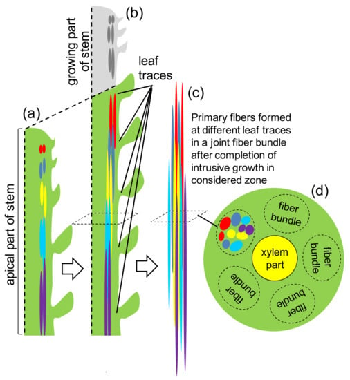
The major consequences of intrusive elongation for fiber quality-related parameters are the following. Firstly, it determines the final size of each individual fiber, both in length and in width. Secondly, it is intrusive elongation that leads to the formation of fiber bundles and dictates their structure. During extensive bidirectional elongation, primary fibers formed at different leaf traces reach each other and form joint bundles [12,14] (Figure 1).

Figure 1.
Development of fiber bundles in the course of intrusive elongation of primary bast fibers. (a) Scheme of stem radial section (several centimeters from the apex). Fibers originated at different leaf traces are designated by different colors. In the lower internodes of the plant the fibers are more elongated; (b) withinseveral days, fibers initiated at different leaf traces elongate by intrusive growth forming a fiber bundle. The additionally grown up stem part is given in grey; (c) the enlarged picture of a fiber bundle; and, (d) a stem cross-section depicting the formed fiber bundle.
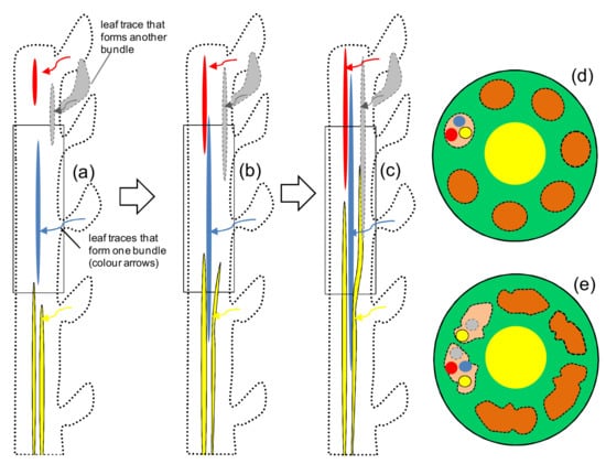
Intrusive elongation leads to the increase of fiber number in the stem cross-section. Due to the substantial cell length, fiber bundles can consist of fiber cells initiated at several leaf traces that were located on the same stem side but at different internodes (Figure 1). Additionally, the direction of intrusive elongation of individual fibers determines the degree of fiber bundle branching (Figure 2). The impact of intrusive elongation for the bundle formation is similarly important for secondary phloem fibers [12]. The importance of intrusive elongation for secondary bast fiber structure was recently confirmed by bundle visualization in planta by means of microtomography [17].

Figure 2.
The direction of intrusive growth affects the bundle structure. (a–c) successive elongation steps of fibers with different direction of growth in considered stem zone; (d) fibers coming from different leaf traces can follow the way made by fibers that originated from the considered leaf trace and have already intruded themselves in between other cells, i.e., slide along “beaten path”. In this case, bundles have “right” shape, (e) some of the fibers make their “own” way (yellow fiber) leading to anastomose between fiber bundles. In this case, different bundles can contain fibers from neighbour leaf traces, not strongly from traces located one bellow other vertically.
Thirdly, special kinds of cell contacts are formed during fiber elongation: intruding of fibers between the other cells leads to the tight packing of fibers within a bundle, with no intercellular spaces. Together with that, the reestablishing of cell surface contacts after their disturbance in the course of fiber intrusive elongation can lead to the special type of “glue” between cells; it may, for example, consist of specific versions of pectic substances able to make gels with peculiar properties. Contacts between fibers are quite important during the retting and further processing steps, permitting bundles the to stay as a whole, while the other cells fall apart. In some cases, the contacts between fibers may be additionally strengthened by lignifications at later stages of fiber development. Fourth, cell enlargement during intrusive growth provides the large cell surface for the further deposition of the thick cell wall that is the major component of mature fibers.
4. Fiber Cell Wall Thickening
Cell wall width in plant fibers can reach 14 µm [18] and is the important determinant of fiber strength [19,20]. Mechanical properties are also influenced by the ratio of cell wall thickness to lumen diameter, called fiber porosity [21]. All of these features are developed during the cell wall thickening stage, which is the most prolonged stage in fiber development and may go on for months.
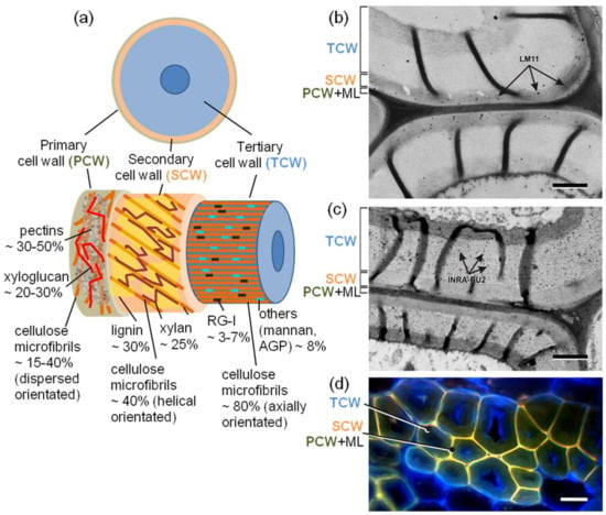
Fiber cell wall is not uniform through its width (Figure 3). Right after the primary cell wall that is formed during the previous stages of fiber development, the secondary cell wall (SCW) is deposited; this is similar to SCW in other plant tissue, like xylem. Together with cellulose, its major constituents are xylan and lignin (Figure 3). The orientation of cellulose microfibrils is helical, with microfibri angle towards major cell axis (MFA) between 20 and 80. The width of SCW in bast fibers can vary significantly: in secondary bast fibers of hemp, it is quite pronounced, while in flax fibers, it is barely detectable. As can be expected from the content of lignin and xylan in jute and kenaf bast fibers, SCW layer is well developed there; however, no direct identification of SCW and TCW layers has been performed yet for these important fiber crops. The developed SCW may have the sublayers that differ in cellulose microfibril orientation and are designated as S1, S2, etc.

Figure 3.
Scheme of structure and composition of different layers of bast fiber cell wall. (a) Scheme of organization and composition of different layers of fiber cell wall; (b,c) immunolocalization of cell wall polysaccharides with (b) LM11 antibody (specific to β-1,4-xylan; [30]) and (c) INRA-RU2 antibody (specific to RG-I backbone; [31]) in hemp stem secondary fibers (photographs by V. Salnikov KIBB KSC RAS); bar 1 µm. Secondary cell wall layer of bast fibers was effectively labeled with LM11 antibody, tertiary cell wall layers of bast fibers, on the contrary, did not bind LM11 antibody, but were labelled by INRA-RU2 antibody. (d) Cross-section of primary phloem fiber bundles of hemp stem; stained with CalcofluorWhite; under UV light (lignified cell wall layers look yellow due to lignin autofluorescence, non-lignified tertiary cell wall layers look blue); bar 20 µm. ML—middle lamellae, AGP—arabinogalactan protein.
Later in fiber development, a very different type of cell wall is deposited in many plant fibers. Since it is formed after PCW and SCW and is very distinct from both, it was suggested to call it tertiary cell wall (TCW) [22,23]. The most distinguishing features of TCW are the high content of cellulose (up to 90%), close to the axial orientation of all cellulose microfibrils, the presence of pectic rhamnogalacturonan-I with long side chains made of galactans, and absence of xylan and lignin. Due to the low content of non-cellulosic polymers in TCW, cellulose microfibrils tend to interact laterally, giving a reason for the increased crystallinity of cellulose. Additional arguments to distinguish secondary and tertiary cell wall come from analysis of transcriptome in fibers isolated at the stage of TCW deposition: the set of expressed genes, both encoding the transcription factors involved in regulation of cell wall formation and the enzymes for biosynthesis and modification of cell wall constituents, is very different from that described for secondary cell wall [24]. The tertiary cell wall is fiber-specific; its other name is G-layer. Composition, architecture, and function of tertiary cell wall have been recently summarized [23]. Deposition of TCW can be induced by stem inclination like it happens in tension wood fibers. But, in bast fibers of many plant species, including most important technical crops, like flax, hemp, ramie, tertiary cell walls are deposited constitutively, in the course of normal plant development.
The proportion between SCW and TCW layers in fibers can be one of the major determinants of the fiber quality. It can be among the reasons for the noted differences in cellulose crystallinity and in MFA between fibers of different origin [21], since these parameters are usually measured for the whole fiber cell wall and thus, give the average numbers for PCW, SCW, and TCW together. Introduction of the term “tertiary cell wall” for plant fibers [22,23] may help to design the experiments accordingly to better understand the role of each cell wall layer in fiber quality-related parameters.
The proportion of SCW to TCW impacts total fiber lignification since it takes place in PCW and SCW, but not TSW (Figure 3). In flax, bast fibers have a negligible layer of the secondary cell wall and the lignification degree is quite low [25,26]. In hemp primary phloem fibers, SCW is more developed than in flax. In secondary phloem fibers, the width of SCW and its proportion in total cell wall area on fiber cross section is higher than in primary fibers [27]. The corresponding higher degree of lignification of secondary phloem fibers in hemp [10] is one of the major reasons for their poorer quality as compared to primary ones.
The overall architecture of each cell wall layer in bast fibers of different origin is quite comparable. For example, the basic design of TCW is similar in different plant species and in xylem and phloem fibers [23]. However, the exact structure of layer-specific polymers may differ. For example, the average length of the side chains in the TCW-specific version of rhamnogalacturonan-I and the degree of backbone substitution is not the same in flax [28] and hemp [27] bast fibers. In lignin, proportions of various monomeric units (H, G, and S) and of various types of linkages between them may vary depending on the fiber source, affecting the fiber application potential [29].
There are other developmental processes in plant stems that occur not in fibers themselves but may affect fiber quality. For example, the intensive secondary growth of stem that occurs in plants, such as hemp, leads to the increased stem circumference, within which the primary fibers are located. The mechanical stresses that emerge in such case may cause partial splitting of fiber bundles to accommodate the increased stem circumference. However, such processes are not within the scope of current review, which deals only with the stages of development of fibers themselves.
5. Problems to Precisely Relate Fiber Developmental Stages to Fiber Yield and Quality
To develop approaches for the improvement of bast fiber yield and quality, the knowledge of the fiber developmental stage and of the key molecular players that are responsible for a certain parameter, are vitally important. However, despite the huge amount of data on fiber yield and quality determination, in most cases, it is not easy to realize the developmental origin of the observed differences. There are several reasons for this situation. One of the problems has the origin in terminology. The word “fiber” is often used for prosenchymatous cells in general, while not taking into account the biological basis. The best-known example is “cotton fiber”, which is actually a very different cell type—a trichome. Together with that, the term “fiber quality” is rather vague. This complex notion may differ in various fields of application; usually it includes such interrelated parameters as strength, fineness, stiffness, and flexibility; it may also consider fiber porosity, adhesive abilities, etc. The other problems are coupled with the arrangement of experiments. The assays of fiber quality are usually performed at plant maturity when the distinct effects of different developmental stages come together, making it difficult to elucidate the impact of the certain developmental process. The comparison of properties of fibers from different plant species, being quite important by itself from a practical point of view, does not give information on the developmental origin of the differences. When conducting the analysis of fibers within the course of plant development, the problems for correct interpretation may come from the way of sampling. “Middle of the stem” or “top part of the plant” collected through the vegetative season, especially when a plant grows intensively, does not permit the direct comparison since each time a sample contains its own set of fibers, initiated at different periods of plant development.
Samples that contain a complex mixture of different tissues, like “the whole stem” and even “fiber-enriched peels”, may give little indication of the processes that occur specifically in fibers, especially while the identification of molecular players that are responsible for quality. Fortunately, the specific mechanical properties of fibers with tertiary cell wall permit to isolate them at an advanced stage of development. Such advantage was used to develop experimental systems for several studies where isolated bast fibers were used [24,27,32,33,34,35]. It is important to avoid enzymatic treatment or retting to obtain isolated fibers for the study of quality-related parameters, so that not to affect the structure of cell wall polymers. It is especially difficult to isolate fibers at the stage of intrusive elongation since fibers are not easily separable from the surrounding tissues and are quite injurable. Laser microdissection combined with cryosectioning was recently developed for bast fibers of flax [16]. The useful system for correct comparison of fibers with different quality is the primary and secondary fibers from the same stem since they have the identical genetic background, similar conditions of growth. However, to separate secondary and primary bast fibers in quantities that are sufficient for biochemical analysis and quality assays is a tedious work.
The modern technologies, such as next-generation sequencing (NGS), which allow for obtaining transcriptome data and are able to advance knowledge on molecular players responsible for fiber quality, have some additional problems. First, quite limited number of fiber crops have sequenced genome. Besides, obtaining of the solid picture by generalizing data from various plant species is entangled by the lack of the effective tools for the comparison of transcriptomic data, especially as a description of the results is often based on the species-specific, or even author-specific designation of genes, without some common reference. The attempt to compare transcriptome data for bast fibers from different sources was performed by Gea Guerriero and co-authors [36], but along with several common genes that were up-regulated in bast fibers (FLAs—fasciclin-like arabinogalactan proteins (flax, hemp, ramie, jute), XTHs—xyloglucan endotransglucosylase/hydrolases (flax, hemp, ramie)), there is no clear information about existence of common molecular players and regulators involved in bast fiber biogenesis. Together with that, a purposeful improvement of the plant fiber properties is hampered by the fact that the key role in the biogenesis of the fiber is played by cell growth and cell wall synthesis, which, being very complex and complicated processes, are still rather poorly understood in plant biology in general.
6. Perspectives of Crop Quality Regulation by Manipulation of Fiber Developmental Stages
The main goals of a modern agriculture are the development and breeding of high-productivity crop varieties with good quality of yield. In addition to the approaches of traditional breeding, the methods of plant molecular biology stepped forward through the last decades. The possibility to change considerably the composition and properties of bast fibers by means of molecular-genetic manipulations is well illustrated by changes in fiber cell wall composition in some mutants and transgenic plants. In flax, such a possibility was demonstrated by ectopic induction of lignin deposition in mutant lines with reduced cellulose content [37] and by changes in mechanical performance of flax stem in transgenic lines with reduced expression of tissue-specific galactosidase involved in fiber cell wall maturation [38].
The extremely important step both for the understanding the mechanisms of bast fibers development and for the elaboration of molecular-genetic approaches to modify fiber properties was the sequencing of fiber crop genomes. They were published for hemp [39], flax [40], jute [41], and ramie [42]. The availability of sequenced genomes, together with new techniques, like modern versions of RNA-sequencing, opened the door for the very informative analysis of whole genome transcriptomes at various stages of bast fiber development [24,35,36,43].
At present, a possibility for a real breakthrough in the field is the identification of key genes regulating fiber yield and quality-related parameters. In this regard, the most realistic, from our point of view, is the identification and further manipulation of the key players for the stages of intrusive growth and of tertiary cell wall deposition, including regulatory elements for the fiber developmental transitions. These stages are “personalized” with respect to fiber development and are not associated with the fundamental processes of phloem and xylem ontogenesis that is inherent to the stage of fibers initiation [9]. The separation in space and time of the stage of intrusive growth and the formation of a tertiary cell wall, as well as the successful development of a methodical pipeline for the extraction of the developing fibers, enabled to obtain information on the transcriptomes of fibers at different stages of biogenesis. Though such advanced studies are still on the way, they have already proved their effectiveness in the elucidation of important genes and proteins that can be addressed in future manipulations of fiber quality.
Identification of key molecular players may help to develop the system of markers to be used in breeding programs. Analysis of polymorphism of genes (including promoter region) that encode the important molecular players may be effective for the search of quality-related gene sequences. The majority of fiber crop varieties are inbred. Such circumstance can lead to broad epiphytoties and decrease the efficiency of selection work. Reliable genetic factors that are associated with fiber yield and quality would help to crossbreed new genetic material successfully. Together with that, large scale studies of genes expressed at various stages of fiber development permit to elucidate gene clusters that are involved in the yield and quality determination in order to advance the genomic selection of fiber crops to improve their yield and quality. Thus, it is important to expand fundamental knowledge of fiber development to achieve goals of modern agriculture.
7. Conclusions
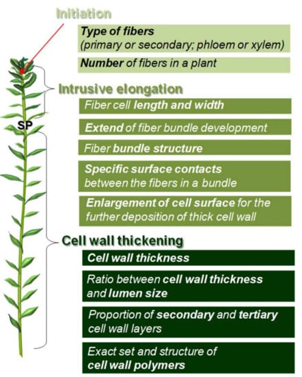
Each stage of fiber cell development within plant organism has the impact on the final yield and quality of fiber crops. The related parameters that are coupled to fiber initiation, elongation and cell wall thickening are summarized in Figure 4. Each of the stages has its own potential for manipulation of the fiber yield and quality. The very important steps undertaken through the last years to elucidate the molecular players responsible for each of the stages open the door for the purposeful improvement of fiber crops and adjustment of plant fiber characteristics for various applications.

Figure 4.
Impact of the key stages of primary bast fiber development on the quality-related parameters. SP-snap point (a marker of fiber developmental transition from elongation to cell wall thickening).
Acknowledgments
We acknowledge Russian Science Foundation for partial supporting the present study-project numbers 17-76-20049 (fiber quality-related processes) and 16-14-10256 (transcriptome analysis). We thank J. Paul Knox (University of Leeds, UK) and Fabienne Guillon (Institut National de la Recherche Agronomique, France) for the kind gifts of LM11 and INRA-RU2 antibodies, and Vadim V. Salnikov (KIBB KazSC RAS, Russia) for providing the photographs used for Figure 3b,d.
Author Contributions
Natalia Mokshina prepared the chapter 4 (“Fiber cell wall thickening”); Tatyana Chernova prepared the chapters 2 and 3 (“Fiber Initiation” and “Fiber Elongation”); Dmitry Galinousky and Oleg Gorshkov wrote the chapters 5 and 6 (“Problems ...” and “Perspectives ...”); Tatyana Gorshkova conceived the study topic, organized, wrote, and reviewed the paper. All authors discussed the review concept and analyzed available data. All authors approved the manuscript.
Conflicts of Interest
The authors declare no conflict of interest. The founding partners had no role in the design of the study; in the collection, analyses, or interpretation of data; in the writing of the manuscript, and in the decision to publish the results.
References
- Ashik, K.P.; Sharma, R.S. A Review on Mechanical Properties of Natural Fiber Reinforced. Hybrid Polymer Composites. J. Miner. Mater. Charact. Eng. 2015, 3, 420–426. [Google Scholar] [CrossRef]
- Pickering, K.L.; Efendy, M.G.A.; Le, T.M. A review of recent developments in natural fiber composites and their mechanical performance. Compos. Part A Appl. Sci. Manuf. 2016, 83, 98–112. [Google Scholar] [CrossRef]
- Namvar, F.; Jawaid, M.; Md Tahir, P.; Mohamad, R.; Azizi, S.; Khodavandi, A.; Rahman, H.S.; Nayeri, M.D. Potential use of plant fibres and their composites for biomedical applications. BioResources 2014, 9, 5688–5706. [Google Scholar] [CrossRef]
- Mohammed, L.; Ansari, M.N.M.; Pua, G.; Jawaid, M.; Islam, M.S. A review on natural fiber reinforced polymer composite and its applications. Int. J. Polym. Sci. 2015, 2015, 243947. [Google Scholar] [CrossRef]
- Akampumuza, O.; Wambua, P.M.; Ahmed, A.; Li, W.; Qin, X.-H. Review of the applications of biocomposites in the automotive industry. Polym. Compos. 2017, 38, 2553–2569. [Google Scholar] [CrossRef]
- Tripathi, K.M.; Vincent, F.; Castro, M.; Feller, J.F. Flax fibers—Epoxy with embedded nanocomposite sensors to design lightweight smart bio-composites. Nanocomposites 2016, 2, 125–134. [Google Scholar] [CrossRef]
- Esau, K. Plant Anatomy, 2nd ed.; John Wiley & Sons: New York, NY, USA, 1965; 767p. [Google Scholar]
- Fahn, A. Plant Anatomy, 4th ed.; Pergamon Press: Oxford, UK, 1990; 588p. [Google Scholar]
- Gorshkova, T.; Brutch, N.; Chabbert, B.; Deyholos, M.; Hayashi, T.; Lev-Yadun, S.; Mellerowicz, E.J.; Morvan, C.; Neutelings, G.; Pilate, G. Plant fiber formation: State of the art, recent and expected progress, and open questions. Crit. Rev. Plant Sci. 2012, 31, 201–228. [Google Scholar] [CrossRef]
- Fernandez-Tendero, E.; Day, A.; Legros, S.; Habrant, A.; Hawkins, S.; Chabbert, B. Changes in hemp secondary fiber production related to technical fiber variability revealed by light microscopy and attenuated total reflectance Fourier transforminfrared spectroscopy. PLoS ONE 2017, 12. [Google Scholar] [CrossRef] [PubMed]
- Hernandez, A.; Westerhuis, W.; van Dam, J.E.G. Microscopic study on hemp bast fibre formation. J. Nat. Fibers 2007, 3, 1–12. [Google Scholar] [CrossRef]
- Snegireva, A.; Chernova, T.; Ageeva, M.; Lev-Yadun, S.; Gorshkova, T. Intrusive growth of primary and secondary phloem fibers in hemp stem determines fiber-bundle formation and structure. AoB Plants 2015, 7. [Google Scholar] [CrossRef] [PubMed]
- Chernova, T.E.; Ageeva, M.V.; Chemikosova, S.B.; Gorshkova, T.A. The formation of primary and secondary fibers in hemp. Bull. All-Rus. Sci. Res. Inst. Bast Crops Process. 2005, 2, 6–13. [Google Scholar]
- Ageeva, M.V.; Petrovská, B.; Kieft, H.; Salnikov, V.V.; Snegireva, A.V.; van Dam, J.E.G.; Emons, A.M.C.; Gorshkova, T.A.; van Lammeren, A.A.M. Intrusive growth of flax phloem fibers is of intercalary type. Planta 2005, 222, 565–574. [Google Scholar] [CrossRef] [PubMed]
- Snegireva, A.V.; Ageeva, M.V.; Amenitskii, S.I.; Chernova, T.E.; Ebskamp, M.; Gorshkova, T.A. Intrusive growth of sclerenchyma fibers. Rus. J. Plant Physiol. 2010, 57, 342–355. [Google Scholar] [CrossRef]
- Mokshina, N.; Gorshkov, O.; Ibragimova, N.; Chernova, T.; Gorshkova, T. Cellulosic fibres of flax recruit both primary and secondary cell wall cellulose synthases during deposition of thick tertiary cell walls and in the course of graviresponse. Funct. Plant Biol. 2017, 44, 820–831. [Google Scholar] [CrossRef]
- Bourmaud, A.; Malvestio, J.; Lenoir, N.; Siniscalco, D.; Habrant, A.; King, A.; Legland, D.; Baley, C.; Beaugrand, J. Exploring the mechanical performance and in-planta architecture of secondary hemp fibres. Ind. Crops Prod. 2017, 108, 1–5. [Google Scholar] [CrossRef]
- Crônier, D.; Monties, B.; Chabbert, B. Structure and chemical composition of bast fibers isolated from developing hemp stem. J. Agric. Food Chem. 2005, 53, 8279–8289. [Google Scholar] [CrossRef] [PubMed]
- Placet, V.; Méteau, J.; Froehly, L.; Salut, R.; Boubakar, M.L. Investigation of the internal structure of hemp fibres using optical coherence tomography and Focused Ion Beam transverse cutting. J. Mater. Sci. 2014, 49, 8317–8327. [Google Scholar] [CrossRef]
- Fidelis, M.E.A.; Pereira, T.V.C.; Gomes, O.F.M.; Silva, F.A.; Filho, R.D.T. The effect of fiber morphology on the tensile strength of natural fibers. J. Mater. Res. Technol. 2013, 2, 149–157. [Google Scholar] [CrossRef]
- Marrot, L.; Lefeuvre, A.; Pontoire, B.; Bourmaud, A.; Baley, C. Analysis of the hemp fiber mechanical properties and their scattering (Fedora 17). Ind. Crops Prod. 2013, 51, 317–327. [Google Scholar] [CrossRef]
- Mellerowicz, E.J.; Gorshkova, T.A. Tensional stress generation in gelatinous fibres: A review and possible mechanism based on cell-wall structure and composition. J. Exp. Bot. 2012, 63, 551–565. [Google Scholar] [CrossRef] [PubMed]
- Gorshkova, T.; Chernova, T.; Mokshina, N.; Ageeva, M.; Mikshina, P. Plant “muscles”: Fibers with a tertiary cell wall. New Phytol. 2018, 218, 66–72. [Google Scholar] [CrossRef] [PubMed]
- Gorshkov, O.; Mokshina, N.; Gorshkov, V.; Chemikosova, S.; Gogolev, Y.; Gorshkova, T. Transcriptome portrait of cellulose-enriched flax fibers at advanced stage of specialization. Plant Mol. Biol. 2017, 93, 431–449. [Google Scholar] [CrossRef] [PubMed]
- Love, G.D.; Snape, C.E.; Jarvis, M.C. Determination of phenolic structures in flax fibre by solid-state 13C-NMR. Phytochemistry 1994, 35, 489–491. [Google Scholar] [CrossRef]
- Gorshkova, T.A.; Salnikov, V.V.; Pogodina, N.M.; Chemikosova, S.B.; Yablokova, E.V.; Ulanov, A.V.; Ageeva, M.V.; van Dam, J.E.G.; Lozovaya, V.V. Composition and distribution of cell wall phenolic compounds in flax (Linum usitatissimum L.) stem tissues, in annals of botany. Ann. Bot. 2000, 85, 477–486. [Google Scholar] [CrossRef]
- Chernova, T.; Mikshina, P.; Salnikov, V.; Ageeva, M.; Ibragimova, N.; Sautkina, O.; Gorshkova, T. Development of Hemp Fibers: The Key Components of Hemp Plastic Composites. In Engineering Nanocomposites—Fiber Crop Composite Production; InTech: Rijeka, Croatia, 2018; 16p, ISBN 978-953-51-5722-9. in press. [Google Scholar]
- Mikshina, P.V.; Gurjanov, O.P.; Mukhitova, F.K.; Petrova, A.A.; Shashkov, A.S.; Gorshkova, T.A. Structural details of pectic galactan from the secondary cell walls of flax (Linum usitatissimum L.) phloem fibres. Carbohyd. Polym. 2012, 87, 853–861. [Google Scholar] [CrossRef]
- Marques, G.; Rencoret, J.; Gutiérrez, A.; del Río, J.C. Evaluation of the chemical composition of different non-woody plant fibers used for pulp and paper manufacturing. Open Agric. J. 2010, 4, 93–101. [Google Scholar] [CrossRef]
- McCartney, L.; Marcus, S.E.; Knox, J.P. Monoclonal antibodies to plant cell wall xylans and arabinoxylans. J. Histochem. Cytochem. 2005, 53, 543–546. [Google Scholar] [CrossRef] [PubMed]
- Ralet, M.C.; Tranquet, O.; Poulain, D.; Moïse, A.; Guillon, F. Monoclonal antibodies to rhamnogalacturonan I backbone. Planta 2010, 231, 1373–1383. [Google Scholar] [CrossRef] [PubMed]
- Hotte, N.S.C.; Deyholos, M.K. A flax fibre proteome: Identification of proteins enriched in bast fibres. BMC Plant Biol. 2008, 8. [Google Scholar] [CrossRef] [PubMed]
- Mokshina, N.; Gorshkova, T.; Deyholos, M.K. Chitinase-Like (CTL) and Cellulose Synthase (CESA) Gene Expression in Gelatinous-Type Cellulosic Walls of Flax (Linum usitatissimum L.) Bast Fibers. PLoS ONE 2014, 9. [Google Scholar] [CrossRef] [PubMed]
- Islam, M.S.; Saito, J.A.; Emdad, E.M.; Ahmed, B.; Islam, M.M.; Halim, A.; Hossen, Q.M.; Hossain, M.Z.; Ahmed, R.; Hossain, M.S.; et al. Comparative genomics of two jute species and insight into fibre biogenesis. Nat. Plants 2017, 3. [Google Scholar] [CrossRef] [PubMed]
- Guerriero, G.; Behr, M.; Legay, S.; Mangeot-Peter, L.; Zorzan, S.; Ghoniem, M.; Hausman, J.-F. Transcriptomic profiling of hemp bast fibres at different developmental stages. Sci. Rep. 2017, 7. [Google Scholar] [CrossRef] [PubMed]
- Guerriero, G.; Behr, M.; Backes, A.; Faleri, C.; Hausman, J.-F.; Lutts, S.; Cai, G. Bast fibre formation: Insights from Next-Generation Sequencing. Procedia Eng. 2017, 200, 229–235. [Google Scholar] [CrossRef]
- Chantreau, M.; Portelette, A.; Dauwee, R.; Kiyoto, S.; Crônier, D.; Morreel, K.; Arribat, S.; Neutelings, G.; Chabi, M.; Boerjan, W.; et al. Ectopic lignification in the flax lignified bast fiber1 mutant stem is associated with tissue-specific modifications in gene expression and cell wall composition. Plant Cell 2014, 26, 4462–4482. [Google Scholar] [CrossRef] [PubMed]
- Roach, M.J.; Mokshina, N.Y.; Badhan, A.; Snegireva, A.V.; Hobson, N.; Deyholos, M.K.; Gorshkova, T.A. Development of cellulosic secondary walls in flax fibers requires beta-galactosidase. Plant Physiol. 2011, 156, 1351–1363. [Google Scholar] [CrossRef] [PubMed]
- Van Bakel, H.; Stout, J.M.; Cote, A.G.; Tallon, C.M.; Sharpe, A.G. The draft genome and transcriptome of Cannabis sativa. Genome Biol. 2011, 12. [Google Scholar] [CrossRef] [PubMed]
- Wang, Z.; Hobson, N.; Galindo, L.; Zhu, S.; Shi, D.; McDill, J.; Yang, L.; Hawkins, S.; Neutelings, G.; Datla, R.; et al. The genome of flax (Linum usitatissimum) assembled de novo from short shotgun sequence reads. Plant J. 2012, 72, 461–473. [Google Scholar] [CrossRef] [PubMed]
- Sarkar, D.; Mahato, A.K.; Satya, P.; Kundu, A.; Singh, S.; Jayaswal, P.K.; Singh, A.; Bahadur, K.; Pattnaik, S.; Singh, N.; et al. The draft genome of Corchorus olitorius cv. JRO-524 (Navin). Genom. Data 2017, 12, 151–154. [Google Scholar] [CrossRef] [PubMed]
- Liu, C.; Zeng, L.; Zhu, S.; Wu, L.; Wang, Y.; Tang, S.; Wang, H.; Zheng, X.; Zhao, J.; Chen, X.; et al. Draft genome analysis provides insights into the fiber yield, crude protein biosynthesis, and vegetative growth of domesticated ramie (Boehmeria nivea L. Gaud). DNA Res. 2017. [Google Scholar] [CrossRef] [PubMed]
- Zhang, N.; Deyholos, M.K. RNASeq analysis of the shoot apex of flax (Linum usitatissimum) to identify phloem fiber specification genes. Front Plant Sci. 2016, 7. [Google Scholar] [CrossRef] [PubMed]
© 2018 by the authors. Licensee MDPI, Basel, Switzerland. This article is an open access article distributed under the terms and conditions of the Creative Commons Attribution (CC BY) license (http://creativecommons.org/licenses/by/4.0/).