Review

Electrospun Nanofibers for Small Molecule Sustained Delivery Targeting Articular Cartilage Regeneration: A Review


by
Frederico Barbosa
1,2,†,
Filipe Miguel
1,2,†,
Margarida F. Domingues
1,2,† and
João Carlos Silva
1,2,*
1
Department of Bioengineering and iBB-Institute for Bioengineering and Biosciences, Instituto Superior Técnico, Universidade de Lisboa, Av. Rovisco Pais, 1049-001 Lisbon, Portugal
2
Associate Laboratory i4HB—Institute for Health and Bioeconomy, Instituto Superior Técnico, Universidade de Lisboa, Av. Rovisco Pais, 1049-001 Lisbon, Portugal
*
Author to whom correspondence should be addressed.
These authors contributed equally to this manuscript.
Fibers 2026, 14(5), 56; https://doi.org/10.3390/fib14050056
Submission received: 11 March 2026 / Revised: 5 May 2026 / Accepted: 8 May 2026 / Published: 11 May 2026

Highlights

What are the main findings?
Overview of the current state of the art of small molecule-loaded electrospun scaffolds for articular cartilage tissue engineering applications.
Comparative analysis of different electrospinning-based strategies for the fabrication of advanced drug delivery platforms.
What are the implications of these findings?
Despite recent promising advances, significant challenges remain, including achieving controlled long-term drug release, minimizing burst release, and optimizing scaffold architecture to ensure adequate mechanical performance and efficient cell infiltration.
Stimuli-responsive systems, predictive machine learning approaches, and advanced electrospinning configurations offer promising strategies to improve drug delivery control and cartilage regeneration outcomes, while enhnacing manufacturing scalability and ensuring reproducibility, thereby bringing these platforms closer to clinical translation.

Abstract

The limited regenerative capacity of articular cartilage (AC) following injury has led to a high prevalence of degenerative AC-related disorders, including osteoarthritis (OA). Current clinical treatments for OA have failed to halt disease progression, driving growing interest in cartilage tissue engineering (CTE) strategies aimed at developing biomimetic substitutes to regenerate damaged AC tissue. Among the available biofabrication techniques, electrospinning has gained attention due to its ability to generate fibrous scaffolds that closely mimic the architecture of the native AC extracellular matrix, while also serving as versatile drug delivery platforms with high surface area and elevated drug loading efficiency. Small molecules, low-molecular-weight therapeutic agents capable of interacting with both cell membrane and intracellular components, can be incorporated into these scaffold systems to target the underlying mechanisms of OA. This review examines the current state of the art of small molecule-loaded electrospun scaffolds for CTE applications. Small molecules targeting pain, inflammation, and cartilage function restoration show considerable therapeutic potential, and their incorporation into coaxial and other advanced electrospinning setups enables controlled and sustained drug release. Recent examples of small molecule-loaded electrospun scaffolds for AC repair demonstrate enhanced chondrogenic differentiation and neo-cartilage formation, supporting their potential as viable CTE strategies. Nevertheless, challenges related to drug release kinetics, scaffold load-bearing properties, manufacturing scalability, reproducibility, and regulatory approval remain critical barriers to clinical translation. Emerging fabrication strategies, AI-assisted optimization, personalized medicine approaches, and stimuli-responsive drug delivery systems offer promising avenues to overcome these limitations and advance the clinical adoption of these platforms.

1. Introduction

Articular cartilage (AC) is a highly specialized connective tissue responsible for distributing mechanical loads within the joints while enabling near-frictionless skeletal movement [1]. Its porous extracellular matrix (ECM), comprised primarily of interstitial water (65–80%), type II collagen fibrils, glycosaminoglycans (GAGs) (e.g., chondroitin sulfate, hyaluronic acid), and proteoglycans (e.g., aggrecan), is responsible for the tissue’s viscoelastic behavior and high tensile and compressive strengths, which are essential for shock absorption and the proper transmission of loads to the underlying subchondral bone [2,3]. The avascularity and reduced chondrocyte population of AC render it especially vulnerable to injury and account for its poor intrinsic regenerative potential [1].
Osteoarthritis (OA), the most prevalent form of arthritis characterized by the gradual degeneration of AC ECM in response to mechanical wear, is the leading cause of adult chronic pain and long-term disability, imposing a substantial social and economic burden in modern aging societies [4,5]. In OA, the mechanical overloading of AC triggers the release of tissue fragments and other damage-associated molecular patterns (DAMPs), which activate macrophages and stimulate the production of pro-inflammatory cytokines and chemokines. These mediators promote the recruitment of additional macrophages and induce chondrocytes to produce metalloproteinases (MMPs) and aggrecanases. This signaling cascade accelerates ECM degradation and synovial inflammation, establishing a self-perpetuating harmful cycle that ultimately leads to the progressive and potentially complete loss of the AC tissue [6,7,8].
Available clinical therapies for the treatment of OA have failed to effectively halt disease progression or directly target its underlying biological mechanisms. Most current approaches focus on symptom management, including pain relief and joint swelling, while others involve surgical procedures aimed at repairing AC defects (e.g., microfractures, allografts), restoring chondrogenic potential (e.g., autologous chondrocyte implantation [ACI], matrix-assisted autologous chondrocyte implantation [MACI]), or performing partial or total joint replacement [9,10,11]. As a result, cartilage tissue engineering (CTE) strategies have gained increasing attention. These approaches typically involve the incorporation of chondrocytes or cartilage progenitors cell types, along with bioactive factors, into biomaterial scaffolds designed to temporarily replace damaged tissue and promote functional AC regeneration [12].
Electrospinning, a highly versatile scaffold fabrication technique used to generate micro- and nano-sized fibers, has been widely reported in the literature for the development of CTE regenerative strategies [13]. By recapitulating the fibrous architecture of the type II collagen fibrils of the tissue’s ECM (major organic component), electrospun scaffolds provide a closer mimicry of the native biomechanical environment of AC. Additionally, the inherently high surface-to-volume ratio of electrospun fibrous scaffolds enhances cell attachment, proliferation, and growth [14].
Particularly, the use of electrospun scaffolds as drug delivery systems has also been extensively highlighted due to their remarkable versatility [15]. Multiple materials and compositions can be employed, enabling the fabrication of fibers with diverse functional and structural properties. Moreover, their high surface area, elevated loading capacity, and high encapsulation efficiency make them particularly attractive platforms for therapeutic delivery [16]. Electrospun scaffolds can enable localized, targeted, and sustained release of bioactive molecules while reducing systemic clearance and facilitating the incorporation of poorly soluble drugs [17].
A wide array of small molecules, defined as low-molecular-weight compounds (<1 kDa), have been explored in the TE field due to their ability to promptly diffuse across the cell membrane and directly interact with intracellular targets, thereby modulating cellular functions more rapidly than many other bio-therapeutic modalities that primarily act through extracellular components (e.g., surface receptors) [18,19]. In addition, small molecules exhibit improved stability, high reproducibility, and relatively low production costs. These advantages have led to their widespread adoption in the pharmaceutical industry, where they account for approximately 90% of approved therapeutic drugs [20]. Given their therapeutic potential, the integration of small molecules into electrospun scaffolds has attracted increasing attention as a promising strategy for CTE. However, despite the growing interest, this topic has yet to be the primary focus of a dedicated review.
In this context, the present work provides a comprehensive analysis of the incorporation of small molecules within electrospun scaffold systems for CTE. An updated overview of the main small molecules used in the context of CTE is presented, along with the different electrospinning technique modalities developed for their delivery. Representative examples of electrospun-based small molecule delivery platforms designed to guide AC repair are highlighted. Finally, current challenges and future perspectives in this field are discussed.

2. Small Molecules for Articular Cartilage Regeneration

Small molecules are a category of chemical compounds with molecular weights ranging from 0.1 to 1 KDa, having emerged as an exciting and powerful tool to modulate cell fate and tissue regeneration [21]. Particularly, cocktails of small molecules have been utilized to directly reprogram fibroblasts into neuronal cells, cardiomyocytes, and induced pluripotent stem cells (iPSCs) [22,23,24,25].
The small size of these molecules facilitates their crossing over the outer plasma membrane and their targeting of extracellular components like surface receptors [26,27] or intracellular proteins like kinases and transcription factors [28]. From a clinical perspective, they offer several advantages as their relatively simple structure facilitates their synthesis, while concomitantly being easy to store, transport and standardize [29]. Additionally, small molecules are able to bypass the immune system, being classified as low-immunogenic [30]. These properties position small molecules as very promising candidates for AC regeneration, as they can offset the downsides of commonly used macromolecules like growth factors. In fact, there has been an emergence of studies that have successfully employed these small molecules to tackle AC regeneration from different approaches (Table 1).

2.1. Generation of Functional Chondrocytes

Restoring functional chondrocytes in the native tissue is one of the cornerstones of AC regeneration that can be achieved either through in vitro expansion or stem cell differentiation under proper culture conditions. As powerful modulators of cell fate, small molecular compounds have been successfully employed to facilitate these routes for chondrocyte generation.
Autologous chondrocyte transplantation is one of the most commonly used techniques for the treatment of OA and AC defects, requiring the in vitro expansion of a sufficient number of chondrocytes prior to implantation to restore native tissue function. However, the dedifferentiation of expanded chondrocytes, represented by a loss of phenotypic function, can lead to the formation of fibrocartilage, hindering its clinical application [31]. Aiming to address this limitation, Kobayashi et al. screened 5822 bioactive compounds that could prevent the dedifferentiation of chondrocytes in monolayer expansion culture. The authors found A-674563 to inhibit the degradation of SOX9 protein, aiding in the expansion of chondrocytes and in the maintenance of their typical characteristics [32].
In 2012, Johnson et al. highlighted kartogenin (KGN) as a small molecule capable of inducing the chondrogenesis of mesenchymal stem/stromal cells (MSCs) in vitro [33]. This factor has since become a key component in cartilage regeneration strategies, as it can induce chondrogenesis by disrupting the interaction between core-binding factor β (CBFβ) and the actin-binding protein filamin A (FLNA). As a result, CBFβ undergoes translocation into the cellular nucleus, where it binds with the RUNX1 transcription factor. Subsequently, this interaction triggers the activation of genes responsible for orchestrating the chondrogenic differentiation process [34]. Various studies followed by trying to create controllable drug delivery systems for this drug, namely by combining it with microspheres or chitosan nanoparticles [35,36]. CHIR99021 is a small molecule WNT protein agonist that Narcisi and colleagues found capable of promoting the chondrogenic capacity of MSCs through the WNT pathway, while also suppressing the hypertrophic propensity of MSC-derived cartilage [37]. The small molecule 6-bromoindirubin-3-oxim is also able to affect the WNT pathway by inhibiting the glycogen synthase kinase-3, leading to an upregulation of cartilage-specific genes [38].

2.2. Inhibition of Inflammation

Inflammation is a hallmark of OA and plays a key role in the onset and progression of the disease. Biomechanical stress can stimulate the release of inflammatory cytokines such as interleukin-1 beta (IL-1β) and tumor necrosis factor alfa (TNFα). These cytokines trigger a cascade of signaling pathways, eventually promoting to the production of MMPs, further leading to the degradation of AC ECM [39]. Due to its importance, several studies employing anti-inflammatory strategies have found success in controlling pain and tissue degeneration [40].
PKF115-584 is a small molecule found to be an inhibitor of the WNT/β-catenin pathway, which is involved in the upregulation of pro-inflammatory cytokines IL-1β and TNFα. It exerts this effect by blocking and disrupting the binding of β-catenin with its transcription factor TCF4 [41]. Landman et al. highlighted its potential to almost completely reverse the IL-1β/TNFα-induced production of MMP3, MMP9 and MMP13 after 4 days of treatment. However, it is important to note that it was unable to reverse the inhibition of chondrogenic markers like aggrecan and collagen type II [42]. IL-1β and TNFα are involved in the activation of the nuclear factor kappa B (NF-κB) signaling pathway, which induces the expression of several pro-inflammatory cytokines and chemokines. Moreover, their critical pro-inflammatory function has been reported both in rheumatoid arthritis and OA patients [43,44]. Nepetin is a small flavonoid molecule that exhibits potent anti-inflammatory properties. In 2018, Chen et al. demonstrated its ability to suppress IL-1β-induced NF-κB activation in retina cells by inhibiting NF-κB degradation [45]. A 2021 study performed by Xu and colleagues built upon these findings by exploring the effects of this small molecule in chondrocytes for OA treatment. In fact, a similar inhibition of the NF-kB pathway was observed, which led to a reduction in the degradation of the main AC ECM components collagen type II and aggrecan [46].

2.3. Pain Treatment

Chronic pain is one of the most relevant characteristics of OA, caused by the increased friction and wear between heavily enervated bones due to the degradation of AC. As a major symptom, pain is a key pharmaceutical target and currently the central focus in OA management. Non-steroidal anti-inflammatory drugs (NSAIDs) and glucocorticoids are commonly used to alleviate OA pain. However, their biggest drawback is the limited long-term efficacy, only being able to provide short-term relief from pain and inflammation. Furthermore, some adverse effects have been highlighted with their prolonged use, such as cardiovascular and renal complications and contribution to cartilage breakdown, among others [47,48].
In recent years, small molecules have emerged as promising candidates for pain management. There are several ongoing clinical trials for this purpose, with the main targets being G-protein coupled receptors (GPCRs) and ion channels [49]. CR845 is a peripherally acting kappa opioid receptor agonist that, in a phase 2 trial, demonstrated a significant reduction in pain and opioid-related side effects in hip OA patients [50]. Another completed clinical trial targeting ion channels employed intra-articular injections of CNTX-4975, a small molecule able to deactivate free terminals of primary afferent pain fibers within the joint, which resulted in a dose-dependent improvement in knee OA-associated pain [50].
Table 1. Examples of studies using small molecules for articular cartilage regeneration strategies.
Small
Molecule
Function/TargetMain ResultsReference
A-674563Chondrocyte generation and phenotype maintenance.Inhibition of SOX9 degradation, expanding chondrocytes maintained their typical characteristics.[32]
KartogeninChondrocyte generation from MSCs.
Interacts with core-binding factor β.
Activates signaling pathways and genes responsible for enhanced MSC chondrogenic differentiation.[33]
CHIR99021MSC differentiation into chondrocytes.
Interacts with the WNT pathway.
Promotes the chondrogenic capacity of MSCs and suppresses the hypertrophic propensity of MSC-based engineered AC tissues.[37]
6-bromoindirubin-3-oximChondrocyte generation.
Interacts with the WNT pathway.
Upregulation of cartilage-specific genes[38]
PKF115-584Anti-inflammation.
Inhibitor of the WNT/β-catenin pathway.
Able to decrease the production of MMP 3, 9 and 13.
However, it was unable to reverse the inhibition of chondrogenic markers.
[42]
NepetinAnti-inflammation.
Suppresses the activation of the NF-κB signaling pathway.
Reduced the degradation of articular cartilage ECM components collagen type II and aggrecan. [46]
CR845Pain management.
Targets kappa opioid receptors.
Resulted in a significant pain reduction and of opioid side-effects in hip OA patients.[50]
CNTX-4975Pain management.
Reversibly deactivates free terminals of primary afferent pain fibers.
Intra-articular injections led to a dose-dependent improvement of pain in knee OA patients.[50]

3. Electrospinning Technique for Small Molecule Delivery Applications

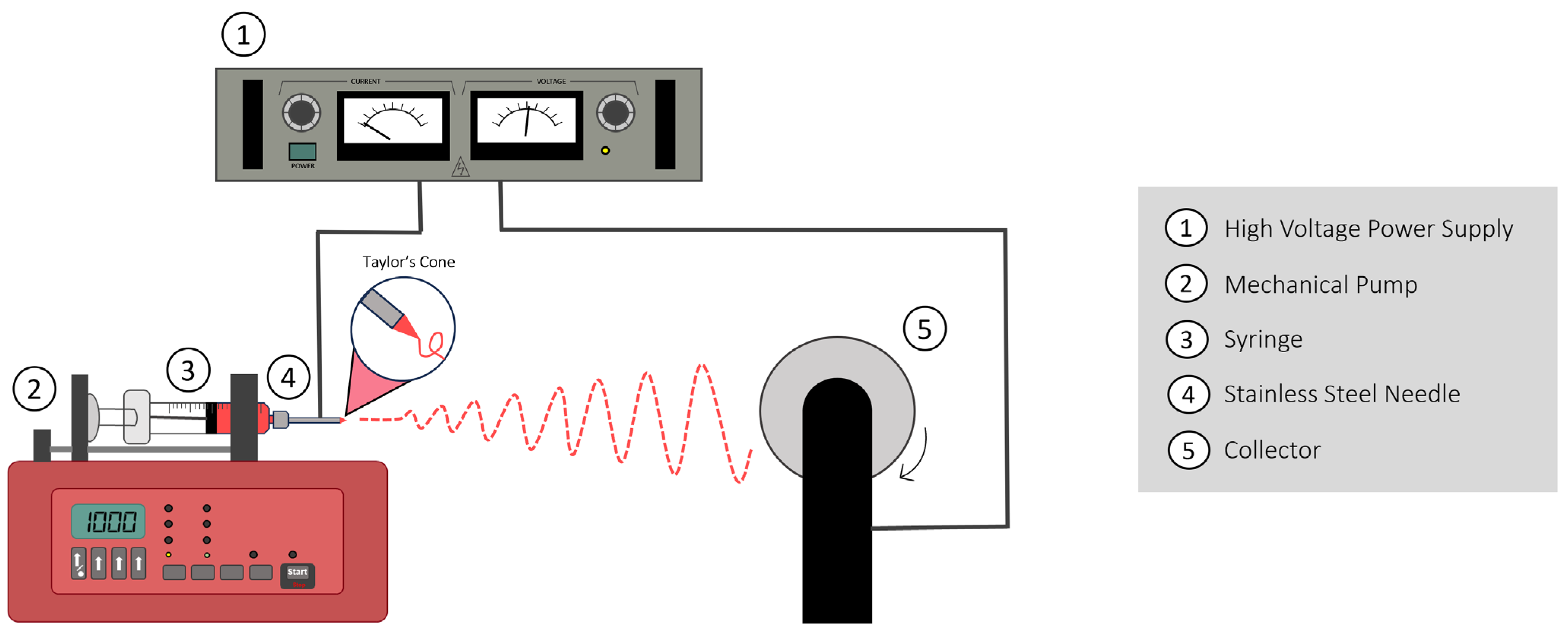
Electrospinning is a scaffold fabrication technique used for producing small-sized fibers—with diameters in the nanometer and micrometer range—capable of mimicking structural ECM features of different human tissues (e.g., type II collagen fibrils size and alignment in AC) as well as assisting in their regeneration (Figure 1) [51,52]. The procedure works as follows: A high electrical voltage is applied to a polymeric solution being ejected at a controlled rate through a small needle, generating repulsive electrostatic forces within the solution which oppose the fluid’s surface tension (creating a conical shape denominated Taylor’s cone). Once a critical point is reached, a jet of charged fluid is ejected from the tip of the cone towards a grounded metal collector. During this process, the electrostatic field forces increase with the distance from the needle, accelerating and stretching the jet, and ultimately leading to the evaporation of the solvent and increase in charge density as the diameter of the jet becomes smaller. As a result, the primary polymer jet is spliced into multiple smaller jets (splaying), with similar splitting processes occurring with these smaller jets until the fibers are deposited layer-by-layer on the surface of the collector [53,54]. Different operational parameters can be adjusted for modifying the morphology and diameter of the generated fibers, namely the applied voltage, flow rate, needle diameter and distance between the tip of the needle and the collector [53,54,55]. Overall, electrospinning is an extremely popular material processing technique, due to its easy manipulation and versatile nature, which enables the production of widely different scaffolds with different biomaterials using the same simple strategy.
A potential application of implantable electrospun fibers is carrying small molecules for targeted drug delivery [16,55]. High attrition rates have plagued drug development in the pharmaceutical industry, with up to 90% of developed drugs failing their respective clinical trials, representing meaningful negative economic and social impacts [56]. One of the key issues is related to molecular obesity, in which potency (more significant biological response at lower doses) is built up inappropriately into a molecule through increased molecular weight and lipophilicity, which ultimately impacts its aqueous solubility [57,58]. In fact, nearly 75% of all drug candidates present low solubility, placing a significant burden on drug development [59,60]. Electrospinning allows the incorporation of poorly soluble drugs into polymeric nanofibers, increasing their surface area and, as a result, enhancing their dissolution rate and drug bioavailability [15,16]. This drug delivery strategy also enables the encapsulation of drugs within the nanofibers, providing protection against degradation/clearance and improving stability. Electrospun fibers can also be tailored to achieve sustained and targeted drug release patterns, optimizing therapeutic outcomes for large molecules [58]. Additionally, this technique can also be applied for combining multiple drugs or therapeutic agents in a single formulation, thus addressing more complex medical conditions [15].
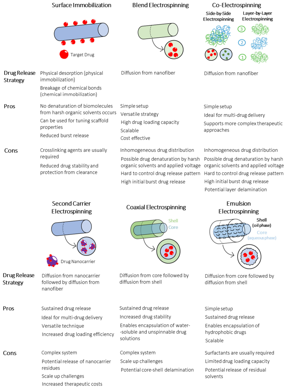
Different electrospinning methods have been employed in the development of targeted drug delivery systems, allowing for a precise and controlled release of drugs at specific sites within the body (Figure 2). In the most conventional one—blend electrospinning—a carrier polymer (usually hydrophilic) and the drug are directly mixed prior to electrospinning [61]. This strategy allows a simple and straightforward method for delivering therapeutic agents, with its success being mainly dependent on drug/polymer affinity (solubility and similar polarity) and degree of drug encapsulation [15,61]. In a study developed by Adeli et al. (2015), Irbesartan-loaded polyvinylpyrrolidone (PVP) monoaxial nanofibers were fabricated via electrospinning for treating hypertension [62]. The combined PVP-Irbesartan drug delivery system yielded an enhancement in relative bioavailability when compared with the plain drug suspension in in vivo rabbit models. A similar process was reported by Zhu et al. (2020) for developing metformin (MET)-loaded polycaprolactone (PCL)/chitosan blended electrospun membranes for guiding bone regeneration. The MET-loaded nanofibers were found to improve the proliferation and osteogenic differentiation of rat bone marrow-derived MSCs in vitro [63]. The use of stimuli-responsive carrier polymers has gained traction as of recently as a potential strategy for achieving on-demand drug release in vivo by controlling changes in environmental conditions at the target site [64]. Different stimuli-responsive fibrous scaffolds have been developed, such as electrospun nanofibers sensitive to temperature, pH, light, magnetic field and reactive oxygen species (ROS), as well as electrically responsive membranes [64,65,66]. In a study performed by Puiggalí-Jou et al. (2018), electrically responsive PCL microfibers loaded with conductive PEDOT nanoparticles (PEDOT NPs) and curcumin (anti-inflammatory role) were generated. By applying pre-defined potential pulses, the PEDOT NPs increased in diameter and migrated to the surface of the microfibers, modifying the structure of the PCL matrix and, in turn, resulting in the timed release of curcumin [65].
A few studies have also reported the development of co-electrospinning systems, in which multiple electrospinning setups are used simultaneously (side-by-side electrospinning) or sequentially (layer-by-layer electrospinning) to produce intertwined and layered monoaxial nanofibers loaded with different bioactive molecules, enabling more complex therapeutic outcomes [67,68]. As an example, Yang et al. (2020) reported the development of co-electrospun lidocaine hydrochloride (LID)-loaded chitosan/mupirocin-loaded PCL nanofibers for wound healing. While the LID-loaded fibers were responsible for addressing pain relief, the mupirocin nanofibers exhibited key anti-inflammatory properties. The developed membranes were found to significantly improve the wound healing process in full-thickness skin defect models [69].
One of the main limitations of blending drugs into monolithic fibers is that biomolecules can potentially denature as a result of the voltage being applied during electrospinning or due to the harsh organic solvents involved [70]. Additionally, inadequate drug/polymer systems are often responsible for a high initial burst drug release and uncontrollable release patterns, which can lead to localized cellular toxicity and reduced therapeutic efficacy [70,71].
To address the potential biomolecule denaturation during electrospinning, the surface immobilization (also known as surface coating or functionalization) of the drug molecules onto the surface of the fibers post-electrospinning—via either physical or chemical interactions—has been reported in the literature [71,72]. This strategy takes advantage of the high surface-to-volume ratio of nanofibers, while also tuning other key properties of the electrospun fibrous meshes, such as their contact angle and cellular adhesion [15]. Several methodologies exist to achieve fiber functionalization via different types of chemical bonding [73]. The most common technique involves the dip coating of the fibers in target solutions for the passive adsorption of bioactive molecules (physical interaction) [72]. Other potential strategies include the modification of the nanofiber surface with hydroxyl, carboxyl, amine or thiol groups, with polydopamine (pDA) surface chemistry in particular offering a reliable alternative for immobilizing antibodies, proteins and peptides onto hydrophobic electrospun membranes [74,75]. These chemical conjugation methods enable a more precise control of the quantity of the incorporated drug within the nanofiber mesh and minimize initial burst drug release [73]. Recently, Wu et al. (2020) fabricated poly(D, L-lactic acid)-poly(ethylene glycol)-poly(D, L-lactic acid) (PELA) electrospun fibrous scaffolds, which were immobilized with bone morphogenetic protein-2 (BMP-2) (with pDA chemistry) to treat acetabulum defects. The BMP-2-loaded PELA implants were able to induce osteogenic differentiation in acetabular defects of in vivo porcine animal models, generating a reduced amount of fibrotic tissue when compared to the uncoated PELA scaffolds [76].
Second carrier electrospinning can be used as an alternative to blend electrospinning, minimizing the problematic initial burst release of drugs that is commonly observed with the latter technique [77]. This strategy involves the loading of the desired bioactive molecules onto secondary carriers, such as microspheres, nanotubes, vesicles, and micelles, before being mixed with the polymeric solutions prior to electrospinning [77,78]. Different drug-loaded particles can be added to the same monoaxial fibers, resulting in multi-drug release [15]. Since the bioactive molecules release from the secondary carrier first, before diffusing out from the nanofibers, a prolonged drug release pattern is obtained [78]. In a study developed by Luo et al. (2018), camptothecin (CPT) (anti-tumorigenic drug) was loaded into promicelles (PMCPT) which, in turn, were embedded in PELA/polyethylene oxide (PEO) monoaxial fibers. Once implanted into in vivo 4T1 breast tumor models, the promicelles were slowly released (sustained release) and self-assembled into folate-targeted and glutathione-sensitive micelles (micelle-generating depot) that were taken up by the cancerous tissue continuously for over a 30-day period. This methodology resulted in significant micelle accumulation in the tumor, leading to stronger tumor inhibitory efficacy and more effective inhibition of tumor metastasis in comparison to the intravenous injection of PMCPT [79].
Similarly to the previously discussed electrospinning methodology, coaxial electrospinning has also been used as a substitute to blend electrospinning, addressing some of the limitations of drug release kinetics of monoaxial blended nanofibers [80,81]. This modified electrospinning setup uses a spinneret with two concentrically aligned capillaries to simultaneously extrude immiscible inner and outer solutions, generating two-phase core–shell fibers. Coaxial electrospinning enables the encapsulation of water-soluble and sensitive drugs in the core, not only protecting the biological activity of these molecules and avoiding issues related with the use of harsh organic solvents but also preventing initial burst release and resulting in a more sustained drug release pattern compared with their monoaxial counterparts [15,80,82]. This strategy also allows for the use of non-spinnable drug solutions, as well as the simultaneous delivery of multiple drugs, by loading the bioactive molecules in both the core and shell regions of the coaxial fibers [15,82]. Most of the core–shell nanofibers used in drug delivery systems are loaded with bioactive molecules in the core. Drug release is performed by nanopath diffusion of the drug as well as the degradation of the shell polymer (which may be accelerated by the swelling of the polymer in the core) [15,83]. In a study performed by Nguyen et al. (2012), coaxial fibers with a poly(lactic acid) (PLA) shell and a salicylic acid (SA)-loaded PCL core, with anti-inflammatory properties, were successfully produced via electrospinning [84]. More recently, Cui et al. (2022) produced dual drug-loaded coaxial electrospun fibers for boosting vascularized bone regeneration. While deferoxamine (DFO), an agent involved in bone angiogenesis, was loaded on the shell, dexamethasone (DEX), an osteogenic factor, was loaded on the core layer (PCL was used as carrier). The combined synergistic effect of DFO and DEX being released from the generated coaxial fibers was found to contribute to the formation of vascularized bone tissue in a rat calvarial defect model [85]. Tri-axial electrospinning setups, which use three aligned capillaries to extrude simultaneously three polymeric solutions, thus enabling the production of three-phase fibers (core–middle layer–shell), have also been reported in the literature, although to a lesser extent [86]. This strategy provides additional potential combinations of biomaterials, fiber structures and drug release kinetics [15].
Emulsion electrospinning is an alternative electrospinning method to coaxial electrospinning that can be applied for generating core–shell fibers using the conventional monoaxial electrospinning setup (with a single uniaxial needle) [87]. In this technique, an organic/oil phase and aqueous phase are mixed, generating an emulsion. Surfactants can also be added to increase the stability of the water-in-oil (W/O) emulsion. Given the increased evaporation rate of the oil phase relative to the aqueous phase, the viscosity of the former increases faster during electrospinning, causing the aqueous phase to migrate to the inner part of the electrospun jet while the oil phase accumulates at the fiber periphery (viscosity gradient). This results in the formation of two-phased fibers, akin to those obtained with coaxial electrospinning [87,88]. Emulsion electrospinning allows the encapsulation of hydrophobic drugs and the sustained release of one or multiple bioactive molecules simultaneously [88]. The ease of scale up of this technique as well as its simplicity and versatility are some of its advantages compared to coaxial electrospinning [15,87]. In a study performed by Liu et al. (2015), platelet-rich plasma (PRP)-loaded PCL/gelatin fibrous scaffolds were produced via emulsion electrospinning for treating cartilage defects in osteochondral injuries. PRP is a concentration of platelets (usually derived from the patient’s own plasma), capable of releasing diverse growth factors (e.g., TGF-β, IGF and PDGF) upon activation. Sustained growth factor release could be observed for over 30 days in vitro. The implanted scaffolds were shown to promote cartilage regeneration, being able to nearly fill in vivo full-thickness cartilage defects with healthy tissue after a 12-week period [89].
Overall, depending on the electrospinning strategy used for developing targeted drug delivery systems, different drug encapsulation approaches are possible (e.g., immobilized on the surface, blended uniformly in the fiber structure, confined in the core section), each with distinct advantages and limitations. Blend electrospinning, while simple, scalable, and cost-effective (factors that favor its clinical translatability), is often associated with inhomogeneous drug distribution and high initial burst release. In contrast, coaxial electrospinning addresses these limitations through its core–shell structure, enabling more sustained release and improved drug stability, albeit at the cost of increased system complexity and scale-up challenges that may hinder its clinical translation. Emulsion electrospinning, on the other hand, offers a more scalable alternative to coaxial systems, maintaining sustained release capabilities while also simplifying the fabrication process, though the requirement for often cytotoxic surfactants and reduced drug loading efficiency (limited by emulsion stability) should be considered. These methodologies therefore lead to diverse drug release profiles, and when selecting optimal drug release platforms for specific applications, careful consideration of both the material/drug composition and the encapsulation technique is crucial, since both factors synergistically impact drug release patterns and, as a result, therapeutic efficacy.

4. Applications of Small Molecule-Loaded Electrospun Nanofibers on Articular Cartilage Regeneration

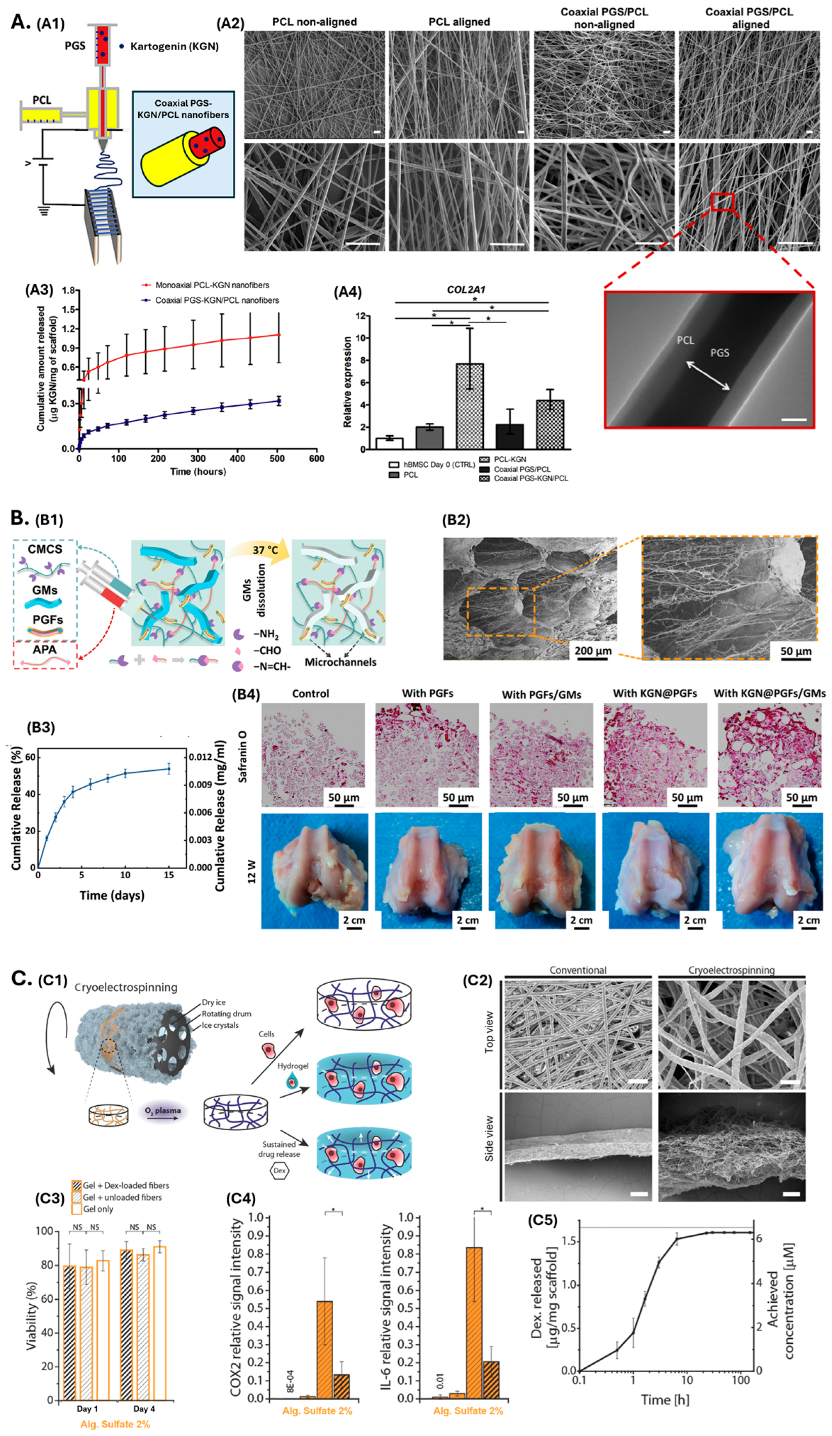
The delivery of small molecules using electrospun scaffolds has emerged as a promising strategy to direct MSC chondrogenic differentiation, enhance cartilage-specific ECM deposition, and modulate inflammatory responses, ultimately improving cartilage repair. Figure 3 illustrates several innovative electrospun-based delivery strategies currently explored for small molecule administration in AC regeneration. Among these molecules, KGN has been the most extensively studied, due to its ability to drive MSC chondrogenic differentiation and exert chondroprotective effects that prevent cartilage degeneration [33].
Silva et al. (2020) [90] developed aligned coaxial nanofibers, composed of a KGN-loaded poly(glycerol sebacate) (PGS) core and a PCL shell, to mimic the anisotropic nanoscale features of AC (Figure 3A). Coaxial electrospinning enabled encapsulation of low-viscosity PGS and controlled small-molecule delivery, while fiber alignment, achieved using parallel copper collectors, significantly enhanced mechanical performance. All electrospun fiber scaffolds exhibited highly porous morphologies resembling AC ECM, with fiber diameters of 505–730 nm, although aligned fibers were consistently thinner. The elastic moduli of aligned monoaxial PCL and coaxial fibers increased to 9.9 ± 0.87 MPa and 11.78 ± 0.73 MPa, respectively, compared to 4.02 ± 0.88 MPa and 5.06 ± 1.51 MPa in non-aligned controls, falling within the range for healthy cartilage (5–25 MPa). This indicates that fiber alignment had a stronger effect on mechanics than the core–shell configuration. KGN release from coaxial scaffolds showed a reduced initial burst within 24 h and a more sustained profile than monoaxial PCL-KGN fibers. After 21 days, total release was 0.32 ± 0.03 and 1.11 ± 0.44 µg KGN/mg for coaxial and monoaxial fibers, respectively, which is attributed to the drug being confined within the fiber core in the coaxial system versus distributed throughout the entire fiber in the monoaxial configuration. Human MSCs cultured on KGN-loaded scaffolds displayed increased proliferation and chondrogenic differentiation, exhibiting upregulation of the cartilage marker genes COL2A1, ACAN, and SOX9, compared to non-loaded scaffolds. Notably, all electrospun scaffold types had increased expression of PRG4, which encodes for the lubricin/superficial zone protein (SZP) present in the superficial layer of AC. Collectively, Silva et al. [90] demonstrated that aligned coaxial PGS/PCL nanofibers provide structural and biochemical cues favorable for MSC chondrogenesis while enhancing control over KGN release.
Elder et al. (2022) [91] blended KGN into PCL/poly(lactic-co-glycolic acid) (PLGA) nanofibers to enhance bone marrow stimulation microfracture (MFx) therapy by stimulating the chondrogenic differentiation of endogenous MSCs and supporting hyaline cartilage regeneration. The incorporation of 2 wt% KGN did not significantly affect fiber diameter (225 ± 24 nm) nor tensile strength (36 ± 6 MPa). Also, in accordance with the study performed by Silva et al. [90], the monoaxial blended fibers promoted an initial burst release of KGN (~30% within 24 h), followed by a slower release phase, reaching ~50% cumulative release by day 28. While the addition of PLGA accelerates the inherently slow degradation of PCL, the excessive initial burst release suggests that further optimization is required to achieve fully sustained KGN delivery matching the time course of cartilage regeneration.
In a more advanced configuration, Liu et al. (2023) [92] incorporated KGN-loaded gelatin/PLA core–shell fibers (PGFs) into aldehydized PEG/carboxymethyl chitosan hydrogels, creating a mechanically reinforced and injectable system (Figure 3B). The gelatin core facilitated drug encapsulation, while the PLA shell provided mechanical integrity to the fibers. Gelatin microrods (GMs) were also added to the hydrogel formulation, resulting in the formation of microchannels within the hydrogels after incubation at 37 °C. In the absence of the fibers (280–980 nm in diameter), the formation of these microchannels led to a collapsed hydrogel. Moreover, increasing fiber content (from 0 to 2%) reduced hydrogel porosity and significantly improved compressive strength, which increased from 36 to 107 kPa. KGN release showed an initial 36% burst release over 3 days, reaching 53.9% over 15 days, and bone-marrow derived MSCs (BMSCs) exhibited higher proliferation (192.9% vs. 167.8% in controls) and migrated through the microchannels. Upregulation of SOX9, COL2, and ACAN gene expression was also observed, as well as increased cartilage-like matrix deposition. Furthermore, in a rabbit cartilage defect model, the authors reported the formation of neocartilage, where the microchannels facilitated rapid tissue growth, highlighting the combined benefits of mechanical reinforcement and localized pharmacologic delivery for cartilage repair.
Fang et al. (2023) [93] developed a biphasic osteochondral–mimetic scaffold loaded with two small molecules. KGN was added to nano-gelatin fibers through blended electrospinning, while MET was added to gelatin/HAp sponges, which were meant to simulate the cartilage and bone parts of the osteochondral tissue, respectively. The KGN-loaded fibers, with an average diameter of 183 ± 57 nm, effectively replicated the tissue fibers alignment and structure of AC, while the bone part exhibited a highly interconnected porous structure. In vitro, MSCs seeded on the cartilage layer exhibited increase expression of chondrogenic marker genes (ACAN, COL2A1, SOX9) and produced abundant sulfated glycosaminoglycans (sGAGs). Alternatively, MSCs seeded on the bone layer demonstrated upregulation of osteogenic markers (ALP, RUNX2, SP7, COL1A1). In vivo, acellular scaffolds promoted osteochondral regeneration, outperforming untreated defects, which showed incomplete trabecular formation and were predominantly filled with fibrous connective tissue. Due to the synergistic effect of MET and KGN, this biphasic scaffold can spatially direct MSC lineage commitment, supporting both AC and subchondral bone regeneration, which highlights the therapeutic potential of a dual-small-molecule delivery strategy for osteochondral repair.
Beyond KGN-based strategies, other small molecules have been incorporated into electrospun constructs for cartilage repair, most notably DEX due to its anti-inflammatory and pro-chondrogenic effects. For instance, Formica et al. (2016) [94] engineered DEX-loaded PCL nanofibers embedded in alginate-sulfate hydrogels (Figure 3C). DEX was incorporated directly into the PCL solution prior to cryoelectrospinning, performed at −78 °C. Unlike conventional electrospinning, cryoelectrospinning promotes ice crystal formation that, upon sublimation, generates ultraporous dense and thin membranes. The resulting cryoelectrospun fibers had an average mesh size of 780 ± 650 µm2, substantially greater than that of conventional electrospun fibers (39 ± 24 µm2). Oxygen plasma treatment improved fiber hydrophilicity, increasing cell infiltration from 20% to 95% of the scaffold depth. Alginate sulfate, selected to mimic the GAG component of cartilage and for its chondrogenic and mitogenic properties, was infiltrated into the plasma-treated DEX-loaded fibers and subsequently crosslinked, resulting in a reinforced alginate-based hydrogel. DEX release reached a plateau within the first 20 h, but the drug retained its anti-inflammatory activity: under IL-1β stimulation, DEX-loaded scaffolds promoted the downregulation of pro-inflammatory markers (COX-2 and IL-6). Moreover, these composite scaffolds resisted the rapid in vivo degradation observed in pure alginate systems, indicating that the addition of DEX-loaded PCL fibers, besides allowing sustained drug release that could rescue chondrocytes from inflammatory environments, also enhances the stability of the hydrogels. Nonetheless, since DEX was mostly eluted in the early phase, the scaffold’s therapeutic efficacy is likely diminished thereafter, suggesting a need for strategies that prolong DEX availability during later stages of cartilage regeneration. Building upon multi-drug delivery concepts, Gupta et al. (2021) [95] incorporated two cartilage-regenerative small molecules into electrospun PCL fibers. The nanofibers featured kaempferol(KAE)-albumin nanoparticles (NPs) embedded within the core, while DEX was incorporated into the shell. KAE enhances proteoglycan synthesis, whereas DEX exerts pro-anabolic and anti-catabolic effects during chondrogenesis. The albumin NPs, with diameters of 50–120 nm, were encapsulated with ~94% efficiency within fibers with diameters of 200–1200 nm. Both drugs showed an initial burst release during the first 24 h, followed by a sustained, low-level release. Fibers with intermediate NP content (4 mg/mL) demonstrated the most controlled drug release kinetics, likely due to their increased crystalline sizes, which reduces water penetration into the polymeric network and thus limits drug elution. In vitro, the optimized formulations promoted high cell viabilities and supported dense GAG production. Although the synergistic chondrogenic effects of the KAE and DEX require further validation, this study illustrates that incorporating multiple small molecules can enhance the therapeutic relevance of electrospun fibers for cartilage regeneration.
Other emerging small molecules include the anti-inflammatory glucose derivative tri-butanoylated N-acetyl-D-glucosamine analog (3,4,6-O-Bu3GlcNAc), which suppresses NF-κB-mediated catabolism in OA. Kim et al. (2016) [96] blended this compound into PLGA electrospun microfibers, produced in either thick (~200 µm) or thin (~20 µm) configurations. Both fiber types displayed degradation half-times of approximately three weeks, and drug release was not influenced by fiber thickness. Chondrocytes from OA donors were seeded onto the drug-loaded fibers and exhibited reduced expression of inflammatory cytokines (IL-1β), matrix-degrading enzymes (MMP-13), and NF-κB signaling components (NFKB1), along with enhanced sGAG deposition, compared to unloaded controls. These outcomes suggest that incorporating similar anti-catabolic small molecules may simultaneously protect cells from inflammatory signaling and promote the production of cartilaginous tissue.
Similarly, the chondroprotective effects of compound K (CK), a ginsenoside-derived small molecule with anti-inflammatory and antioxidative effects, have been explored by Shin et al. (2024) [97] using electrospun scaffolds. CK-loaded PCL nanofibers were coated with gallic-acid-conjugated chitosan (CHI-GA), generating a Janus-like architecture with one hydrophilic bioactive surface and one hydrophobic PCL surface. Fiber diameters decreased from 602.2 ± 339.7 nm in pristine PCL to 471.7 ± 244.4 nm following CK incorporation, likely due to reduced solution viscosity, and then slightly increased to 563.1 ± 308.2 nm after CHI-GA coating. The CHI-GA coated surface significantly enhanced chondrocyte adhesion and integrin expression compared to CK-treated cells alone, while CK delivery reduced the expression of cartilage-degrading genes (ADAMTS-4/-5; MMP-3/-9/-13) and inflammatory mediators (CCL-2/-4/-5; CXCL-2/-3; IL-1β) relative to unloaded fibers. In vivo, CHI-GA/CK/PCL scaffolds improved cartilage preservation and matrix deposition, while suppressing inflammatory gene expression through the activation of the PPARγ pathway.
Figure 3. Examples of electrospun scaffolds used for the delivery of small molecules for AC regeneration. (A) Coaxial aligned PGS-KGN/PCL fibers for CTE. (A1) Scheme of the electrospinning setup used. (A2) SEM images (at two different magnifications) of the non-aligned/aligned monoaxial PCL and coaxial PGS/PCL nanofibers. Scale bar: 5 μm. TEM image of coaxial aligned PGS/PCL nanofibers. Scale bar: 200 nm. (A3) Cumulative KGN release from the nanofibers for 21 days at 37 °C, as a function of time and normalized to the weight of the scaffold (n = 5). (A4) Expression of COL2A1 in hBMSC cultured on the fiber scaffolds after 21 days of chondrogenic differentiation, normalized to GAPDH and calculated as a fold-change relative to the baseline expression of the control sample (hBMSC before scaffold seeding at day 0) (n = 3, * p < 0.05). Reproduced from Silva et al. [90] with permission (Copyright 2020, Elsevier). (B) PEG/carboxymethyl chitosan hydrogels with KGN-loaded gelatin/PLA core–shell fibers. (B1) Schematic illustration for the preparation of nanofiber composite microchannel-containing injectable hydrogels. (B2) Micromorphology and (B3) cumulative release of KGN from the composite hydrogels. (B4) Safranin O staining of hydrogels with in vitro seeded BMSCs after 28 days in culture (top); gross images of cartilage defects with implanted composite hydrogels in rabbit knees at 12 weeks post-operation (bottom). Reproduced from Liu and colleagues [92] with permission (Copyright 2023, Wiley). (C) Cryoelectrospun DEX-loaded PCL fibers used as mechanical reinforcement and drug delivery system in alginate-sulfate hydrogels. (C1) Experimental setup used, consisting of electrospinning on a mandrel held at −78 °C and plasma treatment of the resulting fiber scaffold, with subsequent infiltration with alginate sulfate hydrogel. (C2) SEM images of conventional and cryoelectrospun fibers. Scale bar: 10 μm (top view) and 300 μm (side view). (C3) Cell viability of drug-loaded composites, unloaded composites, and hydrogels alone, at day 4 (* p < 0.05, n = 6). (C4) Expression levels of COX2 and IL-6, relative to the signal measured for the cells seeded into unloaded control scaffolds (* p < 0.05, n = 3). (C5) Release profile of DEX-loaded PCL scaffolds in PBS + 0.1% BSA, measured by HPLC. Reproduced from Formica et al. [94] with permission (Copyright 2016, Wiley). APA: aldehydized polyethylene glycol; CMCS: carboxymethyl chitosan; COL2A1: Collagen Type II alpha 1 chain; COX2: Cyclooxygenase-2; DEX: dexamethasone; GMs: Gelatin microrods; hBMSC: human bone marrow-derived mesenchymal stem/stromal cells; IL-6: Interleukin-6; KGN: Kartogenin; PCL: Polycaprolactone; PGFs: Polylactic acid/gelatin fibers; PGS: Poly (glycerol sebacate).
Fibers 14 00056 g003
In addition to drug encapsulation during fiber formation, small molecules may also be introduced after fabrication. Li et al. (2022) [98] used this strategy by electrospinning poly(L-lactide-co-caprolactone) (PLCL) fibers, applying a pDA coating to enhance hydrophilicity and protein adhesion, and embedding the resulting mats into a citric-acid-doped chitosan hydrogel to form a reinforced composite construct (PDA@PLCL/CC). Icariin (ICA), a small molecule that exhibits chondrogenic and anti-inflammatory effects, was subsequently adsorbed onto the scaffold surface. This architecture produced a highly porous (porosity > 80%), water-absorbent composite with improved mechanical performance and enhanced expression of chondrogenic genes and AC ECM proteins. Furthermore, under a pro-inflammatory environment, ICA-loaded scaffolds reduced chondrocyte fibrosis and downregulated IL-6, IL-1β, iNOS and MMP-3 expression, while preserving matrix deposition. These findings highlight the dual role of ICA in maintaining chondrocyte phenotype and attenuating catabolic degradation, making it an attractive candidate for cartilage-protective drug delivery approaches.
Together, these studies reflect the rising significance of small molecule delivery strategies using electrospun scaffolds, demonstrating how tailored fiber compositions, functional coatings and composite architectures can enable controlled drug delivery that enhances cartilage regeneration. Table 2 provides a comparative summary of the reported studies, highlighting the scaffold properties and main experimental outcomes.

5. Challenges and Future Trends

The development of electrospun scaffolds as delivery systems for small molecules holds significant promise for the future of the CTE field. Nevertheless, their clinical translation is currently hindered by several technical hurdles.
The reliance on volatile organic solvents during electrospinning raises significant concerns regarding residual cytotoxicity, thus requiring their thorough removal from the final constructs [99]. Moreover, the exposure of sensitive therapeutics to high-voltage electric fields can induce molecular degradation, thereby compromising stability and bioactivity and ultimately reducing therapeutic efficacy [100]. Additional challenges include the difficulties in preventing burst release and the limited control over the long-term release kinetics of small molecules [101]. Furthermore, the inherently poor load-bearing properties of electrospun scaffolds, along with their dense, two-dimensional-like fiber packing that restricts cell infiltration, may also limit their effectiveness in supporting AC tissue regeneration [102,103].
In response to these limitations, research in this field has increasingly shifted toward green electrospinning approaches, in which conventional toxic solvents are replaced with more environmentally friendly alternatives, typically water-based systems [104,105]. In addition, the integration of additive manufacturing techniques (e.g., fused deposition modeling [FDM] or 3D melt-extrusion printing) has been explored to combine the recapitulation of the fibrous architecture of native AC ECM, achieved through electrospinning, with mechanically superior and volumetric structures capable of supporting cell infiltration and tissue integration [106,107,108]. Alternative fabrication strategies to conventional electrospinning, such as melt electrowriting (MEW) (near-field electrospinning) and solution blow spinning (SBS), have also been introduced to reduce or completely eliminate the dependence on hig- voltage electric fields for the production of nano- and micro-scale fibrous structures, thereby minimizing the risks of degradation of small molecules incorporated within the scaffolds [109,110,111]. Additionally, emerging machine learning algorithms have been developed to predict drug release kinetics from drug-loaded electrospun nanofibers, representing a promising tool for scaffold optimization and for bridging the gap between preclinical studies and clinical translation [112,113]. Furthermore, stimuli-responsive electrospun scaffolds capable of releasing therapeutics in response to specific local environmental triggers, such as pH or temperature changes, or through external stimuli including electrical, mechanical, or magnetic stimulation, have also been explored [64, 114,,115]. Finally, personalized medicine approaches, in which fiber production is tailored for the precise targeted delivery of specific dosages and drug release profiles, have emerged as a potentially transformative strategy for improving therapeutic efficacy while reducing the likelihood of adverse side effects [116,117].
Beyond the technical challenges outlined above, the clinical translation of small molecule-loaded electrospun scaffolds for CTE faces additional hurdles related to regulatory approval and manufacturing scalability and reproducibility. From a regulatory standpoint, these electrospun constructs must meet stringent requirements set by regulatory agencies such as the FDA and EMA, including extensive preclinical testing and compliance with Good Manufacturing Practices (GMP) standards, with some of these products falling under the category of high-risk medical devices [118,119,120]. Regulatory approval and consequent clinical translation depend on multiple factors, including adequate shelf life, which is tied to the long-term physical and chemical stability of the product, as well as reproducibility and uniformity of drug release, which are directly connected to overall safety and efficacy [120,121]. Additional considerations such as packaging, storage conditions, and sterilization procedures are equally critical in ensuring long-term product stability. Furthermore, electrospinning operational parameters play a critical role in the long-term stability of the resulting scaffolds and should be carefully tuned to preserve their morphological and mechanical properties, while also protecting the incorporated therapeutic agents from adverse conditions (e.g., oxidative damage) [118,122].
From a manufacturing perspective, batch-to-batch reproducibility remains a key challenge in electrospinning, owing to its high sensitivity to environmental conditions and potentially unstable jet dynamics [123]. The development of closed-loop systems to control relative humidity and temperature, the use of AI-assisted live parameter tuning, and the adoption of AC electrospinning, which reduces net charge build-up, are among the approaches being explored to address this issue [124,125,126]. Scalability is also critical for clinical translatability, as traditional single-needle electrospinning may not be suitable for large-scale fiber production. Advanced approaches such as multi-needle and needleless electrospinning are being investigated to increase fiber production rates [127]. The transition to large-scale manufacturing also carries significant cost implications, particularly for more complex electrospinning setups which, together with the expenses associated with extensive preclinical and clinical validation required for regulatory approval, may represent a barrier to widespread clinical adoption [120,128]. Nevertheless, several clinical trials involving drug-loaded electrospun fibers for the treatment of various conditions (e.g., skin, dentistry) are currently ongoing, reflecting significant progress toward the future regulatory approval of electrospun-based products [129,130,131].
Collectively, these emerging trends are expected to overcome many of the current limitations of electrospun drug delivery platforms and further advance the clinical translation of small molecule-loaded scaffolds for CTE.

6. Conclusions

The limited effectiveness of currently available clinical treatments for AC injuries, particularly OA, has led to increasing interest in CTE strategies. In this context, electrospinning has emerged as a widely used scaffold fabrication technique due to the capacity of electrospun constructs to replicate the fibrous architecture of the native AC ECM, while also offering high drug-loading capacity and encapsulation efficiency for the localized and sustained delivery of bioactive molecules. An array of small molecules can be incorporated into these systems due to their significant therapeutic potential.
The current state of the art of small molecule-loaded electrospun scaffolds for CTE applications reflects several important advances. A diverse range of small molecules has been applied to address key hallmarks of OA, including restoration of functional chondrocytes (e.g., A-674563, KGN), inhibition of inflammation (e.g., PKF115-584), and pain management (e.g., CR845), underscoring the broad range of therapeutic targets accessible through this approach. Beyond small molecule selection, scaffold design and fabrication also emerge as defining factors of therapeutic performance, given their impact on drug incorporation, release kinetics, and biological outcomes. Advanced electrospinning approaches, including coaxial and emulsion electrospinning, enable more precise control over drug release profiles and should therefore be prioritized in future scaffold development. In addition, recent studies on small molecule-loaded electrospun scaffolds have reported improved chondrogenic differentiation and neo-cartilage formation, demonstrating the potential of pairing bioactive small molecules with advanced electrospinning setups for cartilage repair.
Despite this, important challenges remain, including controlling long-term drug release kinetics, preventing burst release, and optimizing scaffold architecture for adequate mechanical performance and cell infiltration. Beyond these technical hurdles, the path toward clinical translation is further complicated by regulatory, manufacturing, and economic considerations. Stimuli-responsive systems, predictive machine learning tools, and advanced electrospinning configurations such as needleless setups offer promising avenues to improve drug delivery control, manufacturing scalability, and reproducibility, while alternative fabrication strategies such as MEW may help overcome limitations of conventional electrospinning. The integration of electrospun components within composite structures (e.g., 3D-printed constructs or hydrogels) further represents a promising direction for improving mechanical stability and architectural complexity. Collectively, continued progress in these directions is expected to bring these platforms closer to clinical translation, offering new therapeutic strategies for OA and other AC disorders.

Author Contributions

Conceptualization: J.C.S.; investigation: F.B., F.M., M.F.D. and J.C.S.; writing—original draft preparation: all authors; writing—review and editing: all authors; supervision: J.C.S.; funding acquisition: J.C.S. All authors have read and agreed to the published version of the manuscript.

Funding

The authors acknowledge funding from FCT—Portuguese Foundation for Science and Technology (FCT/MCTES), with dedicated funding from InSilico4OCReg (PTDC/EME-SIS/0838/2021) project grant, and also FCT/MCTES funding to iBB (UDB/04565/2025), Associate Laboratory i4HB (LA/P/0140/2020), and through the PhD scholarships 2022.10572.BD (awarded to FB) and 2024.06319.BD (awarded to FM). J.C.S. acknowledges FCT for financial support under the Assistant Researcher FCT Tenure contract (iBB_U5_SCERG_FCT TENURE (121) iBB).

Institutional Review Board Statement

Not applicable.

Informed Consent Statement

Not applicable.

Data Availability Statement

No new data were created or analyzed in this study.

Conflicts of Interest

The authors declare no conflicts of interest.

References

  1. Wang, M.; Deng, Z.; Guo, Y.; Xu, P. Designing Functional Hyaluronic Acid-Based Hydrogels for Cartilage Tissue Engineering. Mater. Today Bio 2022, 17, 100495. [Google Scholar] [CrossRef] [PubMed]
  2. Jelodari, S.; Ebrahimi Sadrabadi, A.; Zarei, F.; Jahangir, S.; Azami, M.; Sheykhhasan, M.; Hosseini, S. New Insights into Cartilage Tissue Engineering: Improvement of Tissue-Scaffold Integration to Enhance Cartilage Regeneration. BioMed Res. Int. 2022, 2022, 7638245. [Google Scholar] [CrossRef]
  3. Lawless, B.M.; Sadeghi, H.; Temple, D.K.; Dhaliwal, H.; Espino, D.M.; Hukins, D.W.L. Viscoelasticity of Articular Cartilage: Analysing the Effect of Induced Stress and the Restraint of Bone in a Dynamic Environment. J. Mech. Behav. Biomed. Mater. 2017, 75, 293–301. [Google Scholar] [CrossRef]
  4. Steinmetz, J.D.; Culbreth, G.T.; Haile, L.M.; Rafferty, Q.; Lo, J.; Fukutaki, K.G.; Cruz, J.A.; Smith, A.E.; Vollset, S.E.; Brooks, P.M.; et al. Global, Regional, and National Burden of Osteoarthritis, 1990–2020 and Projections to 2050: A Systematic Analysis for the Global Burden of Disease Study 2021. Lancet Rheumatol. 2023, 5, e508–e522. [Google Scholar] [CrossRef]
  5. Coaccioli, S.; Sarzi-Puttini, P.; Zis, P.; Rinonapoli, G.; Varrassi, G. Osteoarthritis: New Insight on Its Pathophysiology. J. Clin. Med. 2022, 11, 6013. [Google Scholar] [CrossRef]
  6. Zhang, H.; Cai, D.; Bai, X. Macrophages Regulate the Progression of Osteoarthritis. Osteoarthr. Cartil. 2020, 28, 555–561. [Google Scholar] [CrossRef]
  7. Knights, A.J.; Redding, S.J.; Maerz, T. Inflammation in Osteoarthritis: The Latest Progress and Ongoing Challenges. Curr. Opin. Rheumatol. 2023, 35, 128–134. [Google Scholar] [CrossRef]
  8. Liu-Bryan, R.; Terkeltaub, R. Emerging Regulators of the Inflammatory Process in Osteoarthritis. Nat. Rev. Rheumatol. 2015, 11, 35–44. [Google Scholar] [CrossRef] [PubMed]
  9. Magni, A.; Agostoni, P.; Bonezzi, C.; Massazza, G.; Menè, P.; Savarino, V.; Fornasari, D. Management of Osteoarthritis: Expert Opinion on NSAIDs. Pain Ther. 2021, 10, 783–808. [Google Scholar] [CrossRef]
  10. Rönn, K.; Reischl, N.; Gautier, E.; Jacobi, M. Current Surgical Treatment of Knee Osteoarthritis. Arthritis 2011, 2011, 454873. [Google Scholar] [CrossRef] [PubMed]
  11. Emami, A.; Namdari, H.; Parvizpour, F.; Arabpour, Z. Challenges in Osteoarthritis Treatment. Tissue Cell 2023, 80, 101992. [Google Scholar] [CrossRef]
  12. Nordberg, R.C.; Bielajew, B.J.; Takahashi, T.; Dai, S.; Hu, J.C.; Athanasiou, K.A. Recent Advancements in Cartilage Tissue Engineering Innovation and Translation. Nat. Rev. Rheumatol. 2024, 20, 323–346. [Google Scholar] [CrossRef]
  13. Yilmaz, E.N.; Zeugolis, D.I. Electrospun Polymers in Cartilage Engineering—State of Play. Front. Bioeng. Biotechnol. 2020, 8, 77. [Google Scholar] [CrossRef] [PubMed]
  14. Rahmati, M.; Mills, D.K.; Urbanska, A.M.; Saeb, M.R.; Venugopal, J.R.; Ramakrishna, S.; Mozafari, M. Electrospinning for Tissue Engineering Applications. Prog. Mater. Sci. 2021, 117, 100721. [Google Scholar] [CrossRef]
  15. Ding, Y.; Li, W.; Zhang, F.; Liu, Z.; Zanjanizadeh Ezazi, N.; Liu, D.; Santos, H.A. Electrospun Fibrous Architectures for Drug Delivery, Tissue Engineering and Cancer Therapy. Adv. Funct. Mater. 2019, 29, 1802852. [Google Scholar] [CrossRef]
  16. Luraghi, A.; Peri, F.; Moroni, L. Electrospinning for Drug Delivery Applications: A Review. J. Control. Release 2021, 334, 463–484. [Google Scholar] [CrossRef]
  17. Halim, N.; Nallusamy, N.; Lakshminarayanan, R.; Ramakrishna, S.; Vigneswari, S. Electrospinning in Drug Delivery: Progress and Future Outlook. Macromol. Rapid Commun. 2025, 46, e2400903. [Google Scholar] [CrossRef]
  18. Chhabra, M. Biological Therapeutic Modalities. In Translational Biotechnology; Elsevier: Amsterdam, The Netherlands, 2021; pp. 137–164. [Google Scholar]
  19. Jin, G.; Palecek, S.P. Inductive Factors for Generation of Pluripotent Stem Cell-Derived Cardiomyocytes. In Engineering Strategies for Regenerative Medicine; Elsevier: Amsterdam, The Netherlands, 2020; pp. 177–242. [Google Scholar]
  20. Govardhanagiri, S.; Bethi, S.; Nagaraju, G.P. Small Molecules and Pancreatic Cancer Trials and Troubles. In Breaking Tolerance to Pancreatic Cancer Unresponsiveness to Chemotherapy; Elsevier: Amsterdam, The Netherlands, 2019; pp. 117–131. [Google Scholar]
  21. Ma, X.; Kong, L.; Zhu, S. Reprogramming Cell Fates by Small Molecules. Protein Cell 2017, 8, 328–348. [Google Scholar] [CrossRef] [PubMed]
  22. Hu, W.; Qiu, B.; Guan, W.; Wang, Q.; Wang, M.; Li, W.; Gao, L.; Shen, L.; Huang, Y.; Xie, G.; et al. Direct Conversion of Normal and Alzheimer’s Disease Human Fibroblasts into Neuronal Cells by Small Molecules. Cell Stem Cell 2015, 17, 204–212. [Google Scholar] [CrossRef]
  23. Wang, H.; Cao, N.; Spencer, C.I.; Nie, B.; Ma, T.; Xu, T.; Zhang, Y.; Wang, X.; Srivastava, D.; Ding, S. Small Molecules Enable Cardiac Reprogramming of Mouse Fibroblasts with a Single Factor, Oct4. Cell Rep. 2014, 6, 951–960. [Google Scholar] [CrossRef]
  24. Hou, P.; Li, Y.; Zhang, X.; Liu, C.; Guan, J.; Li, H.; Zhao, T.; Ye, J.; Yang, W.; Liu, K.; et al. Pluripotent Stem Cells Induced from Mouse Somatic Cells by Small-Molecule Compounds. Science 2013, 341, 651–654. [Google Scholar] [CrossRef]
  25. Lim, K.T.; Lee, S.C.; Gao, Y.; Kim, K.-P.; Song, G.; An, S.Y.; Adachi, K.; Jang, Y.J.; Kim, J.; Oh, K.-J.; et al. Small Molecules Facilitate Single Factor-Mediated Hepatic Reprogramming. Cell Rep. 2016, 15, 814–829. [Google Scholar] [CrossRef]
  26. Jin, Y.; Liu, Q.; Chen, P.; Zhao, S.; Jiang, W.; Wang, F.; Li, P.; Zhang, Y.; Lu, W.; Zhong, T.P.; et al. A Novel Prostaglandin E Receptor 4 (EP4) Small Molecule Antagonist Induces Articular Cartilage Regeneration. Cell Discov. 2022, 8, 24. [Google Scholar] [CrossRef]
  27. Sanchez-Duffhues, G.; Williams, E.; Goumans, M.-J.; Heldin, C.-H.; ten Dijke, P. Bone Morphogenetic Protein Receptors: Structure, Function and Targeting by Selective Small Molecule Kinase Inhibitors. Bone 2020, 138, 115472. [Google Scholar] [CrossRef]
  28. Henley, M.J.; Koehler, A.N. Advances in Targeting ‘Undruggable’ Transcription Factors with Small Molecules. Nat. Rev. Drug Discov. 2021, 20, 669–688. [Google Scholar] [CrossRef]
  29. Zhao, T.; Fu, Y.; Zhu, J.; Liu, Y.; Zhang, Q.; Yi, Z.; Chen, S.; Jiao, Z.; Xu, X.; Xu, J.; et al. Single-Cell RNA-Seq Reveals Dynamic Early Embryonic-like Programs during Chemical Reprogramming. Cell Stem Cell 2018, 23, 31–45.e7. [Google Scholar] [CrossRef] [PubMed]
  30. Lo, K.W.-H.; Ashe, K.M.; Kan, H.M.; Laurencin, C.T. The Role of Small Molecules in Musculoskeletal Regeneration. Regen. Med. 2012, 7, 535–549. [Google Scholar] [CrossRef] [PubMed]
  31. Kon, E.; Verdonk, P.; Condello, V.; Delcogliano, M.; Dhollander, A.; Filardo, G.; Pignotti, E.; Marcacci, M. Matrix-Assisted Autologous Chondrocyte Transplantation for the Repair of Cartilage Defects of the Knee: Systematic Clinical Data Review and Study Quality Analysis. Am. J. Sports Med. 2009, 37, 156–166. [Google Scholar] [CrossRef] [PubMed]
  32. Kobayashi, T.; Fujita, K.; Kamatani, T.; Matsuda, S.; Tsumaki, N. A-674563 Increases Chondrocyte Marker Expression in Cultured Chondrocytes by Inhibiting Sox9 Degradation. Biochem. Biophys. Res. Commun. 2018, 495, 1468–1475. [Google Scholar] [CrossRef]
  33. Johnson, K.; Zhu, S.; Tremblay, M.S.; Payette, J.N.; Wang, J.; Bouchez, L.C.; Meeusen, S.; Althage, A.; Cho, C.Y.; Wu, X.; et al. A Stem Cell–Based Approach to Cartilage Repair. Science 2012, 336, 717–721. [Google Scholar] [CrossRef]
  34. Cai, G.; Liu, W.; He, Y.; Huang, J.; Duan, L.; Xiong, J.; Liu, L.; Wang, D. Recent Advances in Kartogenin for Cartilage Regeneration. J. Drug Target. 2019, 27, 28–32. [Google Scholar] [CrossRef]
  35. Yuan, F.-Z.; Wang, H.-F.; Guan, J.; Fu, J.-N.; Yang, M.; Zhang, J.-Y.; Chen, Y.-R.; Wang, X.; Yu, J.-K. Fabrication of Injectable Chitosan-Chondroitin Sulfate Hydrogel Embedding Kartogenin-Loaded Microspheres as an Ultrasound-Triggered Drug Delivery System for Cartilage Tissue Engineering. Pharmaceutics 2021, 13, 1487. [Google Scholar] [CrossRef]
  36. Kang, M.L.; Ko, J.-Y.; Kim, J.E.; Im, G.-I. Intra-Articular Delivery of Kartogenin-Conjugated Chitosan Nano/Microparticles for Cartilage Regeneration. Biomaterials 2014, 35, 9984–9994. [Google Scholar] [CrossRef] [PubMed]
  37. Narcisi, R.; Arikan, O.H.; Lehmann, J.; ten Berge, D.; van Osch, G.J.V.M. Differential Effects of Small Molecule WNT Agonists on the Multilineage Differentiation Capacity of Human Mesenchymal Stem Cells. Tissue Eng. Part A 2016, 22, 1264–1273. [Google Scholar] [CrossRef] [PubMed]
  38. Baghaban Eslaminejad, M.; Fallah, N. Small Molecule-BIO Accelerates and Enhances Marrow-Derived Mesenchymal Stem Cell In Vitro Chondrogenesis. Iran. J. Med. Sci. 2014, 39, 107–116. [Google Scholar]
  39. Chow, Y.Y.; Chin, K.-Y. The Role of Inflammation in the Pathogenesis of Osteoarthritis. Mediat. Inflamm. 2020, 2020, 8293921. [Google Scholar] [CrossRef]
  40. Zhang, Y.; Pizzute, T.; Pei, M. Anti-Inflammatory Strategies in Cartilage Repair. Tissue Eng. Part B Rev. 2014, 20, 655–668. [Google Scholar] [CrossRef]
  41. Lepourcelet, M.; Chen, Y.-N.P.; France, D.S.; Wang, H.; Crews, P.; Petersen, F.; Bruseo, C.; Wood, A.W.; Shivdasani, R.A. Small-Molecule Antagonists of the Oncogenic Tcf/β-Catenin Protein Complex. Cancer Cell 2004, 5, 91–102. [Google Scholar] [CrossRef]
  42. Landman, E.B.; Miclea, R.L.; van Blitterswijk, C.A.; Karperien, M. Small Molecule Inhibitors of WNT/β-Catenin Signaling Block IL-1β- and TNFα-Induced Cartilage Degradation. Arthritis Res. Ther. 2013, 15, R93. [Google Scholar] [CrossRef]
  43. Roshak, A. Small-Molecule Inhibitors of NF-ΚB for the Treatment of Inflammatory Joint Disease. Curr. Opin. Pharmacol. 2002, 2, 316–321. [Google Scholar] [CrossRef] [PubMed]
  44. Handel, M.L.; Mcmorrow, L.B.; Gravallese, E.M. Nuclear Factor–ΚB in Rheumatoid Synovium. Localization of P50 and P65. Arthritis Rheum. 1995, 38, 1762–1770. [Google Scholar] [CrossRef] [PubMed]
  45. Chen, X.; Han, R.; Hao, P.; Wang, L.; Liu, M.; Jin, M.; Kong, D.; Li, X. Nepetin Inhibits IL-1β Induced Inflammation via NF-ΚB and MAPKs Signaling Pathways in ARPE-19 Cells. Biomed. Pharmacother. 2018, 101, 87–93. [Google Scholar] [CrossRef] [PubMed]
  46. Xu, Z.; Shen, Z.; Wu, B.; Gong, S.; Chen, B. Small Molecule Natural Compound Targets the NF-κB Signaling and Ameliorates the Development of Osteoarthritis. J. Cell. Physiol. 2021, 236, 7298–7307. [Google Scholar] [CrossRef]
  47. Wernecke, C.; Braun, H.J.; Dragoo, J.L. The Effect of Intra-Articular Corticosteroids on Articular Cartilage. Orthop. J. Sports Med. 2015, 3, 2325967115581163. [Google Scholar] [CrossRef]
  48. Sinniah, A.; Yazid, S.; Flower, R.J. From NSAIDs to Glucocorticoids and Beyond. Cells 2021, 10, 3524. [Google Scholar] [CrossRef]
  49. Miller, R.E.; Block, J.A.; Malfait, A.-M. What Is New in Pain Modification in Osteoarthritis? Rheumatology 2018, 57, iv99–iv107. [Google Scholar] [CrossRef]
  50. Stevens, R.M.; Ervin, J.; Nezzer, J.; Nieves, Y.; Guedes, K.; Burges, R.; Hanson, P.D.; Campbell, J.N. Randomized, Double-Blind, Placebo-Controlled Trial of Intraarticular Trans-Capsaicin for Pain Associated With Osteoarthritis of the Knee. Arthritis Rheumatol. 2019, 71, 1524–1533. [Google Scholar] [CrossRef]
  51. Bhardwaj, N.; Kundu, S.C. Electrospinning: A Fascinating Fiber Fabrication Technique. Biotechnol. Adv. 2010, 28, 325–347. [Google Scholar] [CrossRef] [PubMed]
  52. Martins, A.; Araújo, J.V.; Reis, R.L.; Neves, N.M. Electrospun Nanostructured Scaffolds for Tissue Engineering Applications. Nanomedicine 2007, 2, 929–942. [Google Scholar] [CrossRef]
  53. Martins, A.; Reis, R.L.; Neves, N.M. Electrospinning: Processing Technique for Tissue Engineering Scaffolding. Int. Mater. Rev. 2008, 53, 257–274. [Google Scholar] [CrossRef]
  54. Li, W.-J.; Cooper, J.A. Fibrous Scaffolds for Tissue Engineering. In Biomaterials for Tissue Engineering Applications; Springer: Vienna, Austria, 2011; pp. 47–73. [Google Scholar]
  55. Sill, T.J.; von Recum, H.A. Electrospinning: Applications in Drug Delivery and Tissue Engineering. Biomaterials 2008, 29, 1989–2006. [Google Scholar] [CrossRef]
  56. Sun, D.; Gao, W.; Hu, H.; Zhou, S. Why 90% of Clinical Drug Development Fails and How to Improve It? Acta Pharm. Sin. B 2022, 12, 3049–3062. [Google Scholar] [CrossRef]
  57. Hann, M.M. Molecular Obesity, Potency and Other Addictions in Drug Discovery. Medchemcomm 2011, 2, 349–355. [Google Scholar] [CrossRef]
  58. Sebe, I.; Szabó, P.; Kállai-Szabó, B.; Zelkó, R. Incorporating Small Molecules or Biologics into Nanofibers for Optimized Drug Release: A Review. Int. J. Pharm. 2015, 494, 516–530. [Google Scholar] [CrossRef]
  59. Di, L.; Fish, P.V.; Mano, T. Bridging Solubility between Drug Discovery and Development. Drug Discov. Today 2012, 17, 486–495. [Google Scholar] [CrossRef]
  60. Sipos, E.; Csatári, T.; Kazsoki, A.; Gergely, A.; Bitay, E.; Szabó, Z.-I.; Zelkó, R. Preparation and Characterization of Fenofibrate-Loaded PVP Electrospun Microfibrous Sheets. Pharmaceutics 2020, 12, 612. [Google Scholar] [CrossRef]
  61. Ye, K.; Kuang, H.; You, Z.; Morsi, Y.; Mo, X. Electrospun Nanofibers for Tissue Engineering with Drug Loading and Release. Pharmaceutics 2019, 11, 182. [Google Scholar] [CrossRef] [PubMed]
  62. Adeli, E. Irbesartan-loaded Electrospun Nanofibers-based PVP K90 for the Drug Dissolution Improvement: Fabrication, In Vitro Performance Assessment, and In Vivo Evaluation. J. Appl. Polym. Sci. 2015, 132, 42212. [Google Scholar] [CrossRef]
  63. Zhu, J.; Ye, H.; Deng, D.; Li, J.; Wu, Y. Electrospun Metformin-Loaded Polycaprolactone/Chitosan Nanofibrous Membranes as Promoting Guided Bone Regeneration Membranes: Preparation and Characterization of Fibers, Drug Release, and Osteogenic Activity In Vitro. J. Biomater. Appl. 2020, 34, 1282–1293. [Google Scholar] [CrossRef] [PubMed]
  64. Chen, K.; Li, Y.; Li, Y.; Tan, Y.; Liu, Y.; Pan, W.; Tan, G. Stimuli-Responsive Electrospun Nanofibers for Drug Delivery, Cancer Therapy, Wound Dressing, and Tissue Engineering. J. Nanobiotechnol. 2023, 21, 237. [Google Scholar] [CrossRef] [PubMed]
  65. Puiggalí-Jou, A.; Cejudo, A.; del Valle, L.J.; Alemán, C. Smart Drug Delivery from Electrospun Fibers through Electroresponsive Polymeric Nanoparticles. ACS Appl. Bio Mater. 2018, 1, 1594–1605. [Google Scholar] [CrossRef]
  66. Williams, L.; Hatton, F.L.; Willcock, H.; Mele, E. Electrospinning of Stimuli-responsive Polymers for Controlled Drug Delivery: PH- and Temperature-driven Release. Biotechnol. Bioeng. 2022, 119, 1177–1188. [Google Scholar] [CrossRef]
  67. Yu, D.; Wang, M.; Ge, R. Strategies for Sustained Drug Release from Electrospun Multi-layer Nanostructures. WIREs Nanomed. Nanobiotechnol. 2022, 14, e1772. [Google Scholar] [CrossRef]
  68. Kajdič, S.; Planinšek, O.; Gašperlin, M.; Kocbek, P. Electrospun Nanofibers for Customized Drug-Delivery Systems. J. Drug Deliv. Sci. Technol. 2019, 51, 672–681. [Google Scholar] [CrossRef]
  69. Yang, S.; Li, X.; Liu, P.; Zhang, M.; Wang, C.; Zhang, B. Multifunctional Chitosan/Polycaprolactone Nanofiber Scaffolds with Varied Dual-Drug Release for Wound-Healing Applications. ACS Biomater. Sci. Eng. 2020, 6, 4666–4676. [Google Scholar] [CrossRef]
  70. Aavani, F.; Khorshidi, S.; Karkhaneh, A. A Concise Review on Drug-Loaded Electrospun Nanofibres as Promising Wound Dressings. J. Med. Eng. Technol. 2019, 43, 38–47. [Google Scholar] [CrossRef] [PubMed]
  71. CeCe, R.; Jining, L.; Islam, M.; Korvink, J.G.; Sharma, B. An Overview of the Electrospinning of Polymeric Nanofibers for Biomedical Applications Related to Drug Delivery. Adv. Eng. Mater. 2023, 26, 2301297. [Google Scholar] [CrossRef]
  72. Yoo, H.S.; Kim, T.G.; Park, T.G. Surface-Functionalized Electrospun Nanofibers for Tissue Engineering and Drug Delivery. Adv. Drug Deliv. Rev. 2009, 61, 1033–1042. [Google Scholar] [CrossRef] [PubMed]
  73. Rahmani Del Bakhshayesh, A.; Babaie, S.; Niknafs, B.; Abedelahi, A.; Mehdipour, A.; Ghahremani-Nasab, M. High Efficiency Biomimetic Electrospun Fibers for Use in Regenerative Medicine and Drug Delivery: A Review. Mater. Chem. Phys. 2022, 279, 125785. [Google Scholar] [CrossRef]
  74. Goonoo, N.; Bhaw-Luximon, A.; Jhurry, D. Drug Loading and Release from Electrospun Biodegradable Nanofibers. J. Biomed. Nanotechnol. 2014, 10, 2173–2199. [Google Scholar] [CrossRef] [PubMed]
  75. Duque Sánchez, L.; Brack, N.; Postma, A.; Pigram, P.J.; Meagher, L. Surface Modification of Electrospun Fibres for Biomedical Applications: A Focus on Radical Polymerization Methods. Biomaterials 2016, 106, 24–45. [Google Scholar] [CrossRef] [PubMed]
  76. Wu, R.; Gao, G.; Xu, Y. Electrospun Fibers Immobilized with BMP-2 Mediated by Polydopamine Combined with Autogenous Tendon to Repair Developmental Dysplasia of the Hip in a Porcine Model. Int. J. Nanomed. 2020, 15, 6563–6577. [Google Scholar] [CrossRef]
  77. Li, L.; Hao, R.; Qin, J.; Song, J.; Chen, X.; Rao, F.; Zhai, J.; Zhao, Y.; Zhang, L.; Xue, J. Electrospun Fibers Control Drug Delivery for Tissue Regeneration and Cancer Therapy. Adv. Fiber Mater. 2022, 4, 1375–1413. [Google Scholar] [CrossRef]
  78. Lan, X.; Wang, H.; Bai, J.; Miao, X.; Lin, Q.; Zheng, J.; Ding, S.; Li, X.; Tang, Y. Multidrug-Loaded Electrospun Micro/Nanofibrous Membranes: Fabrication Strategies, Release Behaviors and Applications in Regenerative Medicine. J. Control. Release 2021, 330, 1264–1287. [Google Scholar] [CrossRef]
  79. Luo, X.; Chen, M.; Chen, Z.; Xie, S.; He, N.; Wang, T.; Li, X. An Implantable Depot Capable of in Situ Generation of Micelles to Achieve Controlled and Targeted Tumor Chemotherapy. Acta Biomater. 2018, 67, 122–133. [Google Scholar] [CrossRef] [PubMed]
  80. Son, Y.J.; Kim, W.J.; Yoo, H.S. Therapeutic Applications of Electrospun Nanofibers for Drug Delivery Systems. Arch. Pharm. Res. 2014, 37, 69–78. [Google Scholar] [CrossRef] [PubMed]
  81. Li, J.; Liu, Y.; Abdelhakim, H. Drug Delivery Applications of Coaxial Electrospun Nanofibres in Cancer Therapy. Molecules 2022, 27, 1803. [Google Scholar] [CrossRef]
  82. Pant, B.; Park, M.; Park, S.-J. Drug Delivery Applications of Core-Sheath Nanofibers Prepared by Coaxial Electrospinning: A Review. Pharmaceutics 2019, 11, 305. [Google Scholar] [CrossRef]
  83. Imani, R.; Yousefzadeh, M.; Nour, S. Functional Nanofiber for Drug Delivery Applications. In Handbook of Nanofibers; Springer International Publishing: Cham, Switzerland, 2019; pp. 775–829. [Google Scholar]
  84. Nguyen, T.T.T.; Ghosh, C.; Hwang, S.-G.; Chanunpanich, N.; Park, J.S. Porous Core/Sheath Composite Nanofibers Fabricated by Coaxial Electrospinning as a Potential Mat for Drug Release System. Int. J. Pharm. 2012, 439, 296–306. [Google Scholar] [CrossRef]
  85. Cui, J.; Yu, X.; Yu, B.; Yang, X.; Fu, Z.; Wan, J.; Zhu, M.; Wang, X.; Lin, K. Coaxially Fabricated Dual-Drug Loading Electrospinning Fibrous Mat with Programmed Releasing Behavior to Boost Vascularized Bone Regeneration. Adv. Healthc. Mater. 2022, 11, e2200571. [Google Scholar] [CrossRef]
  86. Ghosal, K.; Augustine, R.; Zaszczynska, A.; Barman, M.; Jain, A.; Hasan, A.; Kalarikkal, N.; Sajkiewicz, P.; Thomas, S. Novel Drug Delivery Systems Based on Triaxial Electrospinning Based Nanofibers. React. Funct. Polym. 2021, 163, 104895. [Google Scholar] [CrossRef]
  87. Nikmaram, N.; Roohinejad, S.; Hashemi, S.; Koubaa, M.; Barba, F.J.; Abbaspourrad, A.; Greiner, R. Emulsion-Based Systems for Fabrication of Electrospun Nanofibers: Food, Pharmaceutical and Biomedical Applications. RSC Adv. 2017, 7, 28951–28964. [Google Scholar] [CrossRef]
  88. Lu, Y.; Huang, J.; Yu, G.; Cardenas, R.; Wei, S.; Wujcik, E.K.; Guo, Z. Coaxial Electrospun Fibers: Applications in Drug Delivery and Tissue Engineering. WIREs Nanomed. Nanobiotechnol. 2016, 8, 654–677. [Google Scholar] [CrossRef] [PubMed]
  89. Liu, J.; Nie, H.; Xu, Z.; Guo, F.; Guo, S.; Yin, J.; Wang, Y.; Zhang, C. Construction of PRP-Containing Nanofibrous Scaffolds for Controlled Release and Their Application to Cartilage Regeneration. J. Mater. Chem. B 2015, 3, 581–591. [Google Scholar] [CrossRef] [PubMed]
  90. Silva, J.C.; Udangawa, R.N.; Chen, J.; Mancinelli, C.D.; Garrudo, F.F.F.; Mikael, P.E.; Cabral, J.M.S.; Ferreira, F.C.; Linhardt, R.J. Kartogenin-Loaded Coaxial PGS/PCL Aligned Nanofibers for Cartilage Tissue Engineering. Mater. Sci. Eng. C 2020, 107, 110291. [Google Scholar] [CrossRef]
  91. Elder, S.; Roberson, J.G.; Warren, J.; Lawson, R.; Young, D.; Stokes, S.; Ross, M.K. Evaluation of Electrospun PCL-PLGA for Sustained Delivery of Kartogenin. Molecules 2022, 27, 3739. [Google Scholar] [CrossRef]
  92. Liu, J.; Tang, C.; Huang, J.; Gu, J.; Yin, J.; Xu, G.; Yan, S. Nanofiber Composite Microchannel-Containing Injectable Hydrogels for Cartilage Tissue Regeneration. Adv. Healthc. Mater. 2023, 12, e2302293. [Google Scholar] [CrossRef]
  93. Fang, C.-H.; Lin, Y.-W.; Sun, C.-K.; Sun, J.-S. Small-Molecule Loaded Biomimetic Biphasic Scaffold for Osteochondral Regeneration: An In Vitro and In Vivo Study. Bioengineering 2023, 10, 847. [Google Scholar] [CrossRef]
  94. Formica, F.A.; Öztürk, E.; Hess, S.C.; Stark, W.J.; Maniura-Weber, K.; Rottmar, M.; Zenobi-Wong, M. A Bioinspired Ultraporous Nanofiber-Hydrogel Mimic of the Cartilage Extracellular Matrix. Adv. Healthc. Mater. 2016, 5, 3129–3138. [Google Scholar] [CrossRef]
  95. Gupta, N.; Kamath, S.M.; Rao, S.K.; D, J.; Patil, S.; Gupta, N.; Arunachalam, K.D. Kaempferol Loaded Albumin Nanoparticles and Dexamethasone Encapsulation into Electrospun Polycaprolactone Fibrous Mat–Concurrent Release for Cartilage Regeneration. J. Drug Deliv. Sci. Technol. 2021, 64, 102666. [Google Scholar] [CrossRef]
  96. Kim, C.; Shores, L.; Guo, Q.; Aly, A.; Jeon, O.H.; Kim, D.H.; Bernstein, N.; Bhattacharya, R.; Chae, J.J.; Yarema, K.J.; et al. Electrospun Microfiber Scaffolds with Anti-Inflammatory Tributanoylated N-Acetyl-d-Glucosamine Promote Cartilage Regeneration. Tissue Eng. Part A 2016, 22, 689–697. [Google Scholar] [CrossRef]
  97. Shin, H.H.; Park, J.; Kim, Y.-J.; Kim, D.; Jin, E.-J.; Ryu, J.H. Hydrophilic/Hydrophobic Janus Nanofibers Containing Compound K for Cartilage Regeneration. Int. J. Nanomed. 2024, 19, 1683–1697. [Google Scholar] [CrossRef]
  98. Li, H.; Wang, X.; Shen, Y.; Tang, H.; Tang, X.; Zhang, Y. Chondrogenic and Ameliorated Inflammatory Effects of Chitosan-Based Biomimetic Scaffold Loaded with Icariin. Chin. J. Biotechnol. 2022, 38, 2308–2321. [Google Scholar] [CrossRef] [PubMed]
  99. Avossa, J.; Herwig, G.; Toncelli, C.; Itel, F.; Rossi, R.M. Electrospinning Based on Benign Solvents: Current Definitions, Implications and Strategies. Green Chem. 2022, 24, 2347–2375. [Google Scholar] [CrossRef]
  100. Liu, Z.; Ramakrishna, S.; Liu, X. Electrospinning and Emerging Healthcare and Medicine Possibilities. APL Bioeng. 2020, 4, 030901. [Google Scholar] [CrossRef]
  101. Jose, M.S.; Sumathi, S. A Review of Electrospun Polymeric Fibers as Potential Drug Delivery Systems for Tunable Release Kinetics. J. Sci. Adv. Mater. Devices 2025, 10, 100933. [Google Scholar] [CrossRef]
  102. Gizaw, M.; Faglie, A.; Pieper, M.; Poudel, S.; Chou, S.-F. The Role of Electrospun Fiber Scaffolds in Stem Cell Therapy for Skin Tissue Regeneration. Med One 2019, 4, e190002. [Google Scholar] [CrossRef] [PubMed]
  103. Flores-Rojas, G.G.; Gómez-Lazaro, B.; López-Saucedo, F.; Vera-Graziano, R.; Bucio, E.; Mendizábal, E. Electrospun Scaffolds for Tissue Engineering: A Review. Macromol 2023, 3, 524–553. [Google Scholar] [CrossRef]
  104. Mosher, C.Z.; Brudnicki, P.A.P.; Gong, Z.; Childs, H.R.; Lee, S.W.; Antrobus, R.M.; Fang, E.C.; Schiros, T.N.; Lu, H.H. Green Electrospinning for Biomaterials and Biofabrication. Biofabrication 2021, 13, 035049. [Google Scholar] [CrossRef]
  105. Zhang, Q.; Lin, Z.; Zhang, W.; Huang, T.; Jiang, J.; Ren, Y.; Zhang, R.; Li, W.; Zhang, X.; Tu, Q. Fabrication of Green Poly(Vinyl Alcohol) Nanofibers Using Natural Deep Eutectic Solvent for Fast-Dissolving Drug Delivery. RSC Adv. 2021, 11, 1012–1021. [Google Scholar] [CrossRef]
  106. Smith, J.A.; Mele, E. Electrospinning and Additive Manufacturing: Adding Three-Dimensionality to Electrospun Scaffolds for Tissue Engineering. Front. Bioeng. Biotechnol. 2021, 9, 674738. [Google Scholar] [CrossRef] [PubMed]
  107. Rajzer, I.; Kurowska, A.; Nikodem, A.; Janusz, J.; Jabłoński, A.; Ziąbka, M.; Menaszek, E.; Frankova, J.; Piekarczyk, W.; Fabia, J. 3D Printing and Electrospinning of Drug- and Graphene-Enhanced Polycaprolactone Scaffolds for Osteochondral Nasal Repair. Materials 2025, 18, 1826. [Google Scholar] [CrossRef]
  108. He, W.; Li, C.; Zhao, S.; Li, Z.; Wu, J.; Li, J.; Zhou, H.; Yang, Y.; Xu, Y.; Xia, H. Integrating Coaxial Electrospinning and 3D Printing Technologies for the Development of Biphasic Porous Scaffolds Enabling Spatiotemporal Control in Tumor Ablation and Osteochondral Regeneration. Bioact. Mater. 2024, 34, 338–353. [Google Scholar] [CrossRef]
  109. Yin, Y.; Yang, H.; Han, W.; Guo, C.; Mu, Q.; Yang, H.; Chen, D. Melt Electrowriting for Biomimetic Tissue Engineering: Advances in Scaffold Design, Materials, and Multifunctional Applications. Polym. Adv. Technol. 2025, 36, e70067. [Google Scholar] [CrossRef]
  110. Carriles, J.; Nguewa, P.; González-Gaitano, G. Advances in Biomedical Applications of Solution Blow Spinning. Int. J. Mol. Sci. 2023, 24, 14757. [Google Scholar] [CrossRef] [PubMed]
  111. Zhou, S.; Xing, J.; Sun, L.; Hu, X.; Liu, T.; Liu, Q.; Zhang, Z.; Hao, M.; Liang, B.; Xing, D. Melt Electrowriting: A Promising 3D Printing Technology for Cartilage and Osteochondral Repair. Adv. Ther. 2024, 7, 2300218. [Google Scholar] [CrossRef]
  112. Woodring, R.N.; Gurysh, E.G.; Pulipaka, T.; Shilling, K.E.; Stiepel, R.T.; Pena, E.S.; Bachelder, E.M.; Ainslie, K.M. Supervised Machine Learning for Predicting Drug Release from Acetalated Dextran Nanofibers. Biomater. Sci. 2025, 13, 2806–2823. [Google Scholar] [CrossRef]
  113. Soares, G.O.N.; Santi, V.H.B.D.; Soares, A.C.; Sousa, D.F.S.; de Souza, S.O.L.; Oliveira, O.N., Jr. Machine Learning-Enhanced Nanofiber Systems: A New Frontier in Controlled Drug Release. Nanotechnology 2026, 37, 052001. [Google Scholar] [CrossRef] [PubMed]
  114. Xie, S.; Zhang, J.; Orf, C.; Byars, A.; Zhu, M. Stimuli-Responsive Core-Shell Electrospun Nanofibers for Drug Delivery: A Mini-Review. J. Appl. Polym. Sci. 2025, 142, e56806. [Google Scholar] [CrossRef]
  115. Jariwala, T.; Ico, G.; Tai, Y.; Park, H.; Myung, N.V.; Nam, J. Mechano-Responsive Piezoelectric Nanofiber as an On-Demand Drug Delivery Vehicle. ACS Appl. Bio Mater. 2021, 4, 3706–3715. [Google Scholar] [CrossRef]
  116. O’Meara, C.H.; Nguyen, T.V.; Jafri, Z.; Boyer, M.; Shonka, D.C.; Khachigian, L.M. Personalised Medicine and the Potential Role of Electrospinning for Targeted Immunotherapeutics in Head and Neck Cancer. Nanomaterials 2023, 14, 6. [Google Scholar] [CrossRef]
  117. Mortada, A.M.; Darwesh, A.Y.; Taha, I.E.; Anekalla, T.R.; Mostafa, N.; Maniruzzaman, M. Personalized 3D-Printed Finger Splints Derived from CT Data Incorporating Multi-Drug Bilayer Nanofiber Delivery Systems. AAPS PharmSciTech 2026, 27, 147. [Google Scholar] [CrossRef] [PubMed]
  118. Saleh, A.M.; Selim, R.N.; El-Morsy, M.T.; Mansour, S.A.; Elhady, R.; Ismail, S.F.K.; Gamal, M.; Kenawy, E.-R.S. A Review of Nanofiber-Based Drug Delivery Systems: Fabrication, Characterization, Advances, and Future Prospects. J. Mater. Sci. Polym. 2026, 1, 10. [Google Scholar] [CrossRef]
  119. Younes, H.; Kadavil, H.; Ismail, H.; Adib, S.; Zamani, S.; Alany, R.; Al-Kinani, A. Overview of Tissue Engineering and Drug Delivery Applications of Reactive Electrospinning and Crosslinking Techniques of Polymeric Nanofibers with Highlights on Their Biocompatibility Testing and Regulatory Aspects. Pharmaceutics 2023, 16, 32. [Google Scholar] [CrossRef]
  120. Afzal, M.; Hameed, H.; Zaman, M.; Khan, M.A.; Alvi, M.N.; Ahmad, S.M. Toxicological and Regulatory Aspects and Commercialization. In Electrospraying and Electrospinning in Drug Delivery; Alfatama, M., Yousaf, A.M., Doolaanea, A.A., Shahzad, Y., Sarwar, H.S., Eds.; CRC Press: Boca Raton, FL, USA, 2025; pp. 364–388. [Google Scholar]
  121. Franco, R.F.; Jimenez, P.C. Pharmacological Applications of Electrospun Nanofibers Loaded with Bioactive Natural Compounds and Extracts: A Systematic Review. Drugs Drug Candidates 2025, 4, 8. [Google Scholar] [CrossRef]
  122. Sattler, A.M.; Rodriguez-Salvador, M.; Vazquez-Armendariz, J.; Tejeda Alejandre, R. Patent Landscape of Fiber-Based Fabrication Technologies for Functional Biomaterials: Electrospinning, Forcespinning® and Melt Electrowriting in Tissue Engineering and Drug Delivery (2020 to 2024). J. Funct. Biomater. 2025, 17, 8. [Google Scholar] [CrossRef]
  123. Robinson, A.J.; Pérez-Nava, A.; Ali, S.C.; González-Campos, J.B.; Holloway, J.L.; Cosgriff-Hernandez, E.M. Comparative Analysis of Fiber Alignment Methods in Electrospinning. Matter 2021, 4, 821–844. [Google Scholar] [CrossRef]
  124. Subeshan, B.; Atayo, A.; Asmatulu, E. Machine Learning Applications for Electrospun Nanofibers: A Review. J. Mater. Sci. 2024, 59, 14095–14140. [Google Scholar] [CrossRef]
  125. Wei, D.; Wang, Z.; Lin, H.; Yin, X.P.; Wang, Y. Research Progress in Artificial Intelligence-Assisted Preparation of High-Quality Biomaterials. ACS Omega 2026, 11, 10971–11000. [Google Scholar] [CrossRef] [PubMed]
  126. Al Saif, Y.; Cselkó, R. Revolutionizing Electrospinning: A Review of Alternating Current and Pulsed Voltage Techniques for Nanofiber Production. Processes 2025, 13, 2048. [Google Scholar] [CrossRef]
  127. Ahmadi Bonakdar, M.; Rodrigue, D. Electrospinning: Processes, Structures, and Materials. Macromol 2024, 4, 58–103. [Google Scholar] [CrossRef]
  128. Webber, M.J.; Khan, O.F.; Sydlik, S.A.; Tang, B.C.; Langer, R. A Perspective on the Clinical Translation of Scaffolds for Tissue Engineering. Ann. Biomed. Eng. 2015, 43, 641–656. [Google Scholar] [CrossRef]
  129. Husain, K.; Malik, A.; Kirchens, J.; Choi, G. A Prospective, Blinded, Randomized Controlled Clinical Trial Evaluating the Effect of the Synthetic Electrospun Fiber Matrix in the Treatment of Chronic Diabetic Foot Ulcers. Foot Ankle Surg. Tech. Rep. Cases 2024, 4, 100362. [Google Scholar] [CrossRef]
  130. Golestannejad, Z.; Khozeimeh, F.; Mehrasa, M.; Mirzaeei, S.; Sarfaraz, D. A Novel Drug Delivery System Using Acyclovir Nanofiber Patch for Topical Treatment of Recurrent Herpes Labialis: A Randomized Clinical Trial. Clin. Exp. Dent. Res. 2022, 8, 184–190. [Google Scholar] [CrossRef]
  131. Elgamal, S.G.; El-Shafei, J.M.; Azzazy, H.M.E.-S.; Dwedar, R.A.; El-Khodary, S.A. Evaluation of the Antimicrobial Effect of Pre-Synthesized Novel Antibiotic Electrospun Nanofibers as an Intracanal Delivery Strategy for Regenerative Endodontics: A Randomized Clinical Trial. Open Access Maced. J. Med. Sci. 2022, 10, 303–310. [Google Scholar] [CrossRef]
Figure 1. Schematic representation of a horizontal electrospinning setup, illustrating its main components: (1) high-voltage power supply, (2) mechanical pump, (3) syringe, (4) stainless steel needle, and (5) grounded metal collector. The applied electrical field generates a Taylor cone from which a charged polymer jet is deposited onto the collector to form fibers.
Fibers 14 00056 g001
Figure 2. Summary of the different electrospinning-based strategies for the fabrication of drug delivery platforms, including surface immobilization, blend electrospinning, co-electrospinning, second carrier electrospinning, coaxial electrospinning, and emulsion electrospinning. The drug incorporation method, release mechanism, as well as the main advantages and limitations of each approach are presented, highlighting differences in drug distribution, release profiles, and overall system complexity.
Fibers 14 00056 g002
Table 2. Summary of studies reporting the development of electrospun nanofiber scaffolds encapsulated with small molecule drugs for AC regenerative therapies.
Fiber TypeFiber PropertiesEncapsulated DrugMain ResultsRef.
Coaxial electrospinningFiber core: PGS (80% w/v, + KGN)
Fiber shell: PCL (10% w/v)
Average fiber diameter: 505–730 um
Elastic modulus: 11.78 ± 0.73 MPa
Aligned nanofibers, high porosity and interconnected structure
KGN
(0.2% w/v)
Aligned coaxial fibers mimicked AC anisotropy and ECM porosity, improving mechanical strength and enabling controlled KGN release, compared to monoaxial fibers. Furthermore, the fibers supported hBMSC proliferation and chondrogenic differentiation, with upregulation of COL2A1, ACAN and SOX9.[90]
Blended electrospinningPolymer: PCL/PLLA (1:1, 12% w/v)
Average fiber diameter: 225 ± 24 nm
KGN loading efficiency: 97 ± 16%
Contact angle: 120.7 ± 2.9° Tensile elastic modulus: 36 ± 6 MPa
KGN (20.8 mg/mg polymer blend)KGN-blended fibers exhibited an initial burst release of KGN within 24 h, followed by slower phase, reaching ~50% cumulative KGN release after 28 days.[91]
Coaxial electrospinningPolymer/material: PLA(10% w/v)/Gelatin(10% w/v)
Fiber core: Gelatin (+KGN)
Fiber Shell: PLA
Average fiber diameter: 280–980 nm
KGN (200 mg mL−1)The nanofibers incorporated into microchannel-containing hydrogel acted as drug carriers and mechanical reinforcements, improving hydrogel strength and enabling sustained KGN release (53.9% over 15 days). The system enhanced chondrogenic gene expression (COL2, SOX-9 and Aggrecan) and promoted neocartilage formation in vivo. [92]
Blend electrospinningMaterial: gelatin (10%)
Average fiber diameter: 183 + 57 nm
KGN
(10 M)
(+MET, 50 µM)
Biphasic scaffolds delivered KGN in cartilage-mimicking fibers and MET in bone-mimicking sponges. Fibers promoted MSC chondrogenic marker expression (ACAN, COL2A1, SOX9) and sGAG production.
In vivo, acellular scaffolds supported both cartilage and subchondral bone regeneration.
[93]
Blended (cryo)electrospinningPolymer: PCL (12% w/v)
Mesh size: 780 ± 650 µm2
Fiber mesh thickness: 1500 ± 100 µm
Contact angle: 126° (before) and 0° (after O2 plasma treatment)
DEX (500 µg mL−1)DEX-loaded fibers embedded in alginate-sulfate hydrogels formed a highly porous, reinforced scaffold with improved hydrophilicity and cell infiltration.
DEX release plateaued within 20 h and the composite hydrogel resisted rapid in vivo degradation, though early burst release suggests limited long-term efficacy.
[94]
Blend/second-carrier electrospinning/coaxial electrospiningPolymer: PCL (12% w/v)
Fiber core: KAE-Alb NPs (2, 4 and 6 mg/mL)
Fiber Shell: DEX
Fiber diameters: 200–1200 nm
KAE (1% wt) + DEX (2 mg mL−1)PCL fibers co-loaded with KAE and DEX supported high chondrocyte viability and enhanced GAG production, with drug release showing an initial burst, followed by sustained low-level elution.[95]
Blended electrospinningPolymer: PLGA (20% wt)
Average diameter:
Thick fibers—200 + 20 µm
Thin fibers—20 + 2 µm
3,4,6-O-Bu3GlcNAc (10% wt)The drug-loaded fibers supported OA chondrocytes activity by increasing cartilage-matrix gene expression (COL21, SOX9, ACAN) and sGAG deposition, while also reducing inflammatory and catabolic markers (IL-1β, MMP13, NFKB1).[96]
Blended electrospinningPolymer: PCL (9% wt)
Coating: gallic acid-conjugated chitosan
Average fiber diameter: 471.7 ± 244.4 nm (before coating) and 563.1 ± 308.2 nm (after coating)
Contact angle: 107.8 ± 5.8° (before coating) and 47.5 ± 11.7° (after coating)
Compound k (CK, 0.18% wt)The release of CK reduced the expression of genes involved in cartilage degradation, inflammation, and lipogenesis. In vivo, the scaffolds reduced cartilage degradation by activating PPARγ signaling, which limited lipid accumulation and IL-1β expression.[97]
Surface immobilizationPolymer: PLCL
Coating: PDA
Icariin (ICA)The porous scaffold enhanced chondrocyte matrix production (sGAG, COL II, SOX9). ICA reduced inflammation and catabolic enzyme expression (IL-6, IL-1β, iNOS, MMP-3). Together, they preserved chondrocyte phenotype and reduced cartilage degradation under inflammatory conditions.[98]
Disclaimer/Publisher’s Note: The statements, opinions and data contained in all publications are solely those of the individual author(s) and contributor(s) and not of MDPI and/or the editor(s). MDPI and/or the editor(s) disclaim responsibility for any injury to people or property resulting from any ideas, methods, instructions or products referred to in the content.