Article

Shelf Life Assessment of Fresh Poultry Meat Packaged in Novel Bionanocomposite of Chitosan/Montmorillonite Incorporated with Ginger Essential Oil

1
MEtRiCS, Departamento de Ciências e Tecnologia da Biomassa, Faculdade de Ciências e Tecnologia, FCT, Universidade Nova de Lisboa, Campus de Caparica, 2829-516 Caparica, Portugal
2
LAQV-REQUIMTE, Departamento de Química, Faculdade de Ciências e Tecnologia, Universidade Nova de Lisboa, Campus de Caparica, 2829-516 Caparica, Portugal
*
Author to whom correspondence should be addressed.
Coatings 2018, 8(5), 177; https://doi.org/10.3390/coatings8050177
Submission received: 31 March 2018 / Revised: 26 April 2018 / Accepted: 3 May 2018 / Published: 5 May 2018
(This article belongs to the Special Issue Films and Coatings for Food and Health Applications)

Abstract

:
Active packaging incorporated with natural extracts is a promising technology to extend shelf life of perishable food. Therefore, this study aimed to produce a bionanocomposite based on chitosan reinforced with sodium montmorillonite (MMT) and incorporated with ginger essential oil (GEO). In vitro activity was assessed through migration assay and antimicrobial study against foodborne bacteria. Phenolic compounds were diffused within 48 h of contact, and retained some of their antioxidant activity. Films demonstrated antimicrobial activity against both Gram-positive and -negative bacteria tested. The effect on the shelf life of fresh poultry meat was determined on samples wrapped in the biopolymers and stored under refrigeration for 15 days, through physicochemical and microbiological analyses. Compared to unwrapped poultry meat, samples wrapped in the bionanocomposites showed a reduction in microorganisms count of 1.2–2.6 log CFU/g, maintained color and pH values and thiobarbituric acid reactive substances (TBARS) index increased at a lower rate, extending fresh poultry meat shelf life. The incorporation of GEO enhanced the biopolymer activity, by reducing lipid oxidation and microbiological growth of the poultry meat. In contrast, reinforcement with MMT imprisoned the active compounds in the polymeric chain, hindering its activity. In conclusion, the bionanocomposites tested represent promising substitutes to commercial and unsustainable plastic films.

1. Introduction

Poultry meat is considered a healthy source of high-quality protein, vitamins, minerals and essential polyunsaturated fatty acids (PUFAs), especially the omega (n)-3 fatty acids [1], and still remains one of the cheapest sources of protein [2]. The OECD-FAO agriculture outlook 2016–2025 forecasts an expansion of meat production by 1.8 Mt annually by 2025, mainly due to pork and poultry products [2]. Moreover, the poultry sector is possibly the fastest growing and most flexible of all livestock sectors [1]. Due to its intrinsic composition (i.e., high protein and moisture content) and high pH, the growth of spoilage and pathogenic microorganisms is favored; therefore chicken meat is considered a high perishable food [3,4]. The losses through the production chain are an economic burden to the producer; consequently, food industries are continuously searching for novel technologies/methods to increase the shelf life and overall safety/quality of the poultry products [3].
Biodegradable bioactive food packaging has aroused considerable interest to extend shelf life and guarantee safety of perishable food susceptible to microbial alteration [5]. Through this approach, the industry is capable of overcoming its three currently big problems or concerns: reduce food spoilage and food poisoning [5]; attend to the consumer’s demand for “healthier” and less chemically modified products [3]; and reduce the disposal problem created through the growth of demand and use of petroleum-based plastic materials [6], as biodegradable biopolymers are a more environmentally friendly alternative. In active packaging technology, bioactive substances are incorporated into the system to enhance the quality and to extend the shelf life of products, e.g., meat [7], through three different methods, namely: by direct incorporation of active compounds into the polymeric matrix; as edible films and coatings with bioactive substances; and as activated sachet, patch or tablet [8].
Antimicrobial active packaging with incorporation of essential oils (EOs) has been studied in the past decades as an alternative to traditional technologies to preserve food products [9,10,11]. Despite their good bioactivity, EOs when directly incorporated into food matrices have two main disadvantages: their costs and intense aroma that can change the organoleptic properties of the product, impacting the sensory perception of applied food [5,12]. By incorporating these powerful extracts into polymers, this limitation can be overcome, once small amounts are necessary, enabling these natural additives to be used in the food industry as active packaging [12]. Arkoun and collaborators [5] extended the shelf life of fresh meat by one weak with the use of chitosan-based nanofibers. Fish fillets and chicken breast recorded a higher shelf life with the use of edible films based in chitosan [13] and cellulose acetate [14] incorporated with rosemary essential oil, respectively.
Chitosan (Ch) is the second most abundant polysaccharide in nature after cellulose and considered as a natural biopolymer [15]. It has the advantages of being derived from renewable sources (i.e., found in the exoskeleton of crustaceans and in the cell wall of microorganisms), is biocompatible, biodegradable, non-toxic, able to form membranes, films, gels, and fibers and also exhibits intrinsic antimicrobial properties [9,16]. Biopolymers, including chitosan, are also known for their good oxygen barrier properties [17]—a positive characteristic that plays and important role in the preservation of foodstuffs. Still, being a hydrophilic compound, chitosan films have high permeability to water [18] and, when compared to traditional plastic material, weaker mechanical properties [19]. To enhance such characteristics, nanofillers are generally added [6]. Through the interaction with the polymeric chain, a more tortuous path and stronger structures are created, improving both mechanical and barrier properties [20,21]. Montmorillonite (MMT)—a layered silicate mineral clay—is the most researched and used nanomaterial for this purpose due to its low cost, abundancy, mechanical resistance, swelling and plasticizer ability [22].
Ginger is a spice obtained from the rhizomes of Zingiber officinale (Rosc.), family Zingiberaceae, typically consumed as a fresh paste, slices, dried powder, in candies or as flavoring in culinary products or tea [23]. Essential oils and oleoresins extracted from the species Z. officinale are rich in diverse chemical compounds, such as zingiberene, α-curcumene, monoterpenes camphene, geranial, linalool, 1,8-cineole, which confer bioactivity as antioxidants and anti-inflammatory or antimicrobial extract [24]. Currently, ginger essential oil (GEO) is being used in studies aiming the development of novel active packaging materials to be used in the food industry: chitosan–carboxymethyl cellulose films incorporated with GEO and cinnamon EO [25]; nanofilms based on Tilapia fish skin gelatin and ZnO nanoparticles incorporated with GEO for meat packaging application [26]; nanoemulsion-based edible sodium caseinate coatings applied in fresh meat [27], edible coatings based on hydroxypropyl methylcellulose, beeswax, nanoclay and ginger oil applied in fresh mango cuts [28].
However, to the best of our knowledge, to date, the combination of chitosan, montmorillonite and ginger essential oil have never been produced and tested in fresh poultry meat. Moreover, despite the number of published papers on the development and characterization of chitosan-based films, only a very limited number of studies assessed the antimicrobial potential of chitosan in real food systems [5]. Thus, this work aimed to develop a bionanocomposite on chitosan/MMT and GEO and to evaluate its activity by in vitro and in situ assays.

2. Materials and Methods

2.1. Materials and Reagents

Food grade classification ginger (Zingiber officinale Roscoe) essential oil (Biover, Nazareth, Belgium) was purchased in local market. The biopolymer (chitosan-75% of deacetylation and high molecular weight (31–37 kDa)), ethanol absolute, 1,1,3,3-tetraethoxypropane (TEP) and 2,2-diphenyl-1-picrylhydrazyl (DPPH) were acquired from Sigma-Aldrich (Steinheim, Germany). Folin-Ciocalteu Reagent, trichloroacetic acid (TCA), 2-thiobarbituric acid (TBA) and sodium carbonate anhydrous were obtained from PanReac (Barcelona, Spain), while glacial acetic acid, glycerol, sodium hydroxide (NaOH), and tween 80 from Alfa Aesar (Kandel, Germany). Dimethyl sulfoxide (DMSO) was purchased from Fisher Scientific (Pittsburgh, PA, USA). All microbiological reagents were purchased from Biokar (Allonne, Beauvais, France): Tripto-casein soy broth (TSB), tripto-casein agar (TSA), plate count agar (PCA), brilliant green lactose bile broth and mueller hinton agar (MHA). The nanoclay used (sodium montmorillonite (Cloisite®Na+)), in this paper will be referred as MMT, was kindly donated by BYK Additives & Instruments (Wesel, Germany). The MMT is a commercial montmorillonite on its natural state (with sodium interlayer cation), available as a solid powder with off-white color and odorless, 2.9 g/cm3 density, pH of 9.0 in 2% aqueous dispersion and an interplanar distance d001 = 1.17 nm [29]. All water used was purified using Milli-Q system (Millipore, Billerica, MA, USA) and all chemicals were of analytical reagent grade.

2.2. Films Production

Bionanocomposites were prepared according to Souza et al. [30] and Dias et al. [31]. Chitosan (1.5%, w/v) film form dispersion (FFD) in 1% (v/v) of glacial acetic acid solution was prepared with continuous agitation overnight at room temperature. A plasticizer, glycerol, was added to all samples (30% w/w of polymer). In order to achieve good miscibility and dispersion of the 2.5% (w/w of chitosan) of MMT into chitosan chains an exfoliation process consisting of three agitation cycles (5 min agitation with the ultraturrax (15,000 rpm) (IKA® T18, Staufen, Germany) followed by 15 min (360 W) in an ultrasound bath (Selecta, Barcelona, Spain)) was carried out. Before the last agitation cycle GEO (0.5%; 1% and 2% v/v in FFD) and tween 80 (0.2% w/v in GEO) were added. Biofilms without the nanoreinforcement were produced without the two first agitation cycles. Also films without incorporation of MMT nor GEO were produced in the same conditions. The resulting composite was casted in glass molds (18 cm × 25 cm) or in glass Petri Dish (10 cm diameter) and dried for 72 h at room temperature (relative humidity approximately 50%). Dried films were peeled and stored protected from light at 25 °C until evaluation.

2.3. In Vitro Active Characterization

2.3.1. Migration Assays

Migration assays were performed to determine the diffusion of the active compounds (total phenolic compounds present in the GEO) incorporated into the film towards a fatty food simulant (95% ethanol solution) at 37 ± 2 °C during 10 days [32]. Periodically, total phenol content present in the simulant media and its antioxidant activity were determined by Folin–Ciocalteu method [33] and DPPH assays [34], respectively:
Total phenol content (TPC): 1 mL of simulant was mixed with 3 mL of water and 0.25 mL of Folin–Ciocalteu reagent. After 5 min incubation at the dark, 0.75 mL of sodium carbonate solution 5% (w/v) was added and the system incubated for more 60 min in the same conditions. Absorbance at 760 nm was measured using spectrophotometer UV/Vis (Spekol 1500, Analytikjena, Germany). Calibration curve with known concentration (0–120 mg·L−1) of gallic acid solutions was used to quantify the TPC. Results were expressed in mg gallic acid equivalent (GAE)·L−1 of simulant.
DPPH free radical scavenging assay: antioxidant activity of the active compounds diffused to the simulant media was determined with DPPH assay. Briefly, 1 mL of simulant was mixed with 3 mL of 60 µmol·L−1 DPPH ethanolic solution and incubated in dark for 20 min. The absorbance was read at 517 nm using UV/VIS spectrophotometer and the percentage of inhibition calculated using Equation (1):
Radical   scavenging   ( % ) =   ( Initial   absorbance   Sample   absorbance ) Initial   absorbance   ×   100
In a complementary manner, the diffusion coefficient (D) of phenolic compounds was calculated from the plot of MF,t/MP,0 versus t0.5 using initial migration data according to the model based on the Fick’s second law described in Equation (2) [35]:
M F , t M P , 0 =   2 δ ( D t π ) 0.5
where MF,t (mg GAE) is the total phenol content in the food simulant at time t, MP,0 (mg GAE) is the initial TPC in the packaging film, D (m2·s−1) is the diffusion coefficient of TPC in the packaging film and δ (m) is the thickness of the packaging film.

2.3.2. Antimicrobial Studies

In vitro antimicrobial activity of pure GEO and films was studied by agar diffusion method [36] against Gram-positive bacteria (Bacillus cereus (ATCC11778), Enterococcus faecalis (ATCC29212), Listeria monocytogenes (ATCC15313), Staphylococcus aureus (ATCC6538)); Gram-negative bacteria (Escherichia coli (ATCC8739), Pseudomonas aeruginosa (ATCC9027), Salmonella enterica (ATCC10708)); and one yeast Candida albicans (ATCC10231). Firstly, glycerol stock cultures stored at −80 °C in ultra-low temperature freezer (New Brunswick Scientific, St. Albans, Hertfordshire, UK) were inoculated in TSA or MHA and incubated at 30 °C for B. cereus and C. albicans or at 37 °C for the other microorganisms during 16–24 h for bacteria and 48 h for the yeast. Petri dish (8.5 cm of diameter) containing Mueller Hinton Agar were inoculated with a suspension containing 1 × 10⁸ CFU/mL for the bacteria or 1 × 10⁶ CFU/mL for the yeast (both previously adjusted to match a 0.5 McFarland turbidity standard using a Mc-Farland densitometer (Model Den-1B, Grant Instruments, Cambridge, UK)). For the assessment of pure GEO, wells with 6 mm diameter were cut in the MHA and filled with 50 µL of the pure essential oil or also diluted in DMSO (1:1) for C. albicans. Wells filled with DMSO were used as negative control. For the films, disks (6 mm diameter) were cut and placed onto inoculated plate surface and incubated during 24 h in the fridge (5 ± 2 °C) to allow the migration of the active compounds to MHA, prior to the incubation at 37 °C or 30 °C (depending on the microorganism) for 20 h or 48 h (yeast). Inhibition zone below films disks was also considered as positive antimicrobial activity.
Antimicrobial activity of bionanocomposites was also assessed by viable cell colony count (CFU) method [36,37] against Gram-positive (B. cereus (ATCC11778)) and Gram-negative (S. enterica (ATCC10708)) foodborne bacteria. An amount of 0.2 g of each film was immersed in 4 mL of TSB containing ~106 CFU/mL of the tested bacteria. Subsequently, the tubes were kept shaking (150 RPM) incubated at 37 °C (S. enterica) or 30 °C (B. cereus) during 24 h. Tubes without films were used as control. One hundred microliters of serial dilutions were spread onto TSA plates, incubated for 16–24 h and the number of viable microorganism colonies was then manually counted. Results were expressed as the number of log reductions calculated according to Equation (3):
number   of   log   reductions = log B log A
where B and A are the mean number of bacteria (CFU/mL) in the control samples and treated samples after 24 h incubation, respectively.

2.4. In Situ Active Characterization

Fresh poultry minced meat was purchased in local market and packaged in the biopolymers produced. Approximately 30 g of meat were wrapped on films specimens (5 cm × 18 cm) and stored into plastic boxes with screw cap, under refrigeration (5 ± 2 °C) for 15 days. Meat unwrapped and wrapped in commercial adherent films (polyvinyl chloride (PVC)) were also evaluated as controls. Periodically, meat was characterized in terms of the lipid oxidation, physicochemical and microbiological quality at 0, 3, 7, 10 and 15 days of storage.

2.4.1. Physicochemical Characterization

Physicochemical characterization was evaluated in terms of pH, titratable acidity and moisture according to AOAC method [38]. Color was also determined by measurement of CIE-L*a*b* coordinates (L = 0 (black) to L = 100 (white), −a (greenness) to +a (redness), and −b (blueness) to +b (yellowness)) on the surface of the meat using a CR 410 colorimeter (Minolta Co., Tokyo, Japan) with D65 light source, and visual angle of 10°. Hue angle was calculated according to Equation (4) [32]:
Hue   angle = tan ( b * a * ) 1
where b* and a* are the coordinates measured from the samples.

2.4.2. Thiobarbituric Acid Reactive Substances (TBARS) Index

Lipid oxidation was assessed by TBARS assay [39]. Ten grams of each meat were mixed with 20 mL of trichloroacetic acid (TCA) 7.5% (w/v) and agitated for one hour to extract the malonaldehyde (MDA). Subsequently, 5 mL of the filtrate resulted was combined with 5 mL of 0.02 M 2-thiobarbituric acid (TBA) and heated (95 °C/30 min) in water bath (Memmert, Germany). After cooling, the absorbance was measured at 530 nm in UV/VI spectrophotometer. Calibration curve using known concentrations of MDA (from TEP solution) was used to calculate the TBARS index. Results were expressed as mg of MDA/kg of meat.

2.4.3. Microbiological Growth

Total mesophilic aerobic bacteria (TMAB) and total coliforms were used to evaluate the microbiological quality of the meat packaged. The determination was performed in accordance to ISO 4833-1:2013 [40] and ISO 4831:2006 [41], respectively. Total mesophilic aerobic bacteria counts were performed in Plate Count Agar after incubation at 30 °C for 72 h. Total coliforms counts were performed in brilliant green lactose bile broth after incubation at 30 °C for 48 h to calculate the most probable number (MPN). Results were expressed as log CFU (colony forming units)/g of meat (TMAB) or log MPN/g of meat (total coliforms).

2.5. Statistical Analysis

All experiments were conducted using a completely randomized design with three replications. Statistical analysis of data was performed through a one-way analysis of variance (ANOVA) using Software OriginLab (version 8.5), and when ANOVA was significant (p < 0.05) differences among mean values were processed by the Tukey test. Significance was defined at p < 0.05.

3. Results and Discussion

3.1. In Vitro Active Characterization

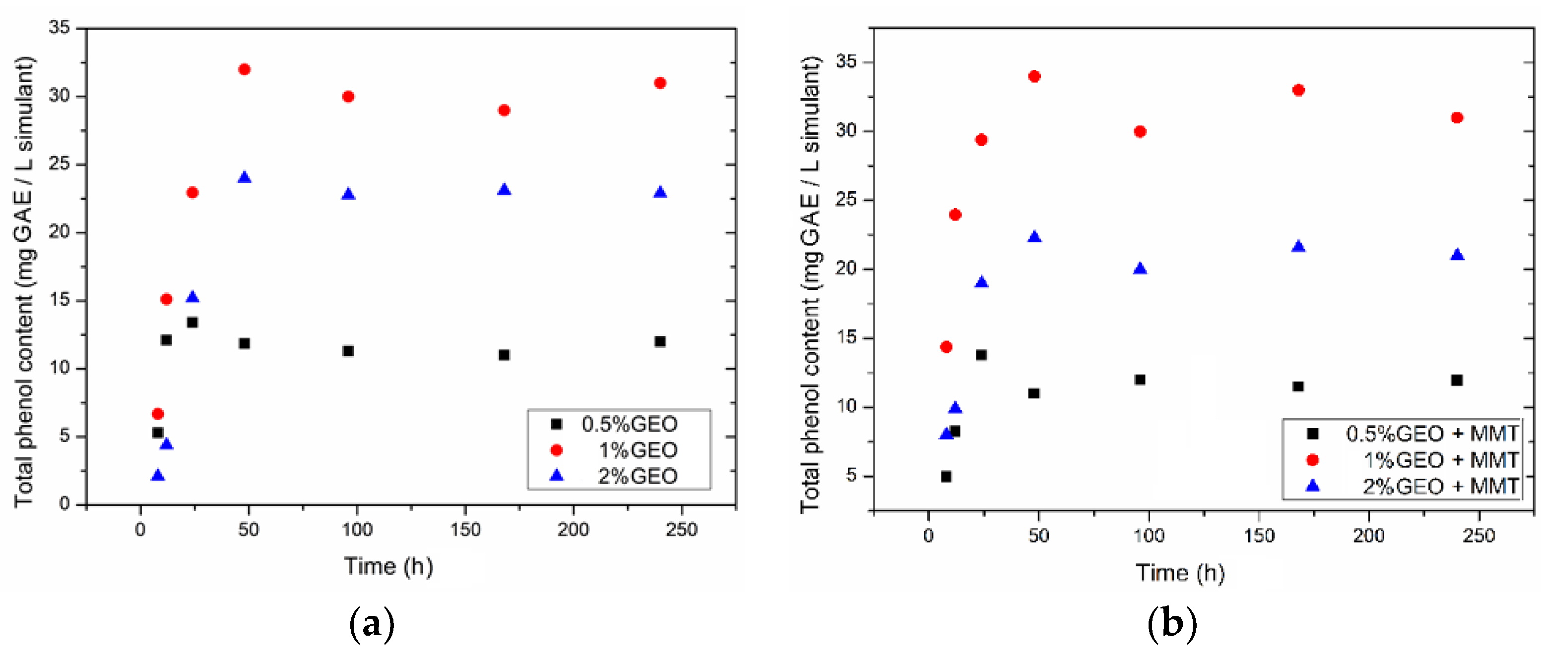
3.1.1. Migration Assay

Diffusion coefficient of TPC, maximum TPC released ratio (maximum total phenol content quantified in the simulant/total phenol content incorporated into the chitosan film) and percentage of radical scavenging are shown in Table 1. The release of phenolic compound towards the 95% ethanol followed an “exponential growth to a maximum” migration pattern, reaching the equilibrium after 24–48 h of contact with the simulant (Figure 1). Similar behavior was reported in the migration of TPC from green tea extract incorporated in ethylene–vinyl alcohol copolymer (EVOH) films to 95% ethanol [42] or from coca extract incorporated in EVOH films to water [43]. The diffusion coefficient did not statistically differ among the bionanocomposites tested (p > 0.05) except Chitosan + MMT + 1% GEO, which showed a significantly higher diffusion coefficient than the ones obtained with the two samples incorporated with 2% GEO (p < 0.05). Films with one percent of GEO and MMT presented the faster release of TPC towards the simulant media. It is also important to point out that the presence of the nanofiller accelerated the diffusion process when 0.5% and 1% of GEO was incorporated, while retarded with 2% GEO. In food systems, the big advantage of active packaging is the possibility to extend the protection time through the gradual release of the bioactive compounds [44]. Therefore, samples with the highest amount of GEO and MMT, from this in vitro migration assay, would be highlighted as the most promising film. However, in comparison with other diffusion coefficient values for similar EOs and films, this study findings are one order of magnitude higher than those obtained for chitosan films incorporated with rosemary essential oil [45], which suggests a smaller release time or lower retention of phenolic compounds from GEO inside chitosan film.
The phenolic compounds released to the simulant media kept some of their antioxidant activity; however, it was not correlated to the TPC present in the simulant, as it was expected and previously reported in the literature [32]. Abdollahi et al. [46] reported a good interaction between chitosan functional groups and rosemary essential oil, furthermore, in the presence of MMT these bonding were enhanced. Such interactions between chitosan × phenolic compounds, and chitosan × phenolic compounds × MMT may also have occurred with present films and can explain the existence of no correlation between TPC in the simulant and the antioxidant activity of compounds released to the simulant. In vitro studies are important to evaluate the activity of novel materials once these can predict the behavior in real situation. However, the food matrix is much more complex than the simulant media, thus in real scenarios the results may be different [32]. Therefore, it is important to also investigate in situ activity of the films.

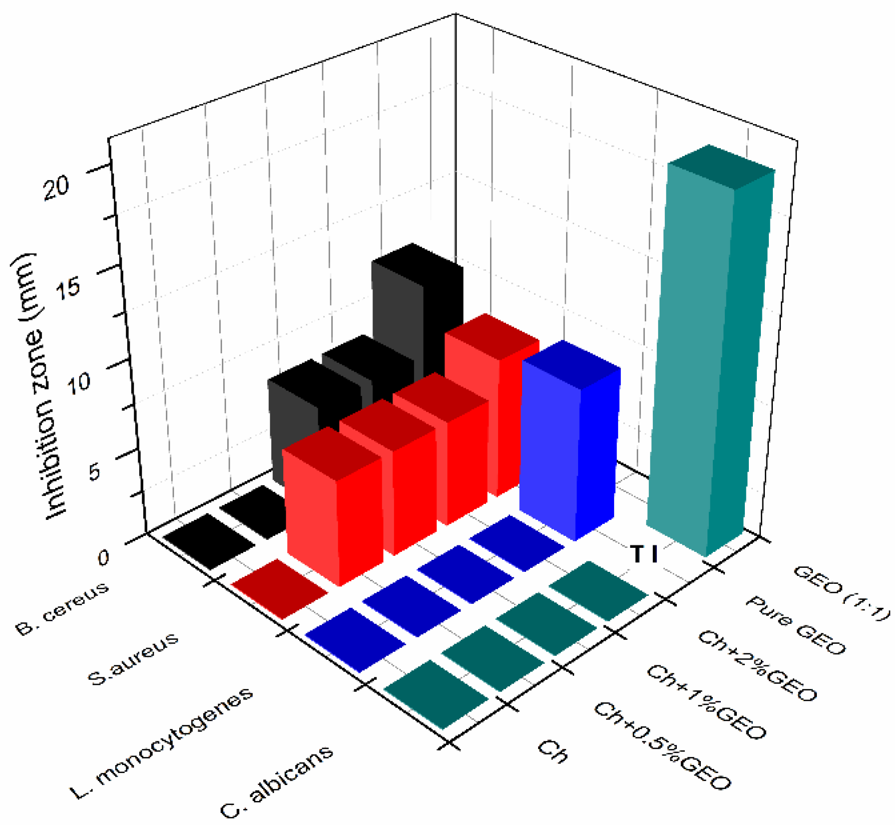
3.1.2. Antimicrobial Studies

Essential oils are well known for their good antimicrobial activity [47], and extensive research and reviews have focused on this matter [48,49,50,51]. In the present study, pure GEO only demonstrated antimicrobial activity against the Gram-positive bacteria (B. cereus, S. aureus and L. monocytogenes) (Figure 2), and no inhibition zone was observed for E. faecalis or any of the Gram-negative foodborne bacteria tested (data are not shown). Regarding the C. albicans, pure GEO led to the complete inhibition of the yeast growth, and when diluted (1:1) an inhibition zone of approximately 20 mm was recorded (Figure 2). This is in accordance with what was previously reported for two ginger essential oils extracted from the plant’s leaves or rhizomes, which in general exhibited better antibacterial activity against the Gram-positive bacteria than against the Gram-negative bacteria [52], probably due to the protection conferred by the lipopolysaccharide layer of the outer membrane of Gram-negative bacteria [53]. Singh et al. [54] found good antifungal activity (average 60% of mycelial growth) of GEO against several Aspergillus spp. and Fusarium moniliforme, which is in agreement with the strong antifungal activity against C. albicans.
The antimicrobial activity of EOs from spices and herbs are believed to be due to their rich content in phenolic compounds [47,54], and the higher susceptibility of Gram-positive bacteria compared to Gram negative bacteria suggests that the microbial targets of oil is the cell wall [24]. Singh et al. [54], Trajano et al. [55] and López et al. [24] also studied the antimicrobial activity of Zingiber officinale essential oil with contradictory conclusions. Trajano et al. [55] only found antibacterial activity of GEO against S. aureus, and no inhibition against L. monocytogens, E. coli, B. cereus, S. enterica, P. aeruginosa and Yersinia enterocolitica, while Singh et al. [54] observed good antimicrobial activity against E. coli, S. aureus, P. aeruginosa and Klebsiella pneumoniae, similar to what was observed by López et al. [24]. The differences from present results and the ones cited may be attributed to the characteristic composition of each GEO tested, once the type and concentration of phenolic compounds are directly correlated to the antimicrobial activity [52]. Also, the extraction procedure applied may have interfered in the final composition of the GEO and consequently on its biological activity.
The films, on the other hand, only presented activity against two of the bacteria tested: B. cereus and S. aureus, and only underneath the disk specimens of the samples incorporated with 1% and 2% of GEO, in the case of B. cereus, and with 0.5%, 1% and 2% of GEO against S. aureus (Figure 2). No activity was observed against Gram negative bacteria or the yeast tested.
Despite its well-known antimicrobial activity, pristine chitosan films did not present an inhibition zone for any of the microorganisms assessed. According to Hafsa et al. [56], the antimicrobial properties of chitosan may become negligible when the polysaccharide is in the form of insoluble films. Similar results were also reported with chitosan film incorporated with propolis extract against several Gram-positive and Gram-negative bacteria, where only the samples incorporated with propolis presented inhibition underneath the disk films and pristine chitosan film did not show antimicrobial activity [57].
Ginger essential oil when incorporated in the films had its antimicrobial activity reduced, probably due to a partial loss of volatile essential oil in the film by evaporation [58] and to the good interaction between the phenolic compounds and the films that entrapped the active compounds in the polymeric chain, lowering its diffusion towards the MHA, and therefore limiting its activity only underneath the disk films [57].
The antimicrobial results for the film in direct contact with the bacteria in the growth media were more promising (Table 1). The maximum mortality reached against Gram-positive (B. cereus) and Gram-negative (S. enterica) foodborne pathogens were respectively 7.3 ad 5.3 log reductions after 24 h contact for pristine chitosan film. Again, films were more efficient against Gram-positive bacteria than Gram negative bacteria, in line with what was previously discussed for the agar diffusion results. In comparison with the films incorporated with GEO and/or MMT, the mortality reduced either with incorporation of the active extracts or the nanoreinforcement (p < 0.05). Thus, the film’s antimicrobial activity is probably due to the intrinsic antimicrobial activity of chitosan, and not from the GEO or MMT added. Chitosan antimicrobial mechanism of action is related to its positive charges (NH3+) that would interfere with the negatively charged residues of macromolecules on the microbial cell surface, causing permeabilization and leakage of intracellular material resulting in cell death [9,59,60]. Montmorillonite and the phenolic compounds from the GEO may have interacted with the amide group of chitosan [60,61] and reduced the number of functional groups to interact with the membrane and display the antimicrobial action.

3.2. In Situ Active Characterization

3.2.1. Physicochemical Characterization

To evaluate the protector effect of the film on the shelf life of the poultry meat, some physicochemical parameters were evaluated through the 15 days of storage (Table 2). The natural deterioration process resulted in increased pH value, with reduction of the acidity, changes on the color (discoloration process) and increase of the moisture content, as can be observed for the meat without packaging (unwrapped). Such changes were less significant for the other samples: samples wrapped in the commercial PVC showed less change than unwrapped samples, but higher changes than samples wrapped with Ch, thus indicating that the biocomposites produced are promising substitutes to commercial and unsustainable plastic films. The incorporation of GEO in the chitosan matrix enhanced significantly the activity of the biopolymers once the samples wrapped in those films showed minimum changes (Table 2). The magnitude of the changes observed in samples wrapped in the biopolymers with MMT, with or without GEO, were in between samples wrapped with Ch and samples wrapped with Ch + GEO.
In terms of color, the meat was evaluated through the calculation of the hue angle (h*) (Table 2). This angle converts the CIE-L*a*b* coordinates a* and b* into angles representing color tonalities starting at 0° and with increasing values rotating counterclockwise: h* of 0° represents the color red, 90° yellow, 180° green and 270° blue [62]. Initial h* observed was 57°, i.e., reddish color, over time the hue angle increased, especially for the control meat and the sample wrapped with the commercial plastic (Table 2). Higher values of hue indicate that the meat color tended to a more yellowish tone, losing its reddish characteristic color. In comparison with the meat protected with the active films, control sample and the meat wrapped in PVC presented higher value of h* in the last day of assessment (p < 0.05). Between the active films incorporated with GEO, despite the amount of essential oil added, the hue angle did not change statistically (p > 0.05), and the meat showed h* values around 55°–60°. Chitosan film and Ch + MMT film also slowed down the discoloration process. Moreover, sample packaged in any of the chitosan films preserved the h* between the day zero and the day 15 (p > 0.05). Thus, the films maintained the original color of the meat, which is a key factor to boost the acceptance of such products by consumer, once color is a valued sensory attribute when accepting or rejecting foodstuff [63]. The decrease of the meat redness, mainly observed in the unwrapped samples and in the samples wrapped with PVC, is directly related to the lipid oxidation process and the degradation of heme molecules with formation of metmyoglobin [14,64,65]. Therefore, chitosan films succeeded in preventing the discoloration process. Two routes can explain the activity of the films: the antioxidant activity of the phenolic compounds that retarded the lipid oxidation together with the protection related to the metal chelator ability of chitosan. Present results are in line with the further TBARS and microbiological study discussed in Section 3.2.2 and Section 3.2.3.
Regarding the pH, control sample reached a pH value around 8.17 after 15 days of refrigerated storage (Table 2). Similar to the results of the color, pH changed less in the wrapped samples. At day 15, the smallest pH value was 5.71 recorded for the meat wrapped in Ch + 1%GEO. The other two concentrations of GEO also kept pH values low. In fact, for those three samples, the pH value between day zero and day 15 did not differed much, and except for Ch + 2% GEO, the change was not statistically significant (p > 0.05). For all the other films, the pH increased along 15 days of storage, and among the samples the value did not change statistically (p > 0.05). Thus, pH values of samples protected with PVC were the same as the pH of the samples protected with Ch, Ch + MMT, or Ch + MMT + GEO. The reduction of the activity of the active films with the incorporation of the MMT is probably due to the diminish migration of the phenolic compounds from GEO to the meat, therefore reducing the protection against microbial development and lipid oxidation, in line with the findings presented in the Section 3.2.2 and Section 3.2.3. The increase in pH values may be attributed to the growth of microorganisms that produce volatile basic components [14]; therefore, the maintenance of the pH indicates the efficiency of the films in the extension of the shelf life of the products. Similar results were reported in literature for chitosan films incorporated with rosemary essential oil applied in fish fillets [66] or for chitosan films incorporated with GEO applied in barracuda fish [67]. In agreement, titratable acidity reduced over the storage time (p < 0.05) (Table 2), which is in line with the increase of pH observed. The highest drop on the titratable acidity occurred for the unwrapped meat, while the samples protected showed less significant changes. Similar to the discussion for pH, the changes in this parameter are attributed to the growth of microorganisms that, through the degradation of the proteins, produce amines that neutralize the acids reducing the titratable acidity [14].
The moisture content of the meat packaged with the chitosan films showed a tendency to maintain or decrease over time (Table 2), especially the meat wrapped with films incorporated with GEO (and with no MMT) (p < 0.05). Such behavior may be attributed to the hydrophilic character of chitosan films that are able to absorb water and exhibit a high water vapor permeability [30], explaining the reduction in the water content of the meat packaged. In a previous work, poultry meat packaged in chitosan films incorporated with rosemary essential oil and montmorillonite also showed similar behavior [45], corroborating the present results.

3.2.2. Thiobarbituric Acid Reactive Substances Index

The oxidation status of the meat was assessed through the measurement of the TBARS index. Change in the secondary products of lipid oxidation was statistically different over time for all treatments (p < 0.05), except for the meat protected with chitosan film incorporated with GEO at the levels of 0.5%, 1% and 2% (p > 0.05) (Table 2). The presence of montmorillonite diminished the protection of the active films, probably due to the good interaction between the nanofiller and the polymer matrix and the phenolic compounds present in the GEO. Thus, the release of the bioactive compounds was diminished, resulting in a weaker protection effect, although a smaller oxidation if compared to the unprotected meat.
Together with the microbial growth, lipid oxidation is one of the most detrimental processes in foodstuffs [66] and the threshold of off-odor perception by consumers corresponds to a TBARS value of 0.5 mg MDA/kg sample [67]. Analyzing present results, up to the seventh day of refrigerated storage, only the meat unwrapped had exceeded this TBARS value threshold and, therefore, would be rejected by the consumers. At day 10 of storage, meat wrapped in PVC, chitosan + MMT, chitosan + MMT + 0.5% and 1% of GEO exceeded the limit. Finally, at the last day of assessment (15th day of storage), meat packaged with chitosan + MMT + 2% GEO overpassed the 0.5 mg MDA/kg meat, while the samples protected with the chitosan film and with chitosan + GEO (0.5%, 1% and 2%) still had not reached the rejection point. This result confirms the potential of the films to be used as preservatives for fresh poultry meat products, once pristine chitosan films and the biopolymers incorporated only with GEO (in all concentrations tested) extended the shelf life of the meat by at least eight days in terms of lipid oxidation.
The protection effect of the films may be attributed either to the good oxygen barrier and the chelator ability of the chitosan [66] and/or to the GEO through the ability of its phenolic compounds to donate electrons and stabilize free radicals acting as antioxidants [14]. Other biopolymers incorporated with different EOs have demonstrated similar results in food matrices [14,66,68,69].

3.2.3. Microbiological Growth

Changes in the total mesophilic aerobic bacteria and total coliforms of unwrapped and wrapped minced poultry meat are presented in Table 3. The meat presented initial TMAB of 5.1 log CFU/g meat, bellow the limit of 6.69 log CFU/g meat established in the Regulation (EC) No 2073/2005 for minced meat [70]. Over time the total mesophilic aerobic bacteria increased for all treatments (p < 0.05), reaching a final counting of 10.1 log CFU/g meat for unwrapped meat and a reduction varying between 1.2 and 2.6 log for the samples protected in the active films. Such results represent an increase in the shelf life time of the product and highlight the films as a tool to preserve the quality and safety of meat products. Moreover, the biopolymers reduced the microbiological growth more than the commercial one, which only showed a reduction of 0.5 log compared to unwrapped samples.
Fresh poultry meat is considered not acceptable for organoleptic (sensory) evaluation at counts greater than 7 log CFU/g [71]. In the present study, the unprotected meat and the samples wrapped in commercial film exceeded this counting since the third day of storage, while the samples protected with the active packaging were either in this limit or below of it. Ginger essential oil enhanced the protection effect of chitosan film, while MMT limited the activity of the films. As previously discussed, the presence of the nanofiller may interfere in the diffusion process of the active compounds and block the functional group of chitosan (amide group), reducing its antimicrobial action. Similar results were observed in fresh chicken breast fillets coated with sodium caseinate incorporated with a nanoemulsion of ginger essential oil [27]. The authors attributed the reduction in the microbial growth to the presence of GEO in the edible coating, and also observed smaller counting with the increase of EO concentration. The protective effect of the bio-based films produced can also be related to the good oxygen barrier properties of chitosan film, once reduced exposure of packaged meat products to high O2 concentration reduces the growth of aerobic microorganisms and oxidation of lipids and myoglobin [7].
The results for total coliforms were in line with the ones reported for TMAB (Table 3). Unwrapped meat presented the higher counting, followed by samples wrapped with PVC. Samples wrapped with the active films showed the lower counting. Again, it was observed that the presence of MMT reduced the antimicrobial activity of the chitosan film (without statistical significance, p > 0.05) and, the presence of GEO enhanced it (with statistical significance, p < 0.05). Although the differences between the total coliform counting for the meat protected with chitosan + 0.5%, 1% or 2% GEO were not statistically different (p > 0.05), a dose response could be observed once the smallest contamination at the 10th day of storage was found for the highest amount of GEO incorporated. Moreover, in comparison to the meat unwrapped or wrapped in PVC, the samples packaged in chitosan film + 2% GEO presented a reduction of 4.5 and 4.2 log in the total coliforms count, i.e., a good antimicrobial action. The phenols, monoterpenes, sesquiterpenes and aldehydes present in EO may be responsible for the results reported. The possible mechanism of action of these oxygenated bioactive compounds may be through the disruption and penetration in the lipid structure of the bacteria cell membrane with further damage of the microorganism enzyme systems [27]. Although GEO did not show antimicrobial activity against Gram-negative bacteria in the in vitro assay, its application in the film caused reduction in the coliform counting in the meat. This is probably due to a synergistic effect of the chitosan and GEO, as discussed by Khanjari, Karabagias and Kontominas [72], that reported lower microbial growth in the raw chicken fillet packaged with N,O-carboxymethyl chitosan incorporated with oregano essential oil, compared to samples packaged in pristine polymer or samples with added pure oregano essential oil. Moreover, the type of food matrix (high moisture content and presence of lipids) facilitated the diffusion of the GEO from the packaging enabling its active action.

4. Conclusions

The preparation of bio-based films demonstrated their potential to be used as primary packaging material to fresh poultry meat, being capable of retarding deterioration process by antimicrobial and antioxidant mechanisms and extending its shelf life time. Although the in vitro results were not as positive as expected, when in direct contact with the food tested, the films demonstrated good efficiency, highlighting the remarkable potential to extend the shelf life of poultry meat products. The incorporation of montmorillonite, at the level tested, diminished the bioactivity of the films produced, while the incorporation of GEO potentialized it. When no mechanical improvements are demanded, the use of MMT is unnecessary. In fact, chitosan film with one percent of GEO is a formula that optimizes the protection effect with an intermediate amount of essential oil, minimizing the negative effects of cost and flavor alterations related to the use of essential oils. Yet, future studies are needed to identify a balanced formula that can optimize the bioactivity of the bionanocomposites with the level of montmorillonite added, for applications that also require mechanical improvements. Further, more research studies with EOs or individual compounds, such as some phenolic compounds, combined with reinforcements, such as MMT, in biopolymers will help to clarify the molecular mechanisms associated with the entrapment/release of the bioactive compounds, helping to define future pathways for the production of bionanocomposites.

Author Contributions

V.G.L.S., A.L.F. and M.P.D. conceived and designed the experiments. V.G.L.S., J.R.A.P. and É.T. Vieira performed the experiments and analyzed the data. V.G.L.S. wrote the paper with contributions from the remaining authors. A.L.F., M.P.D. and I.M.C. supervised the execution of analyses and revised the data and the manuscript.

Funding

This work was supported by CNPq/Brazil [grant number 200790/2014-5]; and MEtRiCS through FCT/MCTES [UID/SEM/04077/2013]; This work was also supported by the Associate Laboratory for Green Chemistry LAQV which is financed by national funds from FCT/MCTES (UID/QUI/50006/2013) and co-financed by the ERDF under the PT2020 Partnership Agreement (POCI-01-0145-FEDER-007265).

Acknowledgments

The authors acknowledge BYK Additives & Instruments for donating the sodium montmorillonite (Cloisite®Na+).

Conflicts of Interest

The authors declare no conflict of interest. The founding sponsors had no role in the design of the study; in the collection, analyses, or interpretation of data; in the writing of the manuscript, and in the decision to publish the results.

References

  1. FAO. Poultry Development Review; FAO: Rome, Italy, 2013; ISBN 978-92-5-108067-2. [Google Scholar]
  2. OECD-FAO Agricultural Outlook 2016–2025; OECD Publishing: Paris, France, 2016; ISBN 9789264253223.
  3. Petrou, S.; Tsiraki, M.; Giatrakou, V.; Savvaidis, I.N. Chitosan dipping or oregano oil treatments, singly or combined on modified atmosphere packaged chicken breast meat. Int. J. Food Microbiol. 2012, 156, 264–271. [Google Scholar] [CrossRef] [PubMed]
  4. Latou, E.; Mexis, S.F.; Badeka, V.; Kontakos, S.; Kontominas, M.G. Combined effect of chitosan and modified atmosphere packaging for shelf life extension of chicken breast fillets. LWT Food Sci. Technol. 2014, 55, 263–268. [Google Scholar] [CrossRef]
  5. Arkoun, M.; Daigle, F.; Holley, R.A.; Heuzey, M.C.; Ajji, A. Chitosan-based nanofibers as bioactive meat packaging materials. Packag. Technol. Sci. 2018, 31, 185–195. [Google Scholar] [CrossRef]
  6. Souza, V.G.L.; Fernando, A.L. Nanoparticles in food packaging: Biodegradability and potential migration to food—A review. Food Packag. Shelf Life 2016, 8, 63–70. [Google Scholar] [CrossRef]
  7. Lee, K.T. Quality and safety aspects of meat products as affected by various physical manipulations of packaging materials. Meat Sci. 2010, 86, 138–150. [Google Scholar] [CrossRef] [PubMed]
  8. Prasad, P.; Kochhar, A. Active packaging in food industry: A Review. IOSR J. Environ. Sci. Toxicol. Food Technol. 2014, 8. [Google Scholar] [CrossRef]
  9. Dutta, P.K.; Tripathi, S.; Mehrotra, G.K.; Dutta, J. Perspectives for chitosan based antimicrobial films in food applications. Food Chem. 2009, 114, 1173–1182. [Google Scholar] [CrossRef]
  10. Otoni, C.G.; Espitia, P.J.P.; Avena-Bustillos, R.J.; McHugh, T.H. Trends in antimicrobial food packaging systems: Emitting sachets and absorbent pads. Food Res. Int. 2016, 83, 60–73. [Google Scholar] [CrossRef]
  11. Malhotra, B.; Keshwani, A.; Kharkwal, H. Antimicrobial food packaging: Potential and pitfalls. Front. Microbiol. 2015, 6, 611. [Google Scholar] [CrossRef] [PubMed]
  12. Yuan, G.; Chen, X.; Li, D. Chitosan films and coatings containing essential oils: The antioxidant and antimicrobial activity, and application in food systems. Food Res. Int. 2016, 89, 117–128. [Google Scholar] [CrossRef] [PubMed]
  13. Alboofetileh, M.; Rezaei, M.; Hosseini, H.; Abdollahi, M. Antimicrobial activity of alginate/clay nanocomposite films enriched with essential oils against three common foodborne pathogens. Food Control 2014, 36, 1–7. [Google Scholar] [CrossRef]
  14. De Melo, A.A.; Geraldine, R.M.; Silveira, M.F.; Torres, M.C.; e Rezende, C.S.; Silva, C.; Fernandes, T.H.; de Oliveira, A.N. Microbiological quality and other characteristics of refrigerated chicken meat in contact with cellulose acetate-based film incorporated with rosemary essential oil. Braz. J. Microbiol. 2012, 43, 1419–1427. [Google Scholar] [CrossRef] [PubMed]
  15. Zivanovic, S.; Chi, S.; Draughon, A. Antimicrobial activity of chitosan. Science 2005, 70, 45–51. [Google Scholar] [CrossRef]
  16. Darder, M.; Colilla, M.; Ruiz-Hitzky, E. Biopolymer-clay nanocomposites based on chitosan intercalated in montmorillonite. Chem. Mater. 2003, 15, 3774–3780. [Google Scholar] [CrossRef]
  17. Ferreira, A.R.V.; Torres, C.A.V.; Freitas, F.; Sevrin, C.; Grandfils, C.; Reis, M.A.M.; Alves, V.D.; Coelhoso, I.M. Development and characterization of bilayer films of FucoPol and chitosan. Carbohydr. Polym. 2016, 147, 8–15. [Google Scholar] [CrossRef] [PubMed]
  18. Srinivasa, P.C.; Ramesh, M.N.; Tharanathan, R.N. Effect of plasticizers and fatty acids on mechanical and permeability characteristics of chitosan films. Food Hydrocoll. 2007, 21, 1113–1122. [Google Scholar] [CrossRef]
  19. Azeredo, H.M.C.; Mattoso, L.H.C.; Avena-Bustillos, R.J.; Filho, G.C.; Munford, M.L.; Wood, D.; McHugh, T.H. Nanocellulose reinforced chitosan composite films as affected by nanofiller loading and plasticizer content. J. Food Sci. 2010, 75, N1–N7. [Google Scholar] [CrossRef] [PubMed]
  20. Mihindukulasuriya, S.D.F.; Lim, L.-T. Nanotechnology development in food packaging: A review. Trends Food Sci. Technol. 2014, 40, 149–167. [Google Scholar] [CrossRef]
  21. Souza, V.G.L.; Pires, J.R.A.; Rodrigues, P.F.; Lopes, A.A.S.; Fernandes, F.M.B.; Duarte, M.P.; Coelhoso, I.M.; Fernando, A.L. Bionanocomposites of chitosan/montmorillonite incorporated with Rosmarinus officinalis essential oil: Development and physical characterization. Food Packag. Shelf Life 2018, 16, 148–156. [Google Scholar] [CrossRef]
  22. Coelho, A.C.V.; Santos, P.D.S.; Santos, H.D.S. Argilas especiais: Argilas quimicamente modificadas-uma revisão. Quim. Nova 2007, 30, 1282–1294. [Google Scholar] [CrossRef]
  23. El-Ghorab, A.H.; Nauman, M.; Anjum, F.M.; Hussain, S.; Nadeem, M. A comparative study on chemical composition and antioxidant activity of ginger (Zingiber officinale) and cumin (Cuminum cyminum). J. Agric. Food Chem. 2010, 58, 8231–8237. [Google Scholar] [CrossRef] [PubMed]
  24. López, E.I.C.; Balcázar, M.F.H.; Mendoza, J.M.R.; Ortiz, A.D.R.; Melo, M.T.O.; Parrales, R.S.; Delgado, T.H. Antimicrobial activity of essential oil of Zingiber officinale Roscoe (Zingiberaceae). Am. J. Plant Sci. 2017, 8, 1511–1524. [Google Scholar] [CrossRef]
  25. Noshirvani, N.; Ghanbarzadeh, B.; Gardrat, C.; Rezaei, M.R.; Hashemi, M.; Le Coz, C.; Coma, V. Cinnamon and ginger essential oils to improve antifungal, physical and mechanical properties of chitosan-carboxymethyl cellulose films. Food Hydrocoll. 2017, 70, 36–45. [Google Scholar] [CrossRef]
  26. Zhang, L.; Liu, A.; Wang, W.; Ye, R.; Liu, Y.; Xiao, J.; Wang, K. Characterisation of microemulsion nanofilms based on Tilapia fish skin gelatine and ZnO nanoparticles incorporated with ginger essential oil: Meat packaging application. Int. J. Food Sci. Technol. 2017, 52, 1670–1679. [Google Scholar] [CrossRef]
  27. Noori, S.; Zeynali, F.; Almasi, H. Antimicrobial and antioxidant efficiency of nanoemulsion-based edible coating containing ginger (Zingiber officinale) essential oil and its effect on safety and quality attributes of chicken breast fillets. Food Control 2018, 84, 312–320. [Google Scholar] [CrossRef]
  28. Klangmuang, P.; Sothornvit, R. Active hydroxypropyl methylcellulose-based composite coating powder to maintain the quality of fresh mango. LWT Food Sci. Technol. 2018, 91, 541–548. [Google Scholar] [CrossRef]
  29. BYK. Cloisite®Na+ Technical Data Sheet. Available online: http://www.byk.com/en/additives/additives-by-name/cloisite-na.php (accessed on 5 February 2018).
  30. Souza, V.G.L.; Fernando, A.L.; Pires, J.R.A.; Rodrigues, P.F.; Lopes, A.A.S.S.; Fernandes, F.M.B. Physical properties of chitosan films incorporated with natural antioxidants. Ind. Crops Prod. 2017, 107, 565–572. [Google Scholar] [CrossRef]
  31. Dias, M.V.; Machado Azevedo, V.; Borges, S.V.; Soares, N.D.F.F.; de Barros Fernandes, R.V.; Marques, J.J.; Medeiros, É.A.A. Development of chitosan/montmorillonite nanocomposites with encapsulated α-tocopherol. Food Chem. 2014, 165, 323–329. [Google Scholar] [CrossRef] [PubMed]
  32. Souza, V.G.L.; Rodrigues, P.F.; Duarte, M.P.; Fernando, A.L. Antioxidant migration studies in chitosan films incorporated with plant extracts. J. Renew. Mater. 2018. [Google Scholar] [CrossRef]
  33. Singleton, V.L.; Orthofer, R.; Lamuela-Raventós, R.M. Analysis of total phenols and other oxidation substrates and antioxidants by means of Folin-ciocalteu. Methods Enzymol. 1999, 299, 152–178. [Google Scholar] [CrossRef]
  34. Brand-Williams, W.; Cuvelier, M.E.; Berset, C. Use of a free radical method to evaluate antioxidant activity. LWT Food Sci. Technol. 1995, 28, 25–30. [Google Scholar] [CrossRef]
  35. Chungy, D.; Papadakis, S.E.; Yamy, K.L. Simple models for assessing migration from food-packaging films. Food Addit. Contam. 2002, 19, 611–617. [Google Scholar] [CrossRef] [PubMed]
  36. Nouri, A.; Yaraki, M.T.; Ghorbanpour, M.; Agarwal, S.; Gupta, V.K. Enhanced antibacterial effect of chitosan film using montmorillonite/CuO nanocomposite. Int. J. Biol. Macromol. 2017, 109, 1219–1231. [Google Scholar] [CrossRef] [PubMed]
  37. ASTM E2149 Standard Test Methods for Determining the Antimicrobial Activity of Immobilized Antimicrobial Agents Under Dynamic Contact Conditions; ASTM International: West Conshohocken, PA, USA, 2001.
  38. AOAC. Official Methods of Analysis of the Association of Official Analytical Chemists, 20th ed.; AOAC International: Gaithersburg, MD, USA, 2016. [Google Scholar]
  39. Rosmini, M.R.; Perlo, F.; Pérez-Alvarez, J.A.; Pagán-Moreno, M.J.; Gago-Gago, A.; López-Santoveña, F.; Aranda-Catalá, V. TBA test by an extractive method applied to “paté”. Meat Sci. 1996, 42, 103–110. [Google Scholar] [CrossRef]
  40. ISO 4833-1:2013 Microbiology of the Food Chain—Horizontal Method for the Enumeration of Microorganisms—Part 1: Colony Count at 30 Degrees C by the Pour Plate Technique; International Organization for Standardization: Geneva, Switzerland, 2013.
  41. ISO 4831:2006(en) Microbiology of Food and Animal Feeding Stuffs—Horizontal Method for the Detection and Enumeration of Coliforms—Most Probable Number Technique; International Organization for Standardization: Geneva, Switzerland, 2006.
  42. Lopez de Dicastillo, C.; Nerin, C.; Alfaro, P.; Catala, R.; Gavara, R.; Hernandez-Munoz, P. Development of new antioxidant active packaging films based on ethylene vinyl alcohol copolymer (EVOH) and green tea extract. J. Agric. Food Chem. 2011, 59, 7832–7840. [Google Scholar] [CrossRef] [PubMed]
  43. Calatayud, M.; López-de-Dicastillo, C.; López-Carballo, G.; Vélez, D.; Hernández Muñoz, P.; Gavara, R. Active films based on cocoa extract with antioxidant, antimicrobial and biological applications. Food Chem. 2013, 139, 51–58. [Google Scholar] [CrossRef] [PubMed]
  44. Bolumar, T.; Andersen, M.L.; Orlien, V. Antioxidant active packaging for chicken meat processed by high pressure treatment. Food Chem. 2011, 129, 1406–1412. [Google Scholar] [CrossRef]
  45. Souza, V.G.L.; Pires, J.R.A.; Vieira, E.T.; Coelhoso, I.M.; Duarte, M.P.; Fernando, A.L. Activity of chitosan-montmorillonite bionanocomposites incorporated with rosemary essential oil: From in vitro assays to aplication in fresh poultry meat (unpublished, manuscript in preparation).
  46. Abdollahi, M.; Rezaei, M.; Farzi, G. A novel active bionanocomposite film incorporating rosemary essential oil and nanoclay into chitosan. J. Food Eng. 2012, 111, 343–350. [Google Scholar] [CrossRef]
  47. Ribeiro-Santos, R.; Andrade, M.; de Melo, N.R.; Sanches-Silva, A. Use of essential oils in active food packaging: Recent advances and future trends. Trends Food Sci. Technol. 2017, 61, 132–140. [Google Scholar] [CrossRef]
  48. Regnier, T.; Combrinck, S.; Du Plooy, W. Essential oils and other plant extracts as food preservatives. In Progress in Food Preservation; Bhat, R., Alias, A.K., Paliyath, G., Eds.; Wiley-Blackwell: Oxford, UK, 2012; pp. 539–579. [Google Scholar]
  49. Teixeira, B.; Marques, A.; Ramos, C.; Neng, N.R.; Nogueira, J.M.F.; Saraiva, J.A.; Nunes, M.L. Chemical composition and antibacterial and antioxidant properties of commercial essential oils. Ind. Crops Prod. 2013, 43, 587–595. [Google Scholar] [CrossRef]
  50. Singh, P.; Singh, S.; Kapoor, I.P.S.; Singh, G.; Isidorov, V.; Szczepaniak, L. Chemical composition and antioxidant activities of essential oil and oleoresins from Curcuma zedoaria rhizomes, part-74. Food Biosci. 2013, 3, 42–48. [Google Scholar] [CrossRef]
  51. López, P.; Sánchez, C.; Batlle, R.; Nerín, C. Solid- and vapor-phase antimicrobial activities of six essential oils: Susceptibility of selected foodborne bacterial and fungal strains. J. Agric. Food Chem. 2005, 53, 6939–6946. [Google Scholar] [CrossRef] [PubMed]
  52. Sivasothy, Y.; Chong, W.K.; Hamid, A.; Eldeen, I.M.; Sulaiman, S.F.; Awang, K. Essential oils of Zingiber officinale var. rubrum Theilade and their antibacterial activities. Food Chem. 2011, 124, 514–517. [Google Scholar] [CrossRef]
  53. Shariatinia, Z.; Fazli, M. Mechanical properties and antibacterial activities of novel nanobiocomposite films of chitosan and starch. Food Hydrocoll. 2015, 46, 112–124. [Google Scholar] [CrossRef]
  54. Singh, G.; Kapoor, I.P.S.; Singh, P.; de Heluani, C.S.; de Lampasona, M.P.; Catalan, C.A.N. Chemistry, antioxidant and antimicrobial investigations on essential oil and oleoresins of Zingiber officinale. Food Chem. Toxicol. 2008, 46, 3295–3302. [Google Scholar] [CrossRef] [PubMed]
  55. Trajano, V.N.; Lima, O.; Leite, E.; Travassos, R. Propriedade antibacteriana de óleos essenciais de especiarias sobre bactérias contaminantes de alimentos. 2009, 29, 542–545. [Google Scholar] [CrossRef]
  56. Hafsa, J.; Smach, M.; Ben Khedher, M.R.; Charfeddine, B.; Limem, K.; Majdoub, H.; Rouatbi, S. Physical, antioxidant and antimicrobial properties of chitosan films containing Eucalyptus globulus essential oil. LWT Food Sci. Technol. 2016, 68, 356–364. [Google Scholar] [CrossRef]
  57. Siripatrawan, U.; Vitchayakitti, W. Improving functional properties of chitosan films as active food packaging by incorporating with propolis. Food Hydrocoll. 2016, 61, 695–702. [Google Scholar] [CrossRef]
  58. Abdollahi, M.; Rezaei, M.; Farzi, G. Original article Improvement of active chitosan film properties with rosemary essential oil for food packaging. Food Sci. Technol. 2012, 47, 847–853. [Google Scholar] [CrossRef]
  59. Verlee, A.; Mincke, S.; Stevens, C.V. Recent developments in antibacterial and antifungal chitosan and its derivatives. Carbohydr. Polym. 2017, 164, 268–283. [Google Scholar] [CrossRef] [PubMed]
  60. Siripatrawan, U.; Harte, B.R. Physical properties and antioxidant activity of an active film from chitosan incorporated with green tea extract. Food Hydrocoll. 2010, 24, 770–775. [Google Scholar] [CrossRef]
  61. Lavorgna, M.; Piscitelli, F.; Mangiacapra, P.; Buonocore, G.G. Study of the combined effect of both clay and glycerol plasticizer on the properties of chitosan films. Carbohydr. Polym. 2010, 82, 291–298. [Google Scholar] [CrossRef]
  62. MINOLTA. Precise Color Communication: Color Control from Perception to Instrumentation; Minolta Co., Ltd.: Osaka, Japan, 2007. [Google Scholar]
  63. Rojas, M.C.; Brewer, M.S. Effect of natural antioxidants on oxidative stability of frozen, vacuum-packaged beef and pork. J. Food Qual. 2008, 31, 173–188. [Google Scholar] [CrossRef]
  64. Contini, C.; Álvarez, R.; O’Sullivan, M.; Dowling, D.P.; Gargan, S.Ó.; Monahan, F.J. Effect of an active packaging with citrus extract on lipid oxidation and sensory quality of cooked turkey meat. Meat Sci. 2014, 96, 1171–1176. [Google Scholar] [CrossRef] [PubMed]
  65. Ghaderi-Ghahfarokhi, M.; Barzegar, M.; Sahari, M.A.; Ahmadi Gavlighi, H.; Gardini, F. Chitosan-cinnamon essential oil nano-formulation: Application as a novel additive for controlled release and shelf life extension of beef patties. Int. J. Biol. Macromol. 2017, 102, 19–28. [Google Scholar] [CrossRef] [PubMed]
  66. Abdollahi, M.; Rezaei, M.; Farzi, G. Influence of chitosan/clay functional bionanocomposite activated with rosemary essential oil on the shelf life of fresh silver carp. Int. J. Food Sci. Technol. 2014, 49, 811–818. [Google Scholar] [CrossRef]
  67. Remya, S.; Mohan, C.O.; Bindu, J.; Sivaraman, G.K.; Venkateshwarlu, G.; Ravishankar, C.N. Effect of chitosan based active packaging film on the keeping quality of chilled stored barracuda fish. J. Food Sci. Technol. 2016, 53, 685–693. [Google Scholar] [CrossRef] [PubMed]
  68. Yang, F.; Hu, S.; Lu, Y.; Yang, H.; Zhao, Y.; Li, L. Effects of coatings of polyethyleneimine and thyme essential oil combined with chitosan on sliced fresh channa argus during refrigerated storage. J. Food Process Eng. 2015, 38, 225–233. [Google Scholar] [CrossRef]
  69. Atarés, L.; Pérez-Masiá, R.; Chiralt, A. The role of some antioxidants in the HPMC film properties and lipid protection in coated toasted almonds. J. Food Eng. 2011, 104, 649–656. [Google Scholar] [CrossRef]
  70. European Commission. Commmission Regulation (EC) No 2073/2005 of 15 November 2005 on microbiological criteria for foodstuffs. Off. J. Eur. Union 2005, L338, 1–19. Available online: https://www.fsai.ie/uploadedFiles/Reg2073_2005%281%29.pdf (accessed on 5 February 2018).
  71. Economou, T.; Pournis, N.; Ntzimani, A.; Savvaidis, I.N. Nisin-EDTA treatments and modified atmosphere packaging to increase fresh chicken meat shelf-life. Food Chem. 2009, 114, 1470–1476. [Google Scholar] [CrossRef]
  72. Khanjari, A.; Karabagias, I.K.; Kontominas, M.G. Combined effect of N,O-carboxymethyl chitosan and oregano essential oil to extend shelf life and control Listeria monocytogenes in raw chicken meat fillets. LWT Food Sci. Technol. 2013, 53, 94–99. [Google Scholar] [CrossRef]
Figure 1. Migration assay–total phenol content diffused from films to simulant over time: (a) Films incorporated with GEO in different concentrations; (b) Films incorporated with MMTNa and GEO in different concentrations. GEO (ginger essential oil), MMT (sodium montmorillonite), GAE (gallic acid equivalent).
Coatings 08 00177 g001
Figure 2. Antimicrobial activity of bionanocomposites and GEO. Chitosan (Ch); Ginger essential oil (GEO); Total inhibition (TI).
Coatings 08 00177 g002
Table 1. In vitro study: migration assay and antibacterial activity.
FilmAntibacterial Activity (Log Reduction)Diffusion Coefficient of TPC (10−11 m2/s)Maximum GEO Diffused/Total Incorporated into FilmsMaximum % Radical Scavenging (DPPH)
B. cereusS. enterica
Ch7.3 ± 0.1 A5.3 ± 0.6 ANo migration
Ch + MMT6.7 ± 1 A3.5 ± 0.7 BNo migration
Ch + 0.5% GEO3.4 ± 0.2 B3.8 ±0.1 B2.06 ± 0.2 A,B0.716.3
Ch + MMT + 0.5% GEO2.3 ± 0.1 B3.2 ±1.0 B3.77 ± 0.48 A,B0.8240.6
Ch + 1% GEO1.5 ± 0.3 B3.8 ± 0.4 B3.15 ± 0.29 A,B0.6810.5
Ch + MMT + 1% GEO3.6 ± 0.8 B3.2 ± 0.3 B11.4 ± 0.63 A1.08.32
Ch + 2% GEO3.6 ± 0.3 B3.6 ± 0.3 B1.24 ± 0.43 B0.3718.9
Ch + MMT + 2% GEO5.3 ± 0.0 A2.3 ± 0.2 B0.13 ± 0.01 B0.316.05
A,B: Different superscripts within the same column indicate statistically significant differences among samples (p < 0.05). Ch—chitosan; MMT—sodium montmorillonite; GEO—ginger essential oil; TPC—total phenol content.
Table 2. Thiobarbituric acid reactive substances (TBARS) and physicochemical evaluation of meat packaged though shelf life time.
ParameterDayUnwrappedPVCChCh + 0.5% GEOCh + 1% GEOCh + 2% GEOCh + MMTCh + MMT + 0.5% GEOCh + MMT + 1% GEOCh + MMT + 2% GEO
TBARS (mg MDA/kg meat)00.11 ± 0.05 a,C0.11 ± 0.05 a,B0.11 ± 0.05 a,B0.11 ± 0.05 a,A0.11 ± 0.05 a,A0.11 ± 0.05 a,A,B0.11 ± 0.05 a,B0.11 ± 0.05 a,C0.11 ± 0.05 a,C0.11 ± 0.05 a,B
30.13 ± 0.06 b,C0.11 ± 0.01 b,B0.19 ± 0.06 b,B0.16 ± 0.02 b,A0.07 ± 0.0 b,B0.10 ± 0.06 b,A,B0.34 ± 0.02 a,B0.15 ± 0.01 b,C0.15 ± 0.03 b,C0.18 ± 0.09 b,B
70.88 ± 0.20 a,B0.07 ± 0.01 c,B0.12 ± 0.02 c,B0.19 ± 0.10 c,A0.05 ± 0.02 c,B0.06 ± 0.02 c,A,B0.49 ± 0.03 b,A,B0.17 ± 0.04 c,C0.16 ± 0.02 c,C0.13 ± 0.02 c,B
101.48 ± 0.33 a,A0.65 ± 0.25 b,c,A0.23 ± 0.06 d,e,A,B0.20 ± 0.02 d,e,A0.04 ± 0.01 e,B0.02 ± 0.01 e,B0.58 ± 0.04 b,c,d,A,B0.78 ± 0.00 b,B0.57 ± 0.00 b,c,d,B0.28 ± 0.03 c,d,e,B
151.97 ± 0.28 a,A0.65 ± 0.02 b,c,d,A0.42 ± 0.12 c,d,A0.25 ± 0.0 c,d,A0.20 ± 0.04 d,A0.15 ± 0.06 d,A1.11 ± 0.54 b,A1.09 ± 0.20 b,A0.83 ± 0.12 b,c,A0.51 ± 0.12 b,c,d,A
Hue (°)057 ± 1 a,E57 ± 1 a,C57 ± 1 a,A57 ± 1 a,B57 ± 1 a,A57 ± 1 a,A57 ± 1 a,A57 ± 1 a,A57 ± 1 a,B57 ± 1 a,B,C
362 ± 0 a,b,D61 ± 1 a,b,c,B59 ± 2 b,c,d,A56 ± 1 d,e,B52 ± 1 e,A53 ± 1 e,B62 ± 0 a,b,A60 ± 2 a,b,c,A58 ± 1 c,d,B62 ± 0 a,A
764 ± 0 a,b,C67 ± 0 a,A59 ± 1 b,c,A56 ± 1 c,d,B57 ± 4 c,d,A57 ± 2 c,d,A62 ± 3 b,c,A59 ± 1 b,c,A64 ± 0 a,b,A53 ± 2 d,C
1067 ± 0 a,B66 ± 1 a,A59 ± 1 c,A64 ± 1 a,b,A58 ± 2 c,A60 ± 1 b,c,A61 ± 2 b,c,A59 ± 1 c,A60 ± 0 b,c,B61 ± 2 b,c,A,B
1570 ± 1 a,A67 ± 1 a,b,A60 ± 2 c,d,A55 ± 0 e,B55 ± 2 e,A57 ± 1 d,e,A62 ± 0 b,c,A60 ± 0 c,d,A57 ± 4 c,d,e,B60 ± 3 c,d,e,A,B
pH05.87 ± 0.11 a,C5.96 ± 0.15 a,C5.87 ± 0.11 a,D5.72 ± 0.02 a,A,B5.71 ± 0.21 a,A5.71 ± 0.21 a,B,C5.96 ± 0.15 a,B5.96 ± 0.15 a,A5.96 ± 0.15 a,B5.96 ± 0.15 a,B
36.09 ± 0.35 a,C6.19 ± 0.08 a,C6.01 ± 0.20 a,C,D5.57 ± 0.07 b,B5.38 ± 0.02 b,B5.45 ± 0.05 b,C6.01 ± 0.00 a,B6.12 ± 0.05 a,A6.03 ± 0.02 a,B5.96 ± 0.09 a,b,B
77.16 ± 0.30 a,B6.50 ± 0.12 b,B6.33 ± 0.02 b,c,B,C5.62 ± 0.01 f,A,B5.74 ± 0.04 e,f,A5.76 ± 0.10 e,f,B5.84 ± 0.02 d,f,B6.02 ± 0.04 c,d,e,A6.10 ± 0.04 c,d,B5.94 ± 0.08 d,e,f,B
107.55 ± 0.02 a,B6.79 ± 0.04 b,B6.51 ± 0.21 b,c,B5.58 ± 0.02 e,B5.60 ± 0.03 e,A,B5.52 ± 0.04 e,B,C6.01 ± 0.04 d,B6.14 ± 0.20 d,A6.29 ± 0.07 c,d,B6.00 ± 0.20 d,B
158.17 ± 0.14 a,A7.26 ± 0.13 b,c,A7.14 ± 0.23 b,c,A5.85 ± 0.2 d,e,A5.71 ± 0.09 e,A6.22 ± 0.01 d,e,A7.27 ± 0.36 b,c,A6.50 ± 0.55 c,d,e,A7.33 ± 0.36 b,A6.61 ± 0.24 b,c,d,A
Acidity (% oleic acid equivalent)01.80 ± 0.39 a,A1.68 ± 0.22 a,A1.80 ± 0.39 a,A2.12 ± 0.11 a,A2.12 ± 0.11 a,A2.12 ± 0.11 a,A1.68 ± 0.22 a,A1.68 ± 0.22 a,A1.68 ± 0.22 a,A1.68 ± 0.22 a,A
31.24 ± 0.37 b,A,B1.32 ± 0.08 b,B1.13 ± 0.01 b,B1.70 ± 0.02 a,B1.75 ± 0.08 a,B1.75 ± 0.03 a,B1.19 ± 0.17 b,B1.04 ± 0.19 b,B1.04 ± 0.09 b,C,D1.26 ± 0.05 b,A,B
70.42 ± 0.14 f,C1.27 ± 0.07 c,d,B0.85 ± 0.05 e,B1.47 ± 0.01 a,b,B,C1.55 ± 0.04 a,C1.41 ± 0.03 a,b,c,C1.49 ± 0.00 a,b,A,B1.22 ± 0.00 c,d,A,B1.33 ± 0.13 b,c,d,B,C1.16 ± 0.02 d,B
100.73 ± 0.15 c,B,C1.15 ± 0.06 b,B,C1.13 ± 0.18 b,c,B1.30 ± 0.20 a,b,C1.46 ± 0.09 a,b,C1.36 ± 0.04 a,b,C1.46 ± 0.10 a,b,A,B1.58 ± 0.23 a,A,B1.52 ± 0.06 a,b,A,B1.65 ± 0.15 a,A
150.66 ± 0.14 b,B,C0.87 ± 0.02 a,b,C1.22 ± 0.30 a,A,B0.73 ± 0.11 a,b,D1.22 ± 0.03 a,D1.11 ± 0.08 a,b,D0.78 ± 0.15 a,b,C1.24 ± 0.34 a,A,B0.76 ± 0.09 a,b,D1.13 ± 0.29 a,b,B
Water content (%)075.5 ± 0.2 a,B75.5 ± 0.2 a,B75.5 ± 0.2 a,A75.5 ± 0.2 a,A75.5 ± 0.2 a,A75.5 ± 0.2 a,A75.5 ± 0.2 a,A75.5 ± 0.2 a,A75.5 ± 0.2 a,A75.5 ± 0.2 a,A
375.4 ± 0.2 a,b,B75.8 ± 0.4 a,B73.4 ± 0.1 c,B,C74.7 ± 0.4 b,A73.3 ± 0.0 c,A,B73.6 ± 0.2 c,B73.8 ± 0.2 c,B73.1 ± 0.4 c,B73.4 ± 0.2 c,B73.0 ± 0.2 c,C
776.0 ± 0.2 a,A,B75.1 ± 0.4 a,B72.5 ± 0.1 b,C73.2 ± 0.3 b,B72.6 ± 0.6 b,B72.8 ± 0.5 b,B,C73.2 ± 0.3 b,B73.1 ± 0.3 b,B72.8 ± 0.0 b,B72.9 ± 0.7 b,C
1076.2 ± 0.4 a,A,B75.6 ± 0.3 a,b,B74.5 ± 0.3 b,c,A,B74.4 ± 0.5 c,A,B72.0 ± 0.1 d,B70.7 ± 0.7 e,C,D73.6 ± 0.2 c,B74.1 ± 0.2 c,B73.4 ± 0.6 c,B74.1 ± 0.2 c,B
1577.3 ± 1.0 a,A76.7 ± 0.1 a,b,A73.4 ± 0.8 c,d,e,B,C71.6 ± 0.8 e,C72.0 ± 2.0 d,e,B71.3 ± 0.4 e,D75.9 ± 0.6 a,b,c,A74.2 ± 0.9 b,c,d,B74.8 ± 0.2 a,b,c,A73.6 ± 0.2 c,d,e,B,C
A–D: Within each parameter, mean values in the same column not sharing upper case superscript letters indicate statistically significant differences among samples (p < 0.05); a–f: Within each parameter, mean values in the same line not sharing lower case superscript letters indicate statistically significant differences among samples (p < 0.05). Thiobarbituric acid reactive substances (TBARS), Malonaldehyde (MDA), Chitosan (Ch), Sodium Montmorillonite (MMT), Ginger Essential Oil (GEO).
Table 3. Microbiological study of meat packaged though shelf life time.
ParameterDayUnwrappedPVCChCh + 0.5% GEOCh + 1% GEOCh + 2% GEOCh + MMTCh + MMT + 0.5% GEOCh + MMT + 1% GEOCh + MMT + 2% GEO
TMAB (log CFU/g meat)05.1 ± 0.1 a,C5.1 ± 0.1 a,C5.1 ± 0.1 a,C5.1 ± 0.1 a,C5.1 ± 0.1 a,B5.1 ± 0.1 a,B5.1 ± 0.1 a,C5.1 ± 0.1 a,C5.1 ± 0.1 a,B5.1 ± 0.1 a,B
38.8 ± 0.1 a,B8.7 ± 0.0 a,B7.1 ± 0.3 b,B7.1 ± 0.4 b,B6.7 ± 0.2 b,A6.8 ± 0.2 b,A6.7 ± 0.0 b,B7.1 ± 0.3 b,B5.3 ± 0.2 c,B5.9 ± 0.0 c,B
710.2 ± 0.1 a,A9.5 ± 0.3 a,b,A8.5 ± 0.2 c,d,A7.4 ± 0.0 e,f,B7.8 ± 0.1 d,f,A8.1 ± 0.1 c,d,e,A7.2 ± 0.7 f,A,B8.7 ± 0.1 b,c,A8.7 ± 0.1 c,A8.3 ± 0.2 c,d,A
1010.1 ± 0.0 a,A9.6 ± 0.1 a,b,A8.7 ± 0.5 c,A8.3 ± 0.2 c,d,A7.5 ± 0.1 e,A7.7 ± 0.5 d,e,A8.5 ± 0.0 c,A8.8 ± 0.0 c,A8.9 ± 0.1 b,c,A8.8 ± 0.0 c,A
Total coliforms (log MPN/g meat)00.8 ± 0.4 a,C0.8 ± 0.4 a,C0.8 ± 0.4 a,C0.8 ± 0.4 a,B0.8 ± 0.4 a,C0.8 ± 0.4 a,B0.8 ± 0.4 a,C0.8 ± 0.4 a,C0.8 ± 0.4 a,D0.8 ± 0.4 a,C
33.0 ± 0.4 b,c,B,C3.4 ± 0.0 a,b,B2.8 ± 0.5 b,c,B3.2 ± 0.2 a,b,c,A4.0 ± 0.0 a,A2.8 ± 0.4 b,c,A2.9 ± 0.5 b,c,B2.9 ± 0.5 b,c,B2.2 ± 0.2 c,C2.2 ± 0.2 c,B
74.5 ± 1.7 a,A,B3.9 ± 0.5 a,b,c,B4.0 ± 0.9 a,b,A,B3.4 ± 0.3 a,b,c,A2.3 ± 0.00 b,c,B2.1 ± 0.1 c,A3.0 ± 0.3 a,b,c,B3.6 ± 0.4 a,b,c,B3.7 ± 0.4 a,b,c,B2.4 ± 0.1 b,c,B
106.7 ± 0.6 a,A6.4 ± 0.0 a,A5.3 ± 1.0 a,A3.2 ± 0.0 b,A2.6 ± 0.6 b,B2.2 ± 0.8 b,A5.9 ± 0.2 a,A6.2 ± 0.2 a,A5.9 ± 0.5 a,A5.9 ± 0.5 a,A
A–D: Within each parameter, mean values in the same column not sharing upper case superscript letters indicate statistically significant differences among samples (p < 0.05); a–f: Within each parameter, mean values in the same line not sharing lower case superscript letters indicate statistically significant differences among samples (p < 0.05). Total mesophilic aerobic bacteria (TMAB), Colony forming units (CFU), Chitosan (Ch), Sodium Montmorillonite (MMT), Most probable number (MPN) Ginger Essential Oil (GEO).

