Research Progress on Biomedical Functional Coatings for Titanium Alloys: A Review

 ,
,
Highlights
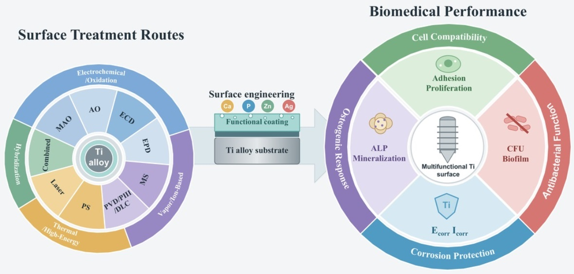
- Nine surface treatments are compared for preparing biomedical coatings on titanium alloys.
- The properties of the coating are affected by its composition, structure, and ion release.
- Ca/P rich coatings promote osteogenesis; Mn/Zn/Cu/Ag rich coatings suppress bacteria.
- Surface treatments should match the intended implant function.
- Ion-release control balances antibacterial effects and cytocompatibility.
- Standardized corrosion, cell, and biofilm tests enable comparisons.
Abstract
1. Introduction
2. Literature Search and Analysis Methods
3. Fabrication Technologies for Biomedical Functional Coatings on Titanium Alloys

| Surface Treatment | Matrix | Reported Antibacterial Outcome | Corrosion Outcome: Icorr (A·cm−2) | Ref. | ||
|---|---|---|---|---|---|---|
| Test Medium | Matrix/Control | After Treatment | ||||
| MAO | CP-Ti | 70.8% (S. aureus) | 0.9 wt% NaCl | 1.63 × 10⁻⁷ | 2.05 × 10⁻⁸ | [56] |
| Ti-6Al-4V | — | 0.9 wt% NaCl | 7.28 × 10⁻⁸ (MAO) | 1.91 × 10⁻⁸ | [57] | |
| AO | Ti-29Nb-13Ta-7.1Zr | — | PBS, pH 7.4 | 7.474 × 10⁻⁶ | 1.40 × 10⁻⁷ | [58] |
| NiTi | >95% at day 1 | PBS | 1.66 × 10⁻⁷ | 6.90 × 10⁻⁹ | [59] | |
| MS | Ti-6Al-4V | — | SBF | 6.79 × 10⁻⁹ | 2.92 × 10⁻⁹ | [60] |
| Ti-6Al-4V | — | 0.5 M NaCl + 2 g·L−1 KF, pH 2 | 2.6 × 10⁻⁸ (TiO2(a)) | 6.5 × 10⁻⁸ (TiO2(a)/graphene) | [61] | |
| Ti-6Al-4V | 30%/40% damaged S. aureus, 2/4 h | — | — | — | [62] | |
| ECD | Ti-6Al-4V ELI | — | HBSS | 1.62 × 10⁻⁶ (0.01 M) | 4.91 × 10⁻⁷ | [63] |
| Porous Ti | — | PBS | 1.83 × 10⁻⁷ | 9.10 × 10⁻⁸ | [64] | |
| EPD | Ti-6Al-4V | E. coli: 15 × 108 CFU·mL−1; S. aureus: 2.2 × 10⁸ CFU·mL−1 | — | — | — | [65] |
| PIII | Ti-6Al-4V | 25.58% (E. coli); 38.20% (B. subtilis) | SBF | 9.07 × 10⁻⁶ | 3.21 × 10⁻⁸ | [66] |
| Ti-6Al-4V | Reduced S. salivarius adhesion | Artificial saliva/SBF | 2.10 × 10⁻⁷/1.30 × 10−7 | 3.00 × 10⁻⁸/2.00 × 10−8 | [67] | |
| NiTi | — | Hanks’ solution | 3.20 × 10⁻⁹ | 2.50 × 10⁻⁹ | [68] | |
| Laser processing | Ti-6Al-4V | — | 3.5% NaCl/Hanks/DMEM | 1.37 × 10⁻⁷/5.72 × 10−8/3.44 × 10−8 | 1.15 × 10⁻⁷/5.71 × 10−8/2.61 × 10−8 | [69] |
| Ti-6Al-4V | — | SBF | 2.459 × 10⁻⁵ | 1.051 × 10⁻⁷ | [70] | |
| Other: ESD | Ti-6Al-4V | — | 3.5% NaCl | 2.64 × 10⁻⁶ | 2.60 × 10⁻⁷ | [71] |
3.1. Micro-Arc Oxidation
3.2. Anodic Oxidation
3.3. Magnetron Sputtering
3.4. Electrochemical Deposition and Related Surface Construction
3.5. Electrophoretic Deposition
3.6. Plasma-Sprayed and Thick Ceramic/Metal Bioactive Coatings
3.7. Physical Vapor Deposition, Plasma Immersion Ion Implantation, Diamond-like Carbon and Near-Surface Modification
3.7.1. Physical Vapor Deposition
3.7.2. Plasma Immersion Ion Implantation
3.7.3. Diamond-like Carbon and Related Near-Surface Modification
3.8. Laser Cladding, Laser Deposition and High-Energy-Beam Surface Processing
3.9. Other Combined Surface-Functionalization and Boundary Methods
3.10. Cross-Technology Comparison, Evidence Limits and Clinical Translation
4. Conclusions
- (1)
- Oxide, calcium-phosphate, metallic, nitride, carbon and composite-ceramic layers can improve the stability of titanium implant interfaces. Their value cannot be established from thickness, pore size or morphology alone. Corrosion, antibacterial activity, cell proliferation and osteogenic response must be measured directly.
- (2)
- No fabrication route is universally superior. MAO and EPD are effective for incorporating Ca/P, Mn, Sr, HAp and other functional constituents. MS and PVD provide dense TiO2, TiN, Ta/Ta2O5 and multielement films. PS and laser cladding are better suited to thicker ceramic or composite layers. Ion implantation, DLC and ESD mainly strengthen near-surface barriers, wear resistance and tribocorrosion performance.
- (3)
- Functional claims should follow the endpoint measured. Ecorr, Icorr, Rp and EIS describe electrochemical behavior; CFU counts, antibacterial rates and strain information support antibacterial claims. CCK-8, MTT and OD measurements address proliferation or cytocompatibility, while ALP activity, mineralization and related tests support osteogenic interpretation. Stronger conclusions require agreement among these endpoints and longer-term models that include corrosion, bacterial challenge and tissue-relevant conditions.
5. Future Perspectives and Outlook
- (1)
- Current studies use different test media, bacterial strains, cell models and evaluation periods, making direct comparison between coating systems difficult. Standardized methods should therefore be established for corrosion current density, antibacterial activity, cell viability, alkaline phosphatase activity and mineralization. Dynamic experimental models that incorporate inflammatory conditions, bacterial challenge and mechanical loading are also needed to better reproduce the implant environment.
- (2)
- Antibacterial elements, such as Ag, Cu, Zn and Mn, can inhibit bacterial adhesion and biofilm formation, but excessive ion release may reduce cytocompatibility. Future coating design should focus on controlled release and suitable elemental concentrations. The balance between antibacterial activity, cell proliferation, osteogenic differentiation and corrosion stability should be evaluated within the same experimental system.
- (3)
- Pores, cracks, residual stress and weak interfacial bonding may cause coating degradation, delamination and particle release. Further research should optimize coating composition, thickness, pore structure and adhesion strength. Graded, multilayer and composite coatings may combine a dense corrosion-resistant inner layer with a bioactive or antibacterial outer layer. Their long-term durability should be examined under wear, cyclic loading and tribocorrosion conditions. For additively manufactured titanium alloys, surface and coating strategies must also accommodate process-dependent roughness, porosity and complex geometry. Comparative work on DMLS- and EBM-fabricated Ti-6Al-4V showed that integrated sandblasting, acid etching and anodization can generate micro/nanoscale topographies with favorable in vitro cell responses [125]. Tribocorrosion-resistant designs likewise require testing under simultaneous mechanical and electrochemical loading. In titanium matrix composites, the response to MAO or thermal oxidation depended on reinforcement architecture and the stability of the oxide-supported subsurface.
Author Contributions
Funding
Institutional Review Board Statement
Informed Consent Statement
Data Availability Statement
Conflicts of Interest
References
- Sarraf, M.; Rezvani Ghomi, E.; Alipour, S.; Ramakrishna, S.; Liana Sukiman, N. A State-of-the-Art Review of the Fabrication and Characteristics of Titanium and Its Alloys for Biomedical Applications. Bio-Des. Manuf. 2022, 5, 371–395. [Google Scholar] [CrossRef] [Scilit] [PubMed]
- Solano-Orrala, D.; Díaz-Cruces, E.; Troconis, J.; Zamora-Ledezma, E.; Rodríguez-Díaz, J.M.; Baltatu, M.S.; Sandu, A.V.; Hermoso-Gil, J.; Alexis, F.; Vizureanu, P.; et al. Advances in β-Titanium Alloys for Safer and Greener Biomedical Implants. Biomater. Adv. 2026, 183, 214755. [Google Scholar] [CrossRef] [Scilit] [PubMed]
- Luo, S.D.; Paisansuthichol, S.; Tran, N.; Qian, M.; Song, T. Biomedical Titanium-Tantalum Alloys for Orthopedic Implant Applications: From Manufacturing to Biological Performance. Bioact. Mater. 2026, 61, 830–878. [Google Scholar] [CrossRef] [Scilit] [PubMed]
- Aufa, A.N.; Hassan, M.Z.; Ismail, Z. Recent Advances in Ti-6Al-4V Additively Manufactured by Selective Laser Melting for Biomedical Implants: Prospect Development. J. Alloys Compd. 2022, 896, 163072. [Google Scholar] [CrossRef] [Scilit]
- Ferraris, S.; Spriano, S. Porous Titanium by Additive Manufacturing: A Focus on Surfaces for Bone Integration. Metals 2021, 11, 1343. [Google Scholar] [CrossRef] [Scilit]
- Han, X.; Ma, J.; Tian, A.; Wang, Y.; Li, Y.; Dong, B.; Tong, X.; Ma, X. Surface Modification Techniques of Titanium and Titanium Alloys for Biomedical Orthopaedics Applications: A Review. Colloids Surf. B Biointerfaces 2023, 227, 113339. [Google Scholar] [CrossRef] [Scilit] [PubMed]
- Rao, Q. Research Progress of the Coatings Fabricated onto Titanium and/or Titanium Alloy Surfaces in Biomaterials for Medical Applications for Anticorrosive Applications. Coatings 2025, 15, 599. [Google Scholar] [CrossRef] [Scilit]
- Kumar, A.; Singh, G. Surface Modification of Ti6Al4V Alloy via Advanced Coatings: Mechanical, Tribological, Corrosion, Wetting, and Biocompatibility Studies. J. Alloys Compd. 2024, 989, 174418. [Google Scholar] [CrossRef] [Scilit]
- Benea, L.; Bogatu, N.; Neaga, V.; Axente, E.R. Influence of Electrochemical Oxidation in H2SO4 and H3PO4 on the Electrochemical Behavior of Ti-6Al-4V ELI Alloy in Artificial Biological Media Mimicking Physiological and Pathological Environments. Materials 2026, 19, 1530. [Google Scholar] [CrossRef] [Scilit] [PubMed]
- Kurtz, M.; Wessinger, A.; Mace, A.; Moreno-Reyes, A.; Gilbert, J. Additively Manufactured Ti-29Nb-21Zr Shows Improved Oxide Polarization Resistance versus Ti-6Al-4V in Inflammatory Simulating Solution. J. Biomed. Mater. Res. A 2023, 111, 1538–1553. [Google Scholar] [CrossRef] [Scilit] [PubMed]
- Kulkarni, M.; Mazare, A.; Gongadze, E.; Perutkova, Š.; Kralj-Iglič, V.; Milošev, I.; Schmuki, P.; Iglič, A.; Mozetič, M. Titanium Nanostructures for Biomedical Applications. Nanotechnology 2015, 26, 062002. [Google Scholar] [CrossRef] [Scilit] [PubMed]
- Yao, Y.; Yi, Z.; Chong, W.J.; Li, K.; Huang, Q.; Liu, Y.; Wen, C.; Wu, H. Micro-Arc Oxidation Surface-Modification of Titanium Alloys for Biomedical Applications: Mechanisms, Advances, Challenges, and Prospects. Prog. Mater. Sci. 2026, 159, 101654. [Google Scholar] [CrossRef] [Scilit]
- Qadir, M.; Li, Y.; Munir, K.; Wen, C. Calcium Phosphate-Based Composite Coating by Micro-Arc Oxidation (MAO) for Biomedical Application: A Review. Crit. Rev. Solid State Mater. Sci. 2018, 43, 392–416. [Google Scholar] [CrossRef] [Scilit]
- Li, G.; Ma, F.; Liu, P.; Qi, S.; Li, W.; Zhang, K.; Chen, X. Review of Micro-Arc Oxidation of Titanium Alloys: Mechanism, Properties and Applications. J. Alloys Compd. 2023, 948, 169773. [Google Scholar] [CrossRef] [Scilit]
- Mesquita, R.C.; Serra, J.B.; Cruz, K.S.; Silva, E.S.; Rodrigues, S.F.; Melo, C.C.; Lobo, A.O.; Marciano, F.R.; Salomão, F.C.C.S.; Reyes, A.E.S. Electrochemical Anodization of β-Ti35Nb5Ta Alloy for Enhanced Surface and Mechanical Properties. J. Alloys Compd. 2025, 1039, 183042. [Google Scholar] [CrossRef] [Scilit]
- Hsieh, K.-H.; Hsu, H.-C.; Wu, S.-C.; Shih, Y.-C.; Yang, H.-W.; Ho, W.-F. Effect of Hydrothermal and Vapor Thermal Treatments on Apatite Inductivity of Titanate Nanotubes on Anodized Ti–5Nb–5Mo Surface. Nanomaterials 2023, 13, 1296. [Google Scholar] [CrossRef] [Scilit] [PubMed]
- Safwat, E.M.; Abdel-Gawad, S.A.; Shoeib, M.A.; El-Hadad, S. Electrochemical Anodization of Cast Titanium Alloys in Oxalic Acid for Biomedical Applications. Front. Chem. Sci. Eng. 2023, 18, 2. [Google Scholar] [CrossRef] [Scilit]
- Meng, Y.; Chen, G.; Yang, Z.; Zhang, Q.; Kurp, P.; Tofil, S.; Wang, B.; Yao, J. Laser Cladded and Laser Surface Nitrided TiZrNb Coatings on Ti-6Al-4 V: Microstructure, Tribological, and Corrosion Behavior. Opt. Laser Technol. 2025, 192, 114064. [Google Scholar] [CrossRef] [Scilit]
- Soni, R.; Pande, S.; Salunkhe, S.; Natu, H.; Abouel Nasr, E.; Shanmugam, R.; Hussein, H.M.A.M. In Vitro and Electrochemical Characterization of Laser-Cladded Ti-Nb-Ta Alloy for Biomedical Applications. Crystals 2022, 12, 954. [Google Scholar] [CrossRef] [Scilit]
- Zhao, H.; Zhao, C.; Xie, W.; Wu, D.; Du, B.; Zhang, X.; Wen, M.; Ma, R.; Li, R.; Jiao, J.; et al. Research Progress of Laser Cladding on the Surface of Titanium and Its Alloys. Materials 2023, 16, 3250. [Google Scholar] [CrossRef] [Scilit] [PubMed]
- Liu, L.; Zeng, D.; Chen, Y.; Zhou, J.; Liao, Y.; Shi, B. Microarc Oxidation Surface of Titanium Implants Promote Osteogenic Differentiation by Activating ERK1/2-miR-1827-Osterix. In Vitro Cell. Dev. Biol. Anim. 2020, 56, 296–306. [Google Scholar] [CrossRef] [Scilit] [PubMed]
- Mukhtar, Z.; Dey, A.; Kundan, N. Optimized Surface Engineering of Ti-6Al-4V: Comprehensive Coating Evaluation for Biomedical Applications. Surf. Interfaces 2025, 56, 105735. [Google Scholar] [CrossRef] [Scilit]
- Viswanathan, S.; Mohan, L.; Chakraborty, M.; Mandal, C.; Bera, P.; Aruna, S.; Anandan, C. Carbon Plasma Immersion Ion Implantation and DLC Deposition on Ni-Ti Alloy. Mater. Manuf. Process. 2018, 33, 1121–1127. [Google Scholar] [CrossRef] [Scilit]
- Valkov, S.; Nikolova, M.P.; Dimitrova, T.V.; Stancheva, M.E.; Dechev, D.; Ivanov, N.; Handzhiyski, Y.; Andreeva, A.; Ormanova, M.; Anchev, A.; et al. Multilayer Ti–Cu Oxide Coatings on Ti6Al4V: Balancing Antibacterial Activity, Mechanical Strength, Corrosion Resistance, and Cytocompatibility. J. Funct. Biomater. 2026, 17, 16. [Google Scholar] [CrossRef] [Scilit] [PubMed]
- Nadian, N.; Nourouzi, S.; Jamshidi Aval, H. Innovative Plasma Spray Coating of HA-Ti-MgO Composite on Ti6Al4V Alloy for Enhanced Performance. J. Mater. Sci. Mater. Med. 2025, 36, 63. [Google Scholar] [CrossRef] [Scilit] [PubMed]
- Kazemi, M.; Ahangarani, S.; Esmailian, M.; Shanaghi, A. Investigating the Corrosion Performance of Ti-6Al-4V Biomaterial Alloy with Hydroxyapatite Coating by Artificial Neural Network. Mater. Sci. Eng. B 2022, 278, 115644. [Google Scholar] [CrossRef] [Scilit]
- Asadi Peyghan, R.; Shekaari, H.; Mokhtarpour, M. Electrophoretic Deposition of Hydroxyapatite Coating by Novel Ethanolamine Based Surface-Active Ionic Liquids on Ti-6Al-4V Alloy for Biomedical Applications. Ceram. Int. 2026, 52, 20040–20065. [Google Scholar] [CrossRef] [Scilit]
- Liu, J.; Pang, C.; Xu, P.; Xu, N.; Li, L. Enhancing Corrosion Resistance and Biocompatibility of Ti-Nb-Mo Alloy Coatings on Ti6Al4V Substrate via Laser Cladding. J. Mater. Eng. Perform. 2025, 34, 23833–23843. [Google Scholar] [CrossRef] [Scilit]
- Kuroda, P.; Grandini, C.; Afonso, C. Surface Characterization of New β Ti-25Ta-Zr-Nb Alloys Modified by Micro-Arc Oxidation. Materials 2023, 16, 2352. [Google Scholar] [CrossRef] [Scilit] [PubMed]
- Mohan, L.; Anandan, C.; Rajendran, N. Electrochemical Behaviour and Bioactivity of Self-Organized TiO2 Nanotube Arrays on Ti-6Al-4V in Hanks’ Solution for Biomedical Applications. Electrochim. Acta 2015, 155, 411–420. [Google Scholar] [CrossRef] [Scilit]
- Stróż, A.; Gawlikowski, M.; Balin, K.; Osak, P.; Kubisztal, J.; Zubko, M.; Maszybrocka, J.; Dudek, K.; Łosiewicz, B. Biological Activity and Thrombogenic Properties of Oxide Nanotubes on the Ti-13Nb-13Zr Biomedical Alloy. J. Funct. Biomater. 2023, 14, 375. [Google Scholar] [CrossRef] [Scilit] [PubMed]
- Li, Q.; Zhao, M.; Li, L.; Dong, L.; Wu, J.; Li, D. Co-Regulation of Cu/Zn Contents Enhanced the Biological and Mechanical Properties of TiN Coated Ti-6Al-4V Alloy. Surf. Coat. Technol. 2020, 395, 125943. [Google Scholar] [CrossRef] [Scilit]
- Rajan, S.T.; Arockiarajan, A.; Vašina, P. Temperature-Engineered Ti-Based Thin-Film Metallic Glass Coatings with Superior Electrochemical and Biological Stability for Bioimplants. Surf. Coat. Technol. 2026, 534, 133693. [Google Scholar] [CrossRef] [Scilit]
- Rahmadani, S.; Anawati, A.; Gumelar, M.; Hanafi, R.; Jujur, I. Optimizing Parameter for Electrophoretic Deposition of Hydroxyapatite Coating with Superior Corrosion Resistance on Pure Titanium. Mater. Res. Express 2022, 9, 115402. [Google Scholar] [CrossRef] [Scilit]
- Correa, D.R.N.; Rocha, L.A.; Ribeiro, A.R.; Gemini-Piperni, S.; Archanjo, B.S.; Achete, C.A.; Werckmann, J.; Afonso, C.R.M.; Shimabukuro, M.; Doi, H.; et al. Growth Mechanisms of Ca- and P-Rich MAO Films in Ti-15Zr-xMo Alloys for Osseointegrative Implants. Surf. Coat. Technol. 2018, 344, 373–382. [Google Scholar] [CrossRef] [Scilit]
- Zhao, S.; Liu, S.; Xue, Y.; Li, N.; Xu, K.; Qiu, W.; Li, X.; Wang, J.; Wu, Q.; Shi, C. Microstructure and Properties of Monolayer Ta and Multilayer Ta/Ti/Zr/Ta Coatings Deposited on Biomedical Ti-6Al-4V Alloy by Magnetron Sputtering. Coatings 2023, 13, 1234. [Google Scholar] [CrossRef] [Scilit]
- Lisoń-Kubica, J.; Taratuta, A.; Wilk, K.; Kolasa, J.; Antonowicz, M.; Paszenda, Z.; Walke, W.; Gümüş, S.; Basiaga, M. The Effect of TiN Coating on the Physicochemical Properties of Ti-13Nb-13Zr Alloy for Biomedical Applications. Langmuir 2025, 41, 13264–13271. [Google Scholar] [CrossRef] [Scilit] [PubMed]
- Mohan, L.; Viswanathan, S.; Latha, S.; John, S.; Bera, P.; Barshilia, H.C. XPS Characterization, Corrosion Behavior, and Enrichment of Calcium Phosphate Phase on Oxide Nanotube Arrays over Ti-13Nb-13Zr Alloy for Biomedical Applications. J. Mater. Eng. Perform. 2026, 35, 28041. [Google Scholar] [CrossRef] [Scilit]
- Gunasekaran, S.; Manjubashini, D.; Gobi Saravanan, K.; Shtansky, D.V.; Subramanian, B. Biocompatibility Screening of Additively Manufactured Ti Alloys with Electrophoretic Deposition of Nano-Hydroxyapatite Composite Layers. Ceram. Int. 2026, 52, 7162–7176. [Google Scholar] [CrossRef] [Scilit]
- Sohrabpoor, H.; Salarvand, V.; Torabpour, M.; Yazdi, M.S.; Asadi, P.; Brabazon, D.; Stein, B.D.; Raghavendra, R. Improving Bioactivity and Corrosion Resistance of Laser-Powder-Bed-Fused Ti6Al4V with Hydroxyapatite and Titanium Oxide Nanocomposite Coatings Applied by Electrophoretic Deposition. J. Mater. Eng. Perform. 2025, 34, 20459–20473. [Google Scholar] [CrossRef] [Scilit]
- Relva, M.; Santo, D.; Alexandre, R.; Faia, P.; Carvalho, S.; Benzarti, Z.; Devesa, S. Electrophoretic Deposition of Green-Synthesized Hydroxyapatite on Thermally Oxidized Titanium: Enhanced Bioactivity and Antibacterial Performance. Appl. Sci. 2025, 15, 8598. [Google Scholar] [CrossRef] [Scilit]
- Tüzemen, Ş.M. Silica-Assisted Electrophoretic Deposition of ZIF-8 on Ti45Nb for Enhanced Corrosion Resistance in PBS. Mater. Lett. 2026, 416, 140685. [Google Scholar] [CrossRef] [Scilit]
- Yildiz, I. Enhanced Surface and Corrosion Properties of Titanium Sheets via Electrophoretic Deposition of Nano Graphene and Boron Doped Hydroxyapatite Coatings for Biomedical Applications. Mater. Res. Express 2025, 12, 125009. [Google Scholar] [CrossRef] [Scilit]
- Alves, A.C.; Durães, C.; Toptan, F. Electrochemical Evaluation of Strontium-Doped Micro-Arc Oxidation Surfaces on Titanium. Metals 2025, 15, 390. [Google Scholar] [CrossRef] [Scilit]
- Sousa, L.; Costa, N.A.; Rossi, A.; Simões, S.; Toptan, F.; Alves, A.C. Micro-Arc and Thermal Oxidized Titanium Matrix Composites for Tribocorrosion-Resistant Biomedical Implants. Surf. Coat. Technol. 2024, 485, 130854. [Google Scholar] [CrossRef] [Scilit]
- Xie, Y.; Wang, X.; Cui, S.; Hu, J.; Wei, Y.; Lian, Y.; Xuan, A.; Yu, B.; Zhang, E. Effect of Micro-Arc Oxidation on Antimicrobial Properties and Biocompatibility of Biomedical Ti-xFe Alloys. Surf. Coat. Technol. 2024, 476, 130174. [Google Scholar] [CrossRef] [Scilit]
- Molaei, M.; Atapour, M. Plasma Electrolytic Oxidation of Additively Manufactured Titanium: Toward Advanced Biomedical Materials. Met. Mater. Int. 2026. Epub ahead of printing. [Google Scholar] [CrossRef] [Scilit]
- Mesquita, R.C.; Serra, J.B.; Silva, E.S.; Rodrigues, S.F.; Batista, B.S.; Alencar, L.M.R.; Melo, C.C.; Menezes, A.S.; Lobo, A.O.; Marciano, F.R. Optimization of Nanostructured Surfaces of the β -Alloy Ti-35Nb-5Ta: The Effect of Electrochemical Anodization on the Functional Performance of Titanium Dioxide Nanotubes. Surf. Coat. Technol. 2025, 503, 131974. [Google Scholar] [CrossRef] [Scilit]
- Zhang, Z.; Liu, H.; Sun, W.; Si, C.; Wang, J. Shot Peening Pre-Treatment for Nano-Nitriding Layer Formation to Enhance Wear and Corrosion Resistance in Medical Titanium Alloys. Mater. Lett. 2024, 368, 136669. [Google Scholar] [CrossRef] [Scilit]
- Xu, J.Y.; Li, K.L.; Tang, J.C.; Zhou, Y.H.; Luo, J.P.; Tang, W.; Lai, Y.X.; Dargusch, M.S.; Yan, M. Additive Manufacturing of Anti-Bacterial and Low-Cost Ti–Mo(–Ag) Alloys Using Elemental Powders through in Situ Laser Alloying. J. Mater. Sci. 2023, 58, 2268–2293. [Google Scholar] [CrossRef] [Scilit]
- Eghbali, N.; Shekargoftar, M.; Rubio Aguado, S.; Barucca, G.; Paternoster, C.; Copes, F.; Mengucci, P.; Sarkissian, A.; Houssiau, L.; Mantovani, D. Enhancing Hemocompatibility of Titanium Alloys through Plasma Immersion Ion Implantation. Mater. Chem. Phys. 2026, 348, 131427. [Google Scholar] [CrossRef] [Scilit]
- Li, H.; Ding, Y.; Hu, X.; Li, W.; Liao, C.; Ding, Z. Microstructure and Performance of Compositionally Graded Ta2O5/Ti Thin Films on Ti6Al4V Alloy Deposited by Magnetron Sputtering. Surf. Coat. Technol. 2024, 494, 131443. [Google Scholar] [CrossRef] [Scilit]
- Hameed, P.; Gopal, V.; Bjorklund, S.; Ganvir, A.; Sen, D.; Markocsan, N.; Manivasagam, G. Axial Suspension Plasma Spraying: An Ultimate Technique to Tailor Ti6Al4V Surface with HAp for Orthopaedic Applications. Colloids Surf. B Biointerfaces 2019, 173, 806–815. [Google Scholar] [CrossRef] [Scilit] [PubMed]
- Gonçalves, V.R.M.; Corrêa, D.R.N.; Cardoso, G.C.; De Almeida, G.S.; Zambuzzi, W.F.; Afonso, C.R.M.; Lisboa-Filho, P.N.; Grandini, C.R. Micro-Arc Oxidation Treatment Applied on the Surface of β TiNb Matrix Composites as a Strategy to Modulate Cellular Behavior. Surf. Coat. Technol. 2025, 506, 132113. [Google Scholar] [CrossRef] [Scilit]
- Luo, B.; Miu, L.; Luo, Y. Titanium Alloys for Biomedical Applications: A Review on Additive Manufacturing Process and Surface Modification Technology. Int. J. Adv. Manuf. Technol. 2025, 137, 3215–3227. [Google Scholar] [CrossRef] [Scilit]
- Zhang, X.; Lv, Y.; Shan, F.; Wu, Y.; Lu, X.; Peng, Z.; Liu, B.; Yang, L.; Dong, Z. Microstructure, Corrosion Resistance, Osteogenic Activity and Antibacterial Capability of Mn-Incorporated TiO2 Coating. Appl. Surf. Sci. 2020, 531, 147399. [Google Scholar] [CrossRef] [Scilit]
- Zhou, C.; Li, S.; Li, Y.; Zhang, M.; Ma, Z. Effect of Pulsed Laser Remelting Power on Wear Resistance and Corrosion Resistance of Biomedical Ti6Al4V Micro-Arc Oxidation Coating. Coatings 2026, 16, 619. [Google Scholar] [CrossRef] [Scilit]
- Sarraf, M.; Musharavati, F.; Jaber, F.; Bushroa, A.R.; Zalnezhad, E.; Nasor, M.; Bae, S.; Chowdhury, M.; Dabbagh, A. Anodization and Nonstructural Surface Characterizations of Fabricated Ti–29Nb–13Ta–7.1Zr Alloy for Biomedical Applications and Its Mechanical, Wettability, Tribological, and Corrosion Properties. Mater. Chem. Phys. 2024, 314, 128825. [Google Scholar] [CrossRef] [Scilit]
- Sun, Y.; Zhao, Y.; Zhang, H.; Rong, Y.; Yao, R.; Zhang, Y.; Yao, X.; Hang, R. Corrosion Behavior, Antibacterial Ability, and Osteogenic Activity of Zn-Incorporated Ni-Ti-O Nanopore Layers on NiTi Alloy. J. Mater. Sci. Technol. 2022, 97, 69–78. [Google Scholar] [CrossRef] [Scilit]
- Ji, P.; Liu, S.; Deng, H.; Ren, H.; Zhang, J.; Sun, T.; Xu, K.; Shi, C. Effect of Magnetron-Sputtered Monolayer Ta and Multilayer Ti-Zr-Ta and Zr-Ti-Ta Coatings on the Surface Properties of Biomedical Ti-6Al-4V Alloy. Mater. Lett. 2022, 322, 132464. [Google Scholar] [CrossRef] [Scilit]
- Kalisz, M.; Grobelny, M.; Świniarski, M.; Mazur, M.; Wojcieszak, D.; Zdrojek, M.; Judek, J.; Domaradzki, J.; Kaczmarek, D. Comparison of Structural, Mechanical and Corrosion Properties of Thin TiO2/Graphene Hybrid Systems Formed on Ti–Al–V Alloys in Biomedical Applications. Surf. Coat. Technol. 2016, 290, 124–134. [Google Scholar] [CrossRef] [Scilit]
- Lenis, J.A.; Rico, P.; Ribelles, J.L.G.; Pacha-Olivenza, M.A.; González-Martín, M.L.; Bolívar, F.J. Structure, Morphology, Adhesion and in Vitro Biological Evaluation of Antibacterial Multi-Layer HA-Ag/SiO2/TiN/Ti Coatings Obtained by RF Magnetron Sputtering for Biomedical Applications. Mater. Sci. Eng. C 2020, 116, 111268. [Google Scholar] [CrossRef] [Scilit] [PubMed]
- Valle, O.A.F.; Verduzco, B.Á.Z.; Garcidueñas, M.G.C.; Castañeda, S.E.L.; García, V.M.S.; Pérez, M.I.D. Controlled Electrodeposition of Silver on Implant-Grade Ti-6Al-4V ELI Alloy: Surface and Corrosion Behavior. J. Mater. Eng. Perform. 2026, 35, 23743–23756. [Google Scholar] [CrossRef] [Scilit]
- Zhou, X.; Fu, X.; Chen, H.; Xiao, Z.; Min, L.; Zhou, Y.; Zhu, X.; Zhang, K.; Tu, C.; Zhang, X. Evaluation and Regulation of the Corrosion Resistance of Macroporous Titanium Scaffolds with Bioactive Surface Films for Biomedical Applications. J. Mater. Chem. B 2019, 7, 3455–3467. [Google Scholar] [CrossRef] [Scilit]
- Heydarian, F.; Mokhtari, M.A.; Bahrami, A.; Karampoor, M.R. Electrophoretic Deposition of Si3N4 Nanoparticles on Anodized Titanium Surfaces for Orthopedic Applications: An Investigation on the Microstructure, Biomedical, and Antibacterial Properties of the Surface. Ceram. Int. 2025, 51, 57420–57437. [Google Scholar] [CrossRef] [Scilit]
- Hussein, M.A.; Ankah, N.K.; Kumar, A.M.; Azeem, M.A.; Saravanan, S.; Sorour, A.A.; Al Aqeeli, N. Mechanical, Biocorrosion, and Antibacterial Properties of Nanocrystalline TiN Coating for Orthopedic Applications. Ceram. Int. 2020, 46, 18573–18583. [Google Scholar] [CrossRef] [Scilit]
- Huang, H.-H.; Shiau, D.-K.; Chen, C.-S.; Chang, J.-H.; Wang, S.; Pan, H.; Wu, M.-F. Nitrogen Plasma Immersion Ion Implantation Treatment to Enhance Corrosion Resistance, Bone Cell Growth, and Antibacterial Adhesion of Ti-6Al-4V Alloy in Dental Applications. Surf. Coat. Technol. 2019, 365, 179–188. [Google Scholar] [CrossRef] [Scilit]
- Mohan, L.; Chakraborty, M.; Viswanathan, S.; Mandal, C.; Bera, P.; Aruna, S.; Anandan, C. Corrosion, Wear, and Cell Culture Studies of Oxygen Ion Implanted Ni-Ti Alloy. Surf. Interface Anal. 2017, 49, 828–836. [Google Scholar] [CrossRef] [Scilit]
- Meng, Y.; Yang, Y.; Zhang, C.; Cui, X.; Liu, E.; Jin, G.; Kang, J.; She, P. Influence of Bioactive Glass Addition on TC4 Laser Cladding Coatings: Microstructure and Electrochemical Properties. Coatings 2023, 13, 1621. [Google Scholar] [CrossRef] [Scilit]
- Xu, N.; Pang, C.; Tong, W.; Liu, J.; Li, L.; Xu, P. Evaluation of Microstructure, Mechanical Properties and Osseointegration Capacity of Laser Cladding β-Type TiNbZr Coatings. Mater. Today Commun. 2024, 41, 110298. [Google Scholar] [CrossRef] [Scilit]
- Burkov, A.A.; Chigrin, P.G.; Dvornik, M.I. Electrospark Cu Ti Coatings on Titanium Alloy Ti6Al4V: Corrosion and Wear Properties. Surf. Coat. Technol. 2023, 469, 129796. [Google Scholar] [CrossRef] [Scilit]
- Wang, X.; Zhang, H.; Liu, B.; Man, T.; Cui, X.; Liu, T.; Zhao, T.; Luhovskyi, Y.; Lu, S.; Nong, Z. Design, Preparation, and Tribological Properties of Novel Wide Temperature Range Self-Lubricating Coatings on TC4 Alloy. Surf. Coat. Technol. 2025, 517, 132852. [Google Scholar] [CrossRef] [Scilit]
- Ma, Y.; Zhang, J.; He, X.; Zhang, H.; Cao, R.; Luhovskyi, Y.; Zhu, M. Achieving Giant Corrosion Resistance of Ti65 Alloy with High-Resistivity Coating by Micro-Arc Oxidation. Mater. Lett. 2025, 398, 138889. [Google Scholar] [CrossRef] [Scilit]
- Nahum, E.Z.; Lugovskoy, S.; Lugovskoy, A.; Kazanski, B.; Sobolev, A. The Study of Hydroxyapatite Growth Kinetics on CP–Ti and Ti65Zr Treated by Plasma Electrolytic Oxidation Process. J. Mater. Res. Technol. 2023, 24, 2169–2186. [Google Scholar] [CrossRef] [Scilit]
- Chen, Z.; Yan, X.; Chang, Y.; Xie, S.; Ma, W.; Zhao, G.; Liao, H.; Fang, H.; Liu, M.; Cai, D. Effect of Polarization Voltage on the Surface Componentization and Biocompatibility of Micro-Arc Oxidation Modified Selective Laser Melted Ti6Al4V. Mater. Res. Express 2019, 6, 086425. [Google Scholar] [CrossRef] [Scilit]
- Garcia-Cabezón, C.; Rodriguez-Mendez, M.; Borrás, V.; Raquel, B.; Cabello, J.; Fonseca, A.; Martin-Pedrosa, F. Application of Plasma Electrolytic Oxidation Coating on Powder Metallurgy Ti-6Al-4V for Dental Implants. Metals 2020, 10, 1167. [Google Scholar] [CrossRef] [Scilit]
- Luz, A.R.; De Lima, G.G.; Santos, E.; Pereira, B.L.; Sato, H.H.; Lepienski, C.M.; Lima, D.B.; Laurindo, C.; Grandini, C.R.; Kuromoto, N.K. Tribo-Mechanical Properties and Cellular Viability of Electrochemically Treated Ti-10Nb and Ti-20Nb Alloys. J. Alloys Compd. 2019, 779, 129–139. [Google Scholar] [CrossRef] [Scilit]
- Zhang, X.; Wang, H.; Li, J.; He, X.; Hang, R.; Huang, X.; Tian, L.; Tang, B. Corrosion Behavior of Zn-Incorporated Antibacterial TiO2 Porous Coating on Titanium. Ceram. Int. 2016, 42, 17095–17100. [Google Scholar] [CrossRef] [Scilit]
- Karbowniczek, J.; Muhaffel, F.; Cempura, G.; Cimenoglu, H.; Czyrska-Filemonowicz, A. Influence of Electrolyte Composition on Microstructure, Adhesion and Bioactivity of Micro-Arc Oxidation Coatings Produced on Biomedical Ti6Al7Nb Alloy. Surf. Coat. Technol. 2017, 321, 97–107. [Google Scholar] [CrossRef] [Scilit]
- Chi, J.; Zhang, H.; Song, S.; Zhang, W.; He, X.; Nong, Z.; Cui, X.; Liu, T.; Man, T. The Impact of Pre- and Post-Treatment Processes on Corrosion Resistance of Micro-Arc Oxidation Coatings on Mg Alloys: A Systematic Review. Materials 2025, 18, 723. [Google Scholar] [CrossRef] [Scilit] [PubMed]
- Ashok Kumar Raju, K.; Biswas, A. Corrosion Behavior of Self-Organized TiO2 Nanotubular Arrays Grown on Ti6Al4V for Biomedical Applications. Mater. Chem. Phys. 2023, 305, 128011. [Google Scholar] [CrossRef] [Scilit]
- Sepúlveda, M.; Capek, J.; Baishya, K.; Rodriguez-Pereira, J.; Bacova, J.; Jelinkova, S.; Zazpe, R.; Sopha, H.; Rousar, T.; Macak, J. Enhancement of Biocompatibility of Anodic Nanotube Structures on Biomedical Ti-6Al-4V Alloy via Ultrathin TiO2 Coatings. Front. Bioeng. Biotechnol. 2024, 12, 810. [Google Scholar] [CrossRef] [Scilit] [PubMed]
- Hang, R.; Liu, Y.; Liu, S.; Bai, L.; Gao, A.; Zhang, X.; Huang, X.; Tang, B.; Chu, P.K. Size-Dependent Corrosion Behavior and Cytocompatibility of Ni–Ti–O Nanotubes Prepared by Anodization of Biomedical NiTi Alloy. Corros. Sci. 2016, 103, 173–180. [Google Scholar] [CrossRef] [Scilit]
- Estrada-Cabrera, E.; Torres-Ferrer, L.R.; Aztatzi-Aguilar, O.G.; De Vizcaya-Ruiz, A.; Meraz-Rios, M.A.; Zarate-Triviño, D.G.; Arizmendi-Morquecho, A.; De Luna Bugallo, A.; Prokhorov, E.; Luna-Barcenas, G. Chitosan-Bioglass Coatings on Partially Nanostructured Anodized Ti-6Al-4V Alloy for Biomedical Applications. Surf. Coat. Technol. 2019, 375, 468–476. [Google Scholar] [CrossRef] [Scilit]
- Hang, R.; Zhao, F.; Yao, X.; Tang, B.; Chu, P.K. Self-Assembled Anodization of NiTi Alloys for Biomedical Applications. Appl. Surf. Sci. 2020, 517, 146118. [Google Scholar] [CrossRef] [Scilit]
- Mohan, L.; Anandan, C.; Rajendran, N. Electrochemical Behavior and Effect of Heat Treatment on Morphology, Crystalline Structure of Self-Organized TiO2 Nanotube Arrays on Ti–6Al–7Nb for Biomedical Applications. Mater. Sci. Eng. C 2015, 50, 394–401. [Google Scholar] [CrossRef] [Scilit] [PubMed]
- Wang, Y.; Li, C.; Yang, H.; Wang, X.; Deng, J.; Yu, X. The Growth of Anti-Friction and Wear-Resistance TiO2 Nanotube Arrays Driven by Residual Stress. Tribol. Int. 2021, 154, 106736. [Google Scholar] [CrossRef] [Scilit]
- Pérez Zapata, K.; Lenis, J.A.; Rico, P.; Ribelles, J.L.G.; Bolívar, F.J. Determination of Synergistic Effect between Roughness and Surface Chemistry on Cell Adhesion of a Multilayer Si-Hydroxyapatite Coating on Ti6Al4V Obtained by Magnetron Sputtering. Thin Solid Films 2022, 760, 139489. [Google Scholar] [CrossRef] [Scilit]
- Ding, Z.; Zhou, Q.; Wang, Y.; Ding, Z.; Tang, Y.; He, Q. Microstructure and Properties of Monolayer, Bilayer and Multilayer Ta2O5-Based Coatings on Biomedical Ti-6Al-4V Alloy by Magnetron Sputtering. Ceram. Int. 2021, 47, 1133–1144. [Google Scholar] [CrossRef] [Scilit]
- Nemati, A.; Saghafi, M.; Khamseh, S.; Alibakhshi, E.; Zarrintaj, P.; Saeb, M.R. Magnetron-Sputtered TixNy Thin Films Applied on Titanium-Based Alloys for Biomedical Applications: Composition-Microstructure-Property Relationships. Surf. Coat. Technol. 2018, 349, 251–259. [Google Scholar] [CrossRef] [Scilit]
- Sánchez-López, J.C.; Godinho, V.; López-Santos, C.; Navarro, P.; Rodríguez-Albelo, L.M.; Sánchez-Pérez, M.; Jiménez-Piqué, E.; Torres, Y. Magnetron Sputtered SS-Ti Coatings for Biomedical Application: A HiPIMS Approach to Improve Corrosion Resistance and Mechanical Behavior. Appl. Surf. Sci. 2025, 680, 161366. [Google Scholar] [CrossRef] [Scilit]
- Rahmati, B.; Sarhan, A.; Zalnezhad, E.; Kamiab, Z.; Dabbagh, A.; Choudhury, D.; Abas, W. Development of Tantalum Oxide (Ta-O) Thin Film Coating on Biomedical Ti-6Al-4V Alloy to Enhance Mechanical Properties and Biocompatibility. Ceram. Int. 2016, 42, 466–480. [Google Scholar] [CrossRef] [Scilit]
- Frutos, E.; Karlík, M.; Jiménez, J.; Langhansová, H.; Lieskovská, J.; Polcar, T. Development of New β/α″-Ti-Nb-Zr Biocompatible Coating with Low Young’s Modulus and High Toughness for Medical Applications. Mater. Des. 2018, 142, 44–55. [Google Scholar] [CrossRef] [Scilit]
- Boudjeda, K.; Bahi, R.; Beliardouh, N.E.; Ramoul, C.E.; Benlala, Y.; Bouzid, K.; Lalaoui, K.; Nayan, N.; Nouveau, C. Analysis of the Anticorrosion Performance and Antibacterial Efficacy of Ti-Based Ceramic Coatings for Biomedical Applications. J. Mater. Eng. Perform. 2024, 33, 10689–10700. [Google Scholar] [CrossRef] [Scilit]
- Hadidi, M.; Bigham, A.; Saebnoori, E.; Hassanzadeh-Tabrizi, S.; Rahmati, S.; Alizadeh, Z.; Nasirian, V.; Rafienia, M. Electrophoretic-Deposited Hydroxyapatite-Copper Nanocomposite as an Antibacterial Coating for Biomedical Applications. Surf. Coat. Technol. 2017, 321, 171–179. [Google Scholar] [CrossRef] [Scilit]
- Chellappa, M.; Vijayalakshmi, U. Electrophoretic Deposition of Silica and Its Composite Coatings on Ti-6Al-4V, and Its in Vitro Corrosion Behaviour for Biomedical Applications. Mater. Sci. Eng. C 2017, 71, 879–890. [Google Scholar] [CrossRef] [Scilit] [PubMed]
- Maciąg, F.; Moskalewicz, T.; Kowalski, K.; Łukaszczyk, A.; Hadzhieva, Z.; Boccaccini, A.R. The Effect of Electrophoretic Deposition Parameters on the Microstructure and Adhesion of Zein Coatings to Titanium Substrates. Materials 2021, 14, 312. [Google Scholar] [CrossRef] [Scilit] [PubMed]
- Fardi, S.R.; Khorsand, H.; Askarnia, R.; Pardehkhorram, R.; Adabifiroozjaei, E. Improvement of Biomedical Functionality of Titanium by Ultrasound-Assisted Electrophoretic Deposition of Hydroxyapatite-Graphene Oxide Nanocomposites. Ceram. Int. 2020, 46, 18297–18307. [Google Scholar] [CrossRef] [Scilit]
- Singh, G.; Sharma, S.; Mittal, M.; Singh, G.; Singh, J.; Changhe, L.; Khan, A.M.; Dwivedi, S.P.; Tahir Mushtaq, R.; Singh, S. Impact of Post-Heat-Treatment on the Surface-Roughness, Residual Stresses, and Micromorphology Characteristics of Plasma-Sprayed Pure Hydroxyapatite and 7%-Aloxite Reinforced Hydroxyapatite Coatings Deposited on Titanium Alloy-Based Biomedical Implants. J. Mater. Res. Technol. 2022, 18, 1358–1380. [Google Scholar] [CrossRef] [Scilit]
- Rocha, R.C.; Galdino, A.G.d.S.; da Silva, S.N.; Machado, M.L.P. Surface, Microstructural, and Adhesion Strength Investigations of a Bioactive Hydroxyapatite-Titanium Oxide Ceramic Coating Applied to Ti-6Al-4V Alloys by Plasma Thermal Spraying. Mater. Res. 2018, 21, e20171144. [Google Scholar] [CrossRef] [Scilit]
- Kuo, T.-Y.; Chin, W.-H.; Chien, C.-S.; Hsieh, Y.-H. Mechanical and Biological Properties of Graded Porous Tantalum Coatings Deposited on Titanium Alloy Implants by Vacuum Plasma Spraying. Surf. Coat. Technol. 2019, 372, 399–409. [Google Scholar] [CrossRef] [Scilit]
- Aruna, S.; Shilpa, M.; Lakshmi, R.; Balaji, N.; Kavitha, V.; Gnanamani, A. Plasma Sprayed Hydroxyapatite Bioceramic Coatings from Coprecipitation Synthesized Powder: Preparation, Characterization and in Vitro Studies. Trans. Indian Ceram. Soc. 2018, 77, 90–99. [Google Scholar] [CrossRef] [Scilit]
- Kaur, S.; Bala, N.; Khosla, C. Characterization of Thermal-Sprayed HAP and HAP/TiO2 Coatings for Biomedical Applications. J. Therm. Spray Technol. 2018, 27, 1356–1370. [Google Scholar] [CrossRef] [Scilit]
- Liu, Y.-C.; Lin, G.-S.; Lee, Y.-T.; Huang, T.-C.; Chang, T.-W.; Chen, Y.-W.; Lee, B.-S.; Tung, K.-L. Microstructures and Cell Reaction of Porous Hydroxyapatite Coatings on Titanium Discs Using a Novel Vapour-Induced Pore-Forming Atmospheric Plasma Spraying. Surf. Coat. Technol. 2020, 393, 125837. [Google Scholar] [CrossRef] [Scilit]
- Alontseva, D.; Safarova, Y.; Voinarovych, S.; Obrosov, A.; Yamanoglu, R.; Khoshnaw, F.; Nessipbekova, A.; Syzdykova, A.; Yavuz, H.I.; Kaliuzhnyi, S.; et al. Microplasma-Sprayed Titanium and Hydroxyapatite Coatings on Ti6Al4V Alloy: In Vitro Biocompatibility and Corrosion Resistance: Part I: Microplasma Spraying Process Coatings with Varying Porosity and Surface Roughness. Johns. Matthey Technol. Rev. 2025, 69, 45–58. [Google Scholar] [CrossRef] [Scilit]
- Galea, S.; Dearnley, P.A.; Mallia, B. Tribocorrosion Response of PVD Molybdenum Nitride (MoN) Coated Ti-6Al-4V. Wear 2025, 582–583, 206325. [Google Scholar] [CrossRef] [Scilit]
- Bahi, R.; Nouveau, C.; Beliardouh, N.E.; Ramoul, C.E.; Meddah, S.; Ghelloudj, O. Surface Performances of Ti-6Al-4V Substrates Coated PVD Multilayered Films in Biological Environments. Surf. Coat. Technol. 2020, 385, 125412. [Google Scholar] [CrossRef] [Scilit]
- Cui, W.; Qin, G.; Duan, J.; Wang, H. A Graded Nano-TiN Coating on Biomedical Ti Alloy: Low Friction Coefficient, Good Bonding and Biocompatibility. Mater. Sci. Eng. C 2017, 71, 520–528. [Google Scholar] [CrossRef] [Scilit] [PubMed]
- Li, Y.; Zhou, T.; Luo, P.; Xu, S. Surface Modification of Ti-49.8at%Ni Alloy by Ti Ion Implantation: Phase Transformation, Corrosion, and Cell Behavior. Int. J. Miner. Metall. Mater. 2015, 22, 868–875. [Google Scholar] [CrossRef] [Scilit]
- Yan, B.; Tan, J.; Wang, D.; Qiu, J.; Liu, X. Surface Alloyed Ti–Zr Layer Constructed on Titanium by Zr Ion Implantation for Improving Physicochemical and Osteogenic Properties. Prog. Nat. Sci. Mater. Int. 2020, 30, 635–641. [Google Scholar] [CrossRef] [Scilit]
- Hatem, A.; Lin, J.; Wei, R.; Torres, R.D.; Laurindo, C.; Soares, P. Tribocorrosion Behavior of DLC-Coated Ti-6Al-4V Alloy Deposited by PIID and PEMS + PIID Techniques for Biomedical Applications. Surf. Coat. Technol. 2017, 332, 223–232. [Google Scholar] [CrossRef] [Scilit]
- Gupta, A.; Hiremath, S.S. Morphological Evolution and Biocorrosion Response of a Hierarchical Micro- Nano-Structured Ti Alloy: Physico-Chemical Characterization and Electrochemical Studies. Surf. Coat. Technol. 2024, 477, 130288. [Google Scholar] [CrossRef] [Scilit]
- Huang, H.-L.; Chang, Y.-Y.; Tsai, M.-T.; Lee, S.-Y.; Chuang, Y.-H.; Yang, M.-X.; Shieh, T.-M.; Su, P. Laser Texturing and Oxidation of TiZrTa Thin Films to Improve the Biocompatible Performance of Titanium Alloys. Surf. Coat. Technol. 2024, 482, 130734. [Google Scholar] [CrossRef] [Scilit]
- Bhardwaj, T.; Shukla, M.; Prasad, N.; Paul, C.; Bindra, K. Direct Laser Deposition-Additive Manufacturing of Ti-15Mo Alloy: Effect of Build Orientation Induced Surface Topography on Corrosion and Bioactivity. Met. Mater. Int. 2020, 26, 1015–1029. [Google Scholar] [CrossRef] [Scilit]
- Bajda, S.; Cholewa-Kowalska, K.; Krzyzanowski, M.; Dziadek, M.; Kopyscianski, M.; Liu, Y.; Rai, A. Laser-Directed Energy Deposition of Bioactive Glass on Ti-6Al-7Nb Titanium Alloy Substrate with Highly Refined Grain Structure. Surf. Coat. Technol. 2024, 485, 130904. [Google Scholar] [CrossRef] [Scilit]
- Mocanu, A.-C.; Miculescu, F.; Stan, G.E.; Tite, T.; Miculescu, M.; Țierean, M.H.; Pascu, A.; Ciocoiu, R.-C.; Butte, T.M.; Ciocan, L.-T. Development of Ceramic Coatings on Titanium Alloy Substrate by Laser Cladding with Pre-Placed Natural Derived-Slurry: Influence of Hydroxyapatite Ratio and Beam Power. Ceram. Int. 2023, 49, 10445–10454. [Google Scholar] [CrossRef] [Scilit]
- Behera, R.R.; Hasan, A.; Sankar, M.R.; Pandey, L.M. Laser Cladding with HA and Functionally Graded TiO2-HA Precursors on Ti–6Al–4V Alloy for Enhancing Bioactivity and Cyto-Compatibility. Surf. Coat. Technol. 2018, 352, 420–436. [Google Scholar] [CrossRef] [Scilit]
- Shen, X.; Shukla, P.; Nath, S.; Lawrence, J. Improvement in Mechanical Properties of Titanium Alloy (Ti-6Al-7Nb) Subject to Multiple Laser Shock Peening. Surf. Coat. Technol. 2017, 327, 101–109. [Google Scholar] [CrossRef] [Scilit]
- Hsiao, V.K.S.; Shih, M.-H.; Wu, H.-C.; Wu, T.-I. Comparative Study of Surface Modification Techniques for Enhancing Biocompatibility of Ti-6Al-4V Alloy in Dental Implants. Appl. Sci. 2024, 14, 10904. [Google Scholar] [CrossRef] [Scilit]
- Kowalczyk, A.; Pura, J.; Sotniczuk, A.; Garbacz, H. Effect of Bulk and Surface Mechanical Treatments on the Tribological Properties of Ti-13Nb-13Zr Alloy for Biomedical Applications. Tribol. Int. 2025, 206, 110605. [Google Scholar] [CrossRef] [Scilit]
- Marques, I.; Alfaro, M.; Saito, M.; da Cruz, N.; Takoudis, C.; Landers, R.; Mesquita, M.; Nociti, F.; Mathew, M.; Sukotjo, C.; et al. Biomimetic Coatings Enhance Tribocorrosion Behavior and Cell Responses of Commercially Pure Titanium Surfaces. Biointerphases 2016, 11, 031008. [Google Scholar] [CrossRef] [Scilit] [PubMed]
- Gu, H.; Ding, Z.; Yang, Z.; Yu, W.; Zhang, W.; Lu, W.; Zhang, L.-C.; Wang, K.; Wang, L.; Fu, Y. Microstructure Evolution and Electrochemical Properties of TiO2/Ti-35Nb-2Ta-3Zr Micro/Nano-Composites Fabricated by Friction Stir Processing. Mater. Des. 2019, 169, 107680. [Google Scholar] [CrossRef] [Scilit]
- Ding, Z.; Zhang, C.; Xie, L.; Zhang, L.; Wang, L.; Lu, W. Effects of Friction Stir Processing on the Phase Transformation and Microstructure of TiO2-Compounded Ti-6Al-4V Alloy. Metall. Mater. Trans. A 2016, 47, 5675–5679. [Google Scholar] [CrossRef] [Scilit]
- Torres, Y.; Sarria, P.; Gotor, F.J.; Gutiérrez, E.; Peon, E.; Beltrán, A.M.; González, J.E. Surface Modification of Ti-6Al-4V Alloys Manufactured by Selective Laser Melting: Microstructural and Tribo-Mechanical Characterization. Surf. Coat. Technol. 2018, 348, 31–40. [Google Scholar] [CrossRef] [Scilit]
- Demirci, S.; Tuncay, M.M. Surface Engineering of Additively Manufactured Ti-6Al-4V Alloys: A Comparative Study on Micro/Nanoscale Topographies for Biomedical Applications. Mater. Today Commun. 2025, 42, 111010. [Google Scholar] [CrossRef] [Scilit]
- Gao, P.; Cai, C.; Sun, H.; Wu, A.; Zhang, Y.; Ding, P.; Jiang, Z.; Xue, M.; Zhao, X.; Lu, L.; et al. Dual-Stimuli Responsive On-Demand CO-Releasing Coating on Titanium Dental Implants for Antibacterial, Anti-Inflammatory and Osteogenic Effects. J. Control. Release 2026, 394, 114929. [Google Scholar] [CrossRef] [Scilit] [PubMed]
- Zhou, M.; Li, G.; Yu, J.; Zhou, Q.; Wang, K.; Kang, J.; Wang, T.; Li, P.; Wei, H. Interfacial Delivery of Carbon Monoxide via Smart Titanium Implant Coating for Enhanced Soft Tissue Integration with Switchable Antibacterial and Immunomodulatory Properties. Bioact. Mater. 2024, 40, 318–333. [Google Scholar] [CrossRef] [Scilit] [PubMed]
- Jagadeeshanayaka, N.; Negi, M.S.; Jambagi, S.C. Tribological and Microstructural Evaluation of HVOF-Sprayed Hydroxyapatite Coatings with Machine Learning-Based Wear Rate Prediction. Tribol. Int. 2026, 213, 111038. [Google Scholar] [CrossRef] [Scilit]




Disclaimer/Publisher’s Note: The statements, opinions and data contained in all publications are solely those of the individual author(s) and contributor(s) and not of MDPI and/or the editor(s). MDPI and/or the editor(s) disclaim responsibility for any injury to people or property resulting from any ideas, methods, instructions or products referred to in the content. |
© 2026 by the authors. Licensee MDPI, Basel, Switzerland. This article is an open access article distributed under the terms and conditions of the Creative Commons Attribution (CC BY) license.
Share and Cite
Ji, C.; Yi, Y.; Wang, B.; Liu, B.; Zhang, H.; Liu, T.; Nong, Z. Research Progress on Biomedical Functional Coatings for Titanium Alloys: A Review. Coatings 2026, 16, 989. https://doi.org/10.3390/coatings16080989
Ji C, Yi Y, Wang B, Liu B, Zhang H, Liu T, Nong Z. Research Progress on Biomedical Functional Coatings for Titanium Alloys: A Review. Coatings. 2026; 16(8):989. https://doi.org/10.3390/coatings16080989
Chicago/Turabian StyleJi, Chunying, Yaxuan Yi, Binhui Wang, Baicheng Liu, Hongliang Zhang, Teng Liu, and Zhisheng Nong. 2026. "Research Progress on Biomedical Functional Coatings for Titanium Alloys: A Review" Coatings 16, no. 8: 989. https://doi.org/10.3390/coatings16080989
APA StyleJi, C., Yi, Y., Wang, B., Liu, B., Zhang, H., Liu, T., & Nong, Z. (2026). Research Progress on Biomedical Functional Coatings for Titanium Alloys: A Review. Coatings, 16(8), 989. https://doi.org/10.3390/coatings16080989