Article

In Vivo Toxicity of Silver Nanoparticles in the Marine Rotifer Brachionus plicatilis: Integrating Metabolic Activity and Generation of Reactive Oxygen Species


by
Thiago Obiedo Garcia
1,2,*,
Analía Ale
3,
Lucas Garcia Da Costa
1,2,
Matheus de Castro Vieira
1,
Victoria Dos Santos Monteiro
1,
Martín Frederico Desimone
4 and
José María Monserrat
1,2
1
Institute of Biological Sciences, Federal University of Rio Grande (FURG), km 8 Itália Avenue, Rio Grande 96203-900, Brazil
2
Programa de Pós-Graduação em Ciências Fisiológicas, Instituto de Ciências Biológicas (ICB), Federal University of Rio Grande (FURG), Rio Grande 96203-900, Brazil
3
Cátedra de Toxicología, Farmacología y Bioquímica Legal, Facultad de Bioquímica y Ciencias Biológicas (FBCB), Universidad Nacional del Litoral (UNL), Consejo Nacional de Investigaciones Científicas y Técnicas (CONICET), Santa Fe 3000, Argentina
4
Instituto de Química y Metabolismo del Fármaco (IQUIMEFA), Facultad de Farmacia y Bioquímica, Universidad de Buenos Aires (UBA), Consejo Nacional de Investigaciones Científicas y Técnicas (CONICET), Buenos Aires 1113, Argentina
*
Author to whom correspondence should be addressed.
Coatings 2026, 16(2), 152; https://doi.org/10.3390/coatings16020152

Submission received: 23 December 2025 / Revised: 15 January 2026 / Accepted: 22 January 2026 / Published: 24 January 2026

Highlights

  • AgNPs exhibit high aggregation and low surface charge in a seawater medium.
  • Exposure to AgNPs reduces metabolic activity in the rotifer B. plicatilis.
  • Silver nanoparticles cause a significant decrease in in vivo ROS levels.
  • Metabolic impairment is an early warning sign of AgNP in vivo toxicity.

Abstract

Silver nanoparticles (AgNPs) have been widely employed across various industrial, medical, and consumer applications due to their unique biocidal properties, raising concerns about their potential impact on biota such as planktonic microinvertebrates, which, in turn, necessitates the rapid development of in vivo nanotoxicological bioassays. Here, we combined physicochemical particle characterization with organismal responses to assess the in vivo nanotoxicity of chemically synthesized AgNPs in the marine rotifer Brachionus plicatilis (Ploimida, Brachionidae). Particles were fully characterized by dynamic light scattering (hydrodynamic diameter and polydispersity), zeta potential, transmission electron microscopy, and UV–Vis spectroscopy in both stock and exposure media. Rotifers were exposed to low AgNP concentrations: 0 (control), 2, and 20 µg/L. After a 24 h exposure, in vivo metabolic activity was quantified via resazurin reduction. Reactive oxygen species (ROS) were measured using the fluorescent probe H2DCF-DA (excitation 485 nm, emission 530 nm), quantified by fluorimeter and fluorescence microscopy. Results showed that AgNP exposure decreased ROS levels at both tested concentrations, a finding that can be linked to reduced aerobic metabolic activity in the rotifers. These findings demonstrate that B. plicatilis provides a rapid and sensitive in vivo toxicity assessment that integrates metabolic and ROS endpoints for nano-ecotoxicity evaluations.

1. Introduction

Nanotechnology is under constant expansion worldwide given the unique properties and multiple applications of the derived manufactured nanomaterials (NMs), particularly in consumer products (e.g., food packaging, textiles, and cosmetics) [1]. This constant growth inevitably generates increasing production of NMs, which will be ultimately released into different environmental matrices [2]. In this context, concern has been raised for aquatic environments, as they constitute one of the most important final sinks [3]. Among the multiple nano-based products, silver nanoparticles or nanosilver (AgNPs) are among the most studied NMs mainly due to their biocide properties, with promising applications in the health and fitness sectors, as well as in food and beverages [4].
The marine environment is constantly affected by the presence of multiple pollutants derived from human activity, among them nanopollutants. In this regard, a review carried out by Corsi et al. [4] highlighted that ecotoxicological effects on the associated biota in cases of AgNP exposure have been less studied in comparison with freshwater systems. In addition, the authors emphasized that most studies considered high nanosilver concentrations, which barely mimic real exposure scenarios. Therefore, there is a critical need for studies assessing not only the realistic effects on marine organisms but also the dynamics and behavior of NMs in high-ionic-strength media, which differ substantially from freshwater environments. The importance of making progress in elucidating AgNP stability in aqueous suspensions lies in the fact that it represents a key factor for advancing nanosafety research [5].
In the field of nanotoxicology, the study of invertebrate organisms as test species has gained importance from the perspective of animal welfare under the “green ecotoxicology” concept. According to Pastorino et al. [6], this approach reinforces the use of invertebrates as alternative models to vertebrates, working toward a more ethically conscious and scientifically robust framework that addresses the 3R principles of reduction, replacement, and refinement in the context of animal testing. In this sense, the authors noted that microorganisms are highly recommended for bioassays evaluating the deleterious effects of pollutants given their critical role in trophic webs, high reproduction rates, and non-selective filter-feeding habits.
According to Corsi et al. [4], nanosilver ecotoxicity has been shown to exert deleterious effects on marine organisms, specifically impacting life history traits, inducing oxidative stress and cytotoxicity, and increasing particle accumulation in tissues. Despite that, little research has been carried out on marine zooplankton; there is some evidence showing altered parameters in microorganisms after exposure to the nanopollutant. For example, in Artemia sp., exposure to AgNPs (0.001–100 mg/L) generated concentration-dependent immobilization with a clear sign of Ag retention inside the gut and adsorption on the external body of the nauplii [7]. Other studies investigated the effects of nanosilver in other organisms, such as microalgae and cyanobacteria [8,9], bivalves [10,11], and crustaceans [12].
Marine rotifers have been studied to a lesser extent, despite the fact that they represent a major component of zooplankton and are useful indicators in ecotoxicological research due to their sensitivity, easy adaptation to laboratory conditions, and short generation times [13]. Brachionus spp. as test species have been poorly assessed in terms of nanotoxicology among the available bibliography. The rotifer Brachionus plicatilis was found to be less sensitive to titanium dioxide NPs (TiO2NPs) than the marine diatom Phaeodactylum tricornutum in terms of mortality [14]. Furthermore, exposure to copper oxide NPs (CuONPs) induced in this species a concentration-dependent response also in terms of mortality [14]. Lastly, for B. plicatilis, it has been reported that exposure to zinc oxide nanoparticles (ZnONPs) resulted in a dose-dependent decrease in the total number of individuals and specific growth rate. In addition, significant zinc bioaccumulation was observed in these organisms, reinforcing the utility of B. plicatilis as a sensitive biological model for assessing the risks of nanopollutants in marine environments [15]. In regard to ZnONPs, a marine rotifer species, B. koreanus, has suffered from reduced reproduction, neurotoxicity, and oxidative stress after exposure [16]. Another study on the genus showed that the species B. manjavacas suffered from size-dependent nanotoxicity: 37 nm particles significantly reduced the growth rate by penetrating the gut wall, whereas larger particles (>3000 nm) remained confined to the digestive tract without causing harm [17]. In the case of nanosilver (and other metal-based NPs), some nanotoxicological studies were carried out with freshwater rotifer species [18,19]; however, it has been well established that the related toxicological mechanisms widely differ in media with low or high ionic strength [4].
The aim of the present study is to evaluate the ecotoxicity of chemically synthesized AgNPs at low and environmentally relevant concentrations in the marine rotifer B. plicatilis through in vivo biomarkers, specifically metabolic activity and reactive oxygen species generation. Furthermore, as intrinsic particle properties such as colloidal stability and surface charge play a critical role in determining ultimate toxicity and environmental fate [20], we carried out a comprehensive characterization of the AgNPs in the marine medium to gain insight into particle behavior.

2. Materials and Methods

2.1. Synthesis and Characterization of Silver Nanoparticles

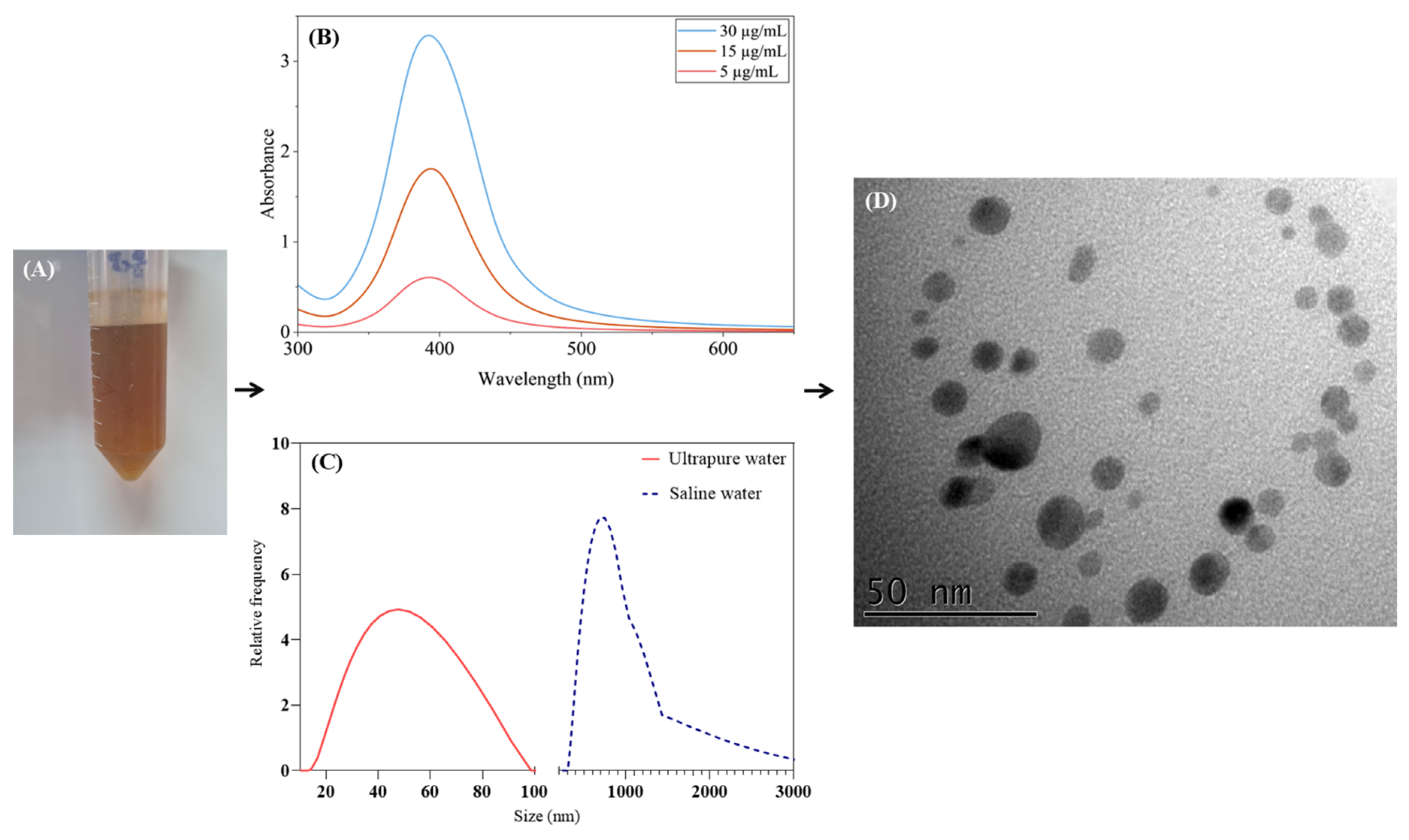
AgNPs were synthesized through a chemical reduction method according to the protocol described by Josende et al. [21]. Briefly, in a glass beaker containing 50 mL of ultrapure water under constant stirring, sodium borohydride (NaBH4, Sigma-Aldrich, Barueri, São Paulo, Brazil) was used as the reducing agent and sodium citrate (Synth) served as the stabilizer. The reduction conditions were modified to adopt a new molar ratio of 2:1 NaBH4:AgNO3, which has been shown to enhance the stability and yield of AgNPs, as proposed by Mulfinger et al. [22]. The reaction was conducted in the absence of light to prevent agglomeration during synthesis [21]. Subsequently, the resulting AgNP stock solution (100 µg/mL) exhibited a characteristic orange-brown coloration (Figure 1A) and was stored at 4 °C.
The characterization of AgNPs was performed employing Transmission Electron Microscopy (TEM), Dynamic Light Scattering (DLS), zeta potential measurements, and UV-Vis spectroscopy. Before each analysis, the stock solution containing AgNPs was dispersed by sonication at 60% amplitude for 5 min (4500 Joules/min) by using a Qsonica Q125 sonicator (Newton, CT, USA) (125 W power). In addition, the UV-Vis spectral analysis was carried out with a Thermo Scientific BioMate 3s spectrophotometer (Madison, WI, USA), using a quartz cuvette, covering wavelengths from 200 to 800 nm, with dilutions as illustrated in Figure 1B. The DLS and zeta potential (Figure 1C and Table 1) were performed in a Litesizer 500 Anton Paar analyzer (São Paulo, SP, Brazil), with the AgNPs diluted in both ultrapure water and marine media (25 ppt salinity) in order to determine the hydrodynamic diameter (HD) and the polydispersity index (PDI) of the colloidal suspensions in each condition. Furthermore, zeta potential was measured using electrode cuvettes (Malvern instruments, Malvern, Worcestershire, UK) to determine the surface charge of the particles in these two media.
Morphological characterization and particle size of chemically synthesized AgNPs were assessed by TEM (JEOL JEM 1400, Akishima, Japan) operating at an accelerating voltage of 100 kV (Figure 1D). A 10× diluted solution of AgNPs was left to dry overnight on copper grids coated with carbon at room temperature. The average size of the AgNPs was determined by counting at least 200 particles across different regions of the copper grid by using the freely available ImageJ software (version 1.54g) [23]. In order to avoid further particle transformation, all the analyses were carried out exactly moments after the AgNPs were released into ultrapure or marine media.

2.2. Organisms and Culture Conditions

Rotifers Brachionus plicatilis (Ploima, Brachionidae) were obtained from the Aquaculture Station of the Federal University of Rio Grande-FURG (Brazil) and maintained at the Institute of Biological Sciences (ICB). The seawater was collected from Cassino Beach (Rio Grande-RS, Brazil, coordinates: 32°12′23.0″ S, 52°10′25.4″ W) and stored in water tanks in FURG. In all cases, prior to use as an exposure medium for rotifers, it was disinfected (2% sodium hypochlorite) and filtered through a 40 µm nylon mesh, followed by the addition of sodium thiosulfate and vigorous aeration to ensure an effective dechlorination. The animals were maintained in 3 L beakers with constant aeration in an incubator (model FT-1010 from Eletrolab, São Paulo, SP, Brazil). Conditions were controlled by keeping the parameters as follows: temperature (25 °C), photoperiod (12:12 light/dark cycle), dissolved oxygen (6.3 ± 0.3 mg/L), salinity (25 ppt), and pH (7.3 ± 0.2). Rotifers were filtered through 50 µm nylon mesh prior to each medium renewal, which was carried out every 48 h. After that, the organisms were resuspended in disinfected seawater to ensure consistency of the medium. The organisms were fed every two days with the microalgae Nannochloropsis oculata (Eustigmatales, Monodopsidaceae) at a concentration of 5 × 10⁶ cells per 1 × 10⁶ rotifers. Additionally, 1 mL aliquots were collected daily from the solution in order to be counted in a Sedgewick Rafter chamber to estimate their density.

2.3. Exposure Conditions

The exposures were conducted in the incubator under the aforementioned controlled conditions, with three independent replicates per treatment unit. Rotifers were fed with algae 4 h prior to the beginning of exposure and then subsequently rinsed with clean seawater to remove residual microalgae from the medium. Next, for metabolism analysis, glass tubes containing 4 mL of rotifers at a density of approximately 450 individuals/mL were prepared. For ROS analysis, 50 mL of rotifers at a density of 500 individuals/mL were prepared. After verifying that oxygen levels remained constant during the 24 h exposure period, aeration was intentionally omitted in both exposures to avoid interference with the organism’s natural behavior given the low volume per experimental unit.
The stock AgNP solution (100 µg/mL) was diluted 100× for 4 mL tubes and 10× for 50 mL tubes to obtain the solutions at 1 and 10 µg/mL, respectively, for each tube. Treatments were prepared by adding 8 or 80 µL of the 1 µg/mL solution into 4 mL tubes and 10 or 100 µL of the 10 µg/mL solution into 50 mL tubes, corresponding to final concentrations of 2 and 20 µg/L of AgNPs, respectively. Each treatment was replicated three times.

2.4. In Vivo Metabolism Assessment

After the 24 h exposure period, 2.4 mL of the rotifer solution was collected from each experimental unit and passed through a 50 µm mesh filter. The retained rotifers were then resuspended in 1.2 mL of disinfected seawater (2:1 filtration ratio) and transferred to a 2 mL microtube prior to distribution into the wells. Each treatment was performed with three independent biological replicates. From each replicate, 150 µL aliquots were pipetted into a microplate, with three technical replicates per biological replicate, for in vivo analysis. Finally, 2 µL of a 1 mM resazurin solution was pipetted directly into the wells, and the plate was incubated for 2 h at 25 °C in the absence of light for subsequent readings. The final rotifer density in each well of the plate was approximately 135 rotifers for the metabolism analysis.
Finally, metabolic activity was measured in a FilterMax F5 fluorimeter (Molecular Devices, San Jose, CA, USA) (wavelengths of 530 and 590 nm for excitation and emission, respectively) assessed through the fluorescence emitted by the conversion of resazurin (Sigma-Aldrich, St. Louis, MO, EUA) into resorufin. Metabolism analysis included an incubation time of 2 h followed by 1 h of readings (every 3 min).

2.5. In Vivo Reactive Oxygen Species Assessment

For reactive oxygen species analysis, 5 mL of rotifer suspension (500 individuals/mL) from each treatment was collected from the 50 mL glass tubes and filtered through a 50 µm mesh filter. The retained organisms were rinsed with 2 mL of seawater and resuspended in 1 mL of clean medium, resulting in a final density of 2500 rotifers/mL in each microtube (5:1 filtration ratio). Subsequently, 12 µL of a 2.6 mM H2DCF-DA solution was added to each tube. The samples were incubated for 1 h in the absence of light at 25 °C. After incubation, 1 mL of the suspension was filtered again through a 50 µm mesh, rinsed with 2 mL of fresh seawater to remove residual H2DCF-DA, and resuspended again for further analysis.
ROS levels were measured in the same FilterMax F5 fluorimeter employed for metabolism analysis. Measurements were carried out with three biological replicates per treatment, and each biological replicate was further analyzed in triplicate wells on the microplate, applying excitation and emission wavelengths of 485 and 520 nm, respectively. Both metabolic and ROS protocols were based on the method of Buitrago et al. [24]. ROS detection relied on the conversion of 2,7-dichlorofluorescin diacetate (H2DCF-DA; Invitrogen, Thermo Fisher Scientific, Waltham, MA, USA) into its final fluorescent form, dichlorofluorescein (DCF). A final density of 375 rotifer/well was achieved for ROS analysis; fluorescence readings were recorded every 1.4 min for a period of 2 h.
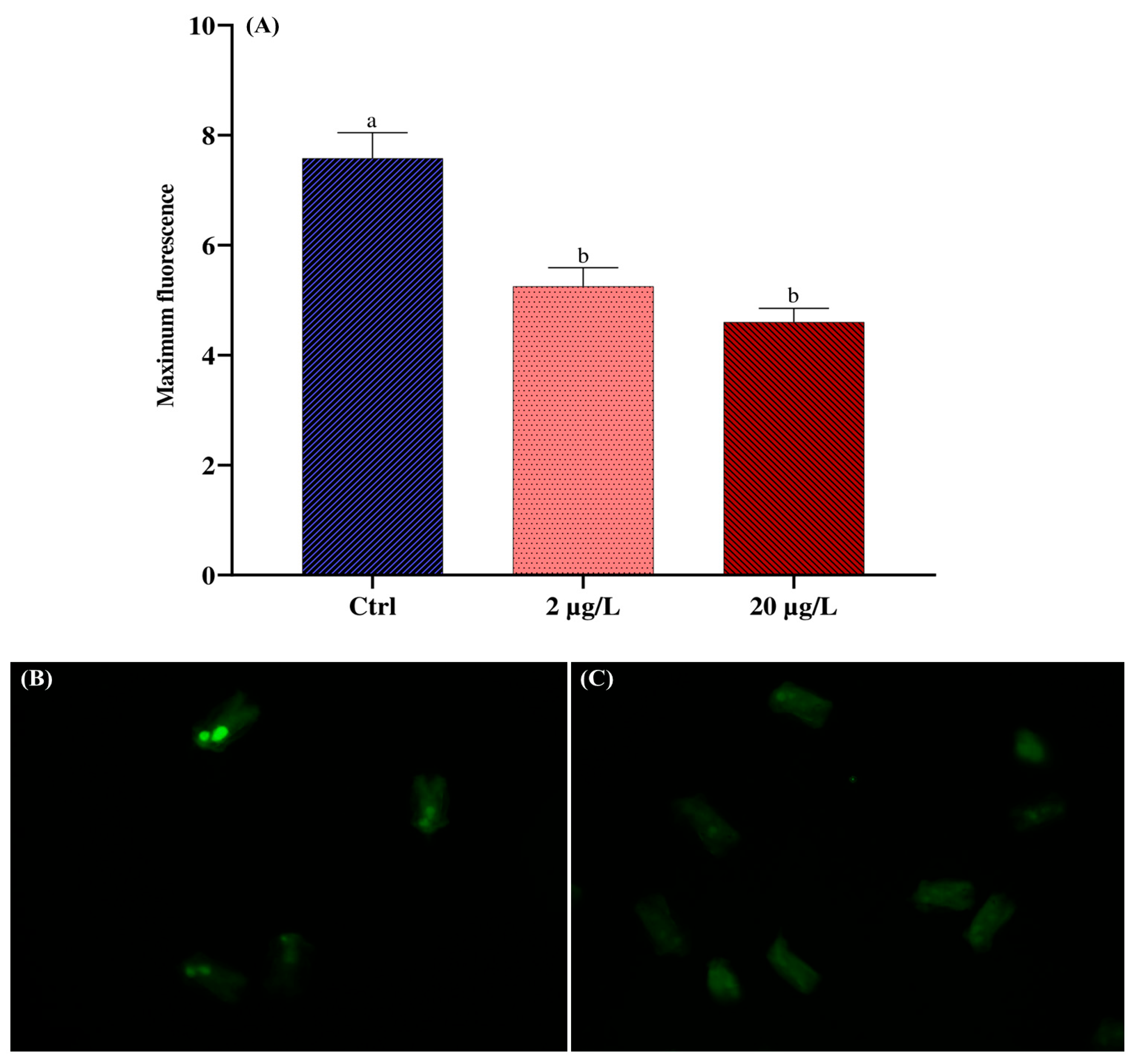
To assess ROS via fluorescence microscopy, an aliquot of 100 µL of rotifers from the prepared tubes for the microplate ROS assay was transferred to a 24-well plate and combined with 50 µL of sodium azide (20 mM) to anesthetize the organisms and facilitate imaging. Each treatment was performed in triplicate. The green fluorescence signal from the oxidized product (DCF) within the organisms was recorded using an Olympus fluorescence microscope (Model IX2-UCB/U-HSTR2, Akishima, Japan) equipped with a UV lamp and a DP72 camera.

2.6. Statistical Analysis

The normality and homogeneity of variances were assessed on the residuals of the ANOVA model using Shapiro–Wilk and Levene tests, respectively. A one-way ANOVA was then performed with AgNP concentrations as the factor and metabolic activity and ROS levels as dependent variables. Since both ROS datasets initially did not meet the assumption of variance homogeneity, the data were square-root-transformed, after which the assumptions were confirmed on the residuals. Post hoc comparisons were conducted using Tukey’s HSD test to identify significant differences at p < 0.05. All analyses were performed with InfoStat software (version 2020e). Data are expressed as mean ± standard error (SE).

3. Results

3.1. Synthesis of Silver Nanoparticles

Chemical synthesis of AgNPs yielded an orange-brown colloidal suspension (Figure 1A). The suspension exhibited a surface plasmon resonance (SPR) peak between 380 and 420 nm, with absorbance intensity increasing proportionally to AgNP concentration (Figure 1B). The particle size distribution appearance in the colloidal suspension ranges from 19.5 nm to 91 nm in ultrapure water and from 332 nm to 3779 nm in saline water as shown in Figure 1C. TEM confirmed nanoparticle formation, revealing predominantly spherical particles with an average core diameter of 8.7 ± 3.0 nm (Figure 1D). Table 1 summarizes AgNP characterization in ultrapure and seawater, including zeta potential, polydispersity index, and hydrodynamic diameter. Physicochemical behavior varied notably between media: in ultrapure water, particles showed high surface charge (−40 ± 3.0 mV) and a diameter of 46.2 ± 15.7 nm, while in seawater, reduced charge (−27 ± 0.5 mV) led to aggregation and a diameter of 1728 ± 103 nm.

3.2. In Vivo Toxicity Assessment

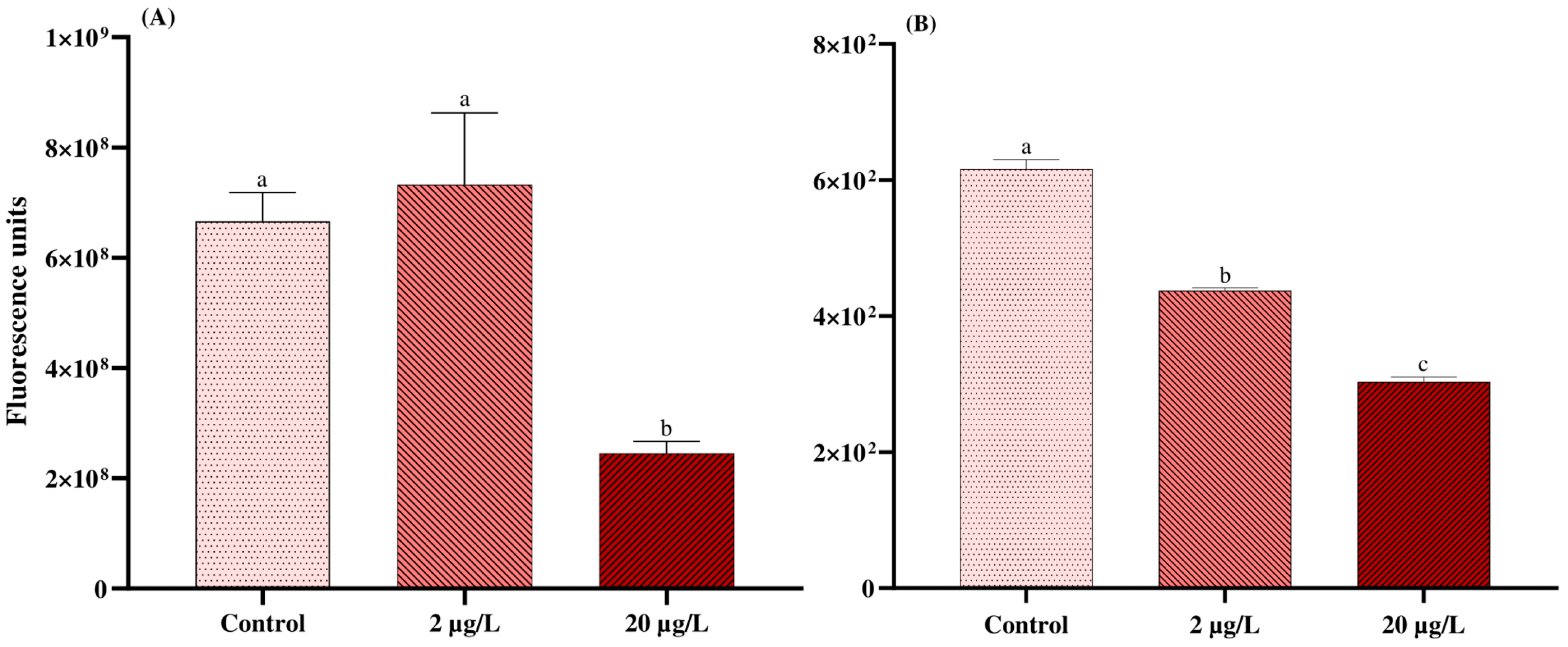
Figure 2 shows the results obtained from metabolism and ROS assays. Exposure to the highest AgNP concentration (20 µg/L) induced a significant decrease in the rotifer metabolic activity (p-value = 0.011). Furthermore, ROS levels decreased in a concentration-dependent manner (p-value < 0.01). These results are consistent with those obtained from fluorescence microscopy for ROS (Figure 3).

4. Discussion

Throughout this study, we carried out the chemical synthesis of AgNPs and fully characterized the particles in both synthesis and exposure media (ultrapure water and seawater). We emphasize the importance of thoroughly characterizing and describing the particle behavior in environmental matrices, which provides a more realistic approach to understanding the fate of AgNPs once they reach the marine ecosystem.
Our study also combines established practices to assess the ecotoxicity of AgNPs toward Brachionus plicatilis with a more environmentally realistic approach. Specifically, we chose to use natural seawater in the experimental design, as it provides a closer approximation to actual marine environmental conditions. Manfra et al. [25] compared the use of natural and artificial seawater in ecotoxicological exposure assays with B. plicatilis and highlighted that each medium can individually influence the toxicity of nanomaterials and their biological effects, underscoring the importance of selecting the most representative medium for ecotoxicity assessment. In this sense, several studies have also adopted natural seawater as a practical and environmentally realistic approach [26,27].
The chemical method employed for AgNP synthesis, with increased sodium borohydride concentration, successfully achieved a more efficient reduction of AgNO3. As a strong reducing agent, sodium borohydride can influence particle size, polydispersity, and surface charge, potentially enhancing colloidal stability [28,29]. Furthermore, this methodological feature improved the production of AgNPs while increasing their stability for studies in media with high ionic strength, such as seawater.
Regarding characterization, our results showed that the AgNPs presented an SPR absorbance band in the UV-Vis spectrum ranging from 380 to 420 nm, with an intensity peak at 400 nm, which agrees with values reported for similar AgNP suspensions [30]. The symmetrical UV-Vis peak indicated the distribution of spherical particles, associated with a relatively low degree of polydispersity [31]. Furthermore, the zeta potential analyses of AgNPs diluted in seawater revealed a reduction in particle surface charges. This behavior is expected for suspensions under high-ionic-strength exposure conditions, where predominant seawater ions (Na+, Mg2+, Ca2+, Cl) partially neutralize the surface charges of AgNPs, thereby weakening electrostatic repulsion and promoting agglomeration [32,33]. This trend is consistent with our hydrodynamic diameter measurements. Nanosilver destabilization is likely to alter the effective particle size distribution and surface reactivity during assays, potentially affecting toxicodynamics and the interpretation of biological effects [34].
We also investigated the in vivo effects of low environmentally relevant concentrations of AgNPs on the marine rotifer B. plicatilis. Accordingly, reported environmental nanosilver concentrations in wastewater treatment plant effluents range between 1 and 10 µg/L, while values in surface waters are substantially lower, around 1–10 ng/L [35]. Considering that effluents are one of the major sources of coastal ecosystem contamination, the experimental concentrations applied in this study (2 and 20 µg/L) were selected to reflect environmentally realistic exposures within the µg/L range and to represent worst-case scenarios. In this context, some investigations have reported that low AgNP concentrations in the range of 1–10 µg/L are capable of exerting toxic effects on the embryonic development of marine animals such as sea urchins and mussels [36,37]. Furthermore, there is a lack of studies on marine rotifers, particularly B. plicatilis, revealing a knowledge gap regarding the ecotoxicity of AgNPs in this species, despite their key role in trophic webs and their high sensitivity to a variety of nanopollutants [14,25].
The in vivo toxicity assessment performed in this study provides valuable information in terms of the primary effects generated by AgNPs in marine scenarios, particularly assessing the sensitivity of B. plicatilis to this nanopollutant. Our results showed an inhibition of metabolic activity in the rotifer upon exposure to the highest AgNP concentrations (20 µg/L), followed by a decrease in ROS levels at both concentrations. These are early responses in the marine rotifer, which could be worsened after long-term exposures, including ecological implications such as major alterations in the trophic webs. In this regard, Snell and Hicks. [17] demonstrated that the absorption of AgNPs in the rotifer body can also reach the amictic eggs of Brachionus manjavacas and accumulate over long periods, reducing offspring fitness.
The metabolic inhibition caused by AgNPs in B. plicatilis could be related to redox processes within the electron transport chain that convert resazurin dye into its fluorescent product, resorufin [24]. This redox process is driven by electrons derived from NADH, NADPH, FADH2, FMNH2, and cytochromes involved in oxidative phosphorylation complexes [38]. Metabolic processes are closely associated with ROS production [39]. As highlighted by Checa et al. [40], mitochondrial complexes I and III are the main sites of electron leakage, leading to the formation of O2•−, which is subsequently converted into H2O2 and OH via the Fenton reaction. Thus, the inhibition of the electron transport chain is expected to diminish this leakage and consequently reduce ROS concentrations, as observed in this study.
It has been suggested that low AgNP concentrations (in the order of µg/L) can activate the antioxidant defense system in freshwater microcrustaceans when the ROS levels increase slightly, acting as a signaling mechanism for the synthesis of reduced glutathione (GSH) and the activation of related enzyme activities such as catalase (CAT) [41]. The activation of these antioxidant defenses could also be a factor that modulates the organism’s physiological balance. However, the decline in ROS and metabolism levels observed under AgNP exposure may also be attributed to mitochondrial activity. This interpretation is supported by previous findings showing that AgNPs can induce mitochondrial membrane depolarization and uncoupling effects, as well as triggering the permeability transition pathway, leading to pore formation and proton efflux from damaged mitochondria [42]. Beyond the bioenergetic aspects discussed above, it is important to consider that AgNPs can also promote structural alterations in mitochondria. Skalska et al. [43] demonstrated that AgNPs directly interfere with mitochondrial function, inducing cristolysis and modifications in the morphology and membrane potential of the organelles. These findings are particularly relevant for the interpretation of our results, as they suggest that the concomitant reduction of metabolism and ROS should not be understood as a decrease in oxidative stress or direct antioxidant activity but rather as a consequence of functional and structural impairment of the organelles. In this context, our data may be related to the fact that exposure to AgNPs not only reduced the metabolic activity but also impaired mitochondrial functional integrity, which could better explain the simultaneous decline in ROS and metabolism observed in our study.
In B. plicatilis, mitochondrial bioenergetic efficiency is a primary driver of population dynamics, as both somatic growth and reproductive output are highly metabolic intensive processes. Doohan [44] demonstrated that reproduction imposes a substantial oxygen demand, with egg production alone costing 36.5 cal × 10−6/h, a value exceeding the basal respiration rate. Furthermore, the authors showed that ovigerous females exhibit a respiratory increase of over 100% that persists post-laying due to sustained metabolic activity. Given this demand, any disruption in mitochondrial energy generation, particularly via oxidative phosphorylation, may act as a bottleneck for population recruitment. Such bioenergetic deficits directly limit the intrinsic rate of increase by compromising the energetic investment, which could also compromise embryonic development [45].
Furthermore, as rotifers rely on efficient nutrient metabolism to fuel their accelerated lifecycles, mitochondrial dysfunction likely compromises the energetic reserves required for both current reproductive effort and future survival. Therefore, the loss of energy generation efficiency does not merely affect individual fitness but serves as a critical limiting factor for the population’s intrinsic rate of increase, directly influencing the sustainability and growth of B. plicatilis cultures.
Finally, we emphasize the need for further research on in vivo toxicity assessment under long-term AgNP exposure, which would offer deeper insight into the ecological resilience of B. plicatilis.

5. Conclusions

Given the promising properties and characteristics of AgNPs, inevitably, it is expected that its production, applications, and further disposal into environmental matrices will increase in the near future. Once introduced into the ecosystem, AgNPs’ environmental fate is a crucial matter that should be studied through a complete characterization of the particles in the exposure media. Here, we discussed the key differences in terms of particle properties between ultrapure water and seawater, with an emphasis on their colloidal behavior in stable versus environmental media. In this regard, ecotoxicological effects of AgNPs are closely linked to their physicochemical characteristics, particularly surface area, particle size, and reactivity, ultimately shaping their toxicological profile.
Ongoing research in nanotoxicology in marine zooplankton, particularly rotifers, should also be further developed. In this sense, in vivo toxicological assessment carried out in this study provided early warning signs of the organisms given the ability of AgNPs to compromise the aerobic metabolic activity in the rotifers and disrupt the cellular energy homeostasis. Consequently, more complex mechanisms could be triggered under chronic AgNP exposures, which should be further elucidated.
Directions for future investigations should integrate bioenergetic markers, together with antioxidant systems and mitochondrial morphology, to clarify whether AgNPs suppress ROS through metabolic inhibition or direct antioxidant effects in B. plicatilis.

Author Contributions

Conceptualization, T.O.G., A.A. and J.M.M.; methodology, T.O.G., A.A. and L.G.D.C.; investigation, T.O.G., A.A., L.G.D.C., M.d.C.V., V.D.S.M. and J.M.M.; writing—original draft preparation, T.O.G.; writing—review and editing, A.A., M.F.D. and J.M.M.; supervision, J.M.M.; funding acquisition, J.M.M. All authors have read and agreed to the published version of the manuscript.

Funding

This research was funded by Conselho Nacional de Desenvolvimento Científico e Tecnológico (CNPq), Brazil (grant-number 308804/2025-2 given to Monserrat, J.M.), and also by the Coordenação de Aperfeiçoamento de Pessoal de Nível Superior—Brasil (CAPES)—Finance Code 001 providing scholarships to the author’s degree (Garcia, T.O.—PhD and Da Costa, L.G.—MsC).

Institutional Review Board Statement

Not applicable.

Informed Consent Statement

Not applicable.

Data Availability Statement

The data supporting the findings of this study are openly available in Zenodo (zenodo.org) at https://doi.org/10.5281/zenodo.18235969.

Acknowledgments

The authors wish to thank Luiz Antônio de Almeida Pinto and Alice Neri, for their aid in DLS measurements, and Juliane Ventura Lima. We also thank the Federal University of Rio Grande (FURG) and Center for Electron Microscopy of the South Zone (CEME-SUL, FURG) for the TEM analyses. Finally, also we thank Juan Rafael Buitrago Ramirez from the Estação Marinha de Aquacultura (EMA, FURG).

Conflicts of Interest

The authors declare that they have no conflicts of interest.

References

  1. Khan, A.U.R.; Adnan, M.; Begum, S.; Nazir, R.; Mussarat, S. Increasing Trend of Silver Nanoparticles as Antibacterial and Anticancer Agent. In Self-Assembly of Materials and Their Applications; IntechOpen: London, UK, 2023; ISBN 978-1-83969-702-9. [Google Scholar]
  2. Cervantes-Avilés, P.; Keller, A.A. Incidence of Metal-Based Nanoparticles in the Conventional Wastewater Treatment Process. Water Res. 2021, 189, 116603. [Google Scholar] [CrossRef]
  3. Willis, K.A.; Serra-Gonçalves, C.; Richardson, K.; Schuyler, Q.A.; Pedersen, H.; Anderson, K.; Stark, J.S.; Vince, J.; Hardesty, B.D.; Wilcox, C.; et al. Cleaner Seas: Reducing Marine Pollution. Rev. Fish Biol. Fish. 2022, 32, 145–160. [Google Scholar] [CrossRef]
  4. Corsi, I.; Desimone, M.F.; Cazenave, J. Building the Bridge from Aquatic Nanotoxicology to Safety by Design Silver Nanoparticles. Front. Bioeng. Biotechnol. 2022, 10, 836742. [Google Scholar] [CrossRef]
  5. Bellingeri, A.; Ale, A.; Rusconi, T.; Scattoni, M.; Lemaire, S.; Protano, G.; Venditti, I.; Corsi, I. Nanosilver Environmental Safety in Marine Organisms: Ecotoxicological Assessment of a Commercial Nano-Enabled Product vs an Eco-Design Formulation. Toxics 2025, 13, 338. [Google Scholar] [CrossRef]
  6. Pastorino, P.; Prearo, M.; Barceló, D. Ethical Principles and Scientific Advancements: In Vitro, in Silico, and Non-Vertebrate Animal Approaches for a Green Ecotoxicology. Green Anal. Chem. 2024, 8, 100096. [Google Scholar] [CrossRef]
  7. An, H.J.; Sarkheil, M.; Park, H.S.; Yu, I.J.; Johari, S.A. Comparative Toxicity of Silver Nanoparticles (AgNPs) and Silver Nanowires (AgNWs) on Saltwater Microcrustacean, Artemia salina. Comp. Biochem. Physiol. Part C Toxicol. Pharmacol. 2019, 218, 62–69. [Google Scholar] [CrossRef]
  8. Pham, T.-L. Effect of Silver Nanoparticles on Tropical Freshwater and Marine Microalgae. J. Chem. 2019, 2019, 9658386. [Google Scholar] [CrossRef]
  9. Dedman, C.J.; Newson, G.C.; Davies, G.-L.; Christie-Oleza, J.A. Mechanisms of Silver Nanoparticle Toxicity on the Marine Cyanobacterium Prochlorococcus under Environmentally-Relevant Conditions. Sci. Total Environ. 2020, 747, 141229. [Google Scholar] [CrossRef]
  10. Algotiml, R.; Gab-Alla, A.; Seoudi, R.; Abulreesh, H.H.; El-Readi, M.Z.; Elbanna, K. Anticancer and Antimicrobial Activity of Biosynthesized Red Sea Marine Algal Silver Nanoparticles. Sci. Rep. 2022, 12, 2421. [Google Scholar] [CrossRef] [PubMed]
  11. Xing, Y.; Pu, X.-M.; Pan, J.-F.; Xu, J.; Liu, C.; Lu, D. From Antioxidant Defense to Genotoxicity: Deciphering the Tissue-Specific Impact of AgNPs on Marine Clam Ruditapes philippinarum. Aquat. Toxicol. 2024, 270, 106883. [Google Scholar] [CrossRef] [PubMed]
  12. Gambardella, C.; Costa, E.; Piazza, V.; Fabbrocini, A.; Magi, E.; Faimali, M.; Garaventa, F. Effect of Silver Nanoparticles on Marine Organisms Belonging to Different Trophic Levels. Mar. Environ. Res. 2015, 111, 41–49. [Google Scholar] [CrossRef]
  13. Rotini, A.; Gallo, A.; Parlapiano, I.; Berducci, M.T.; Boni, R.; Tosti, E.; Prato, E.; Maggi, C.; Cicero, A.M.; Migliore, L.; et al. Insights into the CuO Nanoparticle Ecotoxicity with Suitable Marine Model Species. Ecotoxicol. Environ. Saf. 2018, 147, 852–860. [Google Scholar] [CrossRef]
  14. Clément, L.; Hurel, C.; Marmier, N. Toxicity of TiO2 Nanoparticles to Cladocerans, Algae, Rotifers and Plants—Effects of Size and Crystalline Structure. Chemosphere 2013, 90, 1083–1090. [Google Scholar] [CrossRef]
  15. Mohammadi, S.; Ahmadifard, N.; Atashbar, B.; Nikoo, A.; Manaffar, R. Long-Term Effect of Zinc Oxide Nanoparticles on Population Growth, Reproductive Characteristics and Zinc Accumulation of Marine Rotifer, Brachionus plicatilis. Int. J. Aquat. Biol. 2021, 9, 333–343. [Google Scholar] [CrossRef]
  16. Byeon, E.; Sanpradit, P.; Lee, J.-S.; Jeong, H.; Kim, M.-S.; Hong, M.-S.; Peerakietkhajorn, S.; Sayed, A.E.-D.H.; Lee, J.-S. Size-Dependent Toxicity of Nano- and Microplastics with Zinc Oxide Nanoparticles in the Marine Rotifer Brachionus koreanus. Mar. Pollut. Bull. 2024, 209, 117206. [Google Scholar] [CrossRef]
  17. Snell, T.W.; Hicks, D.G. Assessing Toxicity of Nanoparticles Using Brachionus manjavacas (Rotifera). Environ. Toxicol. 2011, 26, 146–152. [Google Scholar] [CrossRef] [PubMed]
  18. Martins, N.; Pradhan, A.; Pascoal, C.; Cássio, F. Effects of Metal Nanoparticles on Freshwater Rotifers May Persist across Generations. Aquat. Toxicol. 2020, 229, 105652. [Google Scholar] [CrossRef]
  19. Martins, N.; Pradhan, A.; Pascoal, C.; Cássio, F. Can Acclimation of Freshwater Rotifers to Silver Nanoparticles or 5-Fluorouracil Influence Their Multi- and Transgenerational Effects? Sci. Total Environ. 2024, 954, 176326. [Google Scholar] [CrossRef] [PubMed]
  20. El Badawy, A.M.; Silva, R.G.; Morris, B.; Scheckel, K.G.; Suidan, M.T.; Tolaymat, T.M. Surface Charge-Dependent Toxicity of Silver Nanoparticles. Environ. Sci. Technol. 2011, 45, 283–287. [Google Scholar] [CrossRef]
  21. Josende, M.E.; Nunes, S.M.; Müller, L.; Dos Santos Francisco, W.; Gelesky, M.A.; Monserrat, J.M.; Ventura-Lima, J. Multigenerational Effects of Ecotoxicological Interaction between Arsenic and Silver Nanoparticles. Sci. Total Environ. 2019, 696, 133947. [Google Scholar] [CrossRef]
  22. Mulfinger, L.; Solomon, S.D.; Bahadory, M.; Jeyarajasingam, A.V.; Rutkowsky, S.A.; Boritz, C. Synthesis and Study of Silver Nanoparticles. J. Chem. Educ. 2007, 84, 322. [Google Scholar] [CrossRef]
  23. Schneider, C.A.; Rasband, W.S.; Eliceiri, K.W. NIH Image to ImageJ: 25 Years of Image Analysis. Nat. Methods 2012, 9, 671–675. [Google Scholar] [CrossRef]
  24. Ramírez, J.R.B.; Gomes, R.M.M.; Carvalho de Sousa Araujo, A.; Garcia, T.O.; Monserrat, J.M. The Combined Effects of Lipoic Acid and Sodium Acetate on Metabolism, Redox Balance, and Utilization of Yolk Reserves of Artemia sp. Nauplii. Aquaculture 2023, 577, 739936. [Google Scholar] [CrossRef]
  25. Manfra, L.; Rotini, A.; Bergami, E.; Grassi, G.; Faleri, C.; Corsi, I. Comparative Ecotoxicity of Polystyrene Nanoparticles in Natural Seawater and Reconstituted Seawater Using the Rotifer Brachionus plicatilis. Ecotoxicol. Environ. Saf. 2017, 145, 557–563. [Google Scholar] [CrossRef]
  26. Silva, S.M.; Ramos, P.B.; Buitrago, J.R.; da Silva, T.V.N.; Simião, C.S.; Colombo, G.M.; Schmitz, M.; Tesser, M.B.; Prentice, C.; Wasielesky, W.; et al. Zootechnical Performance, Biochemical Response, and Chromaticity in Pacific White Shrimp (Litopenaeus vannamei) (Boone, 1931) after the Inclusion of Lyophilized Açaí (Euterpe oleracea) in the Diet. Aquac. Int. 2020, 28, 1563–1577. [Google Scholar] [CrossRef]
  27. Holanda, M.; Ravagnan, E.; Lara, G.; Santana, G.; Furtado, P.; Cardozo, A.; Wasielesky, W.; Poersch, L.H. Integrated Multitrophic Culture of Shrimp Litopenaeus vannamei and Tilapia Oreochromis niloticus in Biofloc System: A Pilot Scale Study. Front. Mar. Sci. 2023, 10, 1060846. [Google Scholar] [CrossRef]
  28. Al-Ghamdi, H.S.; Mahmoud, W.E. One Pot Synthesis of Multi-Plasmonic Shapes of Silver Nanoparticles. Mater. Lett. 2013, 105, 62–64. [Google Scholar] [CrossRef]
  29. Khatoon, U.T.; Velidandi, A.; Nageswara Rao, G.V.S. Sodium Borohydride Mediated Synthesis of Nano-Sized Silver Particles: Their Characterization, Anti-Microbial and Cytotoxicity Studies. Mater. Chem. Phys. 2023, 294, 126997. [Google Scholar] [CrossRef]
  30. Alzoubi, F.Y.; Ahmad, A.A.; Aljarrah, I.A.; Migdadi, A.B.; Al-Bataineh, Q.M. Localize Surface Plasmon Resonance of Silver Nanoparticles Using Mie Theory. J. Mater. Sci. Mater. Electron. 2023, 34, 2128. [Google Scholar] [CrossRef]
  31. Ider, M.; Abderrafi, K.; Eddahbi, A.; Ouaskit, S.; Kassiba, A. Silver Metallic Nanoparticles with Surface Plasmon Resonance: Synthesis and Characterizations. J. Clust. Sci. 2017, 28, 1051–1069. [Google Scholar] [CrossRef]
  32. Zhong, H. Physicochemical Properties of Protein-Modified Silver Nanoparticles in Seawater. Int. Nano Lett. 2013, 3, 54. [Google Scholar] [CrossRef]
  33. Ershov, V.A.; Ershov, B.G. Oxidative Dissolution and the Aggregation of Silver Nanoparticles in Drinking and Natural Waters: The Influence of the Medium on the Process Development. Toxics 2024, 12, 757. [Google Scholar] [CrossRef]
  34. Bélteky, P.; Rónavári, A.; Zakupszky, D.; Boka, E.; Igaz, N.; Szerencsés, B.; Pfeiffer, I.; Vágvölgyi, C.; Kiricsi, M.; Kónya, Z. Are Smaller Nanoparticles Always Better? Understanding the Biological Effect of Size-Dependent Silver Nanoparticle Aggregation Under Biorelevant Conditions. Int. J. Nanomed. 2021, 16, 3021–3040. [Google Scholar] [CrossRef]
  35. Gottschalk, F.; Sun, T.; Nowack, B. Environmental Concentrations of Engineered Nanomaterials: Review of Modeling and Analytical Studies. Environ. Pollut. 2013, 181, 287–300. [Google Scholar] [CrossRef]
  36. Burić, P.; Jakšić, Ž.; Štajner, L.; Dutour Sikirić, M.; Jurašin, D.; Cascio, C.; Calzolai, L.; Lyons, D.M. Effect of Silver Nanoparticles on Mediterranean Sea Urchin Embryonal Development Is Species Specific and Depends on Moment of First Exposure. Mar. Environ. Res. 2015, 111, 50–59. [Google Scholar] [CrossRef] [PubMed]
  37. Ale, A.; Liberatori, G.; Vannuccini, M.L.; Bergami, E.; Ancora, S.; Mariotti, G.; Bianchi, N.; Galdopórpora, J.M.; Desimone, M.F.; Cazenave, J.; et al. Exposure to a Nanosilver-Enabled Consumer Product Results in Similar Accumulation and Toxicity of Silver Nanoparticles in the Marine Mussel Mytilus galloprovincialis. Aquat. Toxicol. 2019, 211, 46–56. [Google Scholar] [CrossRef]
  38. Allen, J.L.; Kennedy, S.J.; Shaw, L.N. Colorimetric Assays for the Rapid and High-Throughput Screening of Antimicrobial Peptide Activity against Diverse Bacterial Pathogens. Methods Enzym. 2022, 663, 131–156. [Google Scholar] [CrossRef]
  39. Tavallaie, M.; Voshtani, R.; Deng, X.; Qiao, Y.; Jiang, F.; Collman, J.P.; Fu, L. Moderation of Mitochondrial Respiration Mitigates Metabolic Syndrome of Aging. Proc. Natl. Acad. Sci. USA 2020, 117, 9840–9850. [Google Scholar] [CrossRef]
  40. Checa, J.; Aran, J.M. Reactive Oxygen Species: Drivers of Physiological and Pathological Processes. J. Inflamm. Res. 2020, 13, 1057–1073. [Google Scholar] [CrossRef]
  41. Ulm, L.; Krivohlavek, A.; Jurašin, D.; Ljubojević, M.; Šinko, G.; Crnković, T.; Žuntar, I.; Šikić, S.; Vinković Vrček, I. Response of Biochemical Biomarkers in the Aquatic Crustacean Daphnia magna Exposed to Silver Nanoparticles. Environ. Sci. Pollut. Res. 2015, 22, 19990–19999. [Google Scholar] [CrossRef] [PubMed]
  42. Stensberg, M.C.; Madangopal, R.; Yale, G.; Wei, Q.; Ochoa-Acuña, H.; Wei, A.; McLamore, E.S.; Rickus, J.; Porterfield, D.M.; Sepúlveda, M.S. Silver Nanoparticle-Specific Mitotoxicity in Daphnia magna. Nanotoxicology 2014, 8, 833–842. [Google Scholar] [CrossRef]
  43. Skalska, J.; Dąbrowska-Bouta, B.; Frontczak-Baniewicz, M.; Sulkowski, G.; Strużyńska, L. A Low Dose of Nanoparticulate Silver Induces Mitochondrial Dysfunction and Autophagy in Adult Rat Brain. Neurotox. Res. 2020, 38, 650–664. [Google Scholar] [CrossRef] [PubMed]
  44. Doohan, M. An Energy Budget for Adult Brachionus plicatilis Muller (Rotatoria). Oecologia 1973, 13, 351–362. [Google Scholar] [CrossRef] [PubMed]
  45. Jansen, R.P.S.; Burton, G.J. Mitochondrial Dysfunction in Reproduction. Mitochondrion 2004, 4, 577–600. [Google Scholar] [CrossRef] [PubMed]
Figure 1. (A) Tube containing an orange-brown solution of chemically synthesized AgNPs. (B) UV-Vis absorption spectra of AgNPs at 5, 15, and 30 µg/mL, showing a characteristic surface plasmon resonance peak near 400 nm, with intensity increasing with concentration. (C) DLS profiles of AgNPs diluted in ultrapure water (red line) and seawater (25 ppt; blue dotted line), indicating agglomeration effects in the saline medium. (D) TEM images showing spherical AgNP morphology with an average diameter of approximately 8.7 ± 3.0 nm.
Coatings 16 00152 g001
Figure 2. (A) Rotifer metabolism and (B) ROS generation after a 24 h exposure to AgNPs. Data are shown as mean ± SE. Different letters indicate statistical differences among the treatments at p < 0.05.
Coatings 16 00152 g002
Figure 3. (A) Maximum ROS fluorescence intensity in rotifers exposed to AgNPs (Ctrl, 2, and 20 µg/L) quantified by pixel density (mean ± SE). (B) Control rotifers and (C) rotifers exposed to 20 µg/L of AgNPs imaged by fluorescence microscopy. Pixel density was assessed in 10 individuals per treatment. Different letters indicate statistical differences between treatments (p < 0.05).
Coatings 16 00152 g003
Table 1. Characterization parameters of the AgNP colloidal suspension diluted in two different media: ultrapure water (synthesis medium) and seawater (exposure medium, salinity of 25 ppt). Data are expressed as mean ± SD.
MediumZ Potential (mV)Polydispersity Index (%)Hydrodynamic Diameter (nm)
Ultrapure water−40 ± 3.029.6 ± 1.046.2 ± 15.7
Seawater−27 ± 0.525 ± 6.01728 ± 103
Disclaimer/Publisher’s Note: The statements, opinions and data contained in all publications are solely those of the individual author(s) and contributor(s) and not of MDPI and/or the editor(s). MDPI and/or the editor(s) disclaim responsibility for any injury to people or property resulting from any ideas, methods, instructions or products referred to in the content.