Article

Influence of Multiple Firings on the Color Stability and Surface Roughness of Gingival Pink Feldspathic Ceramic

by
Talal M. Alnassar
Department of Prosthetic Dental Science, College of Dentistry, King Saud University, Riyadh 11545, Saudi Arabia
Coatings 2022, 12(12), 1870; https://doi.org/10.3390/coatings12121870
Submission received: 26 October 2022 / Revised: 22 November 2022 / Accepted: 26 November 2022 / Published: 1 December 2022

Abstract

:
The present study was aimed at analyzing the impact of repeated firings on the color stability and surface roughness (Ra) of gingival pink feldspathic porcelain. Twenty specimens (n = 20) were prepared, and repeated firings were carried out. These samples were equally and randomly divided into two groups (n = 10) to assess the color change (ΔE), using a spectrophotometer and Ra using a non-contact profilometer. The ΔE was calculated after the third, fifth, and seventh firings, whereas the Ra was assessed after the first, third, fifth, and seventh firings. The greatest ∆E was observed after the seventh firing (6.86), followed by the fifth firing (3.93). The lowest ∆E was seen after the third firing (2.61). All the inter-group comparisons were statistically significant (p < 0.01). The change in color of gingival pink feldspathic porcelain samples observed after multiple firings could be attributed to pigments in this material becoming unstable, and the possible change in the crystal orientation with increased firings. The highest Ra was observed for the samples after the first firing (1.130 μm), followed by the third firing (0.617 μm) and fifth firing (0.477 μm). The lowest Ra values were seen for the samples after the seventh firing (0.425 μm). All the inter-group comparisons were statistically significant (p < 0.01), except when the Ra values of samples after the fifth and seventh firing were compared (p > 0.01). The decreased Ra could be attributed to the melting of glaze with increased firings, which could have filled the gaps on the porcelain’s surface making it less rough. The present study demonstrated that an increase in repeated firings resulted in an increase in the ∆E values and a decrease in the Ra values of gingival pink feldspathic porcelain samples. The greatest increase in ∆E and the highest reduction in Ra values were noticed after the seventh firing. Future studies, including other parameters (variable thickness of ceramic and firing temperatures), should be conducted to study the impact of repeated firings on the color stability and roughness of gingival pink feldspathic porcelain.

1. Introduction

Dental ceramics are nowadays widely used for various clinical applications due to their high biocompatibility and increased fracture toughness [1]. Dental ceramics are generally divided into etchable glass-matrix, non-etchable polycrystalline ceramics, and hybrid ceramics (etchable and non-etchable) [2]. Feldspathic porcelain belongs to the etchable glass-matrix group and contains a high glass/silica content [3]. Feldspathic porcelain is used in dentistry for esthetic restorations due to its admirable optical and adhesive properties [4]. Several optical properties influence the aesthetics of dental restoration, including its color [5]. Color stability is crucial to assess the success or failure of the prosthetic or restorative treatment [6]. In dentistry, color changes in materials are determined using the Commission Internationale de I’Eclairage (CIE) coloring system, which shows the color parameters (L*, a*, b*) and the color change (ΔE) [7]. Surface roughness is another important property that influences the aesthetics of restorative material. The surface of restorative material is required to be smooth, as a rough surface decreases the material’s flexural strength and increases surface staining and plaque accumulation [7].
Patients receiving implants as part of the prosthodontic treatment often need soft and hard tissue adjustments that are invasive, time-consuming, and expensive [8]. Prosthetic approaches and various materials can overcome these problems, as they have been shown to construct soft and hard tissues effectively [9]. Among the soft tissue materials, gingival-colored pink porcelain stands out due to its superior aesthetics [10]. Pink porcelain has been shown to enhance aesthetics and is effective in masking negotiated surgical outcomes [11,12]. However, to achieve an ideal aesthetic outcome, the restorative material must reproduce the natural color of the tissues.
One major factor that could affect the color of ceramics is firing [13]. During this process porcelain material, while being fabricated, undergoes firing. Repeated firings are also sometimes indicated to improve aesthetics or adjust occlusion [14]. There is a disagreement in the literature regarding the effect of repeated firing on the color of ceramics. Few studies have reported that repeated firing can bring color changes in ceramics [15,16], whereas others have reported no significant color changes after multiple firings were performed on ceramics [17,18]. Concerning surface roughness (Ra), several studies have demonstrated that repeated firings can reduce the roughness of ceramics, making them smooth [7,19]. However, there is a scarcity of data in the literature regarding the effect of repeated firings on gingival feldspathic porcelain’s color and Ra.
Therefore, the present study aimed to analyze the impact of repeated firings on the color stability and Ra of gingival feldspathic porcelain. We hypothesized that the increasing number of firings would affect the color stability of this material. We also hypothesized that increased firings would decrease the material’s Ra.

2. Materials and Methods

The current study received the approval of the research ethics review board at the specialist dental practice and research center.

2.1. Preparation and Grouping of Specimens

Gingival feldspathic porcelain (Cerabien™ ZR Tissue 1; Kuraray Noritake Dental) was used to prepare twenty specimens (6 mm × 4 mm × 2 mm) (n = 20) of 10 mm diameter and 2 mm thickness using the sintering technique. Briefly, the mixing of the ceramic powder and liquid (Cerabien™ ZR forming liquid) was carried out in a vibrator following the manufacturer’s instructions. An absorbent paper was used to remove excess liquid. Teflon molds (Zetalabor; Zhermack) with the required dimensions were utilized to prepare the samples. A vacuum furnace (Ivoclar Programmat P90, Ivoclar Vivadent AG, Schaan, Lichtenstein) was employed for the firing. The recommended parameters for firing were employed (dry-out time: 5 min, pre-drying temperature: 600 °C, vacuum starting temperature: 600 °C, heat rate: 45 °C/min, release vacuum temperature: 930 °C, high temperature: 930 °C, hold time: 1 min and cool time: 4 min). A digital caliper (Guilin Guanglu Measuring Instrument Co, Gullin, China) was utilized post-firing to measure the dimensions of the samples. For the glazing, the paste and liquid were mixed on a clean glass slab. A glaze was then applied and fired as first firing following the suggested parameters (dry-out time of 5 min, the pre-drying temperature of 600 °C, heat rate of 50 °C/min, high temperature of 930 °C, hold time of 30 s, and cool time of 4 min). All the samples were glazed (Cerabien™ ZR Tissue 1-Self glaze; Kuraray Noritake Dental) before each firing, which was accomplished seven times.

2.2. Color Change (ΔE) Assessments

Ten samples (of twenty) were randomly selected and utilized for ΔE assessments after the third, fifth, and seventh firings. One ΔE evaluation was taken after the first firing for each sample. The ΔE was assessed by employing a spectrophotometer (Color Eye 7000A, Spectrophotometer, X-Rite, Pantone, Grand Rapids, MI, USA) which was calibrated before and after each firing. A resin index was fabricated to ensure standardization and ensure reproducibility of the spectrophotometric analysis which was performed on the central portion of the samples. All the ΔE measurements were performed using a gray background (approximately Munsell value 7) with the help of an appropriate silicone seating jig for reproducibility. The L*, a*, b* values were calculated, and then the ΔE was calculated using CIEDE 2000 (DE00) formula.
Δ E 00 = ( Δ L k L S L ) 2 + ( Δ C k C S C ) 2 + ( Δ H k H S H ) 2 + R T ( Δ C k C S C ) ( Δ H k H S H )
The ∆E values were converted into the National Bureau of Standards (NBS) units by utilizing the following formula [20],
NBS units = ΔE* × 0.92
The NBS units, ΔE remarks, and clinical interpretation are presented below in Table 1.

2.3. Ra Analysis

The remaining ten samples were utilized for Ra analysis. A three-dimensional optical non-contact surface microscope (Contour GT-K 3D Optical Microscope, BrukerR, Tucson, AZ, USA) was employed for this assessment. The Ra measurements were performed by placing the specimens flat on the stage of the microscope stage with the help of a pre-fabricated silicone elastomeric polymer (polyvinyl siloxane, 3M ESPE, St. Paul, MN, USA) mold. Like the ΔE assessments, the Ra was measured for all ten samples after the first, third, fifth, and seventh firing. The Ra was calculated five times for each sample, and then the average Ra/sample was calculated at each firing stage. The Vision 64 Control and Analysis Software (BrukerR, Tucson, AZ, USA) was utilized to make the readings precise.

2.4. Statistical Analysis

The data were gathered, entered into spreadsheets, and then analyzed using statistical software (SPSS version 21, IBM, Armonk, NY, USA). The normality of the data was first checked using the Wilk–Shapiro test. The means and standard deviations (SD) of each sample after ΔE and Ra readings were calculated and then compared with the other samples at four different time points (first firing, third firing, fifth firing, and seventh firing). The comparisons among the groups were completed with the analysis of variance (ANOVA) and multiple comparisons (Tukey–Kramer) test. The significance level was set at 1%.

3. Results

3.1. ∆E Assessment Results

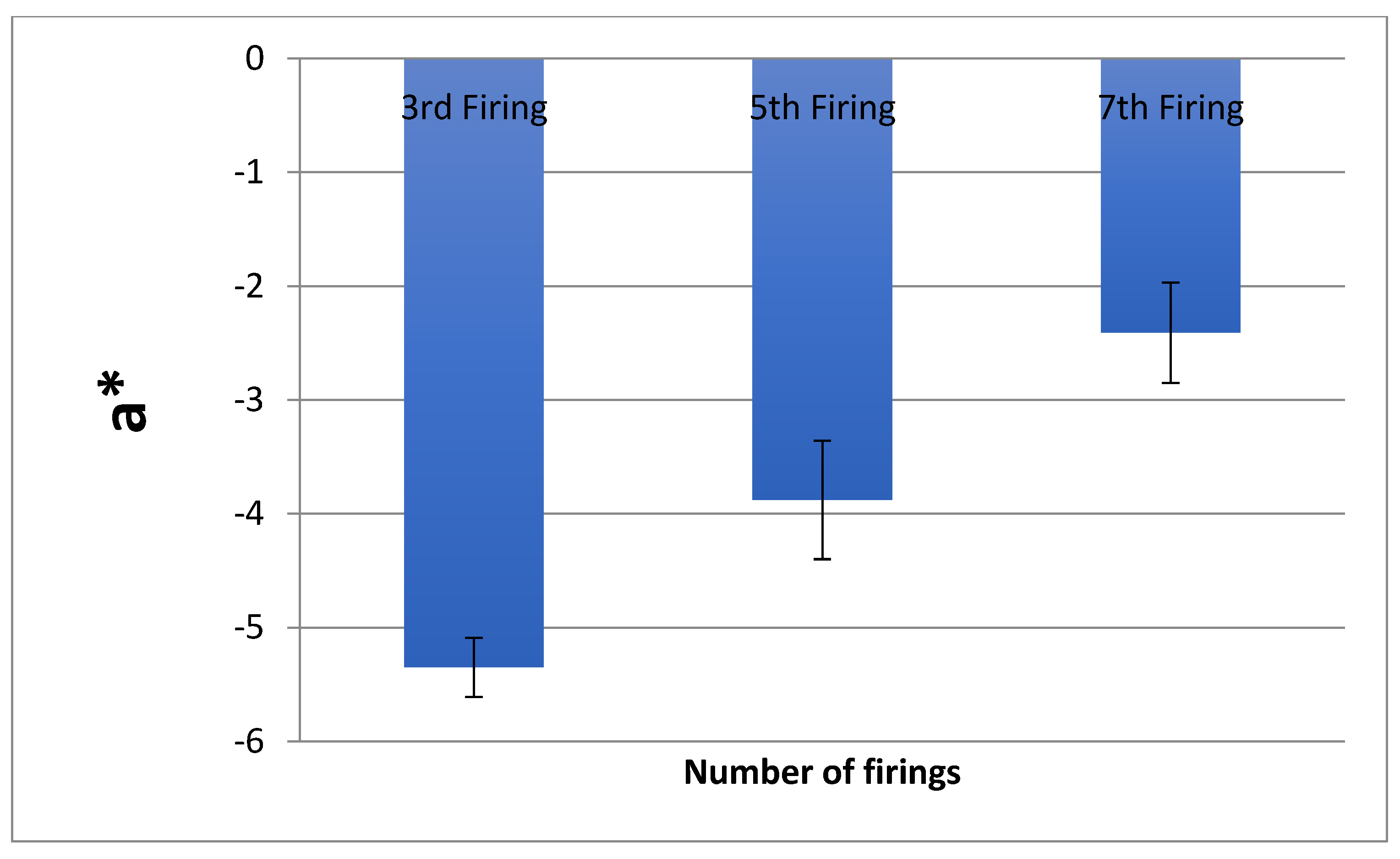
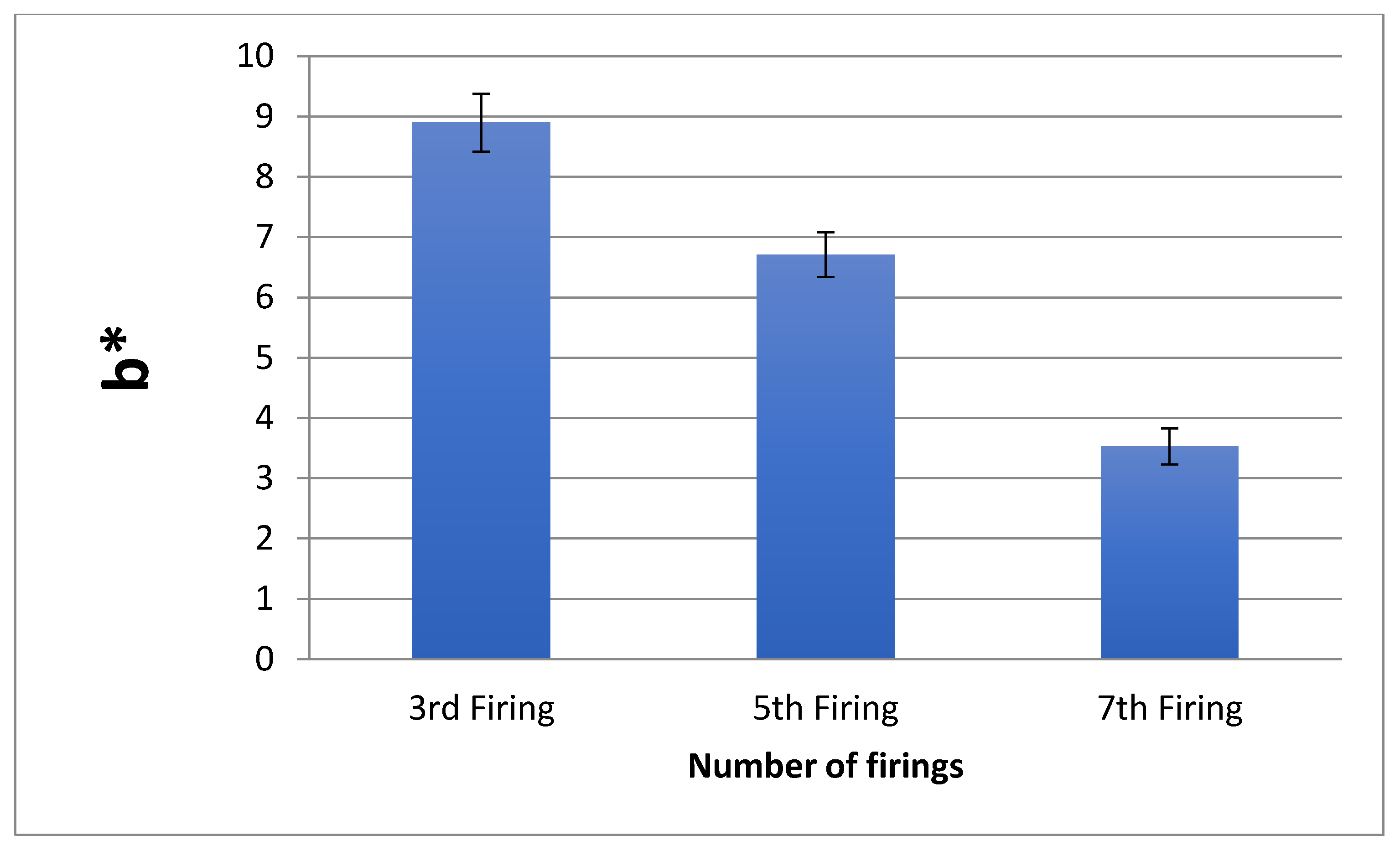
The ∆E values for the ten samples are presented in Table 2. The mean and SD of ∆L, ∆a, ∆b values after each firing are also shown in the same table. The greatest ∆E was observed after the seventh firing (6.86), followed by the fifth firing (3.93), whereas the lowest ∆E was observed after the third firing (2.61) (Figure 1, Figure 2, Figure 3 and Figure 4). All the inter-group comparisons were statistically significant (p < 0.01). After conversion into the NBS units, the greatest value was presented again by the samples, which were fired seven times (6.31), followed by samples that were fired five times (3.61) and three times (2.40).

3.2. Ra Analysis Results

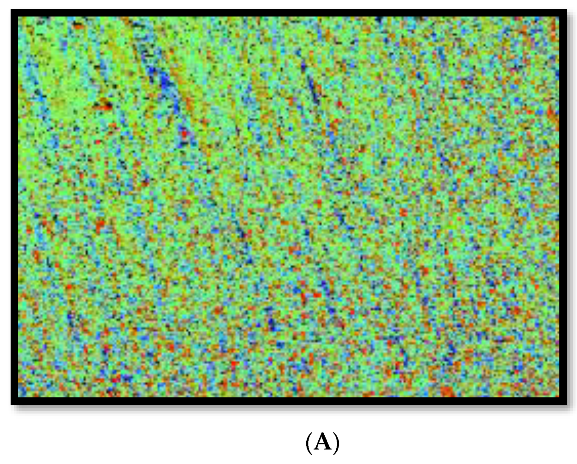
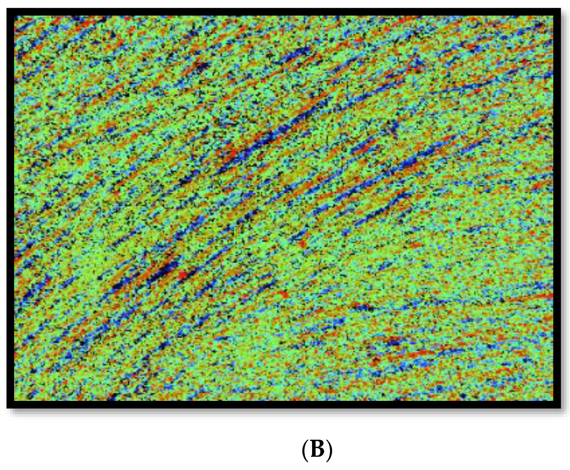
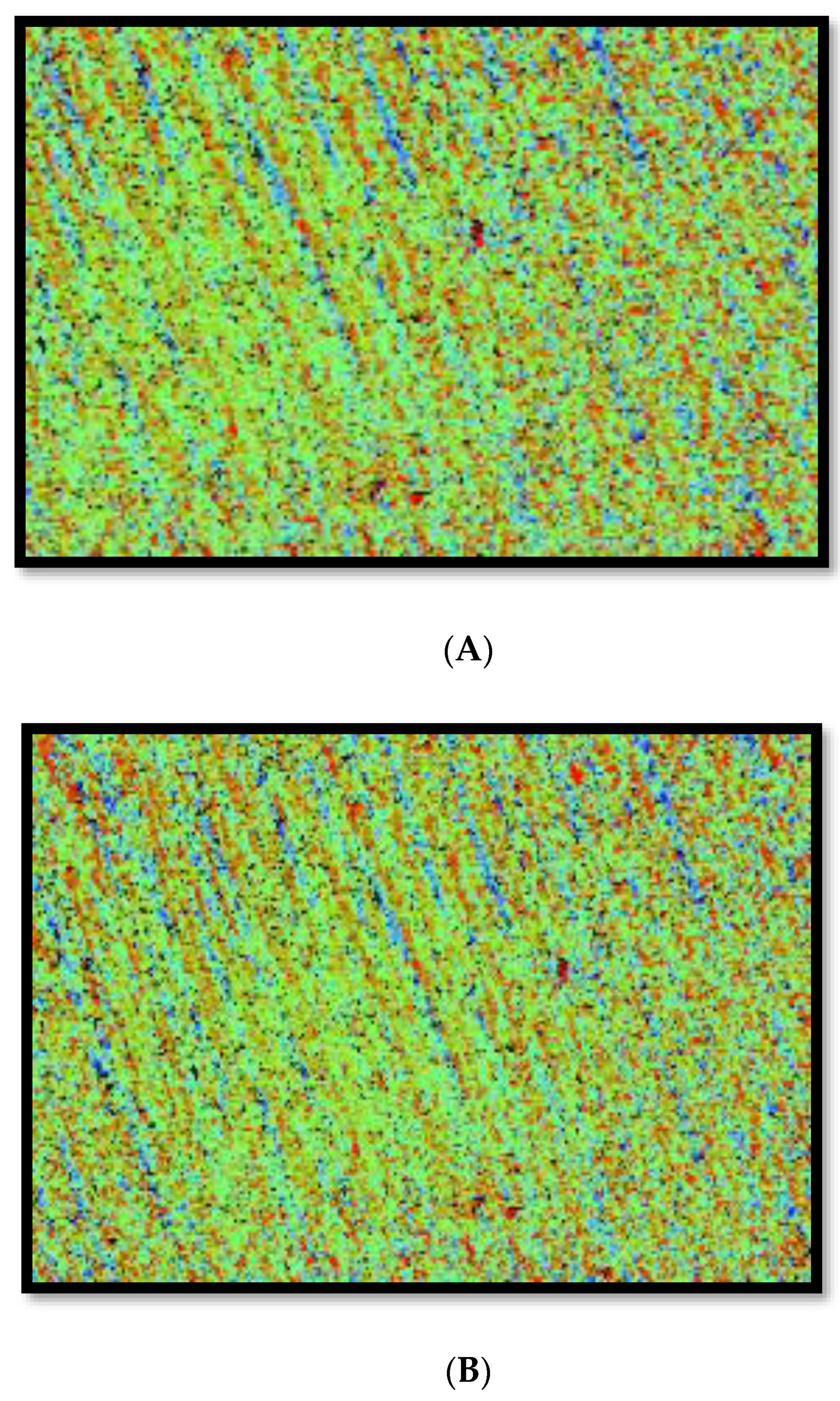
The Ra values for the remaining ten samples are presented in Table 3 (Figure 5 and Figure 6). An inversely proportional relationship between the Ra values and the number of firing cycles was seen in this study. The greatest Ra was observed for the samples after the first firing (1.130), followed by the third firing (0.617) and fifth firing (0.477). The lowest Ra values were observed for the samples after the seventh firing (0.425). All the inter-group comparisons were statistically significant (p < 0.01), except when the Ra values of samples after the fifth and seventh firing were compared (p > 0.01).

4. Discussion

Based on the results of the present study, our hypothesis that an increased number of firings would affect the color stability of the gingival felspathic porcelain was accepted as we observed a direct linear relationship between the number of firings and ΔE of the material. Our second hypothesis, that an increased number of firings would decrease the Ra of the material (making it smoother), was also accepted as we observed an inverse linear relationship between the number of firings and Ra values. Traditionally, the assessment of the changes in the color of dental materials was accomplished using a visual color guide for many years; however, this subjective method led to many imprecisions [21]. Spectrophotometric analysis helps assess minor color changes in dental materials efficiently, and the use of a spectrophotometer reduces the subjective errors associated with colorimetric analysis (performed with bare eyes) [22]. Considering the benefits of a spectrophotometer, we decided to use this device to detect color changes in gingival feldspathic porcelain after a different number of firings.
The CIELab color difference formula has been conventionally used to assess color changes in dental materials [23,24]. However, the CIEDE2000 formula was introduced to improve the CIELab formula and better assess the perceptibility and acceptability of dental materials [25]. Therefore, in the present study ∆E values were calculated using the CIEDE2000 formula. As per the formerly recognized standards, ∆E values between 1 and 3.3 are considered an important difference, but are clinically acceptable; however, an ∆E value > 3.3 is considered clinically unacceptable as it can be perceived by an inexperienced viewer as well [26,27]. The results of our study demonstrated that after the fifth and seventh firings, the ∆E value of gingival feldspathic porcelain samples crossed the clinical acceptability threshold of 3.3. In a previous study, Fehmi et al. also reported that ∆E values of dental ceramics increased after repeated firings, and this increase was above the perceptibility thresholds [14]. Similar results were conveyed by Yilmaz et al., and it was reported in their study that repeated firings damagingly affected the porcelain surface and caused fading in their color [15]. Several other studies have also demonstrated that the number of firings could influence the color stability of dental ceramics, and they should be avoided [28,29]. Our results align with these previously mentioned studies as we also observed that ∆E values increased proportionately with increased firings. The exact reason for this finding is unknown; however, the literature proposes that metal oxides inside dental ceramics act as a pigment and are not color-stable [15]. Therefore, due to repeated firing, the shade of ceramic would change as these metal oxide pigments break and burn out [30]. Another reason that could be attributed to this finding is that repeated firings could alter the crystalline structure of the ceramics resulting in an overall change in their color [31]. It should be noted that the thickness of ceramics could also have an impact on its color stability when subjected to repeated firings [7,32]; however, this factor was not examined in the present study, and the thickness of all porcelain samples was kept standardized at 2 mm.
In the present study, it was observed that Ra values decreased with repeated firings. Previously, Fehmi et al. reported that an increase in the number of firings resulted in reducing the Ra values of dental ceramics (increasing smoothness) [14]. Yilmaz and Ozkan also reported similar findings and demonstrated that Ra values of ceramics decreased with an increase in the number of firings [33]. Our results agree with these previous studies. The reason for this finding could be attributed to the fact that a repeated number of firings can melt the glaze, which could then fill the gap on the porcelain’s surface, making it appear smoother (less rough) [34]. Another reason for this finding could be attributed to the grain size of the gingival feldspathic porcelain. Literature shows that a longer duration of exposure to raised temperatures could affect the grain size of dental ceramics [35,36] which could then affect its surface roughness [37]. Nevertheless, although repeated firing can increase the smoothness of the material, it is generally not recommended to perform extensive chair-side modifications of the restoration [38]. In this aspect, exceptional skills of the clinician and technician are required to warrant the long-term success of the restoration.
Our results should be interpreted cautiously as the present study had certain limitations. One of the limitations was its in vitro design. It should be noted that restorations can act differently in vivo due to the presence of saliva. Additionally, several other influencing factors (like toothbrushing) could change its color and roughness in vivo. Furthermore, we studied the impact of the number of firings on the color stability and Ra of gingival feldspathic porcelain, which was procured commercially from one manufacturer. It is possible that the same material from a different commercial manufacturer can demonstrate slightly different results. We also used gingival feldspathic samples, which were 2 mm thick. It is possible that different thicknesses of these samples could show different ∆E and Ra values. Finally, the reader should note that mineralogical content assessment was not performed in this study. In the future, this assessment could provide important clues about precise minerals which get unstable due to repeated firings, and then steps could be researched to improve their stability (if repeated firings are necessary).

5. Conclusions

Within the present study’s limitations, an increase in the number of firings resulted in an increase in the ∆E values and a decrease in the Ra values of gingival feldspathic porcelain samples. The greatest increase in ∆E and the highest reduction in Ra values were noticed after the seventh firing. Future studies should use a variable thickness of gingival feldspathic porcelain samples, and then study the impact of repeated firings on their color stability and roughness to corroborate the current study’s findings.

Funding

The author would like to acknowledge with much appreciation the College of Dentistry at King Saud University, Research Center (CDRC) for their support.

Institutional Review Board Statement

Not applicable.

Informed Consent Statement

Not applicable.

Data Availability Statement

Data from the study are available on request form the corresponding author.

Conflicts of Interest

Author declares no conflict of interest.

References

  1. Maawadh, A.M.; Almohareb, T.; Al-Hamdan, R.S.; Al Deeb, M.; Naseem, M.; Alhenaki, A.M.; Vohra, F.; Abduljabbar, T. Repair strength and surface topography of lithium disilicate and hybrid resin ceramics with LLLT and photodynamic therapy in comparison to hydrofluoric acid. J. App. Biomat. Func. Mater. 2020, 18, 2280800020966938. [Google Scholar] [CrossRef] [PubMed]
  2. Awad, M.M.; Al Jeaidi, Z.A.; Almutairi, N.; Vohra, F.; Özcan, M.; Alrahlah, A. Effect of self-etching ceramic primer on bond strength of zirconia-reinforced lithium silicate ceramics. J. Adhes. Sci. Technol. 2020, 34, 91–101. [Google Scholar] [CrossRef]
  3. Gracis, S.; Thompson, V.P.; Ferencz, J.L.; Silva, N.R.; Bonfante, E.A. A new classification system for all-ceramic and ceramic-like restorative materials. Int. J. Prosthodont. 2015, 28, 227–235. [Google Scholar] [CrossRef] [PubMed] [Green Version]
  4. Ruales-Carrera, E.; Dal Bó, M.; das Neves, W.F.; Fredel, M.C.; Volpato, C.A.M.; Hotza, D. Chemical tempering of feldspathic porcelain for dentistry applications: A review. Open Ceram. 2022, 9, 100201. [Google Scholar] [CrossRef]
  5. Nistor, L.; Grădinaru, M.; Rîcă, R.; Mărășescu, P.; Stan, M.; Manolea, H.; Ionescu, A.; Moraru, I. Zirconia Use in Dentistry—Manufacturing and Properties. Curr. Health Sci. J. 2019, 25, 28–35. [Google Scholar]
  6. Vohra, F.; Labban, N.; Al-Hussaini, A.; Al-Jarboua, M.; Zawawi, R.; Alrahlah, A.; Naseem, M. Influence of Er; Cr: YSGG laser on shear bond strength and color stability of lithium disilicate ceramics: An in vitro study. Photobiomodul. Photomed. Laser Surg. 2019, 37, 483–488. [Google Scholar] [CrossRef]
  7. AlHamdan, E.M.; Bashiri, A.; Alnashmi, F.; Al-Saleh, S.; Al-shahrani, K.; Al-shahrani, S.; Alsharani, A.; Alzahrani, K.M.; Alqarawi, F.K.; Vohra, F.; et al. Evaluation of Smart Chromatic Technology for a Single-Shade Dental Polymer Resin: An In Vitro Study. Appl. Sci. 2020, 11, 10108. [Google Scholar] [CrossRef]
  8. Alqutub, M.N.; Mukhtar, A.H.; Alali, Y.; Vohra, F.; Abduljabbar, T. Osteogenic Differentiation of Periodontal Ligament Stem Cells Seeded on Equine-Derived Xenograft in Osteogenic Growth Media. Medicina 2022, 58, 1518. [Google Scholar] [CrossRef]
  9. Petropoulou, A.; Pappa, E.; Pelekanos, S. Esthetic considerations when replacing maxillary incisors with implants: A clinical report. J. Prosthet. Dent. 2013, 109, 140–144. [Google Scholar] [CrossRef]
  10. Sonune, S.J.; Kumar, S.; Jadhav, M.S.; Martande, S. Gingival-colored Porcelain: A Clinical Report of an Esthetic-prosthetic Paradigm. Int. J. Appl. Basic Med. Res. 2017, 7, 275–277. [Google Scholar] [CrossRef]
  11. Kalman, L.; MacIntosh, K. The Use of Pink Porcelain to Manage a Malposed Anterior Implant: Case Report. J. Can. Dent. Assoc. 2013, 79, d117. [Google Scholar] [PubMed]
  12. Papaspyridakos, P.; Amin, S.; El-Rafie, K.; Weber, H.P. Technique to Match Gingival Shade when Using Pink Ceramics for Anterior Fixed Implant Prostheses. J. Prosthodont. 2018, 27, 311–313. [Google Scholar] [CrossRef] [PubMed]
  13. Wisniewska, K.; Pichor, W.; Klosek-Wawrzyn, E. Influence of Firing Temperature on Phase Composition and Color Properties of Ceramic Tile Bodies. Materials 2021, 14, 6380. [Google Scholar] [CrossRef] [PubMed]
  14. Fehmi, G.; Ersan, Ç.; Caner, Ö. Influence of repeated firings on the color parameters of two different all-ceramic materials. Color Res. Appl. 2017, 43, 606–611. [Google Scholar] [CrossRef]
  15. Yilmaz, K.; Gonuldas, F.; Ozturk, C. The effect of repeated firings on the color change of dental ceramics using different glazing methods. J. Adv. Prosthodont. 2014, 6, 427–433. [Google Scholar] [CrossRef] [Green Version]
  16. Ozturk, O.; Uludag, B.; Usumez, A.; Sahin, V.; Celik, G. The effect of ceramic thickness and number of firings on the color of two all-ceramic systems. J. Prosthet. Dent. 2008, 100, 99–106. [Google Scholar] [CrossRef]
  17. Bayindir, F.; Ozbayram, O. Effect of number of firings on the color and translucency of ceramic core materials with veneer ceramic of different thicknesses. J. Prosthet. Dent. 2018, 119, 152–158. [Google Scholar] [CrossRef]
  18. Hasssija, J.; Hegde, V.; Sridhar, N. An In Vitro Study on Effect of Ceramic Thickness and Multiple Firings on Colour of Metal Ceramic Restorations. J. Indian Prosthodont. Soc. 2014, 14 (Suppl. 1), 86–92. [Google Scholar] [CrossRef]
  19. Sert, M.; Subasi, M.G.; Donmez, M.B.; Cakmak, G.; Yilmaz, B. Effect of firing cycle and aging on long-term chemical degradation of monolithic CAD-CAM ceramics. J. Prosthet. Dent. 2022, 128, 89 e1–89 e10. [Google Scholar] [CrossRef]
  20. Filho, H.L.; Maia, L.H.; Araújo, M.V.; Eliast, C.N.; Ruellas, A.C. Colour stability of aesthetic brackets: Ceramicand plastic. Aust. Orthod. J. 2013, 29, 13–20. [Google Scholar]
  21. Ibrahim, M.; Farghaly, E.; Badih, R. A comparison of color stability between hybrid ceramic and veneers: An in vitro study. Int. Arab J. Dent. 2019, 10, 25–30. [Google Scholar] [CrossRef]
  22. Javali, M.A.; Abdul Khader, M.; Alqahtani, R.M.; Almufarrij, M.J.; Alqahtani, T.M.; Addas, M.K. Spectrophotometric Analysis of Dental Enamel Staining to Antiseptic and Dietary Agents: In Vitro Study. Int. J. Dent. 2020, 2020, 5429725. [Google Scholar] [CrossRef] [PubMed]
  23. Alzahrani, K.M.; Alhaqbani, M.M.; Basalem, S.A.; Alsubaie, F.; AlRabiah, M.; Alzahrani, A.; AlAali, K.A.; Farooq, I.; Vohra, F.; Abduljabbar, T. Effect of Staining Drinks on the Color Stability of Grit Blasted and Non-Grit Blasted Monolithic Zirconia: An In Vitro Study. Crystals 2022, 12, 1331. [Google Scholar] [CrossRef]
  24. Alnassar, T.M. Color Stability of Monolithic Zirconia in Various Staining Liquids: An In Vitro Study. App. Sci. 2022, 12, 9752. [Google Scholar] [CrossRef]
  25. Gomez-Polo, C.; Portillo Munoz, M.; Lorenzo Luengo, M.C.; Vicente, P.; Galindo, P.; Martin Casado, A.M. Comparison of the CIELab and CIEDE2000 color difference formulas. J. Prosthet. Dent. 2016, 115, 65–70. [Google Scholar] [CrossRef] [PubMed]
  26. Colombo, M.; Cavallo, M.; Miegge, M.; Dagna, A.; Beltrami, R.; Chiesa, M.; Poggio, C. Color stability of CAD/CAM Zirconia ceramics following exposure to acidic and staining drinks. J. Clin. Exp. Dent. 2017, 9, e1297–e1303. [Google Scholar] [CrossRef]
  27. Inokoshi, S.; Burrow, M.F.; Kataumi, M.; Yamada, T.; Takatsu, T. Opacity and color changes of tooth-colored restorative materials. Oper. Dent. 1996, 21, 73–80. [Google Scholar]
  28. Bachhav, V.C.; Aras, M.A. The effect of ceramic thickness and number of firings on the color of a zirconium oxide based all ceramic system fabricated using CAD/CAM technology. J. Adv. Prosthodont. 2011, 3, 57–62. [Google Scholar] [CrossRef] [Green Version]
  29. Mohamed, S.A.; Ahmed, A.F.; Mekkawi, W.O. Effect of Repeated Firings on the Color Change and Surface Roughness of Two All-ceramic Systems. Al-Azhar Dent. J. Girls 2020, 7, 345–354. [Google Scholar] [CrossRef]
  30. Talibi, M.; Kaur, K.; Parmar, H. Do you know your ceramics? Part 2: Feldspathic ceramics. Br. Dent. J. 2022, 232, 80–83. [Google Scholar] [CrossRef]
  31. Dong-Dong, Q.; Lei, Z.; Xiaoping, L.; Wenli, C. Effect of repeated sintering on the color and translucency of dental lithium disilicate-based glass ceramic. Hua Xi Kou Qiang Yi Xue Za Zhi 2015, 33, 50–53. [Google Scholar] [PubMed]
  32. Fathi, A.; Farzin, M.; Giti, R.; Kalantari, M.H. Effects of number of firings and veneer thickness on the color and translucency of 2 different zirconia-based ceramic systems. J. Prosthet. Dent. 2019, 122, 565 e1–565 e7. [Google Scholar] [CrossRef] [PubMed]
  33. Yilmaz, K.; Ozkan, P. Profilometer evaluation of the effect of various polishing methods on the surface roughness in dental ceramics of different structures subjected to repeated firings. Quintessence Int. 2010, 41, e125–e131. [Google Scholar] [PubMed]
  34. Barghi, N. Color and glaze: Effects of repeated firings. J. Prosthet. Dent. 1982, 47, 393–395. [Google Scholar] [CrossRef]
  35. Denry, I.; Holloway, J.A. Ceramics for Dental Applications: A Review. Materials 2010, 3, 351–368. [Google Scholar] [CrossRef] [Green Version]
  36. Stawarczyk, B.; Ozcan, M.; Hallmann, L.; Ender, A.; Mehl, A.; Hämmerlet, C.H. The effect of zirconia sintering temperature on flexural strength, grain size, and contrast ratio. Clin. Oral Investig. 2013, 17, 269–274. [Google Scholar] [CrossRef] [Green Version]
  37. Kulvarangkun, A.; Panyayong, W.; Pumpaluk, P. Experimental study of surface roughness of dental ceramics after polishing with three types of polishing systems. J. Int. Soc. Prev. Communit Dent. 2022, 12, 540–546. [Google Scholar]
  38. Rashid, H. The effect of surface roughness on ceramics used in dentistry: A review of literature. Eur. J. Dent. 2014, 8, 571–579. [Google Scholar] [CrossRef]
Figure 1. ΔE comparison of samples after the third, fifth, and seventh firings.
Coatings 12 01870 g001
Figure 2. L* comparison of samples after the third, fifth, and seventh firings.
Coatings 12 01870 g002
Figure 3. a* comparison of samples after the third, fifth, and seventh firings.
Coatings 12 01870 g003
Figure 4. b* comparison of samples after the third, fifth, and seventh firings.
Coatings 12 01870 g004
Figure 5. Surface roughness micro-graphs for the first (A), and third (B) firing samples.
Coatings 12 01870 g005aCoatings 12 01870 g005b
Figure 6. Surface roughness micro-graphs for the fifth (A), and seventh (B) firing samples.
Coatings 12 01870 g006
Table 1. NBS interpretation of ΔE.
NBS UnitColor Change RemarksClinical Interpretation
0.0–0.5TraceExtremely slight change
0.5–1.5SlightSlight change
1.5–3.0NoticeablePerceivable
3.0–6.0AppreciableMarked change
6.0–12.0MuchExtremely marked changed
>12.0Very muchChange to another color
Table 2. Means and standard deviations for ΔE, ΔL, Δa, and Δb after the third, fifth, and seventh firing.
L*a*b*ΔENBS Units
No. of firingMean ± SDMean ± SDMean ± SDMean ± SD
Third68.01 ± 1.30−5.35 ± 0.268.90 ± 0.482.61 A ± 1.202.40
Fifth70.42 ± 1.40−3.88 ± 0.526.71 ± 0.373.93 B ± 1.663.61
Seventh73.63 ± 1.40−2.41 ± 0.443.53 ± 0.306.86 C ± 2.186.31
Different superscript capital alphabets with mean ΔE values denote significant differences in inter-group comparisons.
Table 3. Means and standard deviations for Ra after the third, fifth, and seventh firing.
No. of FiringsMean ± SDANOVA
First1.130 A ± 0.241<0.01 *
Third0.617 B ± 0.144
Fifth0.477 C ± 0.121
Seventh0.425 C ± 0.146
* significant at p < 0.01, different superscript capital alphabets with mean Ra values denote significant differences in inter-group comparisons (Tukey–Kramer test).
Publisher’s Note: MDPI stays neutral with regard to jurisdictional claims in published maps and institutional affiliations.