Article

Nanoscale ITO Films for Plasmon Resonance-Based Optical Sensors

1
Integrated Optics and Microwave Photonics Laboratory, Tomsk State University of Control System and Radioelectronics (TUSUR), 40, Lenin Avenue, 634050 Tomsk, Russia
2
V.E. Zuev Institute of Atmospheric Optics SB RAS, 1, Academician Zuev Square, 634055 Tomsk, Russia
*
Author to whom correspondence should be addressed.
Coatings 2022, 12(12), 1868; https://doi.org/10.3390/coatings12121868
Submission received: 22 October 2022 / Revised: 21 November 2022 / Accepted: 29 November 2022 / Published: 1 December 2022
(This article belongs to the Topic Optical and Optoelectronic Materials and Applications)

Abstract

:
The developing field of plasmonics has led to the possibility of creating a new type of high-speed, highly sensitive optical sensors for the analysis of chemical and biological media. The functional conducting layers of surface plasmon resonance (SPR) optical sensors are almost always nanoscale thin films of noble metals. To enhance the plasmon resonance, nanostructured films of transparent conductive oxides are introduced into the optical sensors. However, such modified optical sensors operate in the infrared region of the spectrum. In this work, we demonstrate that the use of indium tin oxide (ITO) films with a high concentration of charge carriers makes it possible to shift the surface plasmon resonance into the visible radiation region. The work presents the results of the development of magnetron deposition technology for ITO thin films, with optimal parameters for optical sensors based on surface plasmon resonance operating in the visible range of the spectrum. Their optical and electrical characteristics are investigated. Excitation of the surface and volume plasmon resonance at the dielectric-ITO film interface, using the Kretschman configuration, is studied. It is shown that SPR is excited in the investigated ITO films with a concentration of free charge carriers of the order of 1021–1022 cm−3, when irradiated with a beam of light with TM polarization in the wavelength range of 350–950 nm. At the same time, the addition of various analytes to the surface of an ITO film changes the excitation wavelength of the SPR.

1. Introduction

Resonant excitation of surface plasmons—collective oscillations of metal conduction electrons near the metal/dielectric interface are of great interest in modern physics in the study of metal nanoparticles [1,2,3]. Resonant excitation of surface plasmons is characterized by the presence of a pronounced resonant band in the visible light region, called the surface plasmon resonance (SPR) band.
It is known that, in addition to the characteristics of individual metal particles, the dielectric properties of the environment significantly affect the position of the SPR band [3]. These features underlie the work of chemical and biological sensors [4]. Sensors based on SPR allow real-time analysis of the processes of intermolecular interaction [2,5]. In this case, the dissociation or formation of complexons is accompanied by a change in the refractive index of the environment, which in turn causes a shift in the position of the SPR band.
Traditionally, gold [6], silver [7,8], aluminum [9], and copper [10] are used as plasmonic material. At the same time, a large number of reports indicate the use of highly doped transparent conductive oxides as plasmon materials, among which indium tin oxide (ITO) is widely studied [11,12,13,14,15,16]. It has been demonstrated that transparent conductive oxides are a good candidate for plasmonic materials operating in the infrared frequency range, due to their low optical losses and durability. Most often, ITO films at the same time continue to be used in a place with metal layers, and are produced in the form of an array of nanostructures to enhance SPR [12].
Magnetron sputtering allows a wide range of purposeful changes in optical and electrical properties of ITO films. By changing the electron concentration in ITO films by selecting their doping and deposition modes, we can control the “plasma edge” of absorption and reflection, as well as its shift from the “red border” to the middle infrared range, which is very relevant for substance analysis, which allows plasmon resonance at such wavelengths where the optical sensor sensitivity is the highest [17].
The aim of this work is to develop a technology for ITO thin film magnetron sputterings with the set of parameters optimal for SPR optical sensors operating in the visible range of the spectrum.

2. Materials and Methods

The main plasmonic materials used in modern SPR sensors are noble metals, which have minimal losses and high chemical stability. SPR sensors operate by generating and registering surface electromagnetic waves after the light hits the conductor-insulator interface [18]. The contacting media (the conducting film and the layer of the analyzed substance) must have permittivities of opposite signs. It is known that conductor permittivity takes a negative value at the frequency corresponding to the plasmon resonance [19,20]. The frequency of bulk plasmon resonance is determined by the formula [19]:
ω p = n 0 q 2 m n ε 0 1 / 2 ,
where n0 represents the concentration of free electrons, m n is the effective mass of free charge carriers, and ε0 is the dielectric constant (8.85 × 10⁻¹² F/m).
In thin films of nanometer thickness, the SPR takes place with its frequency defined as [20]:
ω p s = ω p 1 + ε ,
where ε is the dielectric permittivity of the film, calculated as:
ε ε 1 ω p 2 ω T 2 ,
where ε1 is a frequency-independent permeability value, and ωT is the electron relaxation frequency.
The wavelength of the SPR is:
λ p s = 2 π c ω p s ,
where c is the speed of light (3 × 10⁸ m/s).
Thus, to observe the plasmon resonance in the visible and near-infrared region of the optical range, the concentration of free carriers in ITO films must be of the order of 1021–1022 cm−3.
The electrical conductivity of a material depends on the concentration of free carriers in it:
σ = e n 0 μ n ,
where μn is the mobility of charge carriers.
When controlling the process of deposition of thin films, instead of electrical conductivity, the value of volume resistivity is usually used. Using Formulas (1)–(5), and taking into account that the effective mass of conduction electrons in ITO m n = 0.36∙mn [19], the dependence of the surface plasmon resonance wavelength, on the specific volume resistance of the ITO film, was calculated in the range from 100 to 600 μΩ·cm (Figure 1).
Thus, in order to obtain sputter-deposited ITO films with the thickness and concentration of free electrons necessary to initiate SPR, we must choose the composition of the In/Sn metal target, and work out the reactive magnetron sputtering of ITO film deposition to ensure the free carriers concentration of 1021–1022 cm−3 in them.
To study the electrophysical properties, ITO films were deposited on ST-50-1 dielectric citallic substrates. To study the optical properties of ITO films, silica glass KU-1 and polished unalloyed silicon were used as substrates. To study the plasmon resonance, ITO films were deposited on the base of K8 glass prisms, with their faces inclined to the base at a 45° angle.
The surface resistivities of the ITO films were measured by a four-probe technique using a Keithley DMM6500 digital multimeter (USA). Microphotographs of the ITO films were taken using a Hitachi TM-1000 scanning electron microscope (Japan). The phase compositions of the ITO films were studied using an ARL X’TRA X-ray diffractometer (Switzerland). The optical properties were studied using Shimadzu UV-2700 and Shimadzu IRTracer-100 spectrometers (Japan).
The plasmon resonance in ITO films was studied using the Kretschmann configuration, where a thin ITO film was applied to the lower face of an optical glass prism. When a nanoscale ITO film is irradiated with a light beam, an electromagnetic wave is created at the interface between the prism and the electrically conducting film, which excites both the optical mode in the film and the surface plasmons. The radiation spectrum, reflected from the interface between the prism and the electrically conducting film, allows registering of the plasmon resonance excitation. The parameters of plasmon resonance depend on the composition of the analyzed substance, which is applied to the surface of the electrically conducting film. An Avantes AvaSpec-ULS2048-USB2 USB spectrometer (Switzerland) was used as a detector to record the radiation spectrum.

3. Results and Discussion

3.1. ITO Film Deposition Technology

Indium oxide is a wide-gap semiconductor of n-type conductivity [21]. The conductivity of indium oxide is due to the deviation of its composition from stoichiometry [22]. Conductivity electrons in such films are delivered from donor states, and their presence is caused by oxygen vacancies or excess of indium ions. The presence of oxygen vacancies in films of transparent conducting oxides is due to the large oxygen deficit even in equilibrium growth conditions [23]. The formula for indium oxide is In2O3−x(V)x, where V is a doubly charged oxygen vacancy supplying two electrons to the conduction zone. The value of x is usually less than 0.01 [24]. The concentration of free electrons in unalloyed ITO is in the range of 1019−1020 cm−3. The concentration of free electrons in ITO films is increased by doping them with tetravalent tin, as well as by forming oxygen vacancies during film growth, or as a result of high-temperature annealing after the deposition. The above operations form ITO films as In2−ySnyO3−2y−x(V)x.
In addition to increasing the concentration of conduction electrons, high-temperature annealing of ITO films promotes the appearance of a crystalline phase, and an increase in the mobility of charge carriers [25]. The concentration of free electrons thus increases up to 1021 cm−3, and their mobility lies in the range from 10 to 50 cm2/V×s.
Based on the empirical results of the search for the optimal target composition for ITO films deposition by reactive magnetron sputtering of an In/Sn metal target, several conclusions were made. In the area of small values of the Sn dopant metal concentrations (from 0 to 10%), an increase in the charge carrier concentration is observed. This phenomenon is explained by the fact that Sn4+ atoms are donors, and, by substitution of In3+ atoms in a In2O3 lattice, additional electrons appear, which accordingly increase the concentration of free charge carriers. Furthermore, a monotonic decrease in the charge carrier concentration is observed in the span of 10 to 50% Sn content in the In/Sn target. Thus, it was determined that the most optimal target composition for obtaining ITO thin films by reactive magnetron sputtering is 10% Sn and 90% In.
During the research, ITO thin films were deposited in a EPOS-PVD vacuum deposition unit (Russia) by reactive magnetron sputtering of In(90%)/Sn(10%) target in an oxygen-containing atmosphere on pre-cleaned substrates.
The EPOS-PVD unit is equipped with a turbomolecular high vacuum system with automatic control. The deposition devices are represented by a DC magnetron sputtering system with a target diameter of 75 mm, powered by an APEL-M-1.5 power supply and a resistive thermal evaporator. There is a resistive heater with a heating temperature controller up to 500 °C in the working chamber to heat the substrates. Argon and oxygen were used as working gases. A mixture of working gases was fed through a two-channel system based on RRG-12 gas flow regulators. The total working pressure in the chamber during sputtering was (5.3–5.5)∙10−3 Torr, while the oxygen content in the working atmosphere was chosen in the range from 6% to 20%.
In order to ensure minimum reproducible resistance of ITO films on the above setup, the deposition regimes were adjusted with respect to the one developed earlier and described in [26]. Mode correction consisted of ITO film deposition on substrates heated to 300 °C for the same amount of time (20 min), and at the same discharge current of 0.3 A, but with different percentages of oxygen in a working gas mixture. The average film deposition rate was 5 nm/minute. Thus, the thickness of the samples was about 100 nm. A total of 8 series of 5 samples, each deposited at each value of oxygen content in the working gas mixture, were prepared for averaging the results and checking their reproducibility. After ITO film deposition, without evacuation of the working chamber, their subsequent annealing was performed within 40 min at 400 °C.
The results of adjusting the technology of ITO films deposition are presented in Table 1. The analysis of the obtained results from adjusting the ITO film deposition modes showed that the films deposited in the working mixture of argon (90%)/oxygen (10%) have the best characteristics. Conducting high-temperature annealing immediately after film deposition helps to reduce the resistance by more than an order of magnitude.
Conducted studies of the electrophysical characteristics of the ITO films confirmed the described mechanism of their electrical conductivity. The electron-kinetic characteristics of the charge carriers were measured by the Hall effect in the experimental samples, before and after high-temperature annealing (Table 2). The analysis showed that the increase in the electrical conductivity of the films was due to an increase in both the concentration of free charge carriers and their mobility.
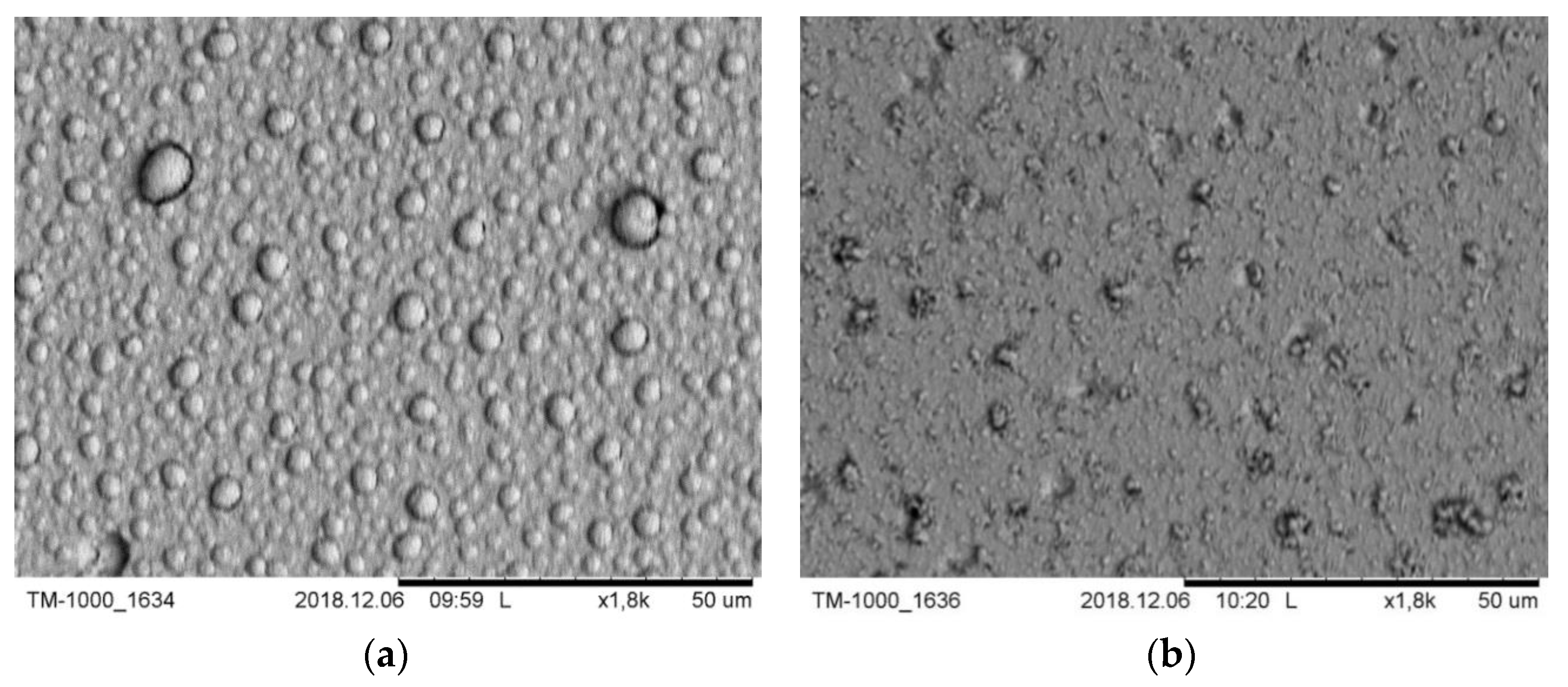
It was revealed that during long high-temperature annealing (more than 20–25 min), there is a significant decrease in the thickness of the sprayed layer (more than 10–15%) relative to the initial thickness. This is probably due to the thickening of the ITO film during its high-temperature heating, which can be seen in the microphotographs of the ITO film sample surfaces, before and after annealing (Figure 2).
The surface morphology of the ITO films is heterogeneous, with the formation of hemispherical structures on the film surface (Figure 2a). These formations are probably caused by atmospheric capture of the working gases during film growth. The size of the hemispherical structures range from 2.5 to 10 μm. As a result of annealing, the film surface flattens out (Figure 2b). The size of such formations reaches 5 μm.
The increase in the charge carrier’s mobility, by an order of magnitude after high-temperature annealing, is most likely caused by an increase in the structural perfection of ITO films. To determine the phase composition, an X-ray phase analysis of the ITO film samples before and after annealing was done (Figure 3).
The analysis of the obtained X-rays showed that there is a partial crystal structure formation in the film volume during ITO film annealing; however, part of the film continues to consist of an amorphous phase.. The orientation of reflection planes (222) prevails and corresponds to the densely packed plane (111) in which direction the most intensive crystal growth occurs.
The crystallite size increases with annealing temperature increase. The significant influence of the annealing mode on the ITO film characteristics is also evidenced by the results of [23,27].
Transmission spectra of ITO films before and after high-temperature annealing are shown in Figure 4.
The transmission spectra analysis of ITO films on quartz glass substrates showed that the maximum optical transmission is observed in the wavelength range of 0.4 to 1 μm. The decrease in transmittance in the wavelength range of 2 to 4 μm is due to the absorption of the substrate. Conducting high-temperature annealing of ITO films leads to a decrease in their optical absorption.
According to Drude’s theory, there is a relationship between the optical and electrical properties of the material due to the interaction of the electric field component of light with the free electrons of transparent conducting oxides [28].
ITO films have high transparency in the visible area of the transmission spectrum. The decrease in transmittance and increase in reflection of light radiation by an ITO film is observed in the infrared region of the spectrum at wavelengths above 1500 nm. This transition, corresponding to the absorption maximum, is associated with the wave excitation of volume plasmon resonance when the frequency of light coincides with the frequency of electrons oscillations in ITO. When the emission wavelength is less than the excitation wavelength of the bulk plasmon resonance, the wave function is oscillatory, and light can pass unimpeded through the ITO film. When the wavelength exceeds the excitation wavelength of the volumetric plasmon resonance, there is a light reflection.
Thus, the study of the electrophysical and optical properties of ITO films obtained by reactive ion-plasma sputtering confirmed the possibility of their use as an active layer for SPR excitation, and its wavelength can be corrected by the concentration of charge carriers in the ITO film.

3.2. Study of Optical Mode Excitation and Plasmon Resonance

To study the excitation of optical modes and plasmon resonance by the Kretschmann configuration, ITO films were deposited on the base of an equilateral glass prism (Figure 5).
A 50 W halogen lamp was used as the radiation source. A Nicol prism was used to give the lamp radiation the necessary polarization: TE-polarization (electric field vector is perpendicular to the radiation incidence plane) or TM-polarization (electric field vector is parallel to the radiation incidence plane). Figure 6 shows the reflection spectra of the prism/ITO film system at different angles between the electric field vector and the incidence plane.
Analysis of the reflection spectra of the prism/ITO film system, at different values of the angle θ, between the electric field vector and the plane of the prism face receiving the radiation, showed that in the studied system with TE polarization (Figure 6, line 3), excitation of the optical mode is observed at the dielectric/ITO film interface. Stable excitation of the plasmon resonance is observed in TM-polarization, when the electric field vector of the incident light is in the incidence plane. The reflection minimum is blurred because of the significant aperture of the incident light beam.
To study the effect of analyte characteristics on the change in plasmon resonance parameters, analytes with different refractive indices were applied to the base of the prism on top of the ITO film: alcohol (n = 1.35), glycerin (n = 1.47), and petrolatum (n = 1.5). Excitation of plasmon resonance was monitored by the reflection spectrum from the prism/ITO film/analyte system. Measurements were performed under specific conditions. Linearly polarized incident light is on the ITO film from the side of the prism. The electric field vector of the incident light is in the incidence plane. When the incidence angle of polarized light on the ITO film is 51°, there is agreement in the wave number of the incident light wave and the surface plasmon wave, which is marked by a significant decrease in the reflection coefficient. The results of the experiments are shown in Figure 7.
Analysis of the reflection spectra of optical radiation from the studied prism/ITO film/analyte system showed the presence of minima related to the SPR excitation [18]. The large resonance width of the spectra is apparently due to the presence of an amorphous phase in the studied ITO films, as a direct consequence of incomplete crystallization. This is confirmed by the X-ray of ITO films subjected to high-temperature annealing after deposition (Figure 3, line 2).
Processing of the obtained spectra of light reflection from the prism/ITO film/analyte system, with different thicknesses of ITO films, made it possible to obtain the dependence of plasmon resonance excitation wavelength change on the used analyte refractive index (Figure 8).
The sensitivity of optical SPR sensors is one of the most important parameters [3,29]. Sensitivity characterizes the change in the value of the sensor output signal when the measured value changes, and is numerically equal to the angle tangent values of the obtained experimental curves inclination (Figure 8) [30]. The obtained dependencies show that 30 nm thick ITO films have a greater sensitivity of the plasmon resonance parameters to the refractive indexes of the analyte changes. It should be noted that the plasmon resonance excitation wavelength itself shifts to the shorter wavelength part of the spectrum when the ITO film thickness increases. This is caused by a decrease in the bulk resistivity of ITO with their increasing thickness (Figure 1).
The shortest plasmon resonance wavelength is observed when the ITO film is in contact with ethyl alcohol with refractive index n = 1.35. When the refractive index of the analyte under study increases, the plasmon resonance wavelength increases too.
Thus, the reflected radiation spectrum from the glass prism/ITO film/analyte interface allows the registering of the excitation of surface or volume plasmon resonance, the parameters of which depend on the analyte properties.

4. Conclusions

In this work, a technological mode of optically transparent low–resistance ITO film deposition with a charge carrier concentration of 1021–1022 cm−3 by reactive magnetron sputtering was developed. Studies of their optical and electrical parameters have been carried out. Irradiation of the interface between a dielectric and a low-resistance ITO film with a TM polarized light beam, with its wavelength range of 350 to 950 nm, by the Kretschman method, showed the excitation of surface plasmon resonance. Registration of plasmon resonance was carried out by the presence of a minimum on the spectrum of reflected radiation, which is caused by partial energy absorption and surface electromagnetic waves excitation. The addition of various analytes to the surface of the ITO film changes the position of the minimum reflection coefficient of optical radiation from the prism/ITO film/analyte system. Thus, surface electromagnetic waves excited in thin ITO films by the Kretschmann method are sensitive to changes in the refractive index of the analyzed liquids. Optical SPR sensors based on ITO films with a thickness of 30 nm showed more than four times greater sensitivity than SPR sensors based on ITO films with 60 nm thickness. The wavelength of the SPR excitation can be adjusted by the concentration of charge carriers in the ITO film. In this work, due to the high concentration of carriers, SPR excitation was observed in the visible spectrum. However, the resonance width of the SPR spectrum is quite large and prevents the creation of optical sensors based on ITO films alone. Further work will be aimed at increasing the sensitivity of optical SPR sensors based on low-resistance ITO films.

Author Contributions

Conceptualization, S.S. and Y.Z.; methodology, Y.Z.; validation, I.Y. and P.T.; formal analysis, S.S.; investigation, K.Z. and A.I.; resources, I.Y. and Y.Z.; data curation, Y.Z.; writing—original draft preparation, Y.Z. and A.I.; writing—review and editing, S.S. and P.T.; visualization, K.Z.; supervision, I.Y.; project administration, Y.Z.; funding acquisition, Y.Z. All authors have read and agreed to the published version of the manuscript.

Funding

The work was carried out with financial support from the Ministry of Science and Higher Education of the Russian Federation under agreement No. 075-03-2020-237/1 from 05.03.2020 (project number FEWM-2020-0040). The study of plasmon resonance excitation was carried out at the expense of the Russian Science Foundation grant No. 21-72-00124.

Institutional Review Board Statement

Not applicable.

Informed Consent Statement

Not applicable.

Data Availability Statement

All data that support the findings of this study are included within the article.

Conflicts of Interest

The authors declare no conflict of interest.

References

  1. Liu, C.; Lü, J.; Liu, W.; Wang, F.; Chu, P.K. Overview of refractive index sensors comprising photonic crystal fibers based on the surface plasmon resonance effect [Invited]. Chin. Opt. Lett. 2021, 19, 102202. [Google Scholar] [CrossRef]
  2. Nguyen, H.H.; Park, J.; Kang, S.; Kim, M. Surface Plasmon Resonance: A Versatile Technique for Biosensor Applications. Sensors 2015, 15, 10481–10510. [Google Scholar] [CrossRef] [PubMed] [Green Version]
  3. Homola, J. Surface Plasmon Resonance Based Sensors; Springer: Berlin/Heidelberg, Germany, 2006. [Google Scholar] [CrossRef]
  4. Noguez, C. Surface Plasmons on Metal Nanoparticles:  The Influence of Shape and Physical Environment. J. Phys. Chem. C 2007, 111, 3806–3819. [Google Scholar] [CrossRef]
  5. Anker, J.; Hall, W.; Lyandres, O.; Shah, N.C.; Zhao, J.; Van Duyne, R.P. Biosensing with plasmonic nanosensors. Nat. Mater. 2008, 7, 442–453. [Google Scholar] [CrossRef] [PubMed]
  6. Saigusa, M.; Tsuboi, K.; Konosu, Y.; Ashizawa, M.; Tanioka, A.; Matsumoto, H. Highly sensitive local surface plasmon resonance in anisotropic Au nanoparticles deposited on nanofibers. J. Nanomater. 2015, 37, 829273. [Google Scholar] [CrossRef] [Green Version]
  7. Liu, Y.; Huang, C.Z. Screening sensitive nanosensors via the investigation of shape-dependent localized surface plasmon resonance of single Ag nanoparticles. Nanoscale 2013, 5, 7458–7466. [Google Scholar] [CrossRef]
  8. Chung, H.Y.; Chen, C.C.; Wu, P.C.; Tseng, M.L.; Lin, W.C.; Chen, C.W.; Chiang, H.P. Enhanced sensitivity of surface plasmon resonance phase-interrogation biosensorby using oblique deposited silver nanorods. Nanoscale Res. Lett. 2014, 9, 476–480. [Google Scholar] [CrossRef] [Green Version]
  9. Su, W.; Zheng, G.; Li, X. Design of a highly sensitive surface plasmon resonance sensor using aluminum based diffraction grating. Opt. Commun. 2012, 285, 4603–4607. [Google Scholar] [CrossRef]
  10. Sang, X.Z.; Zhang, D.W. Research on the SPR properties of copper thin film with regulation of titanium dioxide. Spectrosc. Spectr. Anal. 2016, 36, 2027–2030. [Google Scholar]
  11. Usha, S.P.; Mishra, S.K.; Gupta, B.D. Fabrication and characterization of a SPR-based fibre-optic sensor for the detection of chlorine gas using silver and zinc oxide. Materials 2015, 8, 2204–2216. [Google Scholar] [CrossRef]
  12. Li, Z.; Zhang, Z.; Chen, K. Indium–Tin–Oxide Nanostructures for Plasmon-Enhanced Infrared Spectroscopy: A Numerical Study. Micromachines 2019, 10, 241. [Google Scholar] [CrossRef] [PubMed] [Green Version]
  13. Kamakura, R.; Takeishi, T.; Murai, S.; Fujita, K.; Tanaka, K. Surface-Enhanced Infrared Absorption for the Periodic Array of Indium Tin Oxide and Gold Microdiscs: Effect of in-Plane Light Diffraction. ACS Photonics 2018, 5, 2602–2608. [Google Scholar] [CrossRef]
  14. Li, S.Q.; Guo, P.; Zhang, L.; Zhou, W.; Odom, T.W.; Seideman, T.; Ketterson, J.B.; Chang, R.P.H. Infrared Plasmonics with Indium–Tin-Oxide Nanorod Arrays. ACS Nano 2011, 5, 9161–9170. [Google Scholar] [CrossRef] [PubMed]
  15. Abb, M.; Wang, Y.; Papasimakis, N.; de Groot, C.H.; Muskens, O.L. Surface-Enhanced Infrared Spectroscopy Using Metal Oxide Plasmonic Antenna Arrays. Nano Lett. 2014, 14, 346–352. [Google Scholar] [CrossRef] [Green Version]
  16. Kim, J.; Dutta, A.; Memarzadeh, B.; Kildishev, A.V.; Mosallaei, H.; Boltasseva, A. Zinc Oxide Based Plasmonic Multilayer Resonator: Localized and Gap Surface Plasmon in the Infrared. ACS Photonics 2015, 2, 1224–1230. [Google Scholar] [CrossRef]
  17. Jiang, H.; Zhou, Y.-R.; Liu, F.-Z.; Zhou, Y.-Q. Effect of annealing treatment on characteristics of surface plasmon resonance for indium tin oxide. Acta Phys. Sin.-Chin. Ed. 2018, 67, 177802. [Google Scholar] [CrossRef]
  18. Korotkova, K.; Bainov, D.; Smirnov, S.; Yunusov, I.; Zhidik, Y. Electrical Conductivity and Optical Properties of Nanoscale Titanium Films on Sapphire for Localized Plasmon Resonance-Based Sensors. Coatings 2020, 10, 1165. [Google Scholar] [CrossRef]
  19. Ching-Wei, C.; Yen-Cheng, L.; Chia-Hua, C.; Peichen, Y.; Jia-Min, S.; Ci-Ling, P. Frequency-dependent complex conductrivities and dielectric recponses of indium tin oxide thin films from the visible to the far-infrared. IEEE J. Quantum Electron. 2010, 46, 1746–1754. [Google Scholar] [CrossRef]
  20. Seeger, K. Lattice Reflection in Polar Semiconductors. In Semiconductor Physics, 8th ed.; Hubertus von Riedesel, Ed.; Springer: Heidelberg, Germany, 2002; pp. 352–354. [Google Scholar] [CrossRef]
  21. Granqvist, C.G.; Hultaker, A. Transparent and conducting ITO films: New developments and applications. Thin Solid Films 2002, 411, 1–5. [Google Scholar] [CrossRef]
  22. Untila, G.G.; Kost, T.N.; Chebotareva, A.B.; Timofeev, M.A. Effect of the tin content on the composition and optical and electrical properties of ITO films deposited onto silicon and glass by ultrasonic spray pyrolysis. Semiconductors 2012, 46, 984–990. [Google Scholar] [CrossRef]
  23. Facchetti, A.; Marks, T.J. Transparent Electronics: From Synthesis to Applications, 1st ed.; Wiley: Hoboken, NJ, USA, 2010; p. 452. [Google Scholar]
  24. Amosova, L.P. Electrooptical properties and structural features of amorphous ITO. Semiconductors 2015, 49, 414–418. [Google Scholar] [CrossRef]
  25. Zhidik, Y.S.; Troyan, P.E.; Kozik, V.V.; Kozyukhin, S.A.; Zabolotskaya, A.V.; Kuznetsova, S.A. Investigation of Electrophysical Properties of ITO Films. Russ. Phys. J. 2020, 63, 1139–1143. [Google Scholar] [CrossRef]
  26. Troyan, P.E.; Zhidik, Y.S.; Sakharov, Y.V. Synthesis of conducting films In2O3:Sn with the method of magnetron sputtering and their electrophysical properties. In Proceedings of the 15th International Conference of Young Specialists on Micro/Nanotechnologies and Electron Devices, Novosibirsk, Russia, 30 June–4 July 2014. [Google Scholar] [CrossRef]
  27. Oka, N.; Kawase, Y.; Shigesato, Y. High-rate deposition of high-quality Sn-doped In2O3 films by reactive magnetron sputtering using alloy targets. Thin Solid Films 2012, 520, 4101–4105. [Google Scholar] [CrossRef]
  28. Pasquarelli, R.M.; Ginley, D.S.; O’Hayre, R. Solution processing of transparent conductors: From flask to film. Chem. Soc. Rev. 2011, 40, 5406–5441. [Google Scholar] [CrossRef]
  29. Xia, G.; Zhou, C.; Jin, S.; Huang, C.; Xing, J.; Liu, Z. Sensitivity Enhancement of Two-Dimensional Materials Based on Genetic Optimization in Surface Plasmon Resonance. Sensors 2019, 19, 1198. [Google Scholar] [CrossRef] [Green Version]
  30. Mrozek, P.; Gorodkiewicz, E.; Falkowski, P.; Hościło, B. Sensitivity Analysis of Single- and Bimetallic Surface Plasmon Resonance Biosensors. Sensors 2021, 21, 4348. [Google Scholar] [CrossRef] [PubMed]
Figure 1. The dependency of the SPR wavelength on the resistivity of the ITO film.
Coatings 12 01868 g001
Figure 2. Surface microphotographs of ITO samples: (a) Before high-temperature annealing; (b) After high-temperature annealing.
Coatings 12 01868 g002
Figure 3. X-ray patterns of ITO films: (1) Before high-temperature annealing; (2) After high-temperature annealing.
Coatings 12 01868 g003
Figure 4. Transmission spectra of ITO films in visible spectrum: (1) Before high-temperature annealing; (2) After high-temperature annealing.
Coatings 12 01868 g004
Figure 5. Experimental samples for plasmon resonance excitation: (a) A glass equilateral prism with ITO film deposited on the base; (b) A sample mounted in the experimental setup for plasmon resonance excitation.
Coatings 12 01868 g005
Figure 6. Reflection spectrum of the prism/ITO film system at different values of the angle between the electric field vector and the plane of the prism face receiving the radiation: (1) θ = 0°; (2) θ = 45°; (3) θ = 90°.
Coatings 12 01868 g006
Figure 7. Reflection spectrum of systems glass prism/ITO film/analyte (petrolatum, glycerol, alcohol) system with different thicknesses of ITO films: (a) 30 nm; (b) 60 nm.
Coatings 12 01868 g007
Figure 8. Dependencies of changes in the plasmon resonance excitation wavelength in the prism/ITO film/analyte system on the refractive index of the analyte used: (1) alcohol; (2) glycerol; (3) petrolatum.
Coatings 12 01868 g008
Table 1. Results of ITO film deposition technology correction.
Sample Series NoOxygen in the Gas MixtureρS before Annealing, Ohm/□ρS after Annealing, Ohm/□
120%2475661
218%110785.2
316%102178.9
414%87349.4
512%63913.0
610%3168.6
78%59424.5
86%68137.3
Table 2. Electron-kinetic characteristics of charge carriers in ITO films.
ρV, Ohm·cmn, cm−3μ, cm2/(V·s)
Before annealing3.16 × 10−37.33 × 10202.7
After annealing8.6 × 10−52.25 × 102132.3
Publisher’s Note: MDPI stays neutral with regard to jurisdictional claims in published maps and institutional affiliations.