Article

The Phase Evolution and Photocatalytic Properties of a Ti-TiO2 Bilayer Thin Film Prepared Using Thermal Oxidation

1
Department of Materials Science and Engineering, National Cheng Kung University, Tainan 70101, Taiwan
2
Department of Tropical Agriculture and International Cooperation, National Pingtung University of Science & Technology, Pingtung 91201, Taiwan
3
Department of Agricultural Product Technology, Faculty of Agricultural Technology, Universitas Brawijaya, Malang 65145, Indonesia
4
Department of Plant Industry, National Pingtung University of Science & Technology, Pingtung 91201, Taiwan
5
Kaohsiung Veterans General Hospital, Kaohsiung City 813414, Taiwan
6
Department of Applied Physic, National Pingtung University, Pingtung 90003, Taiwan
7
Department of Materials Engineering, National Pingtung University of Science & Technology, Pingtung 91201, Taiwan
*
Author to whom correspondence should be addressed.
Coatings 2021, 11(7), 808; https://doi.org/10.3390/coatings11070808
Submission received: 6 May 2021 / Revised: 29 June 2021 / Accepted: 30 June 2021 / Published: 3 July 2021

Abstract

:
Ti-TiO2 bilayer thin films were successfully prepared onto a glass substrate using magnetron sputtering with different TiO2 bottom layer conditions. These represent a lack of (as-deposited) and full oxygen content (annealed). Single-layer Ti was additionally used as a control. The influence of oxygen diffusion phenomena of the bottom layer of TiO2 to the upper layer of Ti thin films at different oxidation temperatures on structural, optical, and photocatalytic performance was investigated. X-ray diffraction (XRD) results confirmed that the crystalline phases coexisting on thin-film samples oxidized at 450 °C were TiO, TiO1.4, (bilayer, as-deposited TiO2), anatase (bilayer, annealed TiO2), and rutile (single and bilayer). This finding showed that the film’s phase structure evolution is significantly affected by oxygen diffusion from the bottom layer. Further increasing the thermal oxidation temperature caused a notable decline in the amorphous zone in bilayer thin films based on TEM analysis. Bilayer thin films lead to higher degradation of methylene blue under UV light radiation (63%) than single-layer films (45%) oxidized at 450 °C. High photocatalytic activity performance was found in the bilayer annealed TiO2-Ti thin-film sample. This study demonstrates that the bilayer modification strategy promotes the oxygen-induced bottom layer of TiO2 bilayer thin films.

Graphical Abstract

1. Introduction

Semiconductor-based photocatalytic reactions have been fascinating as a promising technology that relies on the interaction between light and solid semiconductor particles. Titanium dioxide (TiO2) has been acknowledged as the most crucial photocatalyst that strongly resists chemical and photo-corrosion, is non-toxic, and has thermal stability [1]. Several methods have been introduced to synthesize TiO2 thin films, such as the sol-gel method [2], the combustion method [3], the chemical vapor deposition (CVD) method [4], and the sputtering method [5]. The magnetron sputtering technique was approved for providing solid substrate adhesion and a large area with uniform thickness [6,7].
The high-power impulse magnetron sputtering (HiPIMS) technique has demonstrated significant potential in material processing. This technique enables accessing high-density plasmas with excessive quantities of ionized species, high-energy transfer functions, and low duty cycles than direct current magnetron sputtering (DC-MS) [5,8,9]. Various studies have highlighted the favorable impact of reactive HiPIMS on thin-film performance, including denser microstructures, higher hardness, good film adhesion, a high degree of crystallinity, and extended phase stabilities or enhanced surface coverage complex-shaped substrate [10,11,12]. However, due to the excessive ions sputtered during deposition to the target, the HiPIMS approach is less efficient, which decreases the deposition rate [13]. One approach to addressing this issue uses another coating technology. The DC-MS technique generates coatings with a higher deposition rate than the HiPIMS technology. Therefore, the combined HiPIMS and DC-MS method has been suggested to increase deposition rates and improve physical, chemical, and functional properties [14,15,16].
Several studies have recently developed TiO2 films using thermal oxidation from sputtered Ti metal films [17,18]. The thermal oxidation temperature plays a crucial role in the crystal structure, particle morphology, and various TiO2 phases, such as anatase or rutile, and their mixed phases have distinct photocatalytic activity levels [19]. The layered structure of the deposited Ti layers changes from amorphous to crystalline at the anatase phase by thermal oxidation [20]. Oxygen transport plays a crucial role in developing oxide materials using the thermal oxidation technique. Oxygen diffusion in TiO2 thin films has previously been intensively studied based on electrical conductivity measurement [21] and oxygen tracer experiments [22,23]. Several studies have suggested that the oxidation layer is grown during the initial oxidation stage and distributed in the substrate. When the oxygen penetrates the subsurface layer, it is then chemisorbed and formed onto the surface [24], resulting in forming an oxide layer with substantial thickness. However, if the film thickness is greater than 200 nm, it is difficult to obtain the mixed phase (rutile and anatase), because the titanium film does not entirely oxidize down to the inner layer [17]. In addition, increasing the temperatures (700 and 900 °C) to create high oxidation states in thick films results in decreased band gap energy due to oxygen defect formation in the thin films [25]. As a result, the thin-film photocatalytic performance decreases.
Most researchers have focused on the development of thermal oxidation treatment for oxide surface growth and investigated the effects of temperature on the structural, microstructure, optical also Ti/O ratio in single-layer Ti films [25,26]. In contrast, developing a bilayer thin film to perform oxygen diffusion from the bottom to the upper layer has yet to be fully mastered. Therefore, it is necessary to investigate the oxygen diffusion phenomena from TiO2 as the bottom layer, with different conditions representing a lack of and full oxygen content (as-deposited and annealed films, respectively), to the upper layer of Ti thin films. Furthermore, with further thermal oxidation, thin-film coupling between Ti and TiO2 is predicted to contribute to the oxygen diffusion from the bottom to the upper layer, affecting several thin-film properties. In this study, three types of samples were prepared: a single layer of Ti, a bilayer of Ti-TiO2 as a deposit, and a bilayer of annealed Ti-TiO2. Different thermal oxidation temperatures were used to investigate the bottom-layer oxygen dependency on phase evolution, crystalline behavior, microstructures, and photocatalytic properties of Ti-TiO2 bilayer thin films.

2. Materials and Methods

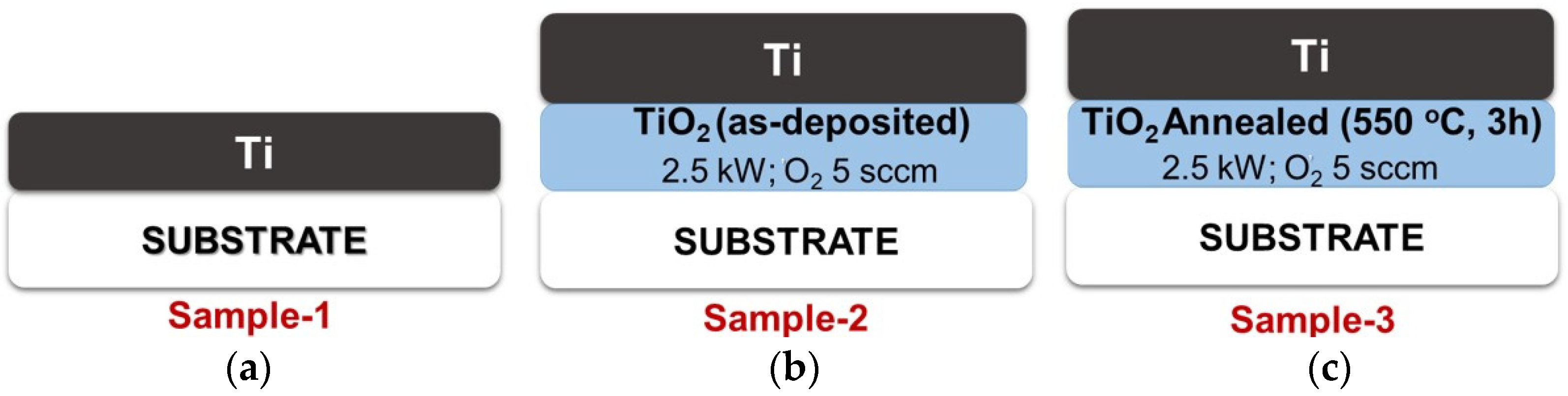
2.1. Preparation of TiO2 Thin Films as a Bottom Layer

A titanium oxide (TiO2) thin film as the bottom layer was deposited onto glass substrates (26 × 76 mm2) using reactive HiPIMS. A pure titanium target (99.99% purity, Ultimate materials Technology Co., Ltd., Hsinchu, Taiwan) with 550 × 125 × 6 mm3 dimensions was used to set the distance between target and substrate at 150 mm. The sputtering power was fixed at 2.5 kW, with a deposition rate of 15 (mm/s) × 2 passes. High-purity argon (99.99%) and oxygen (99.99%) gases were introduced into the vacuum chamber using a mass flow controller with gas flow rates of 200 and 5 sccm, respectively. The combination of oxygen gas flow and sputtering power to develop TiO2 thin films has been observed in previous research. The working pressure was fixed at 2.7 × 10⁻³ Torr. The substrate temperature was controlled at 25 °C. The TiO2 film bottom-layer thickness was controlled at about 40 nm. Both TiO2 films, as-deposited and annealed (550 °C for 3 h in the air), were created to obtain different oxygen contents in the bottom layer. Several parameters, such as sputtering power, oxygen flow, and annealing temperature, used in this research to develop as-deposited and annealed TiO2 were investigated as a preliminary study.

2.2. Ti-TiO2 Bilayer Thin-Film Preparation

Pure Ti films were produced as an upper layer deposited using direct-current (DC) magnetron sputtering with a pure titanium target (99.99% purity, Ultimate materials Technology Co., Ltd, Hsinchu, Taiwan), 3 inches in diameter and 6 mm thick. The distance between target and substrate was set at 100 mm. The base pressure was controlled at 3.2 × 10⁻⁶ Torr, and the working pressure was 6.7 × 10⁻³ Torr. The argon (99.99%) gas flow was controlled at 30 sccm by a mass flow controller. The substrate holder was rotated at 24 revolutions per minute (rpm). Ti upper-layer films were deposited on a TiO2 bottom-layer film at room temperature. The DC power was fixed at 125 W with a 12 min deposition time. The Ti film top-layer thickness was controlled at about 120 nm.
Three types of samples were prepared, as illustrated in Figure 1. Sample 1 was a single-layer Ti film deposited onto glass substrates (Figure 1a). Samples 2 and 3 were a bilayer thin film containing as-deposited TiO2 (without annealing) (Figure 1b) and annealed TiO2 (550 °C for 3 h) (Figure 1c) films as the bottom layer, respectively. Furthermore, the Ti film covered as the upper layer for both samples 2 and 3. Different oxidation temperatures (450 and 550 °C for 3 h) were used to evaluate the effects of the bottom-layer oxygen dependency of Ti-TiO2 bilayer thin films on crystalline behavior, microstructures, optical, and photocatalytic properties.

2.3. Sample Characterization

Film crystallinity was analyzed with X-ray diffraction (XRD; Bruker D8 Advance- AXS Gmbh, Am Studio 2D, Berlin, Germany) using Monochromatic Cu Kα radiation (λ = 1.540598). The machine was operated at 40 kV and 40 mA. The scanning angle 2θ with a value range of 10° ≤ 2θ ≤ 80° and a step size of 0.1°, measuring time at 5 s per step, was used. Microstructural and oxidation layer observations of the thin-film cross-sectional and plane-view morphology grown onto glass substrates were made with field-emission gun transmission electron microscopy (FEG-TEM; FEI E.O Tecnai F20, FEI Company—Thermo Fisher Scientific, Hillsboro, OR, USA) equipped with an energy-dispersive spectrophotometer at an acceleration voltage of 200 kV. Selected-area electron diffraction (SAED) and nano-beam electron diffraction (NBED) were performed using Gatan Software Digital Micrograph software version 3.7.4 (Gatan Inc., Pleasanton, CA, USA).
The thin-film samples’ transmission spectra were monitored on a UV–VIS spectrophotometer (UV/Vis, U-3310, Hitachi Ltd., Hitachi, Japan) with a wavelength of 300–800 nm. X-ray photoelectron spectroscopy (XPS; JEOL, JPS-9010MX, Japan Electronics Corp, Akishima, Japan) was used to examine the films’ quantitative composition on the surface and the internal area. Internal profile experiments were performed by sputter etching at a rate of 28 nm/min in the thin films for 20 s using argon gas. The analyzer was operated at voltage 10 kV and current 5 mA with a Mg Kα radiation line. X-ray photoelectron spectra were referenced to the C 1s peak (Eb = 285 eV) that presented on the sample surface.

2.4. Photocatalytic Activity

The photocatalytic performance of the thin-film samples was evaluated by testing the decolorization of a high-purity C16H18ClN3xH2O (methylene blue (MB)) decomposition solution (Alfa Aesar; Thermo Fisher Scientific, Lancashire, UK) with an initial concentration (C0) of 10 mg/L. Thin-film samples with 26 mm × 38 mm dimensions were immersed into a 20 mL aqueous MB solution (1 mg/L) in a beaker. The distance between the reactor and the lamp was kept at 9 cm. The photocatalytic performance of the Ti-TiO2 bilayer thin film was investigated under 8 watt UV-C lamp irradiation (UV-C T8; λ = 253.7 nm, Philip, Amsterdam, The Netherlands). The dye solution with the photocatalyst was kept in the dark for 10 min to reach adsorption–desorption equilibrium before irradiation. Briefly, from the end of the dark-storage time (t = 0 min, Ct), the dye solution sample was collected at a regular interval of 30 min for 3 h (t = 30–180 min, C30 to C180). The collected sample was analyzed by a UV–VIS spectrophotometer (UV/Vis, U-3310, Hitachi Ltd., Tokyo, Japan) to obtain the absorbance for quantifying the concentration of MB (the maximum absorption wavelength of the dye at 664 nm) [27]. The methylene blue degradation was reported by calculating the percentage of the concentration of the dye that remained (Ct) over the initial dye concentration (C0), obtained from the absorbance standard curve. The degradation performance was evaluated by considering the following Equation (1):
Methylene blue degradation (%) = [1 − (Ct/C0)] × 100
The kinetics of MB photodegradation on the TiO2 surface can be described by the first-order reaction kinetics Equation (2):
In(Ct/C0) = −kt,
where C0 is the initial concentration of MB and Ct is the MB dye concentration at a certain time (mg/L). In the pseudo-first-order photodegradation kinetics, the ln(Ct/C0) linear fit slope in the function of time (t) plot represents the kinetic constant (k) of the photodegradation.

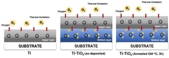
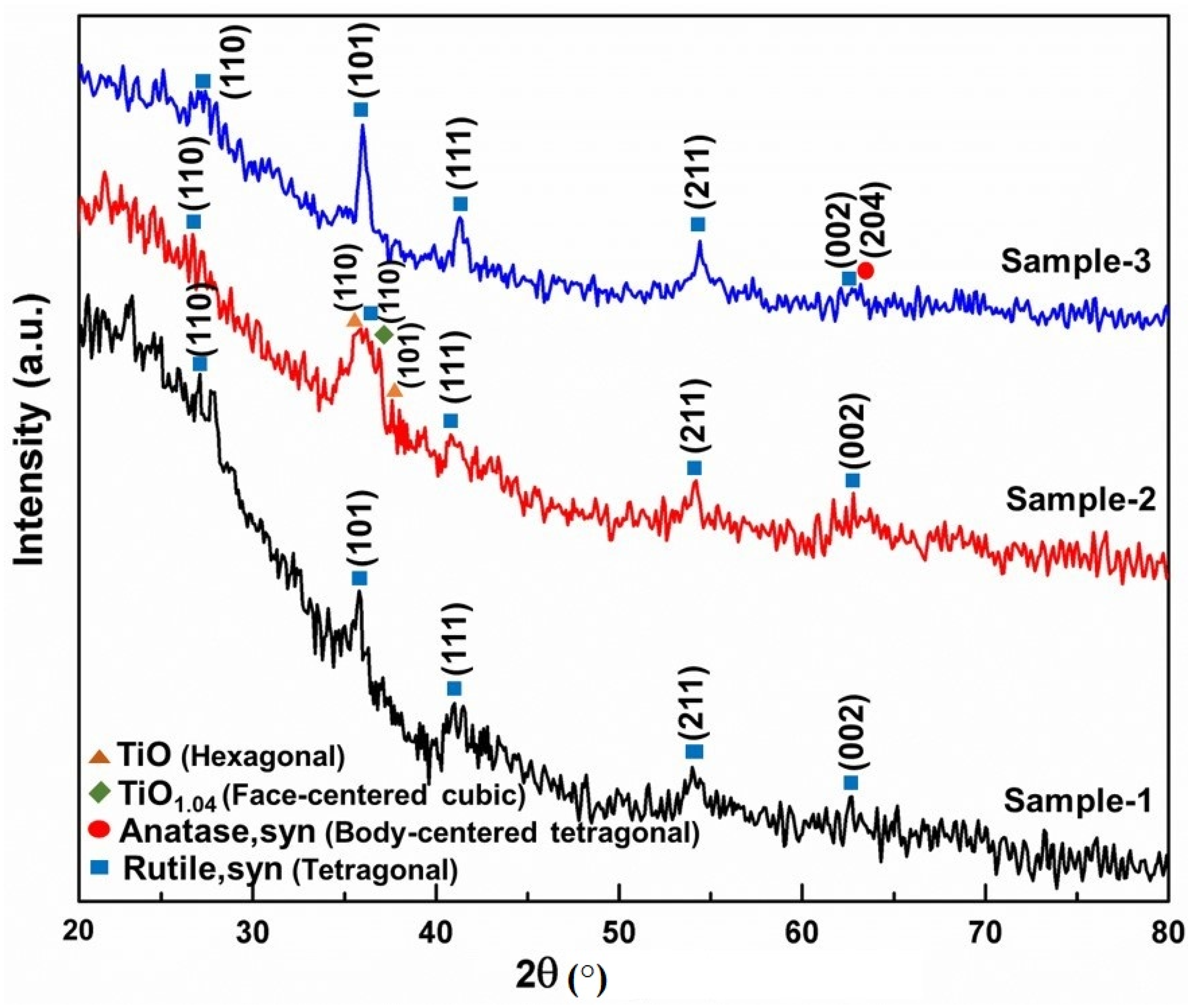
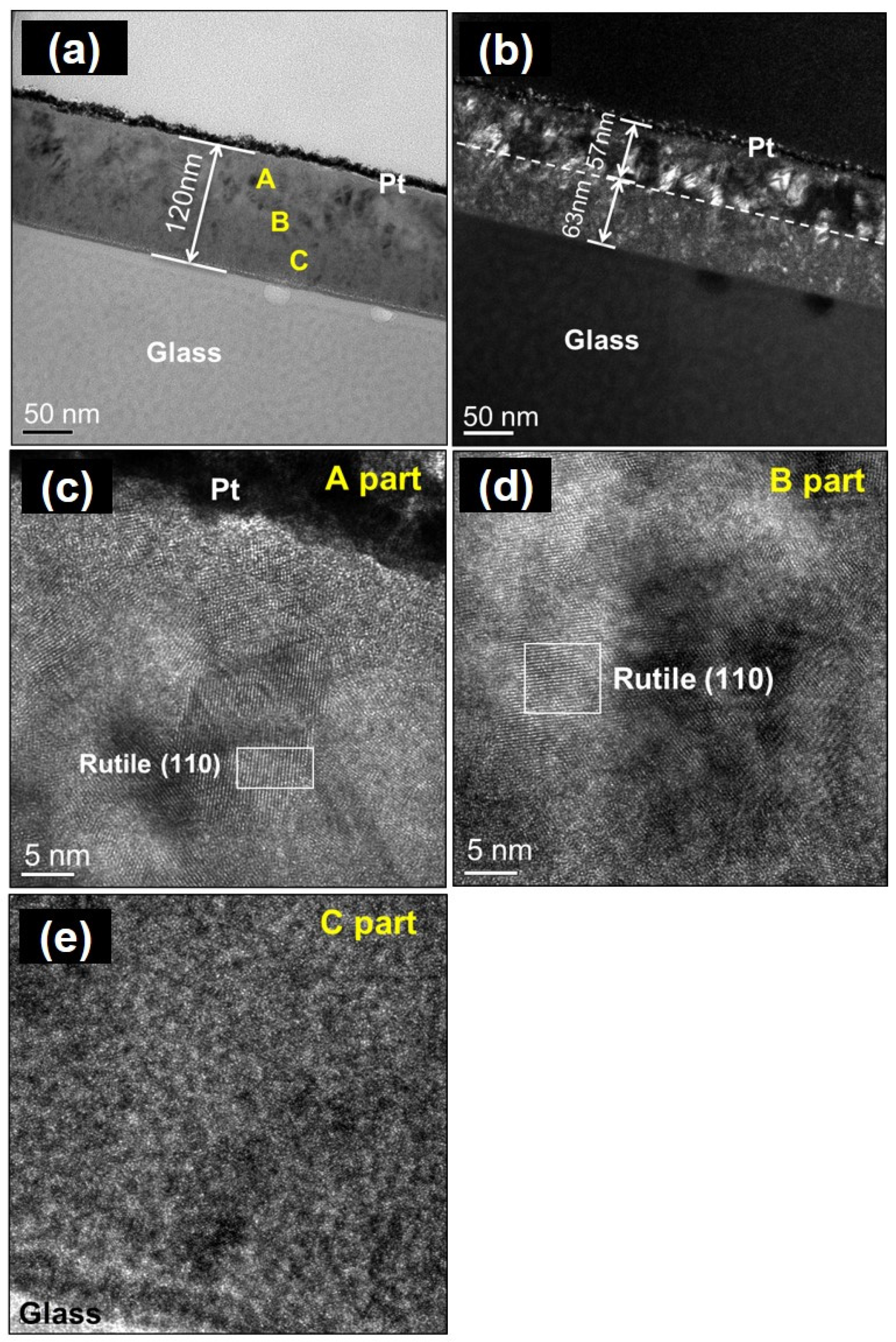
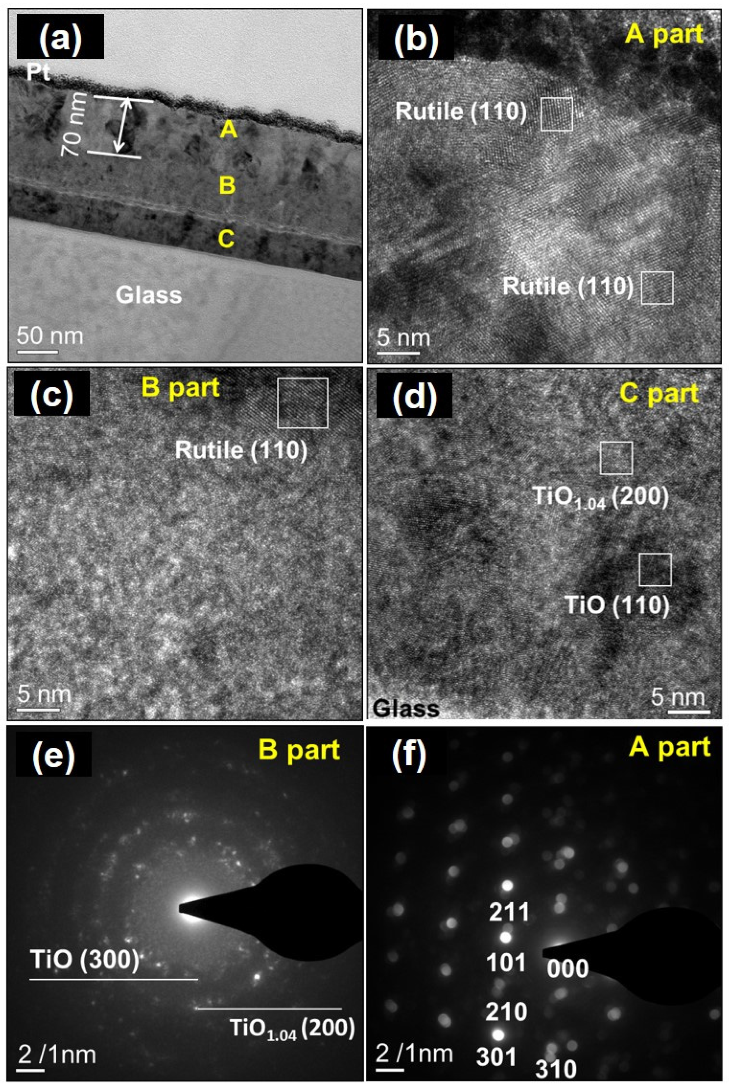
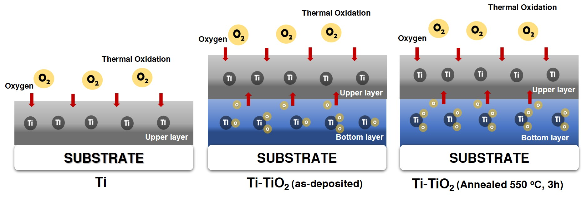
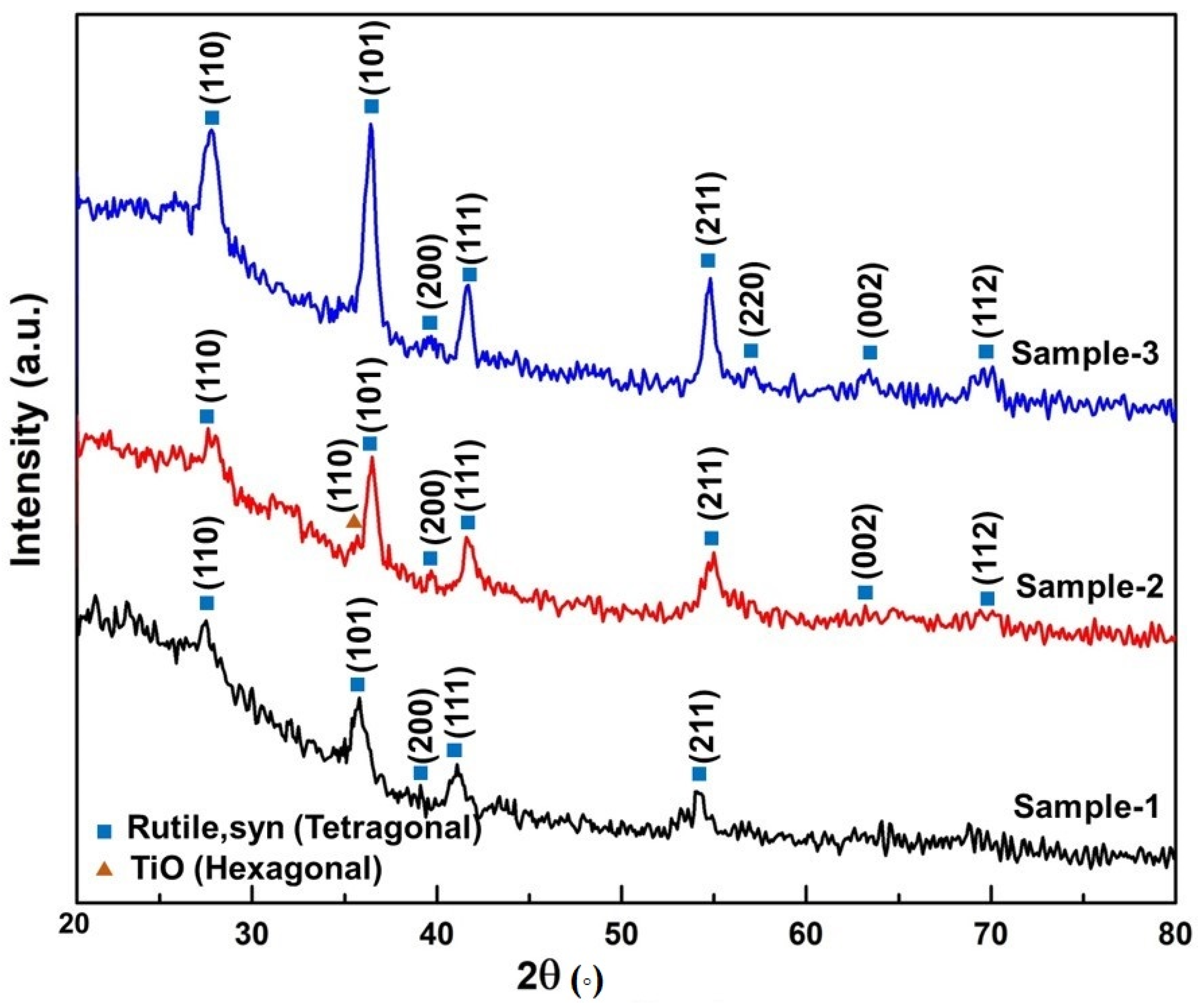
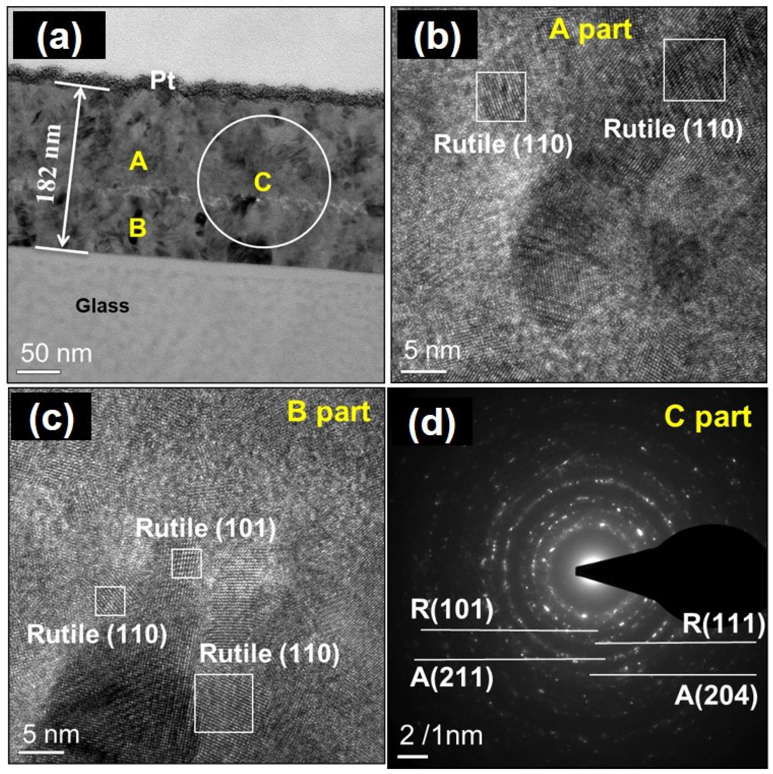
3. Results and Discussion

Figure 2 shows X-ray diffraction patterns of samples that were oxidized in air for 3 h. When the temperature was set at 450 °C, we found that only the TiO2 rutile phase was detected in sample 1. The formation of a single rutile phase at 450 °C is much lower than the formations synthesized using other methods [4,17]. An amorphous phase was found, since XRD peaks showed lower intensities for sample 1, which implied the coexistence of an amorphous structure and crystalline rutile. Three phases coexisted in sample 2: TiO (hexagonal (110)), TiO1.04 (110), and rutile. However, hexagonal TiO and TiO1.04 phases appeared. This is because the titanium dioxide bottom layer does not provide sufficient oxygen for the pure titanium film upper layer during thermal oxidation. This is attributed to the titanium oxide bottom layer being an as-deposited film (insufficient oxygen). This means that a non-stoichiometry TiO layer exists between the upper and the bottom layer. Further study of the microstructure using FE-TEM will be performed to prove this supposition. However, the coexistence of anatase and rutile phases (JCPDS card: 21-1272 and 21-1276) was clearly observed in sample 3. It has to be pointed out that the anatase phase was formed in this investigation. This may be due to the TiO2 bottom layer, which can provide sufficient oxygen to the Ti upper layer. Ting et al. reported that the formation of a significant quantity of oxygen vacancies in the oxidized film accelerates the anatase-to-rutile transformation [25]. Therefore, it is believed that the phase evolution effects on the Ti films oxidized using thermal oxidation depend strongly on the bottom layer.
Figure 3 shows the XRD patterns of samples with different sputtering process parameters annealed at 550 °C. A further increase in temperature to 550 °C resulted in increased diffraction intensity with an associated decrease in the XRD patterns’ amorphous background, indicating improved Ti film crystallinity. Only a rutile crystallization phase was clearly observed in samples 1 and 3 at 550 °C. This indicates complete anatase transition into the rutile phase at 550 °C for samples 1 and 3. As is well known, anatase is a meta-stable phase that may be transformed into the rutile phase during heat treatment [18,28]. For Ti film thermal oxidation, Ti atoms absorb oxygen from the air into the films. The Ti and O atoms react to form TiO2 when the oxidation temperature is 550 °C. However, there was a main rutile phase with a minor TiO phase in sample 2 oxidized at 550 °C. This result indicates that TiO2 (as-deposited) as the bottom layer does not provide sufficient oxygen to the upper layer during thermal oxidation at 550 °C. Chung et al. reported that the formation of TiO was from the Ti thin film after thermal oxidation at 500–550 °C [29].
In the case of the rutile tetragonal crystal structure ( a = b c ) , the lattice constant is calculated using the following formula:
1 d h k l 2 = h 2 + k 2 a + l 2 c 2   ,
where dhkl is the interplanar separation corresponding to Miller indices h, k, and l and a and c are the lattice constants. For all samples, the calculated structural parameters corresponding to peaks (110) and (101) are given in Table 1. When compared with standard rutile TiO2 (a = b = 4.5933 Å and c =2.9592 Å), a slight increase in the lattice constant a and a decrease in the lattice constant c for sample 2 when thermally oxidized at 450 °C were observed. This is due to the TiO and TiO1.04 crystal peaks that represent an oxygen vacancy in a thin-film sample.
High-resolution TEM (HR-TEM) was used to analyze TiO2 thin films to determine whether they were crystalline or amorphous. The TEM specimen microstructures (samples 1, 2, 3) thermally oxidized at 450 °C are shown in Figure 4, Figure 5 and Figure 6, respectively. Figure 4 shows typical bright- and dark-field HR-TEM images of sample 1 thermally oxidized at 450 °C. A film thickness of 120 nm was observed, as shown in Figure 4a. From the dark-field microstructure view, an interface was apparent in the film, separating the crystalline and amorphous zones, as shown in Figure 4b. The oxidation zone in the Ti films was about 57 nm less than the 63 nm amorphous zones. The Ti films thermally oxidized at 450 °C with crystalline structures are exhibited in the upper zone in Figure 4c,d. According to HR-TEM micrograph analysis, the crystalline grains belonged to the rutile crystalline phase (111). Figure 4e clearly shows that the bottom-zone film microstructure is amorphous. However, the film near the surface layer has obviously crystallization. Oxygen diffuses into the internal film zone from the surface by thermal oxidation at 450 °C.
Figure 5 shows sample 2 HR-TEM micrographs and SAED patterns from thermal oxidation at 450 °C for 4 h. The microstructure shows the same interface in the film. It can be divided into two zones. The oxidation thickness in Ti films was about 70 nm at 450 °C for 4 h thermal oxidation, as shown in Figure 5a, which was higher than that for sample 1 (57 nm). According to HR-TEM micrograph analysis, the upper-zone (apart) crystalline grains belong to the rutile crystalline phase (111), as shown in Figure 5b. Two kinds of phase structures coexisted in the interface zone (B part), amorphous and TiO2 grains, as shown in Figure 5c. It clearly shows that the film microstructure is amorphous on the Ti film bottom zone. The TiO2 film bottom layer C part was also analyzed, as shown in Figure 5d. The TiO1.04 (200) and TiO (110) microcrystalline phases coexisted in bottom-layer films. This is attributed to the TiOx film bottom layer deposited onto glass without annealing, which does not readily form the TiO2 crystalline phase stoichiometry. Sekhar et al. reported that TiO2 films deposited using DC reactive magnetron sputtering are amorphous in a continuous gas flow if no bias voltage or heating is applied to the substrate [30,31]. Electron diffraction pattern analysis was carried out to identify crystalline grains, as shown in Figure 5e,f. The SAED pattern displayed a rutile phase in the upper-zone A part (Figure 5f). However, the SAED pattern showed TiO1.04 (200) and TiO (300) planes on the interface-zone B part (Figure 5e). This observation is consistent with the XRD results in the above section. TiO existed in the bottom layer, implying a lack of oxygen content in the film. The oxygen partial pressure influences the crystal formation of Ti films during sputtering [32].
Figure 6 shows HR-TEM micrographs and SAED patterns of sample 3 thermally oxidized at 450 °C. It was mentioned in the above section that sample 3 was TiO2 films deposited on glass substrates as the bottom layer, and it was annealed at 550 °C for 3 h first, and then pure titanium film was deposited on the bottom layer (TiO2 films). In the dark-field view, there was no amorphous zone observed in the films, as shown in Figure 6a. This shows that the film has good crystallinity in all zones, as shown in Figure 6b,c. According to SAED pattern analysis, the crystalline grains belong to the rutile and anatase phases in the films, as shown in Figure 6d. The related literature reported that in the reactive sputtering process, the rutile-and-anatase-mixture-phase TiO2 growth is induced, and the anatase phase increases with the increase in oxygen in the sputter chamber [31,32].
As is well known, anatase phase formation requires sufficient oxygen. Comparing sample 1 to sample 3, the oxygen diffusion from the bottom titanium oxide layer influences the microstructure and phase evolution of Ti films during thermal oxidation. For example, mixture phases (rutile and anatase) with good crystallinity in oxidized Ti films can be obtained when the bottom layer is rutile TiO2 films. Figure 7 shows the amorphous layer thickness of the Ti films on different bottom layers as a function of the oxidation temperature. The amorphous layer thickness was analyzed using TEM. The results showed that the amorphous layer thickness depends strongly on the bottom layer’s oxygen concentration and the oxidation temperature for all samples. The amorphous zone decreased significantly with increasing thermal oxidation temperature. However, a film without an amorphous structure (fully crystalline) was found at 450 °C for sample 3 and at 550 °C for samples 1 and 2. This explains how annealed titanium oxide films as the bottom layer lead to Ti films becoming TiO2 crystalline phases after 450 °C thermal oxidation.
XPS analysis was performed to observe the phase evolution between different sample conditions of Ti-TiO2 bilayer thin films and the oxidation states of the constituent elements present in the samples. Figure 8 displays the spectra of Ti-TiO2 bilayer thin films on glass with different oxidation temperatures. Figure 8a shows the high-resolution XPS spectrum of the Ti 2p peak for samples with different bottom layers at 450 °C. The binding energy curves of Ti 2p with two firm peaks were observed at ~457 and ~462 eV, corresponding to core levels of Ti 2p3/2 and Ti 2p1/2, respectively, which is in good agreement with the recent literature [33,34]. The energy binding differences between the two peaks are dependent on the Ti atoms’ chemical state and the value between these spin-split components [35].
It was observed from the XPS spectra in Figure 8b that increasing the oxidation temperature up to 550 °C shows the binding energy peaks of Ti 2p at single-layer films shifting toward a higher binding energy by ~0.7 eV for both bilayer thin films (samples 2 and 3). Fakhouri et al. [36] proposed that increasing the thin-film layer results in the Ti 2p peak shifting toward lower or high binding energy. This phenomenon may be due to a partial reduction in the titanium cations related to the formation of oxygen vacancies in the lattice or a change in stacked electronic affinity at Ti-TiO2 thin films [36]. According to the literature [37], the chemical states for all thin-film samples (thermally oxidized at 450 and 550 °C) correspond to Ti3+ due to the formation of Ti–O bonds at the binding energy of Ti 2p3/2 of around ~457 eV, characteristic of rutile titanium dioxide that is similar to TiO2. However, the small Ti2+ peaks (456.1 eV) in sample 2 indicated the presence of TiO. The presence of TiO is consistent with the HR-TEM result in Figure 5.
The compositions of Ti-TiO2 bilayer thin films at different thermal oxidation temperatures were identified using XPS. The relative concentrations of titanium and oxygen were analyzed in the Ti-TiO2 bilayer thin films. To understand the difference in oxygen concentration between the surface and the internal zone of the upper Ti oxidized films, the film compositions were analyzed at different positions. The average values of each element are listed in Table 2. The oxygen concentration in the internal zone was lower than that on the surface. For bilayer samples oxidized at 450 °C, the internal and surface zone’s oxygen content was <59% and >69%, respectively. According to XPS analysis results, it is believed that the upper Ti films’ internal zone is not fully oxidized. This means that many oxygen vacancies exist in the films. When the oxidation temperature was enhanced from 450 to 550 °C, the internal zone’s oxygen content increased up to ~60%. The results indicate that the oxygen content in the upper Ti films increases by increasing the oxidation temperature.
UV–VIS spectroscopy was conducted to measure the optical transmission of the Ti-TiO2 bilayer thin films with different samples thermally oxidized at 450 and 550 °C, as shown in Figure 9. The Ti-TiO2 bilayer thin-film transmittance spectra curves indicated that the transmittance spectra gradually shift to a short wavelength as the amorphous thickness in the film increases.
The optical transmission spectra of sample 3 oxidized at 450 °C were exceptionally clear and more than 86% in the visible region. In contrast, sample 1 thermally oxidized at 450 °C had lower transmittance (~41%), while sample 2 had almost no light transmission (0%), as seen in Figure 9a. According to the sample 1 microstructure analysis (Figure 4), oxygen diffusion into the inner film zone slowly occurred at 450 °C, clearly showing that the bottom-zone film microstructure is amorphous. Films with a larger amorphous zone can cause more reflections and light interference, resulting in weak light transmission [38].
The poorest transmittance, especially in the visible region, occurred in sample 2 when oxidized at 450 °C, which is attributed to high absorption in the metallic layer [28], suggesting a sufficient amount of oxygen vacancies that significantly absorbed the incident light [29]. This finding seems consistent with the XRD patterns (Figure 2) and HR-TEM image in Figure 5e, indicating that sample 2 has TiO1.04 (200) and TiO (110) microcrystalline phases that coexist in bottom-layer films. TiO existed in the bottom layer, implying the lack of oxygen content in the film. These phenomena resulted in poor light transmission in sample 2, although it had a lower amorphous thickness than sample 1.
Optical transmittance can be significantly improved while increasing the oxidation temperature up to 550 °C. Due to this phenomenon, titanium metal changes to titanium dioxide after thermal oxidization and increases the oxidation layer of the bilayer thin film. Figure 9b displays bilayer thin films on samples 2 and 3, which showed a significant increase in transmission to ~78% and ~88%, respectively. The transmittance result from sample 3 was higher than the Ti single-layer film thermally oxidized at 600 °C reported by some authors [20,39]. The increasing oxidation temperature has been documented to provide adequate energy to diffuse into the film, improving the TiO2 crystalline strength [40]. However, the transmittance of sample 2 was lower than other samples. It corresponded to TiO (110) instead of the rutile crystallization, as shown in the XRD pattern in Figure 3. As mentioned before, TiO exhibited higher reflective properties due to inadequate oxygen in films. Thin-film transparency improved by increasing the thermal oxidation temperature, which probably removed the residual stress, quantum confinement, and structure order improvement [25], and decreasing the amorphous thickness in the Ti-TiO2 films.
The Ti-TiO2 bilayer thin films’ photocatalytic activity was determined by the degradation of 10 mg/L of MB solution under UV-C light irradiation, as shown in Figure 10. It can be observed that all thin-film samples that were thermally oxidized at 450 and 550 °C exhibited quick adsorption in the dark. This could be attributed to the high specific surface area and small particle size [41]. Figure 10a shows MB degradation (%) of a thin film with different samples oxidized at 450 °C. It was found that all samples provided high photocatalytic degradation performance with a range of 43−63% compared to the substrate. However, increasing the thermal oxidation temperature to 550 °C decreased the photocatalytic activity of samples 2 and 3 (Figure 10b).
According to first-order reaction kinetics, in Figure 10 (inset), the time-dependent ln(Ct/C0) terms are illustrated for all thin-film samples. The calculated reaction rate constants (k) of the thin films thermally oxidized at 450 °C with different sample preparations (samples 1, 2, and 3) were 2.52 × 10⁻³, 4.29 × 10⁻³, and 5.07 × 10⁻³ min−1, respectively (Figure 10a inset), showing a higher rate of degradation for sample 3. The reaction constants (k) gradually increased with a decrease in the amorphous thickness. The amorphous area depended strongly on the bottom-layer annealing temperature and oxygen concentration for all samples. However, after increasing the oxidation temperature to 550 °C, the rate constants (k) of all thin-film samples were stable around 3 × 10⁻³ min−1 (Figure 10b inset). This is attributed to the crystalline phase changing from mixture phases (rutile and anatase) to a single phase (rutile) at 550 °C, as seen in Figure 2 and Figure 3. It is well known that the combination of TiO2 rutile and anatase phases exhibits higher photocatalytic activity in MB solution decomposition than the pure TiO2 phase due to the electrons’ movement from rutile to anatase TiO2 during photoexcitation. This prevents anatase charge recombination, leading to more efficient photogenerated electron–hole pair separation and high photocatalytic performance [42,43]. Kalarivalappil et al. [44] reported that the highest photocatalytic activity is obtained in thin films with an optimum ratio of anatase and rutile phases. However, higher rutile content lessens their photocatalytic activity due to the photogenerated holes and the electrons’ higher recombination rate in the rutile phase.
Figure 11 illustrates the photocatalytic performance of Ti-TiO2 thin films thermally oxidized at different temperatures. It can be clearly seen that both bilayer thin films (samples 2 and 3) had higher photocatalytic performance than the single-layer film (sample 1) due to the lower amorphous thickness in bilayer thin films, as shown in Figure 7. This finding is in agreement with previous research [45].
It was found that the oxidation temperature affects the sample photocatalytic activity. As the thermal oxidation temperature rose from 400 to 450 °C, both bilayer thin films’ degradation rate increased, as seen in samples 2 (57%) and 3 (63%). However, after increasing the thermal oxidation temperature to 550 °C, the bilayer thin-film degradation rate steeply dropped. In addition, sample 2 thermally oxidized at 450 °C showed good photocatalytic activity to degrade MB solution instead of lower optical transmittance. The existence of TiO1.04 (200), TiO (110), and rutile phases in sample 2 contributed to poor transmittance results, while good photocatalytic activity (Figure 7) was due to a higher number of oxygen vacancy defects in the films, contributing to increasing photocatalytic activity. As is well known, oxygen vacancies play a vital role in semiconductor photocatalysis. Therefore, inducing large effects on the electronic, photonic, and photocatalytic properties of TiO2, oxygen deficiencies are critical features [46,47,48].
Elahifard et al. [49] developed TiO2 nano-materials with controllable VO contents (0–2.18%) using an in situ solid-state chemical reduction strategy. The work showed that the bandgap of the resultant rutile TiO2 reduced from 3.0 to 2.56 eV, indicating enhanced visible light absorption. Furthermore, semiconducting photocatalysts are mainly dependent upon the separation of photogenerated electron–hole pairs and transfer of electrons from the photocatalyst into the organic pollutants through the oxygen vacancy defects on the photocatalyst surface [50,51]. Stojadinović et al. [52] reported different photocatalytic activities of Tb3+-doped TiO2 coatings, in which a thicker TiO2 coating oxidation layer has a higher number of oxygen vacancy defects, with the oxide contributing to the increasing photocatalytic activity.
These results indicated that oxygen vacancies are crucial parameters for obtaining high photocatalytic performance. The annealed bottom layer can provide high oxygen diffusion to upper Ti thin films and create a higher number of oxygen vacancy defects in the films, contributing to the increasing photocatalytic activity. Furthermore, an optimum oxidation temperature (450 °C) from TiO2 rutile and anatase phases exhibits higher photocatalytic activity in MB solution degradation than the pure rutile TiO2 phase. Thus, the study demonstrates that this bilayer modification strategy promotes the oxygen-induced TiO2 bilayer thin-film bottom layer that can increase thin films’ transmittance and photocatalytic activity. Furthermore, bilayer thin films could have the potential as photocatalytic coating material, including self-cleaning, self-sterilizing (antimicrobial), and water purification.

4. Conclusions

The bottom-layer oxygen dependency of thin-film samples was investigated in detail by using three types of samples (a single layer of Ti, a bilayer of Ti-TiO2 as-deposited, and a bilayer of Ti-TiO2 annealed thin films). It was found that the film’s phase structure evolution after thermal oxidation was significantly affected by oxygen diffusion from the bottom to the upper layer. Upon further increasing the thermal oxidation temperature, all samples were observed to have a single rutile phase and a notable decline in the amorphous zone in bilayer thin films. The oxygen vacancy is a crucial parameter for obtaining high photocatalytic performance. The annealed bottom layer can provide high oxygen diffusion to upper Ti thin films and create a higher number of oxygen vacancy defects in the films, contributing to increasing photocatalytic activity. This study demonstrates that the bilayer modification strategy promotes the oxygen-induced bottom layer of TiO2 bilayer thin films.

Author Contributions

Methodology and data curation, P.-Y.L.; writing—original draft preparation, writing—review and editing and validation, E.W.; visualization and formal Analysis, T.-C.L.; conceptualization and methodology C.-T.C.; Software and investigation, F.-Y.X.; formal analysis and resources, Y.-T.T.; supervision and funding acquisition, Y.-C.L. All authors have read and agreed to the published version of the manuscript.

Funding

This research received no external funding.

Institutional Review Board Statement

Not applicable.

Informed Consent Statement

Not applicable.

Data Availability Statement

The data presented in this study are available on request from the corresponding author.

Acknowledgments

The author wishes to thank the Department of Materials Engineering, National Pingtung University of Science and Technology, for support.

Conflicts of Interest

The authors declare no conflict of interest.

References

  1. Chen, L.; Graham, M.E.; Li, G.; Gray, K.A. Fabricating highly active mixed phase TiO2 photocatalysts by reactive DC magnetron sputter deposition. Thin Solid Film 2006, 515, 1176–1181. [Google Scholar] [CrossRef]
  2. Yu, J.; Zhao, X.; Zhao, Q. Photocatalytic activity of nanometer TiO2 thin films prepared by the Sol–Gel Method. Mater. Chem. Phys. 2001, 69, 25–29. [Google Scholar] [CrossRef]
  3. Francioso, L.; Presicce, D.S.; Siciliano, P.; Ficarella, A. Combustion conditions discrimination properties of Pt-doped TiO2 thin film oxygen sensor. Sens. Actuators B Chem. 2007, 123, 516–521. [Google Scholar] [CrossRef]
  4. Weihao, L.; Shengnan, C.; Jianxun, W.; Shaohui, J.; Chenlu, S.; Yong, L.; Gaorong, H. Study on structural, optical and hydrophilic properties of FTO/TiO2 tandem thin film prepared by aerosol-assisted chemical vapor deposition method. Surf. Coat. Technol. 2019, 358, 715–720. [Google Scholar] [CrossRef]
  5. Greczynski, G.; Petrov, I.; Greene, J.E.; Hultman, L. Paradigm shift in thin-film growth by magnetron sputtering: From gas-ion to metal-ion irradiation of the growing film. J. Vac. Sci. Technol. A Vac. Surf. Film 2019, 37, 60801. [Google Scholar] [CrossRef] [Green Version]
  6. Jain, R.; Shrivastava, M. Photocatalytic removal of hazardous dye cyanosine from industrial waste using titanium dioxide. J. Hazard. Mater. 2008, 152, 216–220. [Google Scholar] [CrossRef]
  7. Yao, K.S.; Wang, D.Y.; Chang, C.Y.; Weng, K.W.; Yang, L.Y.; Lee, S.J.; Cheng, T.C.; Hwang, C.C. Photocatalytic disinfection of phytopathogenic bacteria by dye-sensitized TiO2 thin film activated by visible light. Surf. Coat. Technol. 2007, 202, 1329–1332. [Google Scholar] [CrossRef]
  8. Ferrec, A.; Keraudy, J.; Jacq, S.; Schuster, F.; Jouan, P.-Y.; Djouadi, M.A. Correlation between mass-spectrometer measurements and thin film characteristics using DcMS and HiPIMS discharges. Surf. Coat. Technol. 2014, 250, 52–56. [Google Scholar] [CrossRef]
  9. Del Giudice, L.; Adjam, S.; La Grange, D.; Banakh, O.; Karimi, A.; Sanjinés, R. NbTiN thin films deposited by hybrid HiPIMS/DC magnetron co-sputtering. Surf. Coat. Technol. 2016, 295, 99–106. [Google Scholar] [CrossRef]
  10. Greczynski, G.; Mráz, S.; Hans, M.; Primetzhofer, D.; Lu, J.; Hultman, L.; Schneider, J.M. Unprecedented Al supersaturation in single-phase rock salt structure VAlN films by Al+ subplantation. J. Appl. Phys. 2017, 121, 171907. [Google Scholar] [CrossRef] [Green Version]
  11. Shimizu, T.; Komiya, H.; Teranishi, Y.; Morikawa, K.; Nagasaka, H.; Yang, M. Pressure dependence of (Ti, Al) N film growth on inner walls of small holes in high-power impulse magnetron sputtering. Thin Solid Film 2017, 624, 189–196. [Google Scholar] [CrossRef]
  12. Zauner, L.; Ertelthaler, P.; Wojcik, T.; Bolvardi, H.; Kolozsvári, S.; Mayrhofer, P.H.; Riedl, H. Reactive HiPIMS Deposition of Ti-Al-N: Influence of the deposition parameters on the cubic to hexagonal phase transition. Surf. Coat. Technol. 2020, 382, 125007. [Google Scholar] [CrossRef]
  13. Lou, B.-S.; Yang, Y.-C.; Qiu, Y.-X.; Diyatmika, W.; Lee, J.-W. Hybrid high power impulse and radio frequency magnetron sputtering system for TiCrSiN thin film depositions: Plasma characteristics and film properties. Surf. Coat. Technol. 2018, 350, 762–772. [Google Scholar] [CrossRef]
  14. Fan, Q.; Liang, Y.; Wu, Z.; Liu, Y.; Wang, T. Microstructure and properties of CrAlSiN coatings deposited by HiPIMS and direct-current magnetron sputtering. Coatings 2019, 9, 512. [Google Scholar] [CrossRef] [Green Version]
  15. Greczynski, G.; Lu, J.; Jensen, J.; Bolz, S.; Kölker, W.; Schiffers, C.; Lemmer, O.; Greene, J.E.; Hultman, L. A Review of metal-ion-flux-controlled growth of metastable TiAlN by HIPIMS/DCMS co-sputtering. Surf. Coat. Technol. 2014, 257, 15–25. [Google Scholar] [CrossRef] [Green Version]
  16. Greczynski, G.; Lu, J.; Jensen, J.; Petrov, I.; Greene, J.E.; Bolz, S.; Kölker, W.; Schiffers, C.; Lemmer, O.; Hultman, L. Strain-free, single-phase metastable Ti0.38Al0.62N alloys with high hardness: Metal-ion energy vs. momentum effects during film growth by hybrid high-power pulsed/Dc magnetron cosputtering. Thin Solid Film 2014, 556, 87–98. [Google Scholar] [CrossRef]
  17. Sartale, S.D.; Ansari, A.A.; Rezvani, S.J. Influence of Ti film thickness and oxidation temperature on TiO2 thin film formation via thermal oxidation of sputtered Ti film. Mater. Sci. Semicond. Process. 2013, 16, 2005–2012. [Google Scholar] [CrossRef]
  18. Zhou, B.; Jiang, X.; Liu, Z.; Shen, R.; Rogachev, A.V. Preparation and characterization of TiO2 thin film by thermal oxidation of sputtered Ti film. Mater. Sci. Semicond. Process. 2013, 16, 513–519. [Google Scholar] [CrossRef]
  19. Munguti, L.; Dejene, F. Influence of annealing temperature on structural, optical and photocatalytic properties of ZnO–TiO2 composites for application in dye removal in water. Nano-Struct. Nano-Objects 2020, 24, 100594. [Google Scholar] [CrossRef]
  20. Astinchap, B.; Moradian, R.; Gholami, K. Effect of sputtering power on optical properties of prepared TiO2 thin films by thermal oxidation of sputtered Ti layers. Mater. Sci. Semicond. Process. 2017, 63, 169–175. [Google Scholar] [CrossRef]
  21. Rothschild, A.; Edelman, F.; Komem, Y.; Cosandey, F. Sensing behavior of TiO2 thin films exposed to air at low temperatures. Sens. Actuators B Chem. 2000, 67, 282–289. [Google Scholar] [CrossRef]
  22. Vaquila, I.; Passeggi, M.C.G.; Ferrón, J. Oxidation process in titanium thin films. Phys. Rev. B 1997, 55, 13925. [Google Scholar] [CrossRef]
  23. Martin, M.; Mader, W.; Fromm, E. Oxidation of iron, aluminium and titanium films in the temperature range 50–200 °C. Thin Solid Film 1994, 250, 61–66. [Google Scholar] [CrossRef]
  24. Aniołek, K. The influence of thermal oxidation parameters on the growth of oxide layers on titanium. Vacuum 2017, 144, 94–100. [Google Scholar] [CrossRef]
  25. Ting, C.-C.; Chen, S.-Y.; Liu, D.-M. Structural evolution and optical properties of TiO2 thin films prepared by thermal oxidation of sputtered Ti films. J. Appl. Phys. 2000, 88, 4628–4633. [Google Scholar] [CrossRef]
  26. Sarvadii, S.Y.; Gatin, A.K.; Kharitonov, V.A.; Dokhlikova, N.V.; Ozerin, S.A.; Grishin, M.V.; Shub, B.R. Oxidation of thin titanium films: Determination of the chemical composition of the oxide and the oxygen diffusion factor. Crystals 2020, 10, 117. [Google Scholar] [CrossRef] [Green Version]
  27. Tayade, R.J.; Natarajan, T.S.; Bajaj, H.C. Photocatalytic degradation of methylene blue dye using ultraviolet light emitting diodes. Ind. Eng. Chem. Res. 2009, 48, 10262–10267. [Google Scholar] [CrossRef]
  28. Hasan, M.M.; Haseeb, A.; Saidur, R.; Masjuki, H.H.; Hamdi, M. Influence of substrate and annealing temperatures on optical properties of RF-sputtered TiO2 thin films. Opt. Mater. 2010, 32, 690–695. [Google Scholar] [CrossRef]
  29. Chung, Y.-L.; Gan, D.-S.; Ou, K.-L.; Chiou, S.-Y. Formation of anatase and TiO from Ti thin film after anodic treatment and thermal annealing. J. Electrochem. Soc. 2011, 158, C319. [Google Scholar] [CrossRef]
  30. Sekhar, M.C.; Kondaiah, P.; Jagadeesh Chandra, S.V.; Mohan Rao, G.; Uthanna, S. Effect of substrate bias voltage on the structure, electric and dielectric properties of TiO2 thin films by DC magnetron sputtering. Appl. Surf. Sci. 2011, 258, 1789–1796. [Google Scholar] [CrossRef]
  31. Sekhar, M.C.; Kondaiah, P.; Radha Krishna, B.; Uthanna, S. Effect of oxygen partial pressure on the electrical and optical Properties of DC magnetron sputtered amorphous TiO2 films. J. Spectrosc. 2013, 1, 3–10. [Google Scholar] [CrossRef]
  32. Horprathum, M.; Eiamchai, P.; Chindaudom, P.; Pokaipisit, A.; Limsuwan, P. Oxygen partial pressure dependence of the properties of TiO2 thin films deposited by DC reactive magnetron sputtering. Procedia Eng. 2012, 32, 676–682. [Google Scholar] [CrossRef] [Green Version]
  33. Güzelçimen, F.; Tanören, B.; Çetinkaya, Ç.; Kaya, M.D.; Efkere, H.İ.; Özen, Y.; Bingöl, D.; Sirkeci, M.; Kınacı, B.; Ünlü, M.B.; et al. The Effect of thickness on surface structure of Rf sputtered TiO2 thin films by XPS, SEM/EDS, AFM and SAM. Vacuum 2020, 182, 109766. [Google Scholar] [CrossRef]
  34. Bharti, B.; Kumar, S.; Lee, H.-N.; Kumar, R. Formation of Oxygen Vacancies and Ti(3+) State in TiO2 Thin Film and Enhanced Optical Properties by Air Plasma Treatment. Sci. Rep. 2016, 6, 32355. [Google Scholar] [CrossRef]
  35. Greczynski, G.; Hultman, L. X-Ray Photoelectron spectroscopy: Towards reliable binding energy referencing. Prog. Mater. Sci. 2020, 107, 100591. [Google Scholar] [CrossRef]
  36. Fakhouri, H.; Arefi-Khonsari, F.; Jaiswal, A.K.; Pulpytel, J. Enhanced visible light photoactivity and charge separation in TiO2/TiN bilayer thin films. Appl. Catal. A Gen. 2015, 492, 83–92. [Google Scholar] [CrossRef] [Green Version]
  37. Gouttebaron, R.; Cornelissen, D.; Snyders, R.; Dauchot, J.P.; Wautelet, M.; Hecq, M. XPS study of TiOx thin films prepared by Dc magnetron sputtering in Ar–O2 gas mixtures. Surf. Interface Anal. Int. J. Devoted Dev. Appl. Tech. Anal. Surf. Interfaces Thin Film 2000, 30, 527–530. [Google Scholar] [CrossRef]
  38. Sério, S.; Jorge, M.E.M.; Maneira, M.J.P.; Nunes, Y. Influence of O2 partial pressure on the growth of nanostructured anatase phase TiO2 thin films prepared by DC reactive magnetron sputtering. Mater. Chem. Phys. 2011, 126, 73–81. [Google Scholar] [CrossRef]
  39. Butt, M.A.; Fomchenkov, S.A. Thermal effect on the optical and morphological properties of TiO2 thin films obtained by Annealing a Ti metal layer. J. Korean Phys. Soc. 2017, 70, 169–172. [Google Scholar] [CrossRef]
  40. Mechiakh, R.; Meriche, F.; Kremer, R.; Bensaha, R.; Boudine, B.; Boudrioua, A. TiO2 thin films prepared by Sol–Gel method for waveguiding applications: Correlation between the structural and optical properties. Opt. Mater. 2007, 30, 645–651. [Google Scholar] [CrossRef]
  41. Peerakiatkhajohn, P.; Butburee, T.; Sul, J.-H.; Thaweesak, S.; Yun, J.-H. Efficient and rapid photocatalytic degradation of methyl orange dye using Al/ZnO nanoparticles. Nanomaterials 2021, 11, 1059. [Google Scholar] [CrossRef]
  42. Wahab, A.K.; Ould-Chikh, S.; Meyer, K.; Idriss, H. On the “Possible” synergism of the different phases of TiO2 in photo-catalysis for hydrogen production. J. Catal. 2017, 352, 657–671. [Google Scholar] [CrossRef]
  43. Wang, P.; Zhou, Q.; Xia, Y.; Zhan, S.; Li, Y. Understanding the charge separation and transfer in mesoporous carbonate-doped phase-junction TiO2 nanotubes for photocatalytic hydrogen production. Appl. Catal. B Environ. 2018, 225, 433–444. [Google Scholar] [CrossRef]
  44. Kalarivalappil, V.; Vijayan, B.K.; Kumar, V. Engineering nanocrystalline titania thin films for high photocatalytic activity. Mater. Today Proc. 2019, 9, 621–626. [Google Scholar] [CrossRef]
  45. Lu, Y.; Matsuzaka, K.; Hao, L.; Hirakawa, Y.; Yoshida, H.; Pan, F.S. Fabrication and photocatalytic activity of TiO2/Ti composite films by mechanical coating technique and high-temperature oxidation. In Proceedings of the ECCM15—15th European Conference on Composite Materials, Venice, Italy, 24–28 June 2012. [Google Scholar]
  46. Bikondoa, O.; Pang, C.L.; Ithnin, R.; Muryn, C.A.; Onishi, H.; Thornton, G. Direct Visualization of defect-mediated dissociation of water on TiO2. Nat. Mater. 2006, 5, 189–192. [Google Scholar] [CrossRef]
  47. Minato, T.; Sainoo, Y.; Kim, Y.; Kato, H.S.; Aika, K.; Kawai, M.; Zhao, J.; Petek, H.; Huang, T.; He, W. The electronic structure of oxygen atom vacancy and hydroxyl impurity defects on titanium dioxide (110) surface. J. Chem. Phys. 2009, 130, 124502. [Google Scholar] [CrossRef]
  48. Shi, H.; Liu, Y.-C.; Zhao, Z.-J.; Miao, M.; Wu, T.; Wang, Q. Reactivity of the defective rutile TiO2 surfaces with two bridging-oxygen vacancies: Water molecule as a probe. J. Phys. Chem. C 2014, 118, 20257–20263. [Google Scholar] [CrossRef]
  49. Elahifard, M.; Sadrian, M.R.; Mirzanejad, A.; Behjatmanesh-Ardakani, R.; Ahmadvand, S. Dispersion of defects in TiO2 semiconductor: Oxygen vacancies in the bulk and surface of rutile and anatase. Catalysts 2020, 10, 397. [Google Scholar] [CrossRef] [Green Version]
  50. Mills, A.; Le Hunte, S. An Overview of semiconductor photocatalysis. J. Photochem. Photobiol. A Chem. 1997, 108, 1–35. [Google Scholar] [CrossRef]
  51. Pan, X.; Yang, M.-Q.; Fu, X.; Zhang, N.; Xu, Y.-J. Defective TiO2 with oxygen vacancies: Synthesis, properties and photocatalytic applications. Nanoscale 2013, 5, 3601–3614. [Google Scholar] [CrossRef] [PubMed]
  52. Stojadinović, S.; Tadić, N.; Radić, N.; Grbić, B.; Vasilić, R. Effect of Tb3+ doping on the photocatalytic activity of TiO2 coatings formed by plasma electrolytic oxidation of titanium. Surf. Coat. Technol. 2018, 337, 279–289. [Google Scholar] [CrossRef]
Figure 1. Sample preparation: (a) sample 1, Ti films; (b) sample 2, bottom layer of TiO2 (as-deposited) + upper layer of Ti films; and (c) sample 3, bottom layer of TiO2 (annealed) + upper layer of Ti films, deposited onto a glass substrate.
Coatings 11 00808 g001
Figure 2. Thin-film X-ray diffraction patterns with different sputtering process parameters thermally oxidized at 450 °C.
Coatings 11 00808 g002
Figure 3. Thin-film X-ray diffraction patterns with different sputtering process parameters thermally oxidized at 550 °C.
Coatings 11 00808 g003
Figure 4. TEM micrographs of a sample 1 thin film thermally oxidized at 450 °C: (a) bright field, (b) dark field, (c) HR-TEM image showing point A, (d) HR-TEM image showing point B, and (e) HR-TEM image showing point C.
Coatings 11 00808 g004
Figure 5. TEM micrographs of a sample 2 thin-film thermally oxidized at 450 °C: (a) plane view of the film, (b) HR-TEM image showing point A, (c) HR-TEM image showing point B, (d) HR-TEM image showing point C, (e) NBED showing point B, and (f) NBED showing point A.
Coatings 11 00808 g005
Figure 6. TEM micrographs of a sample 3 thin-film thermally oxidized at 450 °C; (a) plane view of the film, (b) HR-TEM image showing point A, (c) HR-TEM image showing point B, and (d) NBED showing point C.
Coatings 11 00808 g006
Figure 7. Amorphous thickness of Ti films measured as a function of oxidation temperature.
Coatings 11 00808 g007
Figure 8. XPS curve of the internal area of the Ti-TiO2 bilayer thin film at different oxidation temperatures: binding energy curves of Ti 2p, employing a Shirley-type background oxidized at (a) 450 °C and (b) 550 °C.
Coatings 11 00808 g008
Figure 9. UV–VIS transmittance spectra of a thin film with different samples oxidized at (a) 450 °C and (b) 550 °C.
Coatings 11 00808 g009
Figure 10. Photocatalytic activity of thin-film samples as the function of the irradiation time in MB solution. Inset: first-order reaction rate of thin-film samples with MB. MB degradation of thin-film samples thermally oxidized at (a) 450 °C and (b) 550 °C.
Coatings 11 00808 g010
Figure 11. Photocatalytic activity of TiO2 thin-film samples at different oxidation temperatures irradiated at 180 min.
Coatings 11 00808 g011
Table 1. Optimized lattice parameters of the rutile phase in Ti-TiO2 bilayer thin-film samples as a function of oxidation temperature.
Oxidation Temperature (°C)Samplea
(Å)
c
(Å)
Unit Cell Volume
3)
450Sample 14.55012.968561.4581
Sample 24.61472.940062.6086
Sample 34.59902.964962.7100
550Sample 14.58162.994262.8510
Sample 24.58292.970862.3964
Sample 34.61452.973463.3147
Table 2. Chemical composition of the Ti-TiO2 bilayer thin-film surface and internal area with different samples oxidized for 3 h.
Oxidation Temperature (°C) Element Atomic Concentration (%)
Sample 1Sample 2Sample 3
ababab
450Ti28.3439.1030.4741.2230.7042.60
O71.6660.9069.5358.7869.3057.40
550Ti15.4635.5629.9140.0123.4639.96
O84.5464.4470.0959.9976.5460.04
a Surface area; b internal area.
Publisher’s Note: MDPI stays neutral with regard to jurisdictional claims in published maps and institutional affiliations.

