Article

Clinical Application of Silver Nanoparticles Coated by Benzalkonium Chloride

by
Shakeel Ahmed Ansari
1,* and
Asim Muhammed Alshanberi
2
1
Department of Biochemistry, School of Medicine, Batterjee Medical College for Sciences and Technology, Jeddah 21442, Saudi Arabia
2
Department of Community Medicine and Pilgrims Health Care, Umm Alqura University, Makkah 24382, Saudi Arabia
*
Author to whom correspondence should be addressed.
Coatings 2021, 11(11), 1382; https://doi.org/10.3390/coatings11111382
Submission received: 4 October 2021 / Revised: 21 October 2021 / Accepted: 5 November 2021 / Published: 11 November 2021
(This article belongs to the Special Issue Surface Modification of Nanostructured Materials)

Abstract

:
The present study investigates the surface modification of AgNPs (synthesized by neem leaves) by benzalkonium chloride (BAC). It was observed that 22 × 109 CFU were formed at 0.25 mM AgNPs concentration. However, it was reduced to 14 × 109 CFU for BAC-coated AgNPs at similar experimental conditions. The enzymatic activity of β-glucosidase was significantly enhanced from 0.0625 mM to 0.5 mM concentration of AgNPs, as well as BAC–AgNPs. However, there was no further change of activity beyond this concentration. ZOI of AgNPs and BAC–AgNPs was measured against E. coli, B. subtilis, P. aeruginosa, and S pneumoniae at 0.25 mM and 0.50 mM concentrations of these bioactive agents. ZOI was 3.45 cm and 3.56 cm for AgNPs and BAC–AgNPs at 0.25 mM of these bioactive agents, respectively, against E. coli. However, these values were 4.28 cm and 4.40 cm, respectively, against B. subtilis. ZOI was obtained at 3.36 cm and 3.47 cm, respectively, against P. aeruginosa under similar experimental concentrations. However, ZOI was achieved at 3.44 cm and 3.62 cm, respectively, against S. pneumonia, under similar experimental conditions. Hence, such research findings can be exploited for potential applications in numerous environmental and biomedical fields.

1. Introduction

The development of microbial resistance against multiple antibiotics, coupled with the increase in cost of healthcare has motivated scientists to develop novel and effective antimicrobial therapies continuously, by “nanoparticle-based antimicrobials” [1,2]. Such products are endowed with extended-spectrum activity, which favors far lower proclivity to induce microbial resistance. Nanoparticles own discrete advantages over conventional chemical antimicrobial agents with multidrug resistance [3,4]. Conventionally, the antimicrobial mechanism of action of a chemical agent is not only akin to the specific binding with surface but is also influenced by the metabolism of an agent inside the microorganism [5]. Antimicrobial agents are only useful for a limited duration in medical devices and as prophylaxis in antimicrobial facilities because microorganisms typically evolve drug resistance over many generations. Therefore, variable ways are needed desperately to overcome microbial resistance against antimicrobials for biomedical applications [6,7,8,9].
Ag-based compounds are well known for their strong antimicrobial effects and exhibit strong biocidal effects on a wide variety of pathogenic bacterial strains including E. coli, the most commonly encountered in humans [10,11]. AgNPs possession of amphiphilic hyperbranched macromolecules is an example where it exhibits an effective antimicrobial surface coating agent. Furthermore, they can be tailored for improved efficiency to facilitate their applicability in different fields, especially in the healthcare sector. Nonetheless, nanoparticles developed by green nanotechnology are a newer area with exciting prospects [12,13,14,15].
Green nanotechnology explores proactive influence, not only on the designing of nanomaterials, but most importantly having potential to eliminate (or at least minimize) the pollution caused as a result of conventional methods used for their synthesis [16]. Built on the principles of green technology, it has intense focus on synthesizing nanoscale materials considering a life cycle approach in obtaining nanoproducts. This technology additionally involves the estimation and mitigation where environmental impacts may occur in the product chain. Due to this reason, nanoparticles synthesized via this approach are made to be highly favorable for a plethora of biomedical and biotechnological applications [17].
Silver nanoparticles (AgNPs) have gained substantial interest in diverse fields including medical, food, healthcare, consumer purposes, and industrial purposes, owing to their exclusive physical and chemical properties [18]. The synthesis of AgNPs by green nanotechnology offered a simple, cost-effective, high-yield synthesis and environmentally friendly procedure [19]. The concept of green nanotechnology was introduced to synthesis nanoparticles without using toxic chemicals. This process eliminates the production of undesirable toxic products. Since AgNPs are bioactive and have extended spectrum of antimicrobial properties, they are widely integrated into wound dressings, and are used as an antiseptic and disinfectant in medical applications and in consumer goods [20]. There are various reports of using green (i.e., natural and environmentally friendly) reducing and capping agents for nanomaterial synthesis [21]. Leaves of different plants, such as Azadirachta indica [22], Ocimum tenuiflorum [23], Ficus benghalensis [24], pomegranate peel extract, and cochineal dye [25] have been used for the synthesis of nanoparticles. Essential plant constituents like enzymes such as reductases, phytonutrients, antioxidants, and other protein contents are involved in the biological reduction of the substrates into their corresponding nanoparticles [26]. Nevertheless, benzalkonium chloride is well-suited for improving the antimicrobial efficacy of nanoparticles and good tolerance. It can be used to modify the surface of nanoparticles so that they could be applied in biomedical applications with improved efficacy [27].
Owing to the widespread use of AgNPs in various fields such as biomedical, biotechnological, and environmental safety applications, the present study highlights the synthesis of AgNPs by green technology and its coating by a quaternary ammonium salt, benzalkonium chloride (BAC). The developed AgNPs were characterized by TEM and DLS. Antimicrobial screening of AgNPs, and BAC-coated AgNPs, were observed against a range of gram positive (gm+) bacteria and gram negative (gm) bacteria by colony-forming units, a zone of inhibition, and β-glucosidase activity.

2. Materials and Methods

2.1. Materials

Silver nitrate (AgNO3) and benzalkonium chloride (BAC) were received from Sisco Research Laboratories Pvt. Ltd. (Mumbai, India) for use in the current study. The bacterial growth media, nutrient broth, and nutrient agar were procured from Hi Media, Mumbai, India. The deionized water was preferred for the preparation of reagents and growth media. All the bacterial strains employed in this study were obtained from local hospital. Neem (Azadirachta indica) leaves were collected from nearby garden.

2.2. Green Synthesis of AgNPs

AgNPs were prepared with slight modifications, as per the procedure described by Ahmed et al., 2016 [28]. Fresh neem leaves were initially washed with double-distilled water, followed by air drying at room temperature. Then, 50 gm neem leaves were immersed in 250 mL double-distilled water and boiled for 1 h. The extract obtained was cooled down and filtered with Whatman filter paper No. 1. This step was followed by mixing Azadirachta indica leaf extract and 1 mM AgNO3 in a 1:10 ratio. The solution was kept on magnetic stirrer placed in ice bath for 30 min at room temperature in a dark chamber to cut down the photo-activation of silver nitrate. The reduction of Ag+ to Ag0 was confirmed with the solution turning brown in color, which signifies the formation of AgNPs. The surface of the resulting AgNPs was coated with benzalkonium chloride (0.1% w/v) in an Erlenmeyer glass at room temperature on a magnetic stirrer in a dark chamber.

2.3. Physico-Chemical Characterization of the Synthesized AgNPs

The morphology, size, and dimensions of AgNPs were obtained through the application of JEOL JEM-100F transmission electron microscope with an accelerating voltage of 15 kV. TEM analysis sample was prepared by drop coating diluted NP solution on carbon-coated copper grids at regular atmospheric conditions. The obtained AgNPs were characterized using Dynamic Light Scattering in a Malvern Zetasizer Nano ZS90.

2.4. Antibacterial Assays

The bactericidal effect of AgNPs was studied against four different clinical pathogenic bacteria, namely, E. coli, Bacillus subtilis, Pseudomonas aeruginosa, and Streptococcus pneumonia. AgNPs were dispersed in pre-sterilized millipore water by ultrasonication. Aqueous dispersion of AgNPs of desired concentration was made. The effect of different concentration of AgNPs (0.0625 mM, 0.1250 mM, 0.250 mM, 0.500 mM, and 1.0 mM) on bacteria was performed. Antimicrobial activity through estimation of colony-forming units (CFU) was preferred by plotting the number of bacterial colonies grown on nutrient agar plates against the functional concentration of AgNPs. The biological activity of β–glucosidase was also analyzed at various concentrations of AgNPs [29].

3. Results and Discussion

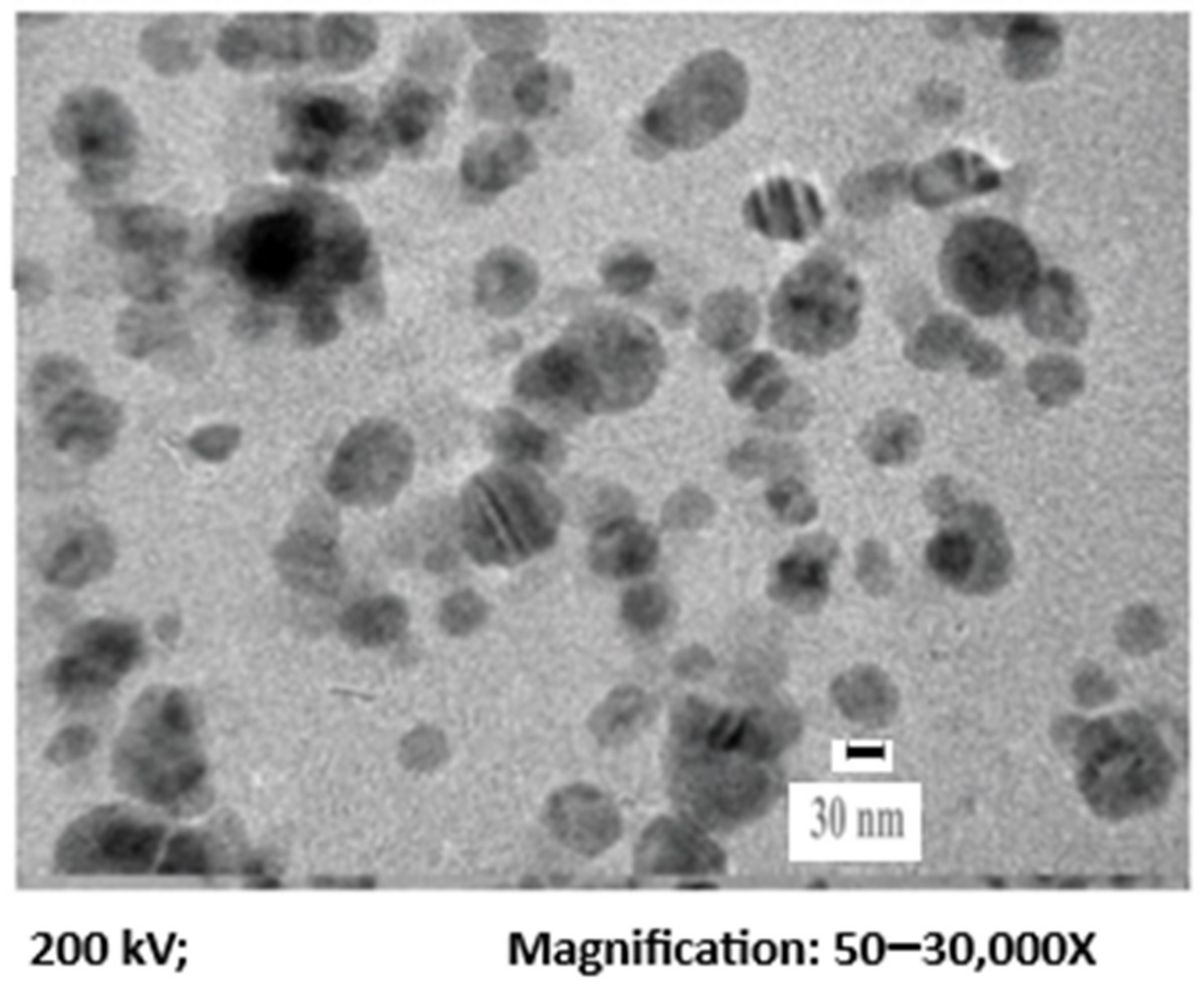
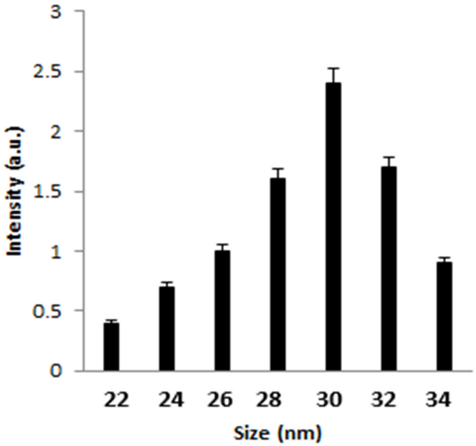
3.1. Physico-Chemical Characterization of the Synthesized AgNPs

TEM image of AgNPs (Figure 1) represent the fineness of the particles with a mean diameter of 30 nm. Dynamic light scattering was also used to determine the size distribution profile of AgNPs. The average mean size of AgNPs was observed as 30 nm (Figure 2).

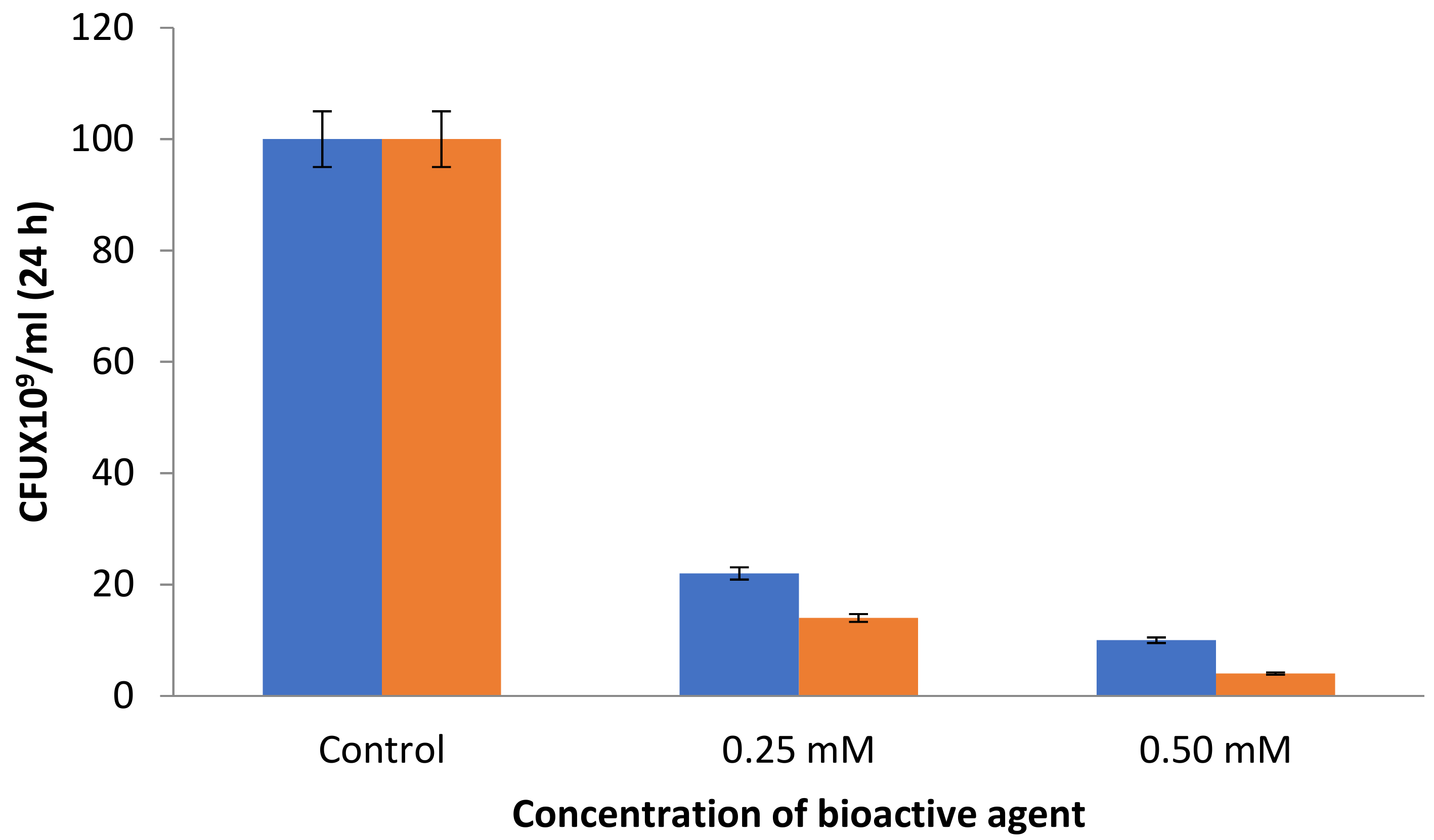
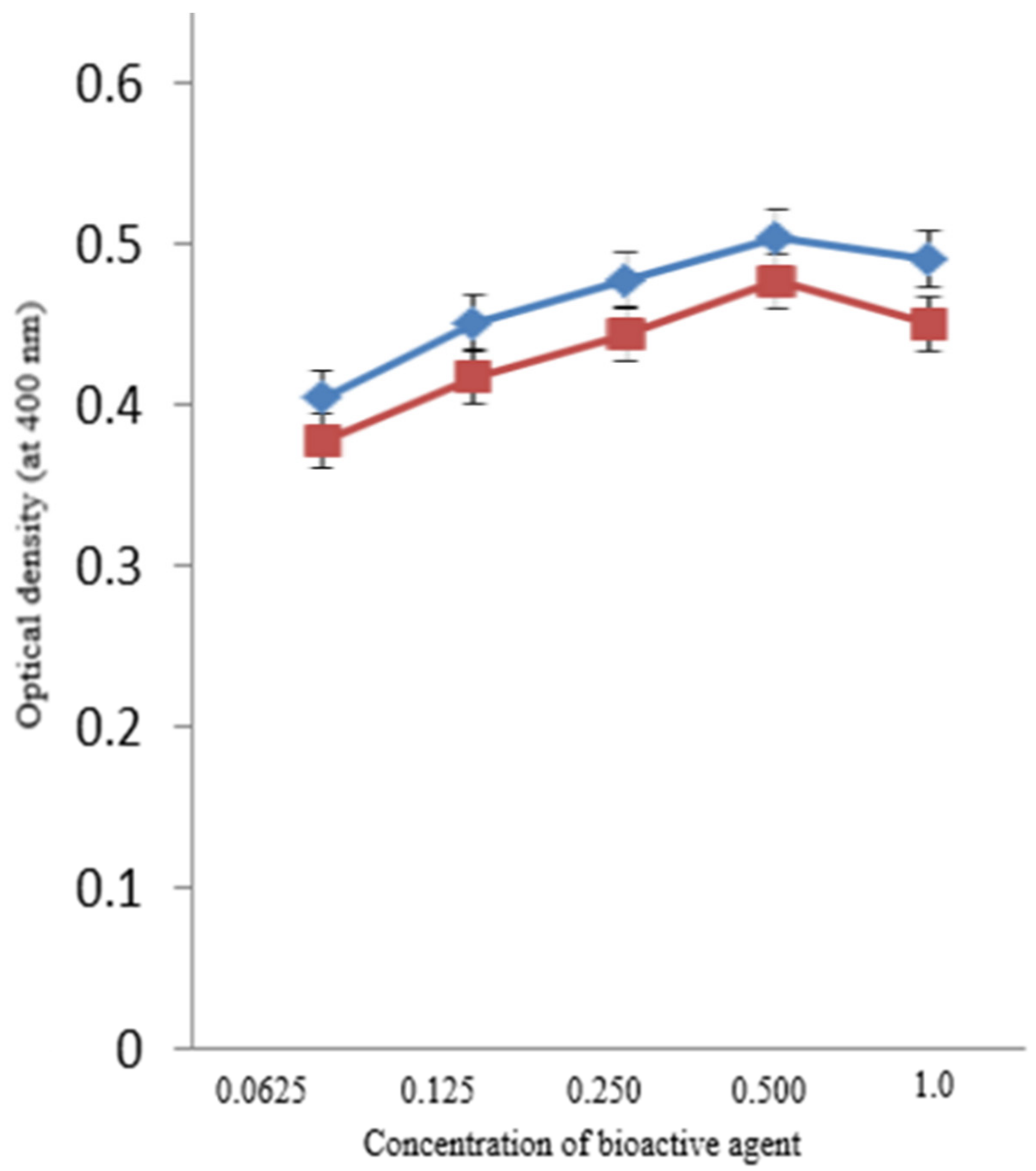
3.2. Antibacterial Assays

The numbers of CFU were reduced significantly with the increased loading of AgNPs. It was observed that 22 × 109 CFU were formed at 0.25 mM AgNPs concentration. However, it was reduced to 14 × 109 CFU for benzalkonium chloride-coated AgNPs at similar experimental conditions (Figure 3).
The possible mechanism could be attributed to the property of benzalkonium chloride that serves as an extended spectrum quaternary ammonium antibacterial agent and is a cationically charged moiety and induces antibacterial action through attraction to the negatively charged bacterial membrane [30]. Similarly, the activity of β-glucosidase was analyzed at various AgNPs concentrations. It was observed that the enzymatic activity of β-glucosidase was enhanced substantially beyond 0.0625 mM till 0.5 mM concentration of AgNPs as well as BAC–AgNPs (Figure 4)
However, there was no further change of activity beyond this concentration. Table 1 suggests the zone of inhibition of AgNPs and BAC–AgNPs against E. coli, B. subtilis, P. aeruginosa, and S pneumoniae at 0.25 and 0.50 mM concentration of these bioactive agents, respectively. ZOI was 3.45 cm and 3.56 cm for AgNPs and BAC–AgNPs at 0.25 mM of these bioactive agents, respectively, against E. coli.
However, the ZOI values were 4.28 cm and 4.40 cm, respectively, against B. subtilis. These values were obtained as 3.36 cm and 3.47 cm, respectively, against P. aeruginosa, and 3.44 cm and 3.62 cm, against S. pneumonia, under similar experimental conditions. When the concentration of these bioactive agents was increased to 0.5 mM, ZOI was reduced significantly for BAC–AgNPs as compared with AgNPs. ZOI was observed as 4.28 cm and 4.40 against E. coli by AgNPs and BAC–AgNPs, respectively. However, ZOI for S. pneumonia was 3.78 cm and 3.91 cm, respectively, for BAC–AgNPs under identical incubation conditions. Mechanistically, the prepared nanoparticles may cause bacterial membrane lysis either directly binding to the negatively charged portion of the lipid molecules or through the release of reactive oxygen species [ROS] which may further trigger intracellular damage through secondary messengers leading to a powerful bactericidal action. Similar mechanism has been anticipated for positively charged nanoparticles against various pathologic bacterial strains [31,32]. However, exact molecular nature of antibacterial action of AgNPs and BAC–AgNPs remains to unclear. Earlier investigators analyzed the different quantities of AgNPs obtained by neem leaves onto agar plates containing bacterial colony. They observed a maximum zone of clearance at 12 μg/mL of AgNPs. The antibacterial activity of such AgNPs was explained by the change in cell membrane permeability or degradation of enzymes in bacteria. The zone of clearance achieved by the developed AgNPs was 6 mm [22]. A similar approach was utilized for analyzing the antimicrobial activity of bleached Typha domingensis fibers, impregnated with AgNPs and benzalkonium chloride. Significant observations were noted in relation to the susceptibility to antimicrobials like S. aureus, E. coli, S. typhimuruim, and S. enteritidis [33].

4. Conclusions

The synthesis of AgNPs by neem leaf in this study satisfies all the conditions of a 100% green chemical process which includes an eco-friendly, rapid, and green approach, characterized by cost-effectiveness, without using external stabilizers or reducing agents. Moreover, the utility of benzalkonium chloride as a quaternary ammonium compound imparted significant improvements in the antibacterial activity of the silver nanoparticles synthesized from the neem leaves. Excellent antimicrobial efficacy was obtained for the bacterial strains, such as E. coli, B. subtilis, P. aeruginosa, and S. pneumonia. Persistent antibacterial activity of such nanoparticles may be analyzed for other microorganisms so that their utility can be extended in biomedical, environmental, and biotechnological sectors. This work will also have a significant impact on developing other nanoparticles by green and clean technologies with substantial environmental benefits.

Author Contributions

S.A.A. was responsible for executing the experiments, writing the original draft of the manuscript. A.M.A. was involved in formal and statistical analysis, validation, reviewing and editing the manuscript. S.A.A. was involved in conceptualization, formal analysis, project administration, resources, reviewing the original and revised drafts from time to time. All authors have read and agreed to the published version of the manuscript.

Funding

This research received no external funding.

Institutional Review Board Statement

Not applicable.

Informed Consent Statement

Not applicable.

Acknowledgments

The authors are thankful to Rukhsana Satar (at Ibn Sina National College for medical Studies, Jeddah, Saudi Arabia) for providing valuable suggestions while preparing the manuscript.

Conflicts of Interest

The authors declare no conflict of interest while submitting this manuscript.

Abbreviations

BACbenzalkonium chloride
CFUcolony-forming units
ZOIzone of inhibition

References

  1. Nikaido, H. Multidrug resistance in bacteria. Ann. Rev. Biochem. 2009, 78, 119–146. [Google Scholar] [CrossRef] [Green Version]
  2. Talebian, S.; Wallace, G.G.; Schroeder, A.; Stellacci, F.; Conde, J. Nanotechnology-based disinfectants and sensors for SARS-CoV-2. Nat. Nanotechnol. 2020, 15, 618–621. [Google Scholar] [CrossRef]
  3. Wang, L.; Hu, C.; Shao, L. The antimicrobial activity of nanoparticles: Present situation and prospects for the future. Int. J. Nanomed. 2017, 12, 1227–1249. [Google Scholar] [CrossRef] [Green Version]
  4. Varier, K.M.; Gudeppu, M.; Chinnasamy, A.; Thangarajan, S.; Balasubramanian, J. Nanoparticles: Antimicrobial applications and its prospects. In Advanced Nanostructured Materials for Environmental Remediation; Environmental Chemistry for a Sustainable World; Naushad, M., Rajendran, S., Gracia, F., Eds.; Springer: Cham, Switzerland, 2019; Volume 25, pp. 321–355. [Google Scholar]
  5. Li, D.; Zhou, B.; Lv, B. Antibacterial therapeutic agents composed of functional biological molecules. J. Chem. 2020, 2020, 6578579. [Google Scholar] [CrossRef] [Green Version]
  6. Hooper, D.C. Mechanisms of action of antimicrobials: Focus on fluoroquinolones. Clin. Infect. Dis. 2001, 32, S9–S15. [Google Scholar] [CrossRef] [PubMed]
  7. Bazaka, K.; Jacob, M.V.; Chrzanowski, W.; Ostrikov, K. Anti-bacterial surfaces: Natural agents, mechanisms of action, and plasma surface modification. RSC Adv. 2015, 5, 48739–48759. [Google Scholar] [CrossRef] [Green Version]
  8. Herman, A.; Herman, A.P. Nanoparticles as antimicrobial agents: Their toxicity and mechanisms of action. J. Nanosci. Nanotechnol. 2014, 14, 946–957. [Google Scholar] [CrossRef] [PubMed]
  9. Almatar, M.; Makky, E.A.; Var, I.; Koksal, F.; Var, I. The role of nanoparticles in the inhibition of multidrug-resistant bacteria and biofilms. Curr. Drug Deliv. 2018, 15, 470–484. [Google Scholar] [CrossRef] [PubMed]
  10. Gudikandula, K.; Maringanti, S.C. Synthesis of silver nanoparticles by chemical and biological methods and their antimicrobial properties. J. Exp. Nanosci. 2016, 11, 714–721. [Google Scholar] [CrossRef]
  11. Franci, G.; Falanga, A.; Galdiero, S.; Palomba, L.; Rai, M.; Morelli, G.; Galdiero, M. Silver Nanoparticles as Potential Antibacterial Agents. Molecules 2015, 20, 8856–8874. [Google Scholar] [CrossRef] [Green Version]
  12. Siddiqi, K.S.; Husen, A.; Rao, R.A.K. A review on biosynthesis of silver nanoparticles and their biocidal properties. J. Nanobiotechnol. 2018, 16, 14–22. [Google Scholar] [CrossRef]
  13. Yaqoob, A.A.; Umar, K.; Ibrahim, M.N.M. Silver nanoparticles: Various methods of synthesis, size affecting factors and their potential applications–a review. Appl. Nanosci. 2020, 10, 1369–1378. [Google Scholar] [CrossRef]
  14. Islam, A.; Jacob, M.V.; Antunes, E. A critical review on silver nanoparticles: From synthesis and applications to its mitigation through low-cost adsorption by biochar. J. Environ. Manag. 2021, 281, 111918–111932. [Google Scholar] [CrossRef] [PubMed]
  15. Syafiuddin, A.; Salmiati; Salim, M.R.; Kueh, A.B.H.; Hadibarata, T.; Nur, H. A review of silver nanoparticles: Research trends, global consumption, synthesis, properties, and future challenges. J. Chin. Chem. Soc. 2017, 64, 732–756. [Google Scholar] [CrossRef]
  16. Verma, A.; Gautam, S.P.; Bansal, K.K.; Prabhakar, N.; Rosenholm, J.M. Green Nanotechnology: Advancement in phytoformulation research. Medicines 2019, 6, 39–49. [Google Scholar] [CrossRef] [PubMed] [Green Version]
  17. Patra, J.K.; Baek, K.-H. Green nanobiotechnology: Factors affecting synthesis and characterization techniques. J. Nanomater. 2014, 2014, 417305. [Google Scholar] [CrossRef] [Green Version]
  18. Ansari, S.A.; Satar, R.; Alam, F.; Alqahtani, M.H.; Chaudhary, A.G.; Naseer, M.I.; Karim, S.; Sheikh, I.A. Cost effective surface functionalization of silver nanoparticles for high yield immobilization of Aspergillus oryzae β-galactosidase and its application in lactose hydrolysis. Proc. Biochem. 2012, 47, 2427–2433. [Google Scholar] [CrossRef]
  19. Roy, A.; Bulut, O.; Some, S.; Mandal, A.K.; Yilmaz, M.D. Green synthesis of silver nanoparticles: Biomolecule-nanoparticle organizations targeting antimicrobial activity. RSC Adv. 2019, 9, 2673–2702. [Google Scholar] [CrossRef] [Green Version]
  20. Ahmad, S.; Munir, S.; Zeb, N.; Ullah, A.; Khan, B.; Ali, J.; Bilal, M.; Omer, M.; Alamzeb, M.; Salman, S.M.; et al. Green nanotechnology: A review on green synthesis of silver nanoparticles—An ecofriendly approach. Int. J. Nanomed. 2019, 14, 5087–5107. [Google Scholar] [CrossRef] [Green Version]
  21. Javed, R.; Zia, M.; Naz, S.; Asida, S.O.; Ain, N.; Ao, Q. Role of capping agents in the application of nanoparticles in biomedicine and environmental remediation: Recent trends and future prospects. J. Nanobiotechnol. 2020, 18, 172–185. [Google Scholar] [CrossRef]
  22. Verma, A.; Mehata, M.S. Controllable synthesis of silver nanoparticles using Neem leaves and their antimicrobial activity. J. Rad. Res. Appl. Sci. 2016, 9, 109–115. [Google Scholar] [CrossRef] [Green Version]
  23. Banerjee, P.; Satapathy, M.; Mukhopahayay, A.; Das, P. Leaf extract mediated green synthesis of silver nanoparticles from widely available Indian plants: Synthesis, characterization, antimicrobial property and toxicity analysis. Biores. Bioproc. 2014, 1, 3–10. [Google Scholar] [CrossRef] [Green Version]
  24. Saware, K.; Venkataraman, A. Biosynthesis and Characterization of Stable Silver Nanoparticles Using Ficus religiosa Leaf Extract: A Mechanism Perspective. J. Clust. Sci. 2014, 25, 1157–1171. [Google Scholar] [CrossRef]
  25. Goudarzi, M.; Mir, N.; Mehdi, M.K.; Bagheri, S.; Mosaoud, S.N. Biosynthesis and characterization of silver nanoparticles prepared from two novel natural precursors by facile thermal decomposition methods. Sci. Rep. 2016, 6, 32539–32549. [Google Scholar] [CrossRef]
  26. Jadoun, S.; Arif, R.; Jangid, N.K.; Meena, R.K. Green synthesis of nanoparticles using plant extracts: A review. Environ. Chem. Lett. 2021, 19, 355–374. [Google Scholar] [CrossRef]
  27. Kim, M.; Weigand, M.R.; Oh, S.; Hatt, J.K.; Krishnan, R.; Tezel, U.; Pavlostathis, S.G.; Konstantinidis, K.T. Widely used benzalkonium chloride disinfectants can promote antibiotic resistance. Appl. Environ. Microbiol. 2018, 84, 17–25. [Google Scholar] [CrossRef] [Green Version]
  28. Ahmed, S.; Saifullah; Ahmad, M.; Swami, B.L.; Ikram, S. Green synthesis of silver nanoparticles using Azadirachta indica aqueous leaf extract. J. Radiat. Res. Appl. Sci. 2016, 9, 1–7. [Google Scholar] [CrossRef] [Green Version]
  29. Ansari, S.A.; Oves, M.; Satar, R.; Khan, A.; Ahmad, S.I.; Jafri, M.A.; Zaidi, S.K.; Alqahtani, M.H. Antibacterial activity of iron oxide nanoparticles synthesized by co-precipitation technology against Bacillus cereus and Klebsiella pneumoniae. Pol. J. Chem. Technol. 2017, 19, 110–115. [Google Scholar] [CrossRef] [Green Version]
  30. Khajav, R.; Sattar, M.; Ashjaran, A. The antimicrobial effect of benzalkonium chloride on some pathogenic microbes observed on fibers of acrylic carpet. Pak. J. Biol. Sci. 2007, 10, 598–601. [Google Scholar] [CrossRef]
  31. Mazur, P.; Skiba-Kurek, I.; Mrowiec, P.; Karczewska, E.; Drozdz, R. Synergistic ROS-associated antimicrobial activity of silver nanoparticles and gentamicin against staphylococcus epidermidis. Int. J. Nanomed. 2020, 15, 3551–3562. [Google Scholar] [CrossRef] [PubMed]
  32. Krce, L.; Sprung, M.; Roncevic, T.; Maravic, A.; Culic, V.C.; Blazeka, D.; Krstulovic, N.; Aviani, I. Probing the mode of antibacterial action of silver nanoparticles synthesized by laser ablation in water: What fluorescence and AFM data tell us. Nanomaterials 2020, 10, 61–72. [Google Scholar] [CrossRef] [PubMed]
  33. Koschevic, M.T.; de Araujo, R.P.; dos Santos Garcia, V.A.; Fakhouri, F.M.; de Oliveira, K.M.P.; Arruda, A.J.; Dufresne, A.; Martelli, S.M. Antimicrobial activity of bleached cattail fibers (Typha domingensis) impregnated with silver nanoparticles and benzalkonium chloride. J. Appl. Polym. Sci. 2021, 138, 55–67. [Google Scholar] [CrossRef]
Figure 1. TEM of the synthesized AgNPS.
Coatings 11 01382 g001
Figure 2. DLS of the synthesized AgNPS.
Coatings 11 01382 g002
Figure 3. CFU by the synthesized AgNPS () and BAC-coated AgNPs ().
Coatings 11 01382 g003
Figure 4. Activity of β-glucosidase with the synthesized AgNPS () and BAC-coated AgNPs ().
Coatings 11 01382 g004
Table 1. Zone of inhibition (diameter, cm) of antibacterial test of AgNPs and Benzalkonium chloride (BAC)-coated AgNPs.
Concentration (mM)E. coliB. subtilisP. aeruginosaS. pneumonia
AgNPsBAC–AgNPsAgNPsBAC–AgNPsAgNPsBAC–AgNPsAgNPsBAC–AgNPs
0.253.45 ± 0.853.56 ± 0.544.28 ± 0.394.40 ± 0.583.36 ± 1.23.47 ± 1.343.44 ± 0.963.62 ± 0.68
0.504.28 ± 0.734.40 ± 0.684.36 ± 0.984.44 ± 0.774.28 ± 0.984.37 ± 1.553.78 ± 0.273.91 ± 0.37
Publisher’s Note: MDPI stays neutral with regard to jurisdictional claims in published maps and institutional affiliations.

