Article

Low-Cost Deposition of Antibacterial Ion-Substituted Hydroxyapatite Coatings onto 316L Stainless Steel for Biomedical and Dental Applications

1
Department of Restorative Dental Sciences, College of Dentistry, Imam Abdulrahman Bin Faisal University, P.O. Box 1982, Dammam 31441, Saudi Arabia
2
Department of Industrial Engineering, Faculty of Engineering, Taibah University, P.O. Box 344, Medina 41411, Saudi Arabia
*
Author to whom correspondence should be addressed.
Coatings 2020, 10(9), 880; https://doi.org/10.3390/coatings10090880
Submission received: 21 July 2020 / Revised: 7 September 2020 / Accepted: 9 September 2020 / Published: 13 September 2020
(This article belongs to the Special Issue Surface Properties of Dental Materials and Instruments)

Abstract

:
Substitutions of ions into an apatitic lattice may result in antibacterial properties. In this study, magnesium (Mg)-, zinc (Zn)-, and silicon (Si)-substituted hydroxyapatite (HA) were synthesized using a microwave irradiation technique. Polyvinyl alcohol (PVA) was added during the synthesis of the substituted HA as a binding agent. The synthesized Mg-, Zn-, and Si-substituted HAs were then coated onto a 316L-grade stainless-steel substrate using low-cost electrophoretic deposition (EPD), thereby avoiding exposure to high temperatures. The deposited layer thickness was measured and the structural, phase and morphological analysis were characterized by Fourier transform infrared spectroscopy (FTIR), X-ray diffraction (XRD) and scanning electron microscope (SEM), respectively. The bacterial adhesion of Staphylococcus aureus was characterized at 30 min, 2 h and 6 h. The results showed homogeneous, uniform thickness (50–70 µm) of the substrate. FTIR and XRD showed the characteristic spectral peaks of HA, where the presence of Mg, Zn and Si changed the spectral peak intensities. The Mg–HA coating showed the least bacterial adhesion at 30 min and 2 h. In contrast, the Si–HA coating showed the least adhesion at 6 h. EPD showed an effective way to get a uniform coating on bio-grade metal implants, where ionic-substituted HA appeared as alternative coating material compared to conventional HA and showed the least bacterial adhesion.

Graphical Abstract

1. Introduction

The selection of metallic materials for dental and biomedical applications depends on various factors such as corrosion behavior, mechanical properties, cost, fabricability and biocompatibility [1]. Dental instruments, orthodontic brackets, wires, bands, ligatures and maxillofacial screws and implants are manufactured using stainless-steel alloy [2,3]. These metal surfaces are prone to bacterial colonization as biofilms [4]. Therefore, metal surfaces must be coated with antibacterial materials. The antibacterial properties of biomaterials have long relied on surface coating and functionalizing techniques such as bioceramic coatings, covalent attachment of polycationic groups [5,6], impregnating nanoparticles with antimicrobial agents [7,8,9] and quaternary ammonium, iodide or silver ions [10,11,12,13].
Among bioceramics, hydroxyapatite (HA) has been substituted with various ions—including cations and anions [14,15,16]. The bioactivity, biocompatibility and antibacterial activity of HA can be increased by substituting zinc (Zn), magnesium (Mg), silicon (Si) [17,18,19]. Several methods for deposition of bioceramics, polymers and composites on metallic implants have been used, these include dip coating [20], pulse laser deposition [21], electrochemical deposition [22], biomimetic deposition [23], plasma spraying [24] and electrophoretic deposition [25]. Among these processes, plasma spraying has been widely used commercially, however, it has some limitations such as material wastage, high dissolution rate, thickness control and phase change in HA [26,27].
Electrophoretic deposition (EPD) is an economical low-tech method that can be used for coatings [28]. It can be performed at room temperature, providing uniform coating thickness, less material wastage, less manpower, ease for complex shapes. Moreover, the rate of deposition, thickness, deposition particle size can be controlled easily by changing the parameters of suspension and power input [29,30,31,32,33].
Previously, HA particulates were coated on the metal substrates using EPD. However, a major drawback of poor adhesion has been observed [28,33]. It has been reported that differences in thermal expansion can generate cracks on the surface of coated layers or at the interface between the coatings and substrates during cooling after sintering [34]. Generally, sintering of the coatings is required as a post-deposition step however polymeric binders are also used to enhance adhesion to the substrate without carrying out high energy sintering steps and to improve the mechanical strength of coatings [35].
Limited data are available in which Zn-, Mg-, and Si-substituted HA has been coated on a metallic substrate using the EPD technique [36]. Therefore, the present study was designed to synthesize Zn–HA, Mg–HA, and Si–HA using microwave irradiation technique and the aim was to deposit these particles on a bio-grade stainless-steel surface using EPD techniques and characterize the structural and antibacterial activities against Staphylococcus aureus in the presence of shear forces. It was hypothesized that these substitutions would help to evade bacterial adhesion and impart antibacterial character to the coated samples.

2. Materials and Methods

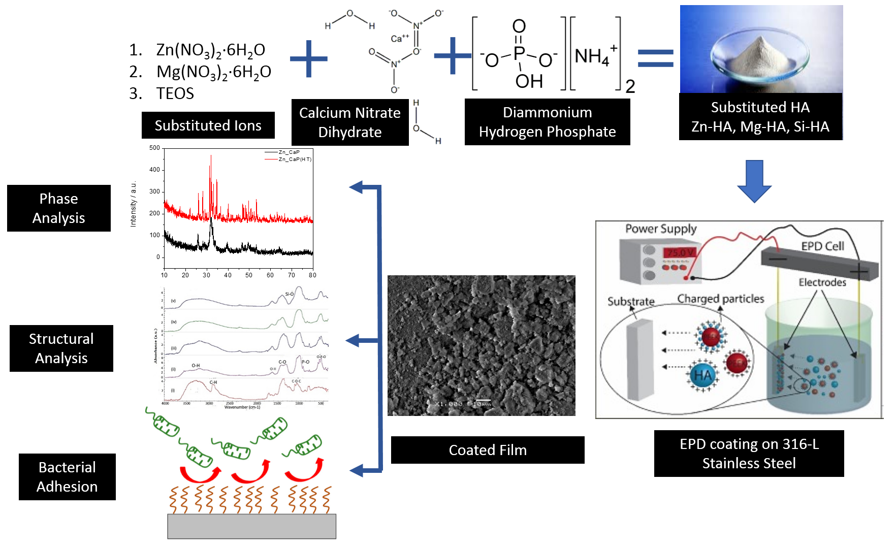
2.1. Synthesis of Substituted HA

All analytical-grade materials were used in this study. The precursors for the synthesis of substituted HA include calcium nitrate tetrahydrate Ca(NO3)2·4H2O (Fluka Honeywelll, Charlotte, NC, USA and diammonium hydrogen phosphate (NH4)2HPO4 (Sigma Aldrich, St. Louis, MO, USA) used as calcium and phosphorous precursors, respectively. Zinc nitrate hexahydrate Zn(NO3)2·6H2O (Merck, Darmstadt, Germany), magnesium nitrate hexahydrate Mg(NO3)2·6H2O (AppliChem GmbH, Darmstadt, Germany) and tetraethyl orthosilicate (TEOS) (Aldrich, St. Louis, MO, USA) were used as zinc, magnesium and silicon substituent salts, respectively. The (Ca + Zn)/P, (Ca + Mg)/P and Ca/(P + Si) ratio was maintained at 1.67 as given in Table 1. Zn–HA, Mg–HA, and Si–HA powders were prepared based on the chemical formulae Ca10−x (xZn(NO3)·6H2O·PO4)6(OH)2, Ca10−x(xMg(NO3)·6H2O(PO4)6(OH)2 and Ca10(PO4)6−x(SiO4)x(OH)2−x, respectively as proposed earlier [37,38,39].
A pure HA was synthesized initially by using a microwave irradiation technique as described earlier by our group [40]. The synthesis was carried out by dissolving required amounts of Ca(NO3)2·4H2O and substituting Zn and Mg ions containing salts in deionized water; (NH4)2HPO4 was separately dissolved in deionized H2O. The pH of both the solutions was adjusted above 11 by adding ammonium hydroxide (NH4OH) as required; (NH4)2HPO4 was added dropwise at 2 mL/min to a solution of Ca(NO3)2·4H2O and substituting ion-containing salt. The pH was continuously monitored and maintained above 11 by adding NH4OH. Whereby, TEOS was dissolved in (NH4)2HPO4 and added dropwise in Ca(NO3)2·4H2O solution and the same procedure was followed. After addition, the solution was left for 30-min stirring. It was then microwave irradiated in a modified domestic microwave oven (Samsung MW101P, Seoul, Korea) at 1000 W for 10 min (30 s ON: 30 s OFF). The suspension was filtered, washed three times with distilled water and oven-dried at 80 °C for 24 h. The obtained powder was ground to carry out deposition and characterization studies.

2.2. Deposition Procedure Used to Prepare Substituted HA Coatings

The electrophoretic technique was carried out on 1 × 2 cm2 stainless-steel 316L substrates (used as cathodes) and carbon anode fixed 1 cm apart. The plates were ground and polished using silicon carbide study (1500, 2000, and 4000), washed and degreased by ultrasonication using methanol (Sigma Aldrich, St Louis, MO, USA). Suspensions for EPD were prepared by adding HA, Zn–HA, Mg–HA, and Si–HA (respectively) in 0.5 wt.% PVA solution. Nitric acid (1.0 M) was added to the solutions until clear solutions were obtained and pH was kept 1–3. Power supply (E Lite 300/300 plus power supply) was regulated at 10 V, suspensions were heated at 80 °C using hotplate and EPD process was carried out for 3 min. During EPD process, current decreased constantly, however, the potential difference across the electrodes remained the same. This happened due to the deposition of HA and substituted HA particles on the electrode and an insulating layer is formed which reduced the electric field influencing electrophoresis for the incoming particles [41]. The as-deposited coatings were air-dried at room temperature for 24 h, later oven-dried at 120 °C for 24 h to evaporate surface absorbed water.
The thickness of coating substrates was measured according to ASTM D6132 [42] using Lendteknet’s ultrasonic pulse–echo meter. The consequential coatings were homogenous, uniform with a thickness of 50–70 µm.

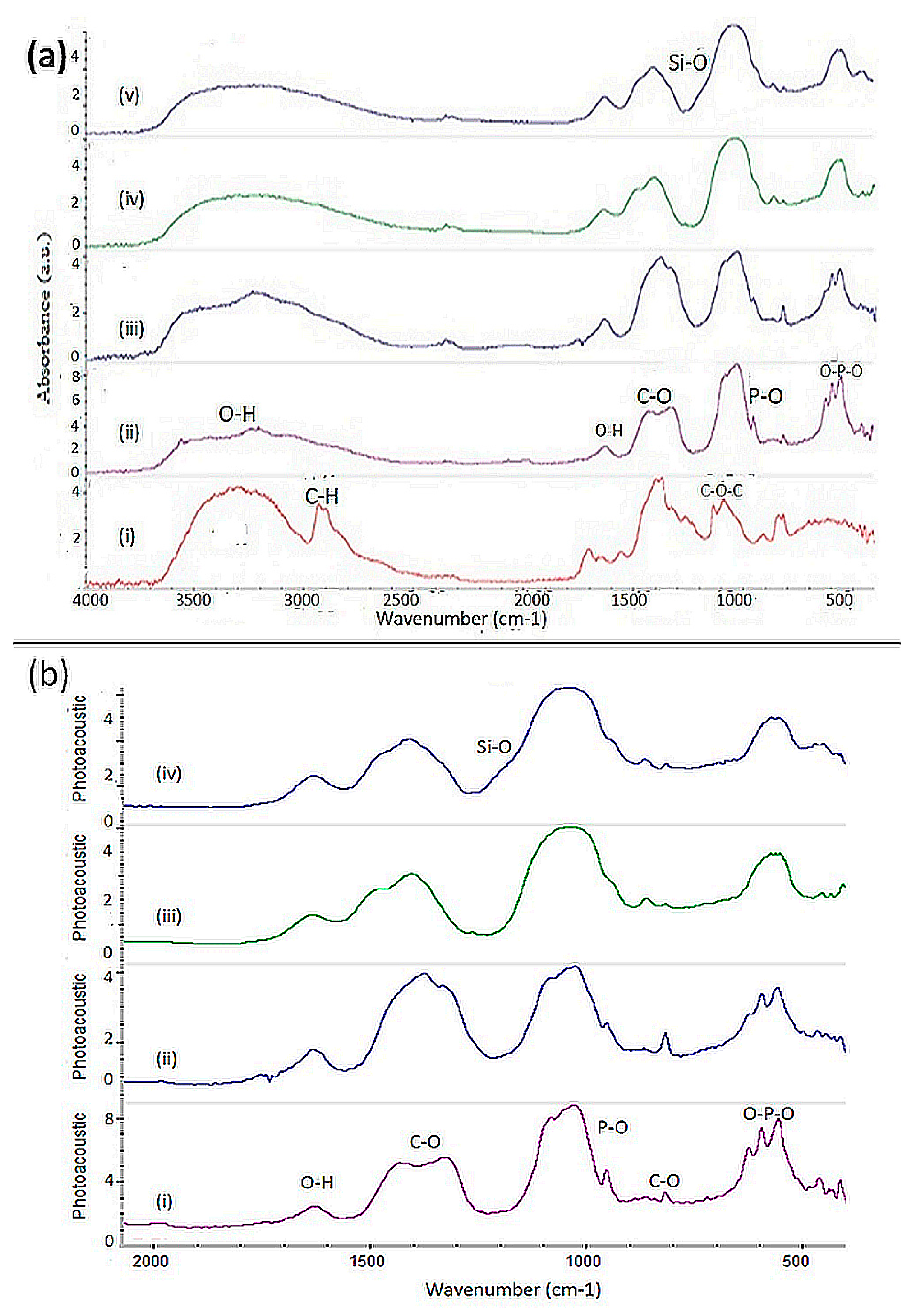
2.3. Fourier Transform Infrared Spectroscopy (FTIR)

Fourier-transform infrared spectroscopy (Thermo Nicolet 6700, Thermo Fischer Scientific, Waltham, MA, USA) with photo-acoustic cell at a resolution of 8 cm−1 and 256 scan numbers were used for the FTIR data collection of PVA, HA, Zn–HA, Mg–HA, and Si–HA. The spectral range was 4000−400 cm−1 and OMINIC software v7.2was used to evaluate the peak positions.

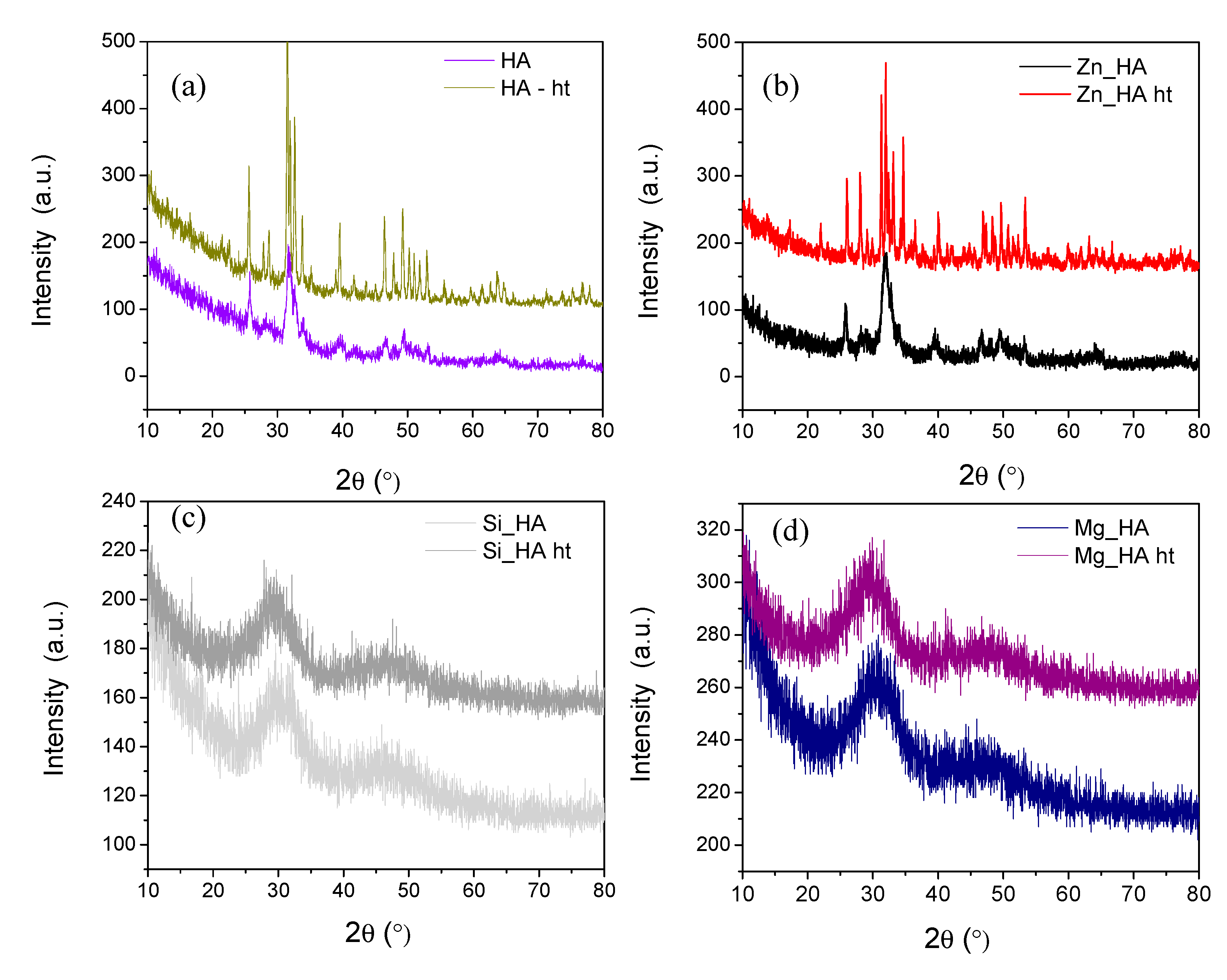
2.4. X-ray Diffraction (XRD)

The X-ray diffraction (XRD) measurements were recorded using a diffractometer (PANalytical XPERT-PRO model, Malvern, Stuttgart, Germany) operated at 40 kV and 40 mA using Cu-Kα radiation. The detector was scanned over a range of angle 2θ = 20°−80° at a step size of 0.02° to identify the HA phase.

2.5. Scanning Electron Microscopic Analysis

The surface morphology of coated samples was investigated by scanning electron microscope (JEOL JSM6490A, JEOL, Tokyo, Japan) at a voltage of 20 V. Samples were gold-coated using JFC 1500 I sputtering device. Images were taken at various magnifications.

2.6. Preparation of Controlled Discs of Substituted Has for Antibacterial Investigations

Compact discs of HA and substituted Has were prepared to be used as control samples for bacterial adhesion testing using hydraulic press HP142009 (Al-Qamar Machinery, Al-Qamar, Lahore, Pakistan). The powders were subjected to a pressure of 550 psi to prepare discs with 8 × 2 mm2, whereby each disc weighed 0.2 ± 0.008 g.

2.7. Bacterial Adhesion Testing

Staphylococcus aureus (American Type Culture Collection, 25,923) was revived from glycerol stock culture on nutrient-agar (N-Agar) and then sub-cultured on tryptone soya broth (TSB) for 24 h at 37 °C in an incubator. The culture dishes were prepared 2 mL of TSB and 20 µL of inoculum. Each type of HA disc and HA coating was subjected to bacterial adhesion testing at 30 min, 2 h and 6 h in triplicate. Shear force was provided by using a shaking incubator (Daihan Scientific, Seoul, Korea) at 120 rpm.
Each disc and plate was taken out at 30 min, 2 h and 6 h in 9 mL 0.9% normal saline solution in 25-mL capped test tubes and sonicated (WiseClean sonicator, PMI-Labortechnik GmbH, Grafstal, Germany) for 2 min at 37 °C to dislodge attached bacteria. Absorbance was measured at 600 nm using a UV-vis spectrophotometer (Bio-Rad Laboratories, Portland, ME, USA).
Each sample material was fixed with 2.5% glutaraldehyde (Merck, Darmstadt, Germany) for 24 h. Sequential dehydration was done step-wise for 30 min in each concentration of alcohol (50%, 60%, 70%, 80%, 90%, and 100%). After this, each sample plate was stained with crystal violet dye (Scharlau, Barcelona, Spain) and counterstained with safranin (K.Y.L Industrial And Manufacturer And Traders Co, Ltd., Tianjin, China) so that bacteria adhering to the coated surface are easily visible. Optika B-600 MET Microscope (Optika, Ponteranica, Italy) was used for microscopic analysis and Optika Pro vision (Optika, Ponteranica, Italy) was used for acquiring digital images at 100X.
Data were analyzed using SPSS Statistics for Windows Software, version 21 (IBM Software, New York, NY, USA). Descriptive statistics were used to calculate the maximum, minimum, mean and standard deviation values. A comparison of two and more than two groups was done by one-way analysis of variance test and followed by post hoc Tukey’s test. All the p values of less than 0.05 were considered statistically significant.

3. Results

3.1. Fourier Transform Infrared Spectroscopy (FTIR)

FTIR data were collected for the deposited HA, Zn–HA + PVA, Mg–HA + PVA and Si–HA + PVA samples. Comparative spectra with complete range (4000–400 cm−1) and selected range (2000–400 cm−1) are given in Figure 1a,b, respectively.
A broad peak attributed to the H-bonded OH stretch of the PVA group at 3200–3570 cm−1 could be seen in Figure 1a(i). The asymmetric and symmetric stretching peaks of C–H were appeared at 2971 and 2910 cm−1, respectively. The spectrum (Figure 1a(ii)) of the control sample (as precipitated HA) showed the deep absorption peaks of PO43− at 1083–1035 cm−1. The peak at 963 cm−1 was due to the asymmetric stretch of the PO43− group. The bending peak of PO43− was visible at 564–604 cm−1. There was a prominent CO32− band from 1343–1490 cm−1 and a bending peak at 827 cm−1. The broad band at 3100–3570 cm−1 represented the OH stretching and peak at 1635 and 633 cm−1 represented the bending peak of OH.
In Figure 1a(iii–v), bands at 1380–1510 cm−1 corresponded to CO32− (B type carbonate substitution) and 1630–1650 cm−1 attributed to bending peak of hydroxyl group. Intense bands at 964, 1030 and 1100 cm−1 corresponded to P–O stretching vibration modes followed by bending peaks at 564 and 602 cm−1 in all three spectra. The change in peak intensities was observed with substituted HA groups, whereby P–O, C–O, and O–H peaks were markedly reduced as shown in Figure 2. The presence of Si–O in the apatite lattice was validated by the appearance of band at 830−1220 cm−1 [37,42] as seen in Figure 1a(v). Variation in peak positions and assignments was observed with substituted HA as tabulated in Table 2. The PVA spectrum showed the characteristic peaks of PVA, whereby 0.5% of PVA was added to all the substituted HA samples prior to EPD. There exists a slight band shifting which was attributed to the addition of PVA.

3.2. X-ray Diffraction (XRD)

Figure 3 shows the XRD spectra of as-deposited and heat-treated coatings. As expected, all the patterns confirmed to the presence of a major phase of HA, as compared to the standards (ICDD 009-0432 and JCPDS-896438) [43,44,45,46,47]. Figure 3a shows at 31.7, 32.2, 32.9, and 39.9 diffraction peaks at 2θ values which correspond to (211), (122), (300), and (310) planes, respectively. No significant phase changes were observed in the as-deposited and heat-treated coatings however, the sharpness of the peak suggested better crystallinity for the case of HA and Zn–HA (Figure 3b) coatings. XRD pattern of Zn–HA coatings exhibited almost all HA peaks, in addition, a very weak diffraction peaks at 26.16° and 54.36° diffraction angle corresponding (220) and (101) plane, respectively match with the Zn peak (JCPDS-040784). Figure 3c,d shows amorphous phases of HA at 2θ ~31.9 and no other phases related to silicon oxide (from Si–HA sample) and stanfieldite (from Mg–HA sample) were observed. Although the XRD patterns of both Si–HA and Mg–HA corresponded to that of HA, however, the diffraction peaks lost intensity, proving a progressive loss of crystallinity [43,44].

3.3. Scanning Electron Microscopic Analysis

The SEM images (Figure 4a–d) show the surface morphology of HA, Zn–HA, Mg–HA and Si–HA-coated samples. Uniform distribution of coating was observed in all samples, in which the particle size was in 5–10 µm.

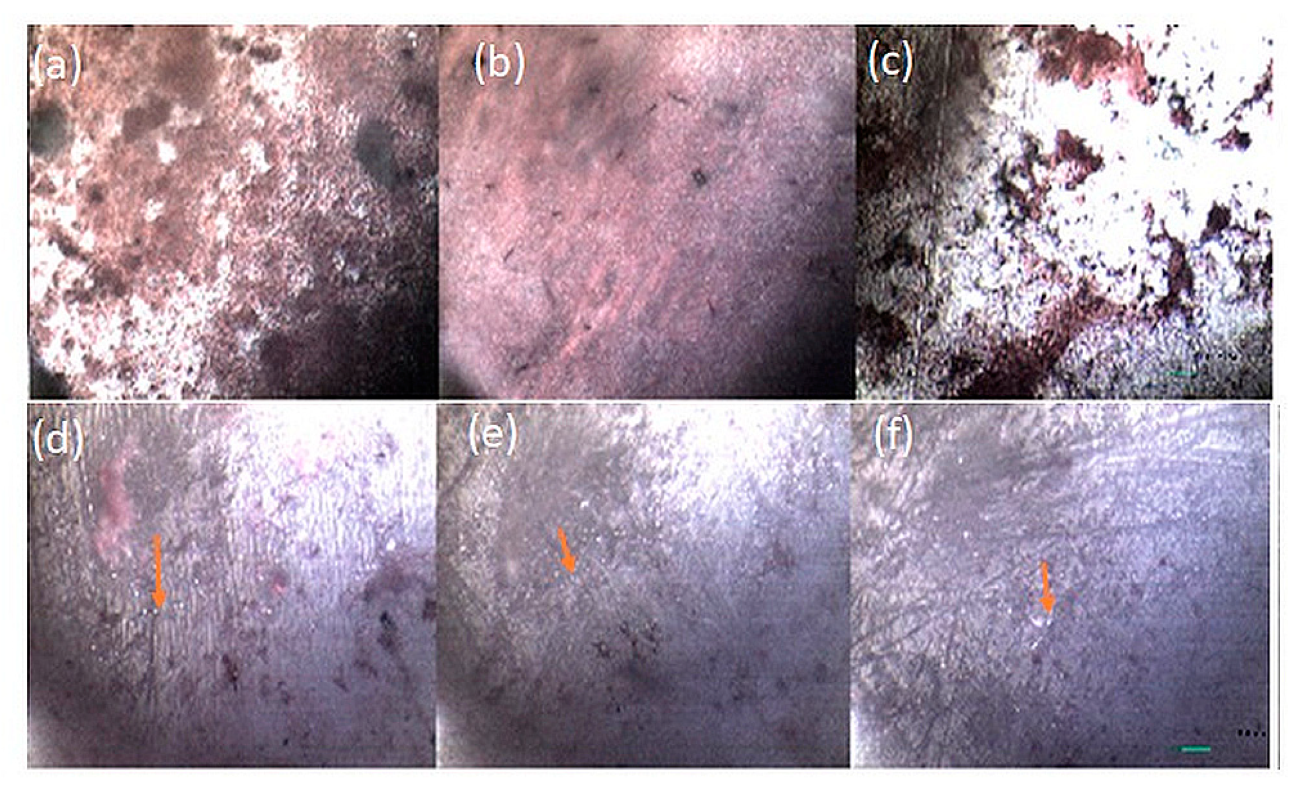
3.4. Antibacterial Adhesion Analysis

The values of absorbance depicted in Table 3 can be correlated with the amount of bacterial adhesion shown at each time interval for both the control discs and coated samples. In the case of control discs, HA and Mg–HA samples showed an increase in absorbance value with the passage of time. In the case of coated samples, initially at 30 min and 2 h, the absorbance value of the silicon sample was higher, but at 6 h the absorbance value was reduced. Absorbance values for HA, Mg–HA and Zn–HA constantly increased from 30 min to 6 h. A statistically significant difference (p ≤ 0.05) was observed between HA and substituted HA groups. Within substituted HA groups, a statistically significant difference (p ≤ 0.05) was observed with a time interval.
Images of stained biofilms (using crystal violet dye and counterstained using safranin) at 1000× are shown in Figure 5, Figure 6 and Figure 7. Mg–HA showed the least adhesion with S. aureus at all the time intervals in both the control disc and coated plate as shown in Figure 5. Zn–HA however showed (Figure 6) a larger number of bacterial colony adherents on the control disc than the coated plate which contained a defined dispersion of thinly beaded colonies. Si–HA offered (Figure 7) most adhesion with S. aureus at 30 min and 2 h with a recession in number at 6 h in both discs and coated plates.

4. Discussion

Metallic implants are inert materials and do not interact with the biologic environment. Hence, coating of HA-based materials on metallic implant surfaces can impart a strong affinity for, and chemical bonding with, the host hard tissue [48]. Substituted HA particles were successfully synthesized using microwave irradiation techniques. The microwave irradiation technique is a quick way to achieve nanoparticles on a large scale [49]. The substituted ions perform a vital role in the biologic responses of HA. The atomic structure of HA allows to substitute almost half of the periodic table elements [50]. Hence, the biologic performance of synthetic HA can be improved and tuned via substitution, doping and grafting of different ions, small functional compounds, peptides and polymers.
A structural and phase analysis of HA and substituted HA was investigated with FTIR and XRD, respectively. It is known that substitution of Mg in HA crystals can reduce the lattice parameters as the ionic radius of Mg2+ is smaller than the ionic radius of Ca2+. Its substitution in HA decreases its crystallinity and can destabilize the apatite structure [51,52]. Similar behavior was observed by XRD pattern in this study. The reduction in the IR absorption band of PO43− was observed with the Mg substitution and the corresponding bands at 1080 and 650–500 cm−1 became broader and intensity was reduced.
Similarly, Zn is substituted at the Ca(II) site in synthetic HA [53]. Its higher substitution in HA decreases crystallinity; therefore, a lower molar percentage was used in this study. It has been reported that 1 wt.% substituted amount of Si in HA was enough to improve bioactivity [54,55]. The appearance of the peak in the XRD spectra at 2θ = 26.16° corresponding to the (220) plane showed the presence of a new phase called parascholzite (CaZn2(PO4)2·2H2O) also known as calcium zinc phosphate in the apatite lattice [56]. The spectral peaks of PO43− and OH became broadened and reduced the crystallinity of the apatite. The v1-stretching P–O peak was merged in a band and a new peak appeared at 941 cm1 attributed to zinc ammonium phosphate. The appearance of a weak band at 1410 cm1 (denoted to NH4+) also supported this suggestion [57]. The CO32− peak presented in pure HA was not observed in the Zn-substituted HA, which is an indication that Zn ions replaced the carbonated group into the HA.
It was found that the substitution of Si into HA (Ca10(PO4)6−x(SiO4)x(OH)2−x) changed the hydroxyl stretching bands at 630 and 3570 cm−1. However, both stretching and bending bands positions were unchanged by the silicate substitution. The CO32− band also appeared at 1500–1410 cm−1. It is reported that the substitution of PO43− by SiO44− reduces the number of hydroxyl groups required for charge balance [58]. Furthermore, hydrogen-bonded OH groups attached to the Si were expected to occur, while Si–O–Si bending mode frequencies were observed at 827 and 470 cm−1. Therefore, the intensity of the v3 carbonate peak was increased. The results of this study are in accordance with previous studies [59,60].
This study assessed the antibacterial activity of three types of substituted HAs with S. aureus in the presence of shear force. For that, an in-depth analysis of the antibacterial activity of each substituted HA was done with the help of a UV-vis spectrophotometer. A time-based study was conducted to investigate an increase or decrease in the number of bacterial colonies on the substrate. It has been observed from previous studies on bacterial adhesion that the time frame from zero to 6 h is critical in bacterial attachment on the substrate [61]. Therefore, in the present study, the antibacterial activity of zinc, magnesium and silicon-substituted HA was conducted at up to 6 h.
In the case of Zn–HA, an initial attachment was deterred however zinc substitution did not seem to affect proliferation rate at 2-h and 6-h incubation time periods. Availability of Si–O seemed to aid in the initial attachment of bacterial colonies onto the coated plates and discs as Si–HA showed a large number of bacterial attachments at 30 min and 2 h in both the discs and coated samples. However, silicon did not aid in further proliferation of S. aureus, which led to the recession in their number at 6 h.
It appears that Mg–HA and Zn–HA had a better propensity of deterring initial bacterial attachment. On the other hand, silicon substitution had an antibacterial effect at a 6-h incubation time interval. Bacterial adhesion follows a complex mechanism and depends upon the physicochemical properties of bacterial cells and substrate [62]. It has been reported that S. aureus has a dominant electron donor character at intermediate pH (pH 5, pH 7 and pH 9) and shows greater attachment to the acidic and predominantly hydrophobic substrate [63]. Bacterial adhesion depends not only on the hydrophobicity of the bacterial cell wall [64] but also on environmental factors like pH, ionic strength, medium composition and temperature. Overall, Si–HA showed the most adhesion and Mg–HA showed the least adhesion. Zn–HA had a better antibacterial profile on coated samples in comparison to its disc sample that can be due to the thinner film of Zn–HA. This means that thinner films are better suited for the provision of antibacterial property to the coatings. Furthermore, the inhibitory effect of Zn-substituted HA on the development of bacteria is established and in another study, Zn(NO3)2·6H2O have shown its impact on the reduction of the numbers of S. aureus bacteria [38].
Further studies should be conducted on the same lines with different bacterial strains. Therefore, a much clearer image of the antibacterial profile of these coated materials can be acquired. Moreover, substitution with other elements should also be tried and their antibacterial activities should be studied in detail.

5. Conclusions

The role of each substitution in imparting antibacterial property to HA coatings becomes prominent in this study. Microwave irradiation techniques were successfully used to synthesize HA and substituted HA (Zn–HA, Mg–HA, and Si–HA). EPD—a low-cost deposition technique—was used to coat as-synthesized HA and substituted HA powders on biomedical-grade 316L stainless-steel surface. Spectroscopic and X-ray diffraction analysis confirmed the presence of substituted ions in HA. Uniform coatings were achieved through the EPD process. Magnesium-substituted HA offered better antibacterial activity against S. aureus in comparison to zinc and silicon-substituted HA. The bacterial adhesion was influenced by both the bacterial chemistry and the physicochemical properties of the coated substrates. The environmental factors also seemed to play an important role in bacterial attachment. Mg–HA not only showed lesser bacterial adhesion, but also stopped bacterial proliferation. Therefore, out of Zn–HA, Mg–HA and Si–HA, Mg–HA has great prospects as antibacterial, bioactive bioceramic coatings on metallic biomedical and dental implants.

Author Contributions

Conceptualization, A.S.K. and M.A.; methodology, M.A.; validation, M.A. and A.S.K.; investigation, M.A.; writing—original draft preparation, M.A.; writing—review and editing, A.S.K.; supervision, A.S.K. All authors have read and agreed to the published version of the manuscript.

Funding

This research received no external funding.

Acknowledgments

The authors would like to thank Musaddaq (Interdisciplinary Research Center in Biomedical Materials, COMSATS University Islamabad, Lahore Campus, Pakistan) for helping in synthesis of substituted hydroxyapatite.

Conflicts of Interest

The authors declare no conflict of interest.

References

  1. Eliaz, N. Corrosion of metallic biomaterials: A review. Materials 2019, 12, 407. [Google Scholar] [CrossRef] [PubMed] [Green Version]
  2. Gritsch, K.; Laroche, N.; Bonnet, J.-M.; Exbrayat, P.; Morgon, L.; Rabilloud, M.; Grosgogeat, B. In vivo evaluation of immediately loaded stainless steel and titanium orthodontic screws in a growing bone. PLoS ONE 2013, 8, e76223. [Google Scholar] [CrossRef] [PubMed] [Green Version]
  3. Salehi, P.; Babanouri, N.; Roein-Peikar, M.; Zare, F. Long-term antimicrobial assessment of orthodontic brackets coated with nitrogen-doped titanium dioxide against Streptococcus mutans. Prog. Orthod. 2018, 19, 35. [Google Scholar] [CrossRef] [PubMed] [Green Version]
  4. Hosman, A.H.; van der Mei, H.C.; Bulstra, S.K.; Kuijer, R.; Busscher, H.J.; Neut, D. Influence of Co-Cr particles and Co-Cr ions on the growth of staphylococcal biofilms. Int. J. Artif. Organs 2011, 34, 759–765. [Google Scholar] [CrossRef] [PubMed]
  5. Tiller, J.C.; Liao, C.J.; Lewis, K.; Klibanov, A.M. Designing surfaces that kill bacteria on contact. Proc. Natl. Acad. Sci. USA 2001, 98, 5981–5985. [Google Scholar] [CrossRef] [Green Version]
  6. Cen, L.; Neoh, K.G.; Kang, E.T. Antibacterial activity of cloth functionalized with N-alkylated poly(4-vinylpyridine). J. Biomed. Mater. Res. Part A 2004, 71A, 70–80. [Google Scholar] [CrossRef]
  7. Shi, Z.; Neoh, K.G.; Kang, E.T.; Wang, W. Antibacterial and mechanical properties of bone cement impregnated with chitosan nanoparticles. Biomaterials 2006, 27, 2440–2449. [Google Scholar] [CrossRef]
  8. Schmidmaier, G.; Lucke, M.; Wildemann, B.; Haas, N.P.; Raschke, M. Prophylaxis and treatment of implant-related infections by antibiotic-coated implants: A review. Injury 2006, 37, S105–S112. [Google Scholar] [CrossRef]
  9. Van de Belt, H.; Neut, D.; Schenk, W.; van Horn, J.R.; van der Mei, H.C.; Busscher, H.J. Infection of orthopedic implants and the use of antibiotic-loaded bone cements—A review. Acta Orthop. Scand. 2001, 72, 557–571. [Google Scholar] [CrossRef]
  10. Yorganci, K.Y.; Krepel, C.K.; Weigelt, J.W.; Edmiston, C.E. In vitro evaluation of the antibacterial activity of three different central venous catheters against gram-positive bacteria. Eur. J. Clin. Microbiol. Infect. Dis. 2002, 21, 379–384. [Google Scholar] [CrossRef]
  11. Nohr, R.S.; Gavin Macdonald, J. New biomaterials through surface segregation phenomenon: New quaternary ammonium compounds as antibacterial agents. J. Biomater. Sci. Polym. Ed. 1994, 5, 607–619. [Google Scholar] [CrossRef] [PubMed]
  12. Tyagi, M.; Singh, H. Preparation and antibacterial evaluation of urinary balloon catheter. Biomed. Sci. Instrument 1997, 33, 240–245. [Google Scholar]
  13. Ewald, A.; Glückermann, S.K.; Thull, R.; Gbureck, U. Antimicrobial titanium/silver PVD coatings on titanium. Biomed. Eng. Online 2006, 5, 22. [Google Scholar] [CrossRef] [PubMed] [Green Version]
  14. Gul, H.; Khan, M.; Khan, A.S. Bioceramics: Types and clinical applications. In Handbook of Ionic Substituted Hydroxyapatites; Elsevier: Amsterdam, The Netherlands, 2020; pp. 53–83. [Google Scholar]
  15. Capuccini, C.; Torricelli, P.; Sima, F.; Boanini, E.; Ristoscu, C.; Bracci, B.; Socol, G.; Fini, M.; Mihailescu, I.N.; Bigi, A. Strontium-substituted hydroxyapatite coatings synthesized by pulsed-laser deposition: In vitro osteoblast and osteoclast response. Acta Biomater. 2008, 4, 1885–1893. [Google Scholar] [CrossRef] [PubMed]
  16. Xue, W.; Moore, J.L.; Hosick, H.L.; Bose, S.; Bandyopadhyay, A.; Lu, W.W.; Cheung, K.M.C.; Luk, K.D.K. Osteoprecursor cell response to strontium-containing hydroxyapatite ceramics. J. Biomed. Mater. Res. Part A 2006, 79A, 804–814. [Google Scholar] [CrossRef] [PubMed]
  17. Webster, T.J.; Massa-Schlueter, E.A.; Smith, J.L.; Slamovich, E.B. Osteoblast response to hydroxyapatite doped with divalent and trivalent cations. Biomaterials 2004, 25, 2111–2121. [Google Scholar] [CrossRef]
  18. Thian, E.S.; Huang, J.; Vickers, M.E.; Best, S.M.; Barber, Z.H.; Bonfield, W. Silicon-substituted hydroxyapatite (SiHA): A novel calcium phosphate coating for biomedical applications. J. Mater. Sci. 2006, 41, 709–717. [Google Scholar] [CrossRef]
  19. Kalita, S.J.; Bhatt, H.A. Nanocrystalline hydroxyapatite doped with magnesium and zinc: Synthesis and characterization. Mater. Sci. Eng. C 2007, 27, 837–848. [Google Scholar] [CrossRef]
  20. Wang, Y.; Chen, L.; Yang, H.; Guo, Q.; Zhou, W.; Tao, M. Large-area self assembled monolayers of silica microspheres formed by dip coating. Mater. Sci. 2010, 28, 467–478. [Google Scholar]
  21. Paital, S.; He, W.; Dahotre, N. Laser pulse dependent micro textured calcium phosphate coatings for improved wettability and cell compatibility. J. Mater. Sci. Mater. Med. 2010, 21, 2187–2200. [Google Scholar] [CrossRef]
  22. Yang, G.-L.; He, F.-M.; Hu, J.-A.; Wang, X.-X.; Zhao, S.-F. Biomechanical comparison of biomimetically and electrochemically deposited hydroxyapatite-coated porous titanium implants. J. Oral Maxillofac. Surg. 2010, 68, 420–427. [Google Scholar] [CrossRef] [PubMed]
  23. Liu, F.; Xu, J.; Wang, F.; Zhao, L.; Shimizu, T. Biomimetic deposition of apatite coatings on micro-arc oxidation treated biomedical NiTi alloy. Surf. Coat. Technol. 2010, 204, 3294–3299. [Google Scholar] [CrossRef]
  24. Dey, A.; Mukhopadhyay, A.K.; Gangadharan, S.; Sinha, M.K.; Basu, D. Development of hydroxyapatite coating by microplasma spraying. Mater. Manufac. Proc. 2009, 24, 1321–1330. [Google Scholar] [CrossRef]
  25. Stoch, A.; Brozek, A.; Kmita, G.; Stoch, J.; Jastrzebski, W.; Rakowska, A. Electrophoretic coating of hydroxyapatite on titanium implants. J. Mol. Struct. 2001, 596, 191–200. [Google Scholar] [CrossRef]
  26. Ellies, L.G.; Nelson, D.G.A.; Featherstone, J.D.B. Crystallographic changes in calcium phosphates during plasma-spraying. Biomaterials 1992, 13, 313–316. [Google Scholar] [CrossRef]
  27. Sui, J.-L.; Li, M.-S.; Lu, Y.-P.; Bai, Y.-Q. The effect of plasma spraying power on the structure and mechanical properties of hydroxyapatite deposited onto carbon/carbon composites. Surf. Coat. Technol. 2005, 190, 287–292. [Google Scholar] [CrossRef]
  28. Besra, L.; Liu, M. A review on fundamentals and applications of electrophoretic deposition (EPD). Prog. Mater. Sci. 2007, 52, 1–61. [Google Scholar] [CrossRef]
  29. Rodriguez, H.H.; Vargas, G.; Cortes, D.A. Electrophoretic deposition of bioactive wollastonite and porcelain-wollastonite coatings on 316L stainless steel. Ceram. Int. 2008, 34, 1303–1307. [Google Scholar] [CrossRef]
  30. Sharma, S.; Soni, V.; Bellare, J. Chitosan reinforced apatite–wollastonite coating by electrophoretic deposition on titanium implants. J. Mater. Sci. Mater. Med. 2009, 20, 1427–1436. [Google Scholar] [CrossRef]
  31. Corni, I.; Ryan, M.P.; Boccaccini, A.R. Electrophoretic deposition: From traditional ceramics to nanotechnology. J. Eur. Ceram. Soc. 2008, 28, 1353–1367. [Google Scholar] [CrossRef]
  32. Wang, Z.; Chen, F.; Huang, L.; Lin, C. Electrophoretic deposition and characterization of nano-sized hydroxyapatite particles. J. Mater. Sci. 2005, 40, 4955–4957. [Google Scholar] [CrossRef]
  33. Iqbal, N.; Nazir, R.; Asif, A.; Chaudhry, A.A.; Akram, M.; Fan, G.Y.; Akram, A.; Amin, R.; Park, S.H.; Hussain, R. Electrophoretic deposition of PVA coated hydroxyapatite on 316L stainless steel. Cur. Appl. Phys. 2012, 12, 755–759. [Google Scholar] [CrossRef]
  34. Wei, M.; Ruys, A.; Swain, M.; Kim, S.; Milthorpe, B.; Sorrell, C. Interfacial bond strength of electrophoretically deposited hydroxyapatite coatings on metals. J. Mater. Sci. Mater. Med. 1999, 10, 401–409. [Google Scholar] [CrossRef] [PubMed]
  35. Abu Bakar, M.S.; Cheng, M.H.W.; Tang, S.M.; Yu, S.C.; Liao, K.; Tan, C.T.; Khor, K.A.; Cheang, P. Tensile properties, tension-tension fatigue and biological response of polyetheretherketone-hydroxyapatite composites for load-bearing orthopedic implants. Biomaterials 2003, 24, 2245–2250. [Google Scholar] [CrossRef]
  36. Arcos, D.; Vallet-Regí, M. Substituted hydroxyapatite coatings of bone implants. J. Mater. Chem. B 2020, 8, 1781–1800. [Google Scholar] [CrossRef]
  37. Gibson, I.R.; Best, S.M.; Bonfield, W. Chemical characterization of silicon-substituted hydroxyapatite. J. Biomed. Mater. Res. 1999, 44, 422–428. [Google Scholar] [CrossRef]
  38. Thian, E.; Konishi, T.; Kawanobe, Y.; Lim, P.; Choong, C.; Ho, B.; Aizawa, M. Zinc-substituted hydroxyapatite: A biomaterial with enhanced bioactivity and antibacterial properties. J. Mater. Sci Mater. Med. 2013, 24, 437–445. [Google Scholar] [CrossRef]
  39. Omema, U.; Khalid, H.; Chaudhry, A.A. Magnesium-substituted hydroxyapatite. In Handbook of Ionic Substituted Hydroxyapatites; Elsevier: Amsterdam, The Netherlands, 2020; pp. 197–216. [Google Scholar]
  40. Nazir, R.; Iqbal, N.; Khan, A.S.; Akram, A.; Asif, A.; Chaudhry, A.A.; Rehman, I.U.; Hussain, R. Rapid synthesis of thermally stable hydroxyapaptite. Ceram. Int. 2012, 38, 457–462. [Google Scholar] [CrossRef]
  41. Mehboob, H.; Awais, M.; Khalid, H.; Ch, A.A.; Siddiqi, S.A.; Rehman, I. Polymer-assisted deposition of hydroxyapatite coatings using electrophoretic technique. Biomed. Eng. Appl. Basis Commun. 2014, 26, 1450073. [Google Scholar] [CrossRef]
  42. ASTM D6132–13 Standard Test Method for Nondestructive Measurement of Dry Film Thickness of Applied Organic Coatings Using an Ultrasonic Coating Thickness Gage; ASTM International: West Conshohocken, PA, USA, 2017.
  43. Khan, N.I.; Ijaz, K.; Zahid, M.; Khan, A.S.; Abdul Kadir, M.R.; Hussain, R.; Rehman, A.; Darr, J.A.; Rehman, I.U.; Chaudhry, A.A. Microwave assisted synthesis and characterization of magnesium substituted calcium phosphate bioceramics. Mater. Sci. Eng. C 2015, 56, 286–293. [Google Scholar] [CrossRef]
  44. Aminian, A.; Solati-Hashjin, M.; Samadikuchaksaraei, A.; Bakhshi, F.; Gorjipour, F.; Farzadi, A.; Moztarzadeh, F.; Schmücker, M. Synthesis of silicon-substituted hydroxyapatite by a hydrothermal method with two different phosphorous sources. Ceram. Int. 2011, 37, 1219–1229. [Google Scholar] [CrossRef]
  45. Selvaraju, S.; Ramalingam, S.; Rao, J.R. Inorganic apatite nanomaterial: Modified surface phenomena and its role in developing collagen based polymeric bio-composite (Coll-PLGA/HAp) for biological applications. Coll. Surfaces B Biointerfaces 2018, 172, 734–742. [Google Scholar] [CrossRef] [PubMed]
  46. Khalajabadi, S.Z.; Abu, A.B.H.; Ahmad, N.; Kadir, M.R.A.; Ismail, A.F.; Nasiri, R.; Haider, W.; Redzuan, N.B.H. Biodegradable Mg/HA/TiO2 nanocomposites coated with MgO and Si/MgO for orthopedic applications: A study on the corrosion, surface characterization, and biocompatability. Coatings 2017, 7, 154. [Google Scholar] [CrossRef] [Green Version]
  47. Qasim, I.; Mumtaz, M.; Nadeem, K.; Abbas, S.Q. Zinc Nanoparticles at Intercrystallite Sites of (Cu0.5Tl0.5) Ba2Ca3Cu4O12−δ Superconductor. J. Nanomater. 2016, 2016, 9781790. [Google Scholar] [CrossRef] [Green Version]
  48. Catauro, M.; Barrino, F.; Blanco, I.; Piccolella, S.; Pacifico, S. Use of the sol–gel method for the preparation of coatings of titanium substrates with hydroxyapatite for biomedical application. Coatings 2020, 10, 203. [Google Scholar] [CrossRef] [Green Version]
  49. Khalid, H.; Suhaib, F.; Zahid, S.; Ahmed, S.; Jamal, A.; Kaleem, M.; Khan, A.S. Microwave-assisted synthesis and in vitro osteogenic analysis of novel bioactive glass fibers for biomedical and dental applications. Biomed. Mater. 2018, 14, 015005. [Google Scholar] [CrossRef]
  50. Hughes, J.M.; Rakovan, J. The crystal structure of apatite, Ca5(PO4)3(F,OH,Cl). Rev. Mineral. Geochem. 2002, 48, 1–12. [Google Scholar] [CrossRef]
  51. Bertoni, E.; Bigi, A.; Cojazzi, G.; Gandolfi, M.; Panzavolta, S.; Roveri, N. Nanocrystals of magnesium and fluoride substituted hydroxyapatite. J. Inorg. Biochem. 1998, 72, 29–35. [Google Scholar] [CrossRef]
  52. Laurencin, D.; Almora-Barrios, N.; de Leeuw, N.H.; Gervais, C.; Bonhomme, C.; Mauri, F.; Chrzanowski, W.; Knowles, J.C.; Newport, R.J.; Wong, A. Magnesium incorporation into hydroxyapatite. Biomaterials 2011, 32, 1826–1837. [Google Scholar] [CrossRef] [Green Version]
  53. Ma, X.; Ellis, D.E. Initial stages of hydration and Zn substitution/occupation on hydroxyapatite (0001) surfaces. Biomaterials 2008, 29, 257–265. [Google Scholar] [CrossRef]
  54. Vallet-Regí, M.; Arcos, D. Silicon substituted hydroxyapatites. A method to upgrade calcium phosphate based implants. J. Mater. Chem. 2005, 15, 1509–1516. [Google Scholar] [CrossRef]
  55. Gasqueres, G.; Bonhomme, C.; Maquet, J.; Babonneau, F.; Hayakawa, S.; Kanaya, T.; Osaka, A. Revisiting silicate substituted hydroxyapatite by solid-state NMR. Magn. Reason. Chem. 2008, 46, 342–346. [Google Scholar] [CrossRef] [PubMed]
  56. Ofudje, E.A.; Adeogun, A.I.; Idowu, M.A.; Kareem, S.O. Synthesis and characterization of Zn-Doped hydroxyapatite: Scaffold application, antibacterial and bioactivity studies. Heliyon 2019, 5, e01716. [Google Scholar] [CrossRef] [Green Version]
  57. Ijaz, K.; Khalid, H.; Chaudhry, A.A. Zinc-substituted hydroxyapatite. In Handbook of Ionic Substituted Hydroxyapatites; Elsevier: Amsterdam, The Netherlands, 2020; pp. 217–236. [Google Scholar]
  58. Moreno-Perez, B.; Matamoros-Veloza, Z.; Rendon-Angeles, J.C.; Yanagisawa, K.; Onda, A.; Pérez-Terrazas, J.E.; Mejia-Martínez, E.E.; Burciaga Díaz, O.; Rodríguez-Reyes, M. Synthesis of silicon-substituted hydroxyapatite using hydrothermal process. Boletín de la Sociedad Española de Cerámica y Vidrio 2020, 59, 50–64. [Google Scholar] [CrossRef]
  59. Catauro, M.; Barrino, F.; Dal Poggetto, G.; Crescente, G.; Piccolella, S.; Pacifico, S. New SiO2/caffeic acid hybrid materials: Synthesis, spectroscopic characterization, and bioactivity. Materials 2020, 13, 394. [Google Scholar] [CrossRef] [PubMed] [Green Version]
  60. El Yacoubi, A.; Massit, A.; Fathi, M.; El Idrissi, B.C.; Yamni, K. Characterization of silicon-substituted hydroxyapatite powders synthesized by a wet precipitation method. IOSR J. Appl. Chem. 2014, 7, 24–29. [Google Scholar] [CrossRef]
  61. An, Y.H.; Friedman, R.J. Concise review of mechanisms of bacterial adhesion to biomaterial surfaces. J. Biomed. Mater. Res. 1998, 43, 338–348. [Google Scholar] [CrossRef]
  62. Carniello, V.; Peterson, B.W.; van der Mei, H.C.; Busscher, H.J. Physico-chemistry from initial bacterial adhesion to surface-programmed biofilm growth. Adv. Coll. Interface Sci. 2018, 261, 1–14. [Google Scholar] [CrossRef]
  63. Zmantar, T.; Bettaieb, F.; Chaieb, K.; Ezzili, B.; Mora-Ponsonnet, L.; Othmane, A.; Jaffrezic, N.; Bakhrouf, A. Atomic force microscopy and hydrodynamic characterization of the adhesion of staphylococcus aureus to hydrophilic and hydrophobic substrata at different pH values. World J. Microbiol. Biotechnol. 2011, 27, 887–896. [Google Scholar] [CrossRef]
  64. Van Loosdrecht, M.; Lyklema, J.; Norde, W.; Schraa, G.; Zehnder, A. The role of bacterial cell wall hydrophobicity in adhesion. Appl. Environ. Microbiol. 1987, 53, 1893–1897. [Google Scholar] [CrossRef] [Green Version]
Figure 1. (a) Comparative IR spectra showing the characteristic peaks of (i) polyvinyl alcohol (PVA), (ii) HA, (iii) Zn–HA, (iv) Mg–HA and (v) Si–HA. PVA spectrum shows the hydroxyl band (3570–3200 cm−1), C–H (2971, 2910 cm−1) and C–O–C band (1150–950 cm−1). Spectral peak of HA appears at 3570–3100 cm−1 (stretching O–H), 1635 cm−1, 633 cm−1 (bending O–H), 1490–1343 cm−1 (carbonate), 1083–1035 cm−1, 963 cm−1 (stretching P–O) and 604–564 cm−1 (bending O–P–O). Change in peak intensity observes with substituted HA groups, whereby the characteristic peak of Si–O appears in Si–HA at 1220–830 cm−1; (b) selected range of comparative spectra of (i) HA, (ii) Zn–HA, (iii) Mg–HA and (iv) Si–HA.
Coatings 10 00880 g001
Figure 2. Peak height analysis showing a change in peak/band intensities after substitution of HA with Zn, Mg and Si. Change in peak intensity exhibits the influence of substituted ions on the HA structure. The significant change in peak intensity observes at O–P–O (604–564 cm−1), P–O (1100–960 cm−1), v3 C–O (827 cm−1), v1 C–O (1383–1450 cm−1) and O–H (1640 cm−1).
Coatings 10 00880 g002
Figure 3. XRD spectra of as-deposited and heat-treated (ht) coatings. (a) HA; (b) Zn–HA; (c) Si–HA; (d) Mg–HA.
Coatings 10 00880 g003
Figure 4. SEM images of (a) HA, (b) Zn–HA, (c) Mg–HA and (d) Si–HA showing the covering of substrate with deposited particles. Particles are in the micron range.
Coatings 10 00880 g004
Figure 5. Optical microscopic images of Mg–HA discs at (a) 30 min, (b) 2 h and (c) 6 h and Mg–HA-coated samples at (d) 30 min, (e) 2 h and (f) 6 h of Mg–HA. Arrows indicate the presence of S. aureus on substrate surface. Scale bar ~150 µm.
Coatings 10 00880 g005
Figure 6. Optical microscopic images of Zn–HA discs at (a) 30 min, (b) 2 h and (c) 6 h and Zn–HA-coated samples at (d) 30 min, (e) 2 h and (f) 6 h of Mg–HA. Arrows indicate the presence of S. aureus on substrate surface. Scale bar ~150 µm.
Coatings 10 00880 g006
Figure 7. Optical microscopic images of Si–HA discs at (a) 30 min, (b) 2 h and (c) 6 h and Si–HA-coated samples at (d) 30 min, (e) 2 h and (f) 6 h of Mg–HA. Arrows indicate the presence of S. aureus on substrate surface. Scale bar ~150 µm.
Coatings 10 00880 g007
Table 1. Concentration of precursors used in syntheses of hydroxyapatite (HA), Zn–HA, Mg–HA, and Si–HA.
GroupsCa(NO3)2.4H2O Conc.
(M)
(NH4)2HPO4 Conc.
(M)
Conc. Of Substituted Salts
(M)
HA0.1050.062
Zn–HA0.100.059Zn(NO3)2·6H2O = 0.008
Mg–HA0.08230.063Mg(NO3)2·6H2O = 0.021
Si–HA0.1020.047TEOS = 0.018
Table 2. FTIR bands and peak assignments as observed in this study.
Peak AssignmentHASi–HAMg–HAZn–HA
Degenerated bending (v2) phosphate470
Triply degenerated bending mode, v4, of the O–P–O bond564–604500–650 (band shape) *500–650 (band shape) *564, 600
Bending peak of C–O827827 **827 **835
Asymmetric stretching peak (v1) of PO43−963941 ***
triply degenerated vibration,v3 peak of P–O1083–10351180–920 *1130–920 *1050–1110
Stretching mode, v1, of the CO31343–14901500–14101440
Bending peak of O–H1635 and 633164016401642, 625 *
Si–O–Si1220–830, 430–470
Si in Si–HA876
* degeneration of the ν3 PO4 3− domain at 1020 cm−1, v4 O–P–O band at 560–604 cm−1 due to the SiO44− in the environment of the phosphate ions. ** peak almost diminished. *** additional peak of Zn appears.
Table 3. Absorbance mean (SD) values for discs and coated HA, Zn–HA, Mg–HA and Si–HA samples at 30 min, 2 h and 6 h of bacterial adhesion.
TimeControl DiscsCoated Plates
HAZn–HAMg–HASi–HAHAZn–HAMg–HASi–HA
30 min0.086 (0.008)0.0000.015 (0.009)0.090 (0.006)0.09 (0.003)0.019 (0.008)0.006 (0.000)0.059 (0.002)
2 h0.095 (0.002)0.026 (0.008)0.017 (0.009)0.11 (0.006)0.14 (0.003)0.063 (0.005)0.020 (0.001)0.162 (0.005)
6 h0.18 (0.002)0.024 (0.004)0.042 (0.007)0.094 (0.005)0.15 (0.006)0.081 (0.009)0.086 (0.002)0.025 (0.007)

