Article

A Comparative Clinical Study of the Self-Adhering Flowable Composite Resin Vertise Flow and the Traditional Flowable Composite Resin Premise Flowable

Department of Conservative Dentistry with Endodontics, Faculty of Medical Sciences in Zabrze, Medical University of Silesia, 40-007 Katowice, Poland
*
Author to whom correspondence should be addressed.
Coatings 2020, 10(8), 800; https://doi.org/10.3390/coatings10080800
Submission received: 21 June 2020 / Revised: 5 August 2020 / Accepted: 17 August 2020 / Published: 18 August 2020
(This article belongs to the Special Issue Surface Properties of Dental Materials and Instruments)

Abstract

:
Background. The aim of this research was to carry out a comparative clinical study of a self-adhesive, light-curing composite material called Vertise Flow and a traditional flow material called Premise flowable used in combination with dedicated bonding systems. In order to standardize the clinical environment, the stability of oral hygiene (analyzed with the oral hygiene index and the approximal plaque index) was taken into consideration. Methods. The study involved 37 patients with 64 fillings. They were distributed into three groups: 22 fillings in Group I, 22 fillings in Group II and 20 fillings in Group III. In Group I (G I), Vertise Flow material was applied without the use of an etching agent or a bonding system; in Group II (G II), Premise flowable material was applied without the use of an etching agent, but with the use of the OptiBond All-In-One seventh-generation bonding system; in Group III (G III), Premise flowable material was applied after etching and treatment of the hard tooth tissues using a fifth-generation OptiBond Solo Plus bonding system. Then, at appropriate time intervals (0, i.e., right after filling and after 6, 12 and 24 months), the fillings were subjected to clinical evaluation, conducted according to the Ryge scale criteria with the use of registration by means of a fluorescent high-intensity visible light beam produced by a camera (Vista Proof). Results. The quality of fillings performed with the use of comparable materials was subjected to clinical evaluation using the Ryge scale of fillings after 6, 12, and 24 months; the examination showed significant differences between the tested materials. The Vertise Flow material used without an etching agent or a bonding system (G I) presented the weakest results with respect to marginal adaptation and smoothness among those evaluated in this study. The intensification of the degradation continued over time until the final clinical observation at 24 months. The results confirmed that the best quality was achieved with the Premise flowable material after etching and treatment of the hard tooth tissues using a fifth-generation OptiBond Solo Plus bonding system (G III) relative to others for all time points during the observation. Conclusions. The used preparation scheme and bonding system have an impact on the final quality of the composite filling. There is a need to carry out a qualitative clinical evaluation of dental restorative materials under uniform conditions using evaluation scales.

1. Introduction

Nowadays, composite is the most commonly used material for dental repair that simultaneously meets mechanical, strength and esthetic requirements. However, in order for it to connect to the tissues of the tooth permanently and tightly, a bonding system must be used before its application. Dentin adhesives are divided into three groups, according to the type of adhesion to dentine. The first group is composed of adhesives that modify the smear layer, which is a kind of natural base; its dental tubules are closed. In the second group, the smear layer is partly dissolved and modified; the compounds of the adhesive system do not cause the tissue’s demineralization. A newly created, impregnated layer closes the dental tubules. The third group is composed of systems that require complete smear layer removal and etching with the use of acids or with chelation. In this, the dental tubules are opened. We can observe demineralized intertubular dentine with the matrix of collagen opened and dental tubules enlarged [1]. Resin tags with a hybrid layer provide a strong connection to tooth tissues and protection against bacteria and other irritants [2]. This is why these adhesive agents are used as an alternative to calcium hydroxide for biologic treatment of pulp [3,4,5]. As compounds with both hydrophobic and hydrophilic groups it is possible for adhesive systems to connect to organic and non-organic elements of dentine—as well as react with composite material. Further developments have led to the creation of the seventh generation of bonding systems, and the next step is to use compobonds, composite materials that already include an integral bonding system. Bonding systems contained within the composite material were introduced in 2009. Their distinctive feature is that they do not require etching or the use of any bonding system [6]. The elimination of etching with the use of orthophosphoric acid—and the absence of complicated bonding procedures—are expected to bring tangible benefits, namely, the shortening and simplification of the whole procedure, as well as the absence of postoperative hypersensitivity. This could mark the beginning of an eighth generation of bonding systems. ‘Compobonds’ is one of the proposed names for materials that combine the properties of self-etching bonding systems and nanofiller resins [7,8]. Their ability to connect to tooth tissues chemically and micromechanically is attributed to acidic monomers that are included in the flow composite [9,10,11]. Products from this group include Vertise Flow (Kerr) and Fusio Liquid Dentin (Pentron). From the available articles, we could not find a simple answer to the question of the durability and quality of adhesion of dental materials belonging to this group [12,13]. In his research, Hamdy did not assess self-adhering flowable resin composite positively with regard to microleakage [13]. On the other hand, the results of Oz et al. showed positive results from 5-year clinical observations of 47 I Class fillings. Oz et al. compared Vertise Flow with the conventional flowable resin composite used with an etch-and-rinse system. They assessed overall retention, marginal adaptation, marginal staining, surface luster and color match. They observed similar degradation of fillings when marginal adaptation and marginal discoloration were taken into consideration [14]. These promising results have led the authors to carry out research with the use of more combinations of bonding systems and the Ryge scale, as well [14]. The aim of the present research was to perform a comparative clinical study of a self-adhesive, light-curing composite material called Vertise Flow and a traditional flow material called Premise flowable used in combination with dedicated bonding systems. In order to standardize the clinical environment, the stability of the oral hygiene (analyzed with oral hygiene index and approximal plaque index) was taken into consideration, because insufficient oral cavity hygiene promotes degradation of all Black’s class fillings [15]. In clinical trials, it is difficult to maintain constant conditions in long-term observations, so oral hygiene becomes important.

2. Materials and Methods

Clinical examination included adults of both genders who had no health contraindications that could have prevented their participation in conservative treatment. Before research started, all patients were informed about its aim and the risks it could carry. They were acquainted with the information for the patient and gave written approval for taking part in the research (in accordance with the proposal required by the Bioethical Commission of Silesian Medical University (KNW/0022/KB1/19a/12 of 06.03.2012).

2.1. Inclusion Criteria

Cavities were required to be classified as Black’s Class I caries affecting pits and fissures on the occlusal surfaces of molars and on lingual surface maxillary incisors in adult patients of Medical Center Reden in Dąbrowa Górnicza, Poland (not more than two in one patient). Included patients had no contraindications that would prevent the conservative treatment from taking place and were able to give their own consent for the examination.

2.2. Exclusion Criteria

Cavities diagnosed on the lingual surface of proximal surfaces of molars and premolars (Class II), the proximal surfaces of anterior teeth without/with involvement of the incisal angle (Class III, Class IV) or at the gingival third of the facial and lingual surfaces of anterior and posterior teeth (Class V) were excluded.
The study involved 37 patients with 64 fillings. The average age of patients was 33.09 (SD = 12.21). Eight women and 14 men were examined in Group I. The average age was 34.54 years (SD = 10.70) (women: 40.75 ± 11.98; men: 31.00 ± 7.96). In Group II, 10 women and 12 men were examined. The average age was 34.13 years (SD = 13.62) (women: 37.30 ± 11.99; men: 31.50 ± 14.33). In Group III, 6 women and 14 men were examined. The average age was 30.35 years (SD = 11.64) (women: 32.33 ± 8.47; men: 29.50 ± 12.67). Patient qualification and the application of 64 fillings on the occlusal surfaces of 60 molar teeth and in the blind holes of four incisors were performed. This amounted to 22 fillings in Group I, 22 fillings in Group II and 20 fillings in Group III. There was at least one filling in cavities on lingual surface maxillary incisors in each group.
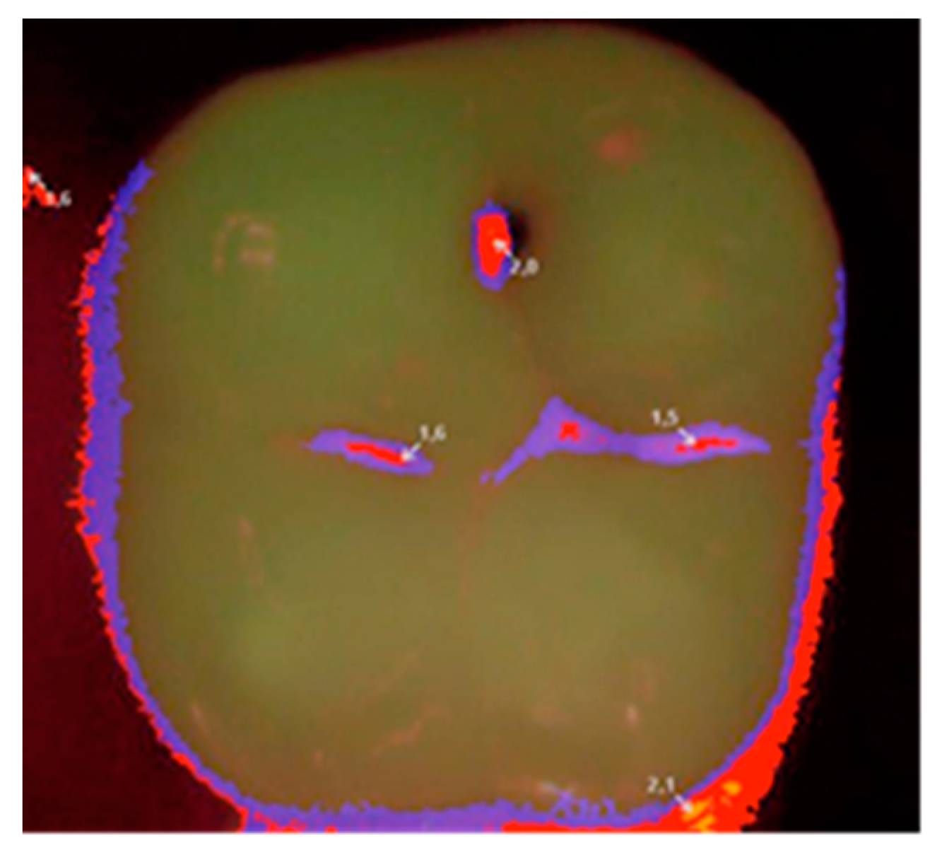
Each patient was qualified on the basis of a clinical statement that confirmed the presence of a small carious lesion, further validated by diagnostic results of Diagnodent (KaVo, Biberach, Germany) and a Vista Proof (Dürr Dental, Bietigheim-Bissingen, Germany) camera. An initial Vista Proof camera image (Figure 1) showing the extent and intensity of caries was recorded, and the corresponding numeric values (maximum) were saved.
Qualification of patients, cavity preparation and filling as well as clinical assessment were performed by one operator. The operator was trained before he started the procedures, and he was supervised by two independent, trained researchers with regard to strict adherence to the clinical management scheme. The method of randomization and qualification of the patients to allocate them to a given clinical group, which determined the choice of the applied material, was based on the drawing of cards by the patients from a limited poll.
Previously prepared cavities of Black’s Class I were filled with materials applied in accordance with the manufacturer’s recommendations in three configurations:
  • Group I (G I): Vertise Flow material without the use of an etching agent or a bonding system;
  • Group II (G II): Premise flowable material without the use of an etching agent, but with the use of the OptiBond All-In-One seventh-generation bonding system;
  • Group III (G III): Premise flowable material after etching and treatment of the hard tooth tissues using a fifth-generation OptiBond Solo Plus bonding system.
The composition and application mode of materials used in the study are presented in Table 1 [16,17].
Group I: 22 Black’s Class I cavities were prepared traditionally, rinsed with water and dried with a maximum airstream for 5 s. Subsequently, the Vertise Flow material (without an etching agent or a bonding system) in the shade chosen by means of the colorant was applied to the cavities with the use of an application tip and distributed with a moderate pressure using a supplied brush for 15–20 s. Any excess of the composite was removed.
Group II: 22 Black’s Class I cavities were prepared traditionally, rinsed with a stream of water and dried (not overdried: the state of drying was controlled by leaving a shiny surface). The OptiBond All-In-One bonding system was shaken vigorously before application, and then 2–3 drops were transferred to a clean container. A large amount of the bonding system (without prior use of an earlier application) was applied to the enamel/dentine surface with the use of a disposable applicator. The preparation was rubbed into the surface by performing brushing movements for 20 s. OptiBond All-In-One was applied again and rubbed in for 20 s. It was dried first with a gentle stream of air and then with a medium force jet for at least 5 s. Finally, it was hardened with lamp light for 10 s. Premise flowable in the shade chosen by means of the colorant was applied to the cavity prepared in this way along the axial and gingival margins. It was distributed with a brushing motion in such a way that it filled the margins of the cavities. Any excess of the composite was removed.
Group III: 20 Black’s Class I cavities were prepared traditionally, rinsed with a stream of water and dried (not overdried). Enamel was etched with 37% orthophosphoric acid. Then, the cavity was thoroughly rinsed so that the etcher was removed completely. The OptiBond Solo Plus bonding system was vigorously shaken before application, and then 2–3 drops were transferred to a clean container. After drying (not overdrying), the bonding system was then applied to the enamel/dentine surface with a disposable applicator. Then, it was dried with a gentle stream of air for 3 s and hardened with lamp light for 20 s. Premise flowable material in the shade chosen by means of the colorant was applied to the cavity prepared in this way along the axial and gingival margins. It was distributed with a brushing motion in such a way that it filled the margins of the cavity. Any excess of the composite was removed.
In all groups, the single layers of materials did not exceed 2 mm, and each layer was hardened with lamp light for 20 s. The light intensity of the lamp was maintained at 800 MW/cm2 and was regularly checked with a radiometer. If A3.5 shade or universal opaque was used, the polymerization time was extended to 40 s. During the material’s polymerization, protective glasses were used, and the purity of the optical fiber was controlled. The end of the light-guide was placed at a distance of about 2–5 mm from the exposed material, and after starting the light exposure, it was close to it for 1–2 s. The fillings in all groups (I, II, and III) were finished and polished with KerrHawe OptiDisc, Occlubrusch or the HiLuster system (Kerr) and rinsed with a stream of water.
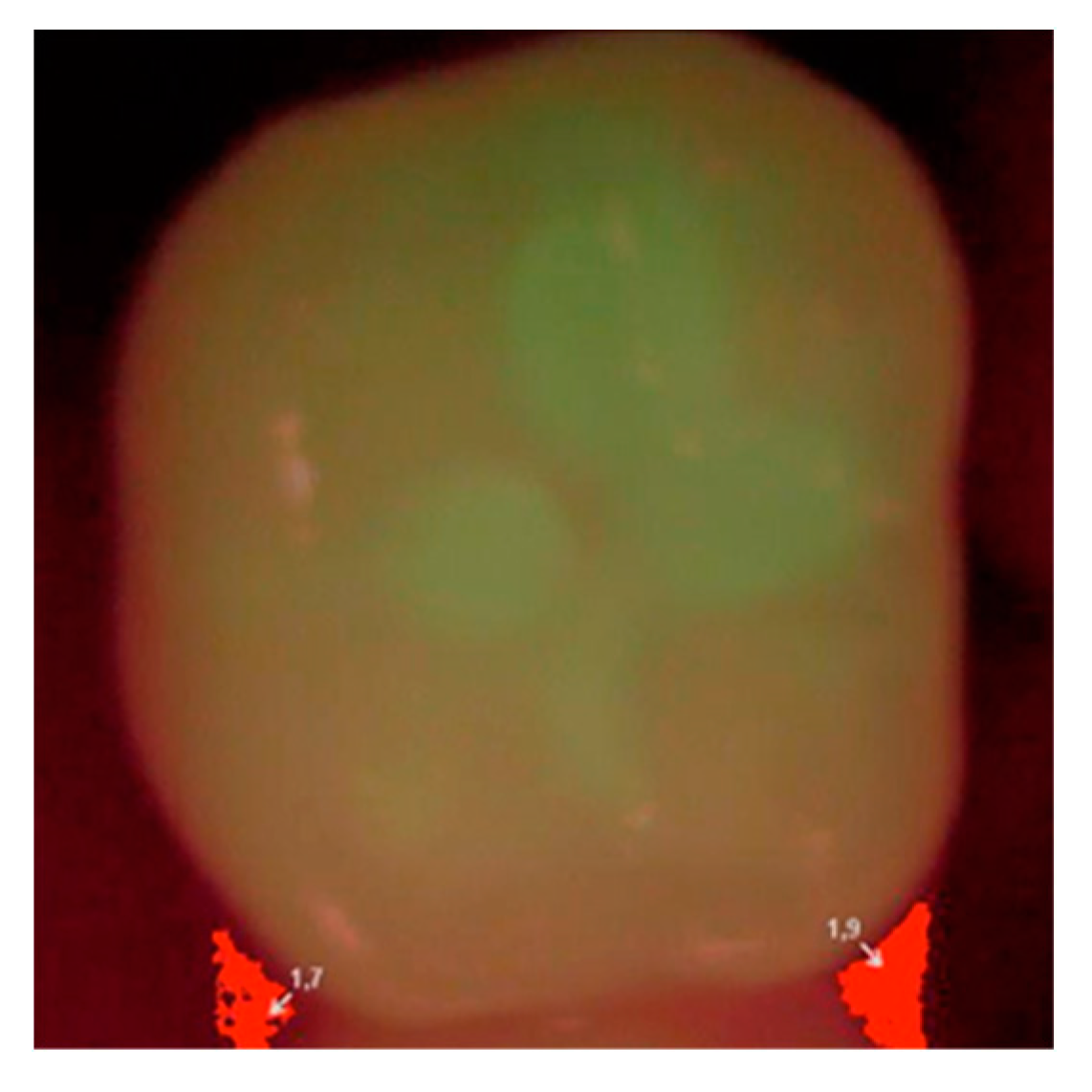
Then, at appropriate time intervals (0, i.e., right after filling, and after 6, 12, and 24 months), the patients were subjected to clinical examination, and the fillings were evaluated according to the Ryge scale criteria [14] with the use of additional visual registration by means of a Vista Proof camera (
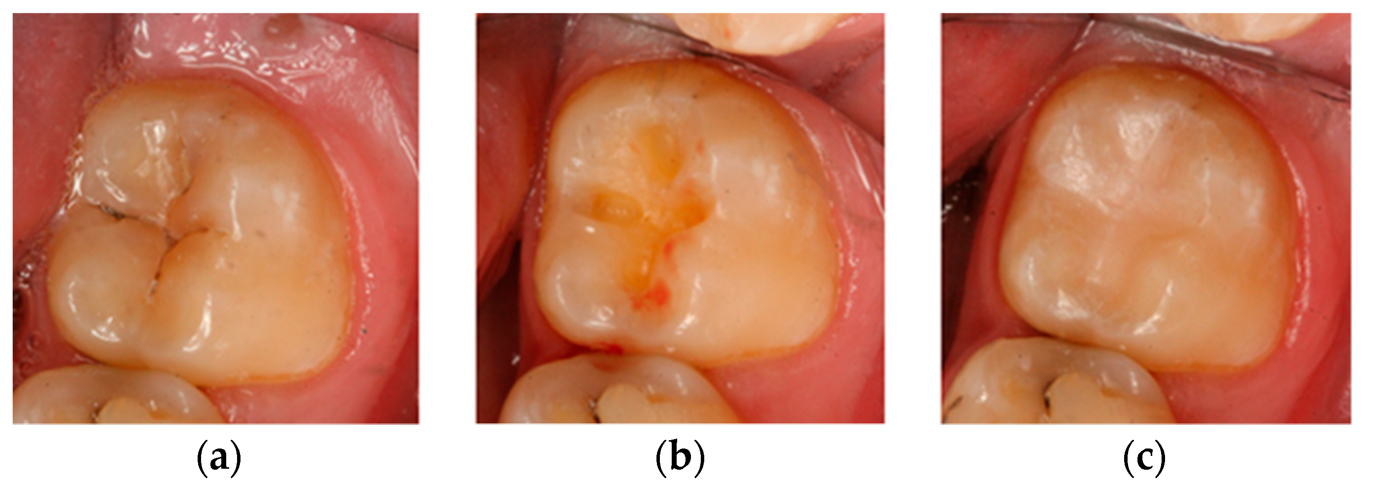
Figure 2). During each follow-up visit, an intraoral photograph was taken for the record with a Canon EOS 30D digital camera with a Canon EF 100 mm f/2.8 L Macro IS USM lens and a Canon Macro Ring Lite MR-14EX. The photographs were taken before and after the cavities were prepared, as well as after filling each cavity with a given material (Figure 3). Control tests were complemented by a plaque/calculus evaluation using Greene and Vermilion’s oral hygiene index (OHI) index and Lange’s approximal plaque index (API) [18].
All obtained results on a four-level Ryge scale (values from 0 to 3) were grouped in a table. The Ryge scale is presented in Table 2.

2.3. OHI-S (Oral Hygiene Index-Simplified) by Green and Vermillion

The debris index and the calculus index are two components of this index. Each of these indices is based on numeric determinations representing the amount of debris or calculus found on the preselected six tooth surfaces.
The examination was performed with the use of a dental mirror. Six teeth were examined, including 16 and 26 on the buccal surfaces, 26 and 46 on the lingual surfaces and 11 and 31 on the labial surfaces. The result of the examination was assigned a value between 0 and 3:
0-
No debris or calculus;
1-
Soft debris or supragingival calculus, covering not more than one-third of the exposed surface;
2-
Soft debris or supragingival calculus, covering not more than two-thirds of the exposed tooth surface or presence of flecks of subgingival calculus around the cervical portion of the tooth or both;
3-
Soft debris or supragingival calculus covering more than two-thirds of the exposed tooth surface or a continuous heavy band of subgingival calculus around the cervical portion of the tooth or both.
The results obtained for particular teeth (surfaces) were added and then divided by the number of examined teeth. The possible results vary between 0 and 3 for the debris and calculus indices and between 0 and 6 for the OHI-S values.

2.4. API (Approximal Plaque Index) by Lange

A yes/no decision was made with regard to whether the examined interproximal surfaces are covered by plaque (+) or not (−). The index is calculated as per the following formula:
API = no .   of   plaque   ( + )   sites no .   of   sites   examined × 100 %
According to the results, the oral hygiene conditions of individuals can be determined as follows:
  • 100–70%: poor oral hygiene;
  • 69–40%: insufficient oral hygiene;
  • 39–25%: pretty good oral hygiene;
  • <25%: optimum hygiene.

2.5. Statistical Analysis

In order to check the differences in API and OHI across the groups over time, an analysis of variance for mixed samples was performed. To check the exact differences between measurements in groups, a series of Bonferroni post hoc comparisons was performed. In order to check the differences in the status of the fillings on the Ryge scale after 6, 12, and 24 months, an analysis with the Friedman test was performed. To check the differences in fillings with regard to the groups, an analysis with the Kruskal–Wallis test was conducted. To check the exact differences between the results in groups, the Mann–Whitney U-test was performed.

3. Results

3.1. Group I

The results obtained right after the application of fillings, as indicated by the Ryge scale, demonstrate appropriate adaptation (no deficiencies or interfacial opening visible) of all fillings in relation to all assessment categories, that is, the structure of the surface, anatomic shape and marginal adhesion.
After six months of observation in Group I, the obtained values changed. In terms of the surface structure, in one case, the value ‘1′ was defined as ‘slight discoloration’, and in one case, the value ‘1′ was defined as ‘slightly rough surface’. In the area of marginal adhesion, in one case, the value ‘1′ was defined as ‘discoloration of the hard tooth tissues along the filling’, and in 11 cases, the value ‘1′ was defined as ‘visible superficial damage to the margin of the filling’ (Table 3).
After 12 months of observation, in the area of the surface structure, in one case, the value ‘1′ was defined as ‘slight discoloration’; in one case, the value ‘2′ was defined as ‘strongly discolored surface’; and in three cases, the value ‘1′ was defined as ‘slightly rough surface’. In the area of marginal adhesion, in 2 cases, the value ‘1′ was defined as ‘discoloration of hard tooth tissues along the filling’, and in 13 cases, the value ‘1′ was defined as ‘visible superficial damage to the margin of the filling’ (Table 3).
After 24 months of observation, in the area of the surface structure, in one case, the value ‘2′ was defined as ‘strongly discolored surface’, and in 11 cases, the value ‘1′ was defined as ‘slightly rough surface’. In the area of marginal adhesion, in 5 cases, the value ‘1′ was defined as ‘discoloration of hard tooth tissues along the filling’, and in 13 cases, the value ‘1′ was defined as ‘visible superficial damage to the margin of filling’, while in 1 case, the value ‘2′ was defined as ‘damage to the edge of the filling’. It was found that the number of indications according to the Ryge scale increased with the passage of time, reaching a maximum at 24 months (Table 3).

3.2. Group II

After 6 months of observation, the obtained values changed. In the area of the surface structure, in three cases, the value ‘1′ was defined as ‘slight discoloration’. In the area of marginal adhesion, in two cases, the value ‘1′ was defined as ‘discoloration of hard tooth tissues along the filling’, and in two cases, the value ‘1′ was defined as ‘visible superficial damage to the margin of the filling’ (Table 4).
After 12 months of observation, in the area of the surface structure, in three cases, the value of ‘1′ was defined as ‘slight discoloration’, and in the area of marginal adhesion, in three cases, the value ‘1′ was defined as ‘discoloration of hard tooth tissues along the filling’, while in four cases, the value ‘1′ was defined as ‘visible superficial damage to the margin of the filling’ (Table 4).
After 24 months of observation, in the area of the surface structure, in three cases, the value of ‘1′ was defined as ‘slightly rough surface’. Within the anatomic shape, in one case, the value ‘1′ was described as ‘partial occlusion points, yet with a possibility of correction’. In the area of marginal adhesion, in five cases, the value ‘1′ was defined as ‘discoloration of hard tooth tissues along the filling’, and in five cases, the value ‘1′ was defined as ‘visible superficial damage to the margin of the filling’. It was also found that, as far as surface structure is concerned, the number of indications of the Ryge scale did not increase with time, while in the area of anatomic shape and marginal adhesion, the number of indications of the Ryge scale increased and reached the maximum at 24 months (Table 4).

3.3. Group III

After 6 months of observation, the obtained values were as follows: in the area of the surface structure, in one case, the value ‘1′ was defined as ‘slightly rough surface’. In the area of marginal adhesion, in one case, the value ‘1′ was defined as ‘discoloration of the hard tooth tissues along the filling’, and in one case, the value ‘1′ was defined as ‘visible superficial damage to the margin of filling’ (Table 5).
After 12 months of observation, in the area of the surface structure, in one case, the value ‘1′ was defined as ‘slightly rough’, and in the case of marginal adhesion, the value ‘1′ was defined as ‘discoloration of hard tooth tissues along the fill’, and in one case, value ‘1′ was defined as ‘visible superficial damage to the edge of the fill’ (Table 5).
After 24 months of observation, in the area of the surface structure, one obtained value ‘1′ was defined as ‘slightly rough surface’. In the area of marginal adhesion, in two cases, the value ‘1′ was defined as ‘discoloration of hard tooth tissues along the filling’, while in three cases, the value ‘1′ was defined as ‘visible superficial damage to the margin of filling’. It was also found that, in the area of the surface structure, the number of indications of the Ryge scale did not increase with the passage of time, while in the marginal adhesion area, the number of Ryge scale indications increased slightly, reaching the maximum in 24 months (Table 5).
Subsequently, in order to check the differences in the fillings analyzed in relation to the Ryge scale in individual months, divided into groups, the analysis was carried out with the Kruskal–Wallis test. From the results of the analysis, it was found that there were significant differences between groups in fillings assessed by the Ryge scale after 6 months (χ2(2) = 7.63; p < 0.05; ε2 = 0.14), after 12 months (χ2(2) = 10.07; p < 0.01; ε2 = 0.18) and after 24 months (χ2(2) = 17.39; p < 0.001; ε2 = 0.31). In order to check the exact differences, the Mann–Whitney U-test was carried out. From the results, it was found that there were significant differences between fillings assessed by the Ryge scale after 6 months between Groups I and III (Z = −2.61; p < 0.05; r = 0.35). The level of fillings assessed by the Ryge scale after 6 months in Group I was higher than that in Group III (M = 0.12, SD = 0.14 vs. M = 0.02, SD = 0.06). The differences between these measurements were moderate, with r = 0.35 (Table 6).
From the results, it was found that there were significant differences between fillings assessed by the Ryge scale after 12 months between Groups I and III (Z = −3.13; p < 0.01; r = 0.43). The level of fillings assessed with the Ryge scale after 12 months in Group I was higher than that in Group III (M = 0.17, SD = 0.17 vs. M = 0.03, SD = 0.07). The differences between these measurements were moderate, as stated on the basis of r = 0.43 (Table 7).
From these results, it was found that there were significant differences between the fillings assessed on the Ryge scale after 24 months between Groups I and II (Z = −3.30; p < 0.01; r = 0.44) and between Groups I and III (Z = −3.65; p < 0.001; r = 0.50). The level of fillings assessed with the Ryge scale after 24 months in Group I was higher than that in Group II (M = 0.29, SD = 0.19 vs. M = 0.10, SD = 0.13). The differences between these measurements were moderate, with r = 0.44. The level of fillings assessed with the Ryge scale after 24 months in Group I was higher than that in Group III (M = 0.29, SD = 0.19 vs. M = 0.07, SD = 0.14). The differences between these measurements were high, as based on r = 0.50 (Table 8).

3.4. Oral Hygiene

3.4.1. OHI Index

The average value of the OHI index in qualified patients at the beginning of the study was 0.58 (SD = 0.32), and it decreased during subsequent follow-ups to reach a value of 0.53 (SD = 29) after 6 months of observation, 0.50 (SD = 0.31) after 12 months and 0.40 (SD = 0.22) after 24 months (Table 9).
In relation to individual clinical groups, the values were as follows: in Group I, the average OHI on the day when the filling procedure was performed amounted to 0.59 (SD = 0.32); after 6 months, it was 0.54 (SD = 0.27); after 12 months, it was 0.50 (SD = 0.39); and after 24 months, it was 0.48 (SD = 0.28). In Group II, the average OHI index on the day when the filling was performed was 0.61 (SD = 0.37); after 6 months, it was 0.51 (SD = 0.30); after 12 months, it amounted to 0.48 (SD = 0.27); and after 24 months, it was 0.38 (SD = 0.20). In Group III, the mean OHI index on the day of the filling was 0.52 (SD = 0.26); after 6 months, it was 0.56 (SD = 0.30); after 12 months, it was 0.51 (SD = 0.28); and after 24 months, it was 0.34 (SD = 0.16). While analyzing changes in OHI over time—and taking into consideration division into groups—it was observed that the value of the indicator decreased over time: the highest was at the beginning of the treatment, and the lowest was found at the last follow-up visit (Table 10).
In order to check the differences in the OHI values between groups and over time, an analysis of variance in a mixed sample scheme was carried out. It was found that there were significant differences for the main effect, i.e., OHI hygiene measurements over time (F(3.171) = 9.98; p < 0.001; η2 = 0.15). In order to check the exact differences, a series of Bonferroni post hoc tests of comparisons was carried out, based on which it was found that the OHI hygiene level (24 m) was significantly lower than OHI (0 m), OHI (6 m) and OHI hygiene (12 m) (M = 0.40, SD = 0.22 vs. M = 0.58, SD = 0.32 and M = 0.53, SD = 0.29 and M = 0.50, SD = 0.31). The differences between these measurements were small, as stated on the basis of η2 = 0.15 (Table 9).

3.4.2. API Index

Similarly, the values of the API indicator were analyzed. The mean value of the index in qualified patients was 51.71 (SD = 20.44), and the mean values at specific time points were as follows: after 6 months, it was 46.72 (SD = 17.14); after 12 months, it was 45.17 (SD = 15.22); and after 24 months, it was 38.71 (SD = 13.89). As in the case of the OHI index, the average (API) value decreased over time (Table 11).
Taking into account the division into clinical groups, the values developed as follows: In Group I, the average API on the day when the filling was performed amounted to 55.36 (SD = 22.37); after 6 months, it was 46.93 (SD = 16.22); after 12 months, it was 44.63 (SD = 17.29); and after 24 months, it was 42.22 (SD = 15.38). In Group II, the average API on the day the filling was performed amounted to 51.54 (SD = 20.44); after 6 months, it was 46.94 (SD = 17.49); after 12 months, it was 43.14 (SD = 11.85); and after 24 months, it was 34.97 (SD = 13.93). In Group III, the average API on the day the filling was performed amounted to 47.88 (SD = 18.54); after 6 months, it was 46.24 (SD = 18.65); after 12 months, it was 48.26 (SD = 16.73); and after 24 months, it was 39.37 (SD = 11.49). There was a decrease in the API value. At the beginning of the treatment, 69–40% of the results were interpreted as ‘average hygiene’, while at the last follow-up visit, 39–25% of the results in Groups II and III were assessed as ‘quite good hygiene’ (Table 12).
In order to check the differences in API hygiene measurements between groups and over time, analysis of variance in a mixed sample scheme was carried out. Based on the results of the analysis of variance in the mixed sample scheme, it was found that there were significant differences in the main effect, i.e., in the measurements of API hygiene over time (F(3.171) = 6.48; p < 0.001; η2 = 0.10). To verify the discrepancies in detail, a series of Bonferroni post hoc tests of comparison was carried out, based on which it was found that the API hygiene level (24 m) was significantly lower than the hygiene level of API (0 m) and API (6 m) (M = 38.71; SD = 13.89 vs. M = 51.71, SD = 20.44 and M = 46.72, SD = 17.14). The differences between these measurements were small, as indicated by η2 = 0.10 (Table 12).
A summary of the results confirmed significant differences between the tested materials. Vertise Flow material used without etching agent or a bonding system (G I) presented the weakest results with respect to marginal adaptation and smoothness among those evaluated in this study. The intensification of the degradation continued over time until the final clinical observation at 24 months. The results confirmed that the best quality was obtained by using the Premise flowable material after etching and treatment of the hard tooth tissues with a fifth-generation OptiBond Solo Plus bonding system relative to others for all time points of observation.

4. Discussion

The development of successive generations of bonding systems with improved adhesion and affinity to tooth tissues, which we have been observing over the years, mainly boils down to the simplification of the bonding procedure itself. The number of components of the bonding system is reduced, the use of etching is a thing of the past owing to the presence of acid monomers in the primer, and finally, the binding system is included in the composite material itself. It appears that such progress could eliminate errors made during the application of bonding systems and, as a consequence, protect the dentine–pulp complex against excessive mechanical and chemical interference. The exact and meticulous procedure of using a bonding agent in compliance with the recommended time to perform each stage of the procedure, while simultaneously paying close attention so as not to dry the dentin, may actually seem demanding and prone to mistakes [19].
However, does easier bonding also mean an effective connection to tooth tissues? If we take into account the strength of such a connection, it is not necessarily true. On the other hand, we noted that the fourth-generation systems still dominate in terms of the durability of connection to tooth tissues [20]. Nonetheless, when it comes to posttreatment hypersensitivity, leaving the hybrid layer to react only with the binding system may reduce the negative feelings associated with tooth filling [21]. Giacomini sheds a different light on the above issue: depending on the type of dentin, she prefers other bonding systems. Her research shows that dentin with small tubules, which is located in the peripheral regions (tooth necks, incisal margins), acquires a greater strength in its connection to new universal bonding systems than to previously used ones [22]. How that connection behaves over time is not without significance. The hybrid layer, which is largely responsible for the quality of the connection, should be of good quality at the time of its creation and, more important, should maintain these properties over time [23]. Matrix metalloproteinases (MMPs) play an important role in this process: endogenous enzymes present in dentin cause degradation of the collagen matrix. This may be triggered by adhesive procedures that use simplified total-etch (TE) binding systems [24] or strongly acidic self-etch (SE) systems [25]. MMP activity and the lack or presence of MMP inhibitors have an impact not only on the appearance of secondary caries at the material–tooth tissue boundary, but also on the presence and activity of primary caries.
The Vertise Flow material—which was the inspiration behind the research in this study—is an innovative and one of the first composite materials that contain a functional GPDM monomer (glycerol phosphate dimethacrylate), which the procedure of applying classical etching and bonding to be omitted. For this reason, there are still only a few reports evaluating this material in the literature [11,12,13]. It is even more difficult to find a study that compares the Vertise Flow material with other traditional materials and includes laboratory and clinical or clinical tests lasting for at least 24 months. Vichi et al. clinically studied Vertise Flow in vivo, applied in small Class I cavities in accordance with the manufacturer’s instructions. They then evaluated those defects using the Ryge scale after one day, seven days, one month, three months and six months. After three months, three out of 40 fillings already had the indication ‘1′ in the marginal adhesion zone (presence of discoloration, fissures), one of which was qualified for replacement within a year [11]. This was considered to be satisfactory clinical behavior. The obtained result could be influenced by the fact that the researchers carefully checked the occlusion before and after the defect and eliminated the cases in which the extent of the filling was in the areas of occlusion and articulation points (functional areas). Nevertheless, it is similar to the fact that the Ryge scale indications already seem worse in the area of marginal adhesion in the 6th month. In clinical studies, Chałas et al. assessed Vertis Flow with the four-level Ryge scale immediately after performing 67 fillings in small Black’s Class I defects outside the occlusion zone and small Black’s Class V cervical lesions. They obtained very good results for all parameters of the analyzed categories [26]. Similarly, after three months of observation of the Vertise Flow material in 30 Black class I defects assessed with the Ryge scale, Marzec et al. assessed 100% of the fillings to be clinically acceptable with 100% retention [27]. The clinical study of Kucukyilmaza is also worth mentioning, as it compared, after 24 months, groove sealants, Vertise Flow and a traditional flow material, applied with the use of a bonding system. The retention of the Vertise Flow material after that time was the lowest and amounted to 62.9%. For comparison, the retention of groove sealants ranged between 80.6% and 73.1%, and a traditional flow with the bonding system was as high as 95.7% [28]. In the face of a small number of clinical trials, it is worth paying attention to the works of Celik, who clinically tested another self-adhesive flow type compomer with the commercial name Fusio Liquid Dentine. Forty cervical cavities were filled with that material, while another 40 were filled with the G-aenial nanohybrid material using a fourth-generation binding system (OptiBond FL), which he treated as a control group. The condition of 27 out of 40 Fusio Liquid Dentine fillings after six months was assessed as clinically unacceptable, whereas in the control group, 40 out of 40 fillings were still satisfactory. The author defined the success rate as 33% and considered the clinical evaluation of the tested material to be unacceptable [29].
This study involved an assessment of fillings in small Black’s Class I cavities applied in accordance with the manufacturer’s recommendations in three configurations: the Vertise Flow material without an etching agent or a bonding system (G I), the Premise flowable material without the use of an etching agent but with the use of the OptiBond All-In-One binding system (G II) and the Premise flowable material after etching and treatment of hard tooth tissues with a fifth-generation OptiBond Solo Plus bonding system (G III). Then, at appropriate time intervals (0, i.e., right after filling and after 6, 12, and 24 months), these fillings were subjected to clinical evaluation performed according to the Ryge scale and with the use of a fluorescent high-intensity visible light beam produced by a camera (Vista Proof). Similar to Chałas’s study, very good results were obtained in all analyzed categories on the four-level Ryge scale immediately after the filling with the Vertise Flow material. After six months of observation, as many as 12 of the 22 fillings had the indication ‘1′ in the marginal adhesion zone (presence of discolorations, fissures). For comparison, Vichi obtained 3 out of 40 in the same area [11]. In other material groups, the results were much better. Premise flowable with a seventh-generation bonding system scored 4 out of 22 and that with a fifth-generation bonding system scored 2 out of 20. After 24 months, as many as 20 fillings with Vertise Flow had indications in the marginal adhesion zone. For comparison, after 24 months, Premise flowable with a seventh-generation bonding system had a score of 10 and that with a fifth-generation bonding system scored only 5. Vertise Flow material used without an etching agent or a bonding system (G I) presented the weakest results with respect to marginal adaptation and smoothness among those evaluated in this study. The intensification of the degradation continued over time until the final clinical observation at 24 months. Kucukyilmaza made similar observations, as he reported that after 24 months, Vertise Flow had the worst retention among the materials examined [26]. Therefore, the above assessments are in line with Celik’s study, which, after six months, showed that 27 out of 40 fillings applied with another material that was also self-adhesive, i.e., Fusio Liquid Dentine, were clinically unacceptable [29].
In the case of small cavities, although the application of flow type materials is an acceptable solution [30] due to their mechanical properties, they should not be used solo for molar reconstruction, especially in the areas of large forces (occlusal contacts) [31]. In these areas, flow type materials should be used as liners, for extended sealing of fissures, for repairs of minor damage to the margins of fillings and for restorations in fissures and anatomic openings. Helvatjoglu-Antoniades et al. believe that their use as liners reduces the incidence of microleakage and the risk of debonding [32]. Chałas et al. reported that flow type materials should be placed in class I and II cavities, but with a narrow outline, not exposed to large occlusion loads [26]. In our own clinical and laboratory studies, it was found that the Vertise Flow material is the weakest when it comes to the strength of the connection to the tooth tissues as compared with other flow materials. However, the author of the study believes that poor results are not only caused by material properties, but also a consequence of abandoning selective enamel etching and, instead, bidding everything on the acid monomers included in the composition of the material itself. It is encouraging that companies are trying to introduce new technologies and facilitate the work of the dentist, but progress in general means that, sometimes, it takes two steps backward before taking one step forward. From the results of this study, the omission of the enamel etching procedure at the current stage of development of dental materials may be considered to be these ‘two steps backward’.
Oral hygiene indicators are widely used and included among the basic criteria for describing the current state of hygiene of the patient [18]. However, it is interesting to observe how their values are shaped in the case of those patients who are constantly subjected to dental control. The authors of the study observed that both OHI and API improved their average values at the beginning of treatment (OHI = 0.58, API = 51.71) and after 24 months (OHI = 0.40 and API = 38.71). In her doctoral dissertation, Czaplińska showed that in orthodontic patients who were motivated every day and instructed to improve their toothbrushing, the API index was lower (more favorable) at the end of the treatment than at the beginning [33]. The same was confirmed in the studies by Śmiech-Słomkowska, by Jabłońska-Zrobek and also by Bardal et al. [34,35]. It is difficult to maintain constant conditions in clinical trial observations of composite materials. Therefore, the analysis of the level of oral hygiene with the OHI and/or API index can be an extremely important element in standardizing the conditions of observation in compared groups of patients during long-term observation.

5. Conclusions

The used preparation scheme and bonding system have an impact on the final quality of the composite filling. There is a need to carry out a qualitative clinical evaluation of the dental restorative materials under uniform conditions by using evaluation scales.

Author Contributions

A.M.: data curation, formal analysis, investigation, writing—original draft; A.T.: formal analysis, resources, writing—review and editing; H.T.: conceptualization, project administration; M.T.: methodology, project administration, supervision. All authors have read and agreed to the published version of the manuscript.

Funding

This research received no external funding.

Conflicts of Interest

The authors declare no conflict of interest.

References

  1. Van Meerbeek, B.; Perdigão, J.; Lambrechts, P.; Vanherle, G. The Clinical performance of adhesives. J. Dent. 1998, 26, 1–20. [Google Scholar] [CrossRef]
  2. Sundfels, R.H.; Valentini, T.A.; de Alexandre, R.S.; Briso, A.L.F.; Sundefeld, M.L.M.M. Hybrid layer thickness and resin tag length of a self-etching adhhesive bonded to sound dentin. J. Dent. 2005, 33, 675–681. [Google Scholar] [CrossRef]
  3. Buczkowska-Radlińska, J.; Szyszka, L.; Wojtarowicz, R. The long-term evaluation of the complications of biological pulp treatment with using calcium hydroxide materials for direct and indirect pulp capping. Pol. J. Environ. Stud. 2007, 16, 39–42. [Google Scholar]
  4. Costa, C.A.; Teixeira, H.M.; Lopes do Nascimento, A.B.; Hebling, J. Biocompalibity of resin- based dental materials applied as liners in deep cavities prepared in human teeth. J. Biomed. Mater. Res. B Appl. Biomater. Res. 2007, 81, 175–184. [Google Scholar] [CrossRef] [PubMed]
  5. Koliniotou-Koumpia, E.; Papadimitrriou, S.; Tziafas, D. Pulpal responses after appliction of current adhesive systems to deep cavities. Clin. Oral. Investig. 2007, 11, 313–320. [Google Scholar] [CrossRef]
  6. Garcia, R.N.; Morelli, A.E.; da Silva, B.S.; Giongo, B.M.; Pollheim, C.P.; Largura, G.S.; Mensch, L.F.; Costa, M.F.; Candiotto, N.R.; Gomes, A.C.R. Bonding performance of a self-adhering flowable composite to substrates used in direct technique. RSBO 2013, 10, 343–349. [Google Scholar]
  7. Poitevin, A.; De Munck, J.; Van Ende, A.; Suyama, Y.; Mine, A.; Peumans, M.; Van Meerbeek, B. Bonding effectiveness of self-adhesive composites to dentin and enamel. Dent. Mater. 2013, 29, 221–230. [Google Scholar] [CrossRef] [PubMed]
  8. Hanabusa, M.; Mine, A.; Kuboki, T.; Momoi, Y.; Van Landuyt, K.L.; Van Meerbeek, B.; De Munck, J. TEM interfacial characterization of an experimental self-adhesive filling material bonded to enamel/dentin. Dent. Mater. 2011, 27, 818–824. [Google Scholar] [CrossRef]
  9. Fu, J.; Kakuda, S.; Pan, F.; Hoshika, S.; Ting, S.; Fukuoka, A.; Bao, Y.; Ikeda, T.; Nakaoki, Y.; Selimovic, D.; et al. Bonding performance of a newly developed step-less all-in-one system on dentin. Dent. Mater. J. 2013, 32, 203–211. [Google Scholar] [CrossRef] [Green Version]
  10. Juloski, J.; Goracci, C.; Rengo, C.; Giovannetti, A.; Vichi, A.; Vulicevic, Z.R.; Ferrari, M. Enamel and dentin bond strength of new simplified adhesive materials with and without preliminary phosphoric acid-etching. Am. J. Dent. 2012, 25, 239–243. [Google Scholar]
  11. Vichi, A.; Goracci, C.; Ferrari, M. Clinical study of the self-adhering flowable composite resin Vertise Flow in Class I restorations: Six-month follow-up. Int. Dent. 2010, 12, 14–23. [Google Scholar]
  12. Oz, F.D.; Ergin, E.; Cakir, F.Y.; Gurgan, S. Clinical evaluation of a self-adhering flowable resin composite in minimally invasive class I cavities: 5-year result of a double blind randomized, controlled clinical trial. Acta Stomatol. Croat. 2020, 54, 10–21. [Google Scholar] [CrossRef]
  13. Hamdy, T.M. Interfacial microscopic examination and chemical analysis of resin-dentin interface of self-adhering flowable resin composite. F1000Research 2017, 6, 1688. [Google Scholar] [CrossRef] [PubMed]
  14. Turkun, L.Ş. Clinical evaluation of a self-etching and a one-bottle adhesive system at two years. J. Dent. 2003, 31, 527–534. [Google Scholar] [CrossRef]
  15. Nedeljkovic, I.; Teughels, W.; De Munck, J.; Van Meerbeek, B.; Van Landuyt, K.L. Is secondary caries with composites a material- based problem? Dent. Mater. 2015, 31, e247–e277. [Google Scholar] [CrossRef]
  16. Sabbagh, J.; Dagher, S.; Osta, N.E.; Souhaid, P. Randomized Clinical Trial of a Self-Adhering Flowable Composite for Class I Restorations: 2-Year Resluts. Int. J. Dent. 2017, 5041529. [Google Scholar] [CrossRef]
  17. Moosavi, H.; Kimyai, S.; Forghani, M.; Khodadadi, R. The clinical effectivness of various adhesive systems: An 18-month evaluation. Oper. Dent. 2013, 38, 134–141. [Google Scholar] [CrossRef]
  18. Trzcionka, A.; Twardawa, H.; Mocny-Pachońska, K.; Tanasiewicz, M. Oral cavity status of long-term hemodialized patients vs. their socio-economic status. Med. Prac. 2020, 71, 1–10. [Google Scholar] [CrossRef]
  19. Van Meerbeek, B.; Van Landuyt, K.; De Munck, J.; Hashimoto, M.; Peumans, M.; Lambrechts, P.; Yoshida, Y.; Inoue, S.; Suzuki, K. Technique-sensitivity of contemporary adhesives. Dent. Mater. J. 2005, 24, 1–13. [Google Scholar] [CrossRef]
  20. De Munck, J.; Mine, A.; Poitevin, A.; Van Ende, A.; Cardoso, M.V.; Van Landuyt, K.L.; Peumans, M.; Van Meerbeek, M. Meta-analytical review of parameters involved in dentin bonding. J. Dent. Res. 2012, 91, 351–357. [Google Scholar] [CrossRef]
  21. Peumans, M.; Munck, J.; Van Landuyt, K.; Lambrechts, P.; Van Meerbeek, B. Three-year clinical effectiveness of a two-step self-etch adhesive in cervical lesions. Euro. J. Oral. Sci. 2005, 113, 512–518. [Google Scholar] [CrossRef] [PubMed]
  22. Giacomini, M.C.; Scaffa, P.M.C.; Chaves, L.P.; Vidal, C.M.P.; Machado, T.N.; Honório, H.M.; Tjäderhane, L.; Wnag, L. Role of Proteolytic Enzyme Inhibitors on Carious and Eroded Dentin Associated With a Universal Bonding System. Oper. Dent. 2017, 42, 188–196. [Google Scholar] [CrossRef] [PubMed]
  23. Łukomska-Szymańska, M.; Sokołowski, J.; Łapińska, B. Degradation of a hybrid layer–review of literature. J. Stoma 2017, 70, 88–94. [Google Scholar] [CrossRef]
  24. Mazzoni, A.; Pashley, D.H.; Nishitani, Y.; Breschi, L.; Mannello, F.; Tjäderhane, L.; Toledano, M.; Pashley, E.L.; Tay, F.R. Reactivation of inactivated endogenous proteolytic activities in phosphoric acid-etched dentine by etch-and-rinse adhesives. Biomaterials 2006, 27, 4470–4476. [Google Scholar] [CrossRef] [PubMed]
  25. De Munck, J.; Van den Steen, P.E.; Mine, A.; Van Landuyt, K.L.; Poitevin, A.; Opdenakker, G.; Van Meerbeek, B. Inhibition of enzymatic degradation of adhesive-dentin interfaces. J. Dent. Res. 2009, 88, 1101–1106. [Google Scholar] [CrossRef] [PubMed]
  26. Chałas, R.; Kamińska-Pikiewicz, K.; Zubrzycka-Wróbel, J.; Sochaczewska-Dolecka, A.; Bachanek, T. Compobond–composite or bond? Proprietary clinical observation of Vertise Flow material. Dent. Forum 2015, 42, 11–15. [Google Scholar]
  27. Marzec, L.; Grodoń, G.; Skałecka-Sądel, A.; Skośkiewicz-Malinowska, K. Initial clinical evaluation of fillings made of Vertise™ Flow composite material. Mag. Stomatol. 2013, 4, 121–124. [Google Scholar]
  28. Kucukyilmaz, E.; Selcuk, S. Evaluation of different fissure sealant materials and flowable composites used as pit-and-fissure sealants: A 24-month clinical trial. Pediatr. Dent. 2015, 37, 468–473. [Google Scholar]
  29. Çelik, E.; Uzer, B.A.; Fatma, Y. Six-month Clinical Evaluation of a Self-adhesive Flowable Composite in Noncarious Cervical Lesions. J. Adhes. Dent. 2015, 17, 361–368. [Google Scholar]
  30. Helvatjoglu-Antoniades, M.; Papadogiannis, Y.; Lakes, R.S.; Dionysopoulos, P.; Papadogiannis, D. Dynamic and static elastic moduli of packable and flowable composite resins and their development after initial photo curing. Dent. Mater. 2006, 22, 450–459. [Google Scholar] [CrossRef]
  31. Baroudi, K.; Silikas, N.; Watts, D.C. Time-dependent viscoelastic creep and recovery of flowable composites. Eur. J. Oral. Sci. 2007, 115, 517–521. [Google Scholar] [CrossRef] [PubMed]
  32. Leevailoj, C.; Cochran, M.A.; Matis, B.A.; Moore, B.K.; Platt, J.A. Microleakage of posterior packable resin composites with and without flowable liners. Oper. Dent. 2001, 26, 302–307. [Google Scholar] [PubMed]
  33. Czaplińska, J. Stan Uzębienia, Przyzębia I Higieny Jamy Ustnej U Dzieci I Młodzieży Leczonych Aparatami Stałymi [Rozprawa Doktorska]; Uniwersytet Medyczny im. K. Marcinkowskiego: Poznań, Polska, 2012. [Google Scholar]
  34. Śmiech-Słomkowska, G.; Jabłońska-Zrobek, J. Higiena jamy ustnej pacjentów leczonych stałymi aparatami ortodontycznymi. Mag. Stomatol. 2003, 12, 19–20. [Google Scholar]
  35. Bardal, P.A.P.; Olympio, K.P.K.; de Bastos, J.R.M.; Henriques, J.F.C.; Buzalaf, M.A.R. Education and motivation in oral health: Preventing disease and promoting health in patients undergoing orthodontic treatment. Dental Press J. Orthod. 2011, 16, 95–102. [Google Scholar] [CrossRef]
Figure 1. Image from a Vista Proof camera showing Tooth 47 qualified for treatment.
Coatings 10 00800 g001
Figure 2. Image from a Vista Proof camera showing Tooth 47 in an early assessment, just after filling.
Coatings 10 00800 g002
Figure 3. Images showing Tooth 37 (a) before cavity preparation; (b) after cavity preparation; and (c) after filling.
Coatings 10 00800 g003
Table 1. Composition and application mode of materials used in the study.
MaterialManufacturerCompositionApplication Mode
CompositeVertise FlowKerr Corporation, Orange, CA, USAMatrix: glycerol-phosphate dimethacrylate (GPDM), urethane
dimethacrylate (UDMA), bisphenol A
glycidyl dimethacrylate (bis-GMA) and other methacrylate comonomers, photoinitiators.
Fillers: 70% by weight. Ytterbium fluoride, barium aluminosilicate glass, prepolymerized fillers, colloidal silica
Brush the thin layer (<0.5 mm) with moderate pressure (15–20 s), light cure (20 s), place additional layers (≤2 mm), light cure (20 s)
Premise
Flowable
Kerr Corporation, Orange, CA, USAMatrix: bis-GMA, triethylene glycol dimethacrylate (TEGDMA), ethoxylated bis-phenol-A-dimethacrylate, light cure initiators, stabilizers.
Fillers: 72.5% by weight. Barium glass, prepolymerized fillers, silica nanoparticles
Apply the thin layer (≤2 mm), light cure (10 s)
Adhesive SystemOptiBond All-In-OneKerr Corporation, Orange, CA, USAGPDM, mono- and difunctional monomers, water, acetone, ethanol, nanofillers, camphorquinone (CQ)Apply one layer, scrub it using a microbrush (20 s), repeat the procedure, dry with air (5 s), light cure (10 s)
OptiBond Solo PlusKerr Corporation, Orange, CA, USAEtchant: 37.5% phosphoric acid, bis-GMA, hydroxyethyl methacrylate (HEMA), glycol dimethyl acrylate (GDMA), GPDM, ethanol, CQ, 2-(ethylhexyl)-4-(dimetylamino) benzoate (ODMAB), 2,6-di-tert-butyl-4-methylphenol (BHT), fumed silicone dioxide, gamma-methacryloxypropyltrimethoxysilane (A174), barium aluminoborosilicate, Na2Si6 FEtch (15 s), rinse (15 s), dry (15 s), apply the adhesive, scrub it using a microbrush (20 s), dry with air (3 s), light cure (20 s)
Table 2. Ryge scale.
Criteria0123
Texture of the surface (criteria: color, smoothness, luster of restoration)The restoration surface is smooth, the restoration matches the adjacent tooth structure in color, no discolorationsThe restoration surface is slightly rough, but it is possible to smoothen it by polishing or filling with an excess and the failure can be corrected during the first control visitThe surface is rough, strongly discolored, no possibility of correctionThe surface is broken, very strongly discolored, the filling needs to be immediately changed
Anatomic form (criteria: reconstruction of anatomic form, cusps and proximal surfaces)Restoration with appropriate anatomic form, cusps reconstructed, incisal edges, proximal contact and occlusal contacts preservedRestoration with insufficiently shaped incisal edge, partial points of occlusal contact, but there is a possibility of correctionRestoration insufficiently shaped, insufficient occlusion, there is no possibility of its correctionRestoration insufficiently shaped, occlusal trauma
Marginal adaptation (criteria: discolorations, fissures, secondary caries)No margin gap, microleakage or discoloration of tooth tissues along the margin of the fillingVisible superficial damage of the filling’s margin and discolorations of hard tooth tissues along the fillingDamage of the filling’s margin, visible dentine or liner, possible discoloration of hard tooth tissuesMobile filling, fracture of tooth wall, secondary marginal caries, filling needs to be immediately changed
Table 3. Number of obtained Ryge scale indications in Group I after 6, 12 and 24 months.
Observation TimeSurface StructureAnatomic ShapeMarginal Adaptation
ColorSmoothnessReconstructionDiscolorationSealing
6 months110111
12 months330213
24 months2110515
Table 4. Number of obtained Ryge scale indications in Group II after 6, 12, and 24 months.
Observation TimeSurface StructureAnatomic ShapeMarginal Adhesion
ColorSmoothnessReconstructionDiscolorationSealing
6 months30022
12 months30034
24 months30155
Table 5. Number of obtained Ryge scale indications in Group III after 6, 12, and 24 months.
Observation TimeSurface StructureAnatomic ShapeMarginal Adhesion
ColorSmoothnessReconstructionDiscolorationSealing
6 months01011
12 months01011
24 months01023
Table 6. Results of the Mann–Whitney U-test for the Ryge scale after 6 months.
GroupMean (M)Standard Deviation (SD)
G I0.120.14
G II0.050.11
G III0.020.06
Table 7. Results of the Mann–Whitney U-test for the Ryge scale after 12 months.
GroupMean (M)Standard Deviation (SD)
G I0.170.17
G II0.080.12
G III0.030.07
Table 8. Results of the Mann–Whitney U-test for the Ryge scale after 24 months.
GroupMean (M)Standard Deviation (SD)
G I0.290.19
G II0.100.13
G III0.070.14
Table 9. Average values and standard deviations of OHI index value in time.
TimeMean (M)Standard Deviation (SD)
00.580.32
6 months0.530.29
12 months0.500.31
24 months0.400.22
Table 10. Average values and standard deviations of Greene and Vermilion’s oral hygiene index (OHI) index values over time with regard to group division.
Time GroupMean (M)Standard Deviation (SD)
OHI 0G I0.590.32
G II0.610.37
G III0.520.26
OHI 6G I0.540.27
G II0.510.30
G III0.560.30
OHI 12G I0.500.39
G II0.480.27
G III0.510.28
OHI 24G I0.480.28
G II0.380.20
G III0.340.16
Table 11. Average values and standard deviations of Lange’s approximal plaque index (API) index values over time.
TimeMean (M)Standard Deviation (SD)
051.7120.44
6 months46.7217.14
12 months45.1715.22
24 months38.7113.89
Table 12. Average values and standard deviations of API index values over time with regard to group division.
Time GroupMeanStandard Deviation (SD)
API 0G I55.3622.37
G II51.5420.44
G III47.8818.54
API 6G I46.9316.22
G II46.9417.49
G III46.2418.65
API 12G I44.6317.29
G II43.1411.85
G III48.2616.73
API 24G I42.2215.38
G II34.9713.93
G III39.3711.49

