Article

The Mechanisms of Degradation of Titanium Dental Implants

by
Agnieszka Ossowska
* and
Andrzej Zieliński
Department of Materials Engineering and Bonding, Gdansk University of Technology, 80233 Gdańsk, Poland
*
Author to whom correspondence should be addressed.
Coatings 2020, 10(9), 836; https://doi.org/10.3390/coatings10090836
Submission received: 29 July 2020 / Revised: 22 August 2020 / Accepted: 26 August 2020 / Published: 28 August 2020
(This article belongs to the Special Issue Recent Developments of Electrodeposition Coating)

Abstract

:
Titanium dental implants show very good properties, unfortunately there are still issues regarding material wear due to corrosion, implant loosening, as well as biological factors—allergic reactions and inflammation leading to rejection of the implanted material. In order to avoid performing reimplantation operations, changes in the chemical composition and/or modifications of the surface layer of the materials are used. This research is aimed at explaining the possible mechanisms of titanium dissolution and the role of oxide coating, and its damage, in the enhancement of the corrosion process. The studies of new and used implants were made by scanning electron microscopy and computer tomography. The long-term chemical dissolution of rutile was studied in Ringer’s solution and artificial saliva at various pH levels and room temperature. Inductively coupled plasma mass spectrometry (ICP-MS) conjugated plasma ion spectrometry was used to determine the number of dissolved titanium ions in the solutions. The obtained results demonstrated the extremely low dissolution rate of rutile, slightly increasing along with pH. The diffusion calculations showed that the diffusion of titanium through the oxide layer at human body temperature is negligible. The obtained results indicate that the surface damage followed by titanium dissolution is initiated at the defects caused by either the manufacturing process or implantation surgery. At a low thickness of titanium oxide coating, there is a stepwise appearance and development of cracks that forms corrosion tunnels within the oxide coating.

1. Introduction

Titanium alloys possess good strength properties and high resistance to the most aggressive environments such as hydrochloric acid or sulfuric acid [1,2,3]. The compact, stable oxide layer [4] is responsible for corrosion resistance and biotolerance, effectively stopping the anodic pickling of the substrate [5,6,7]. Another important function is to chemically stabilize the implant in the living organism [4,8,9,10]. The more compact and bonded the passive layer is to the substrate, the better the corrosion resistance. In the case of thick oxide layers, an improvement in tribological properties may also be observed [7]. According to Hanawa et al. [11], the top sublayer of the titanium oxide layer inhibits metal ion release [12,13] and its transformation in vitro. Additionally, the oxide layer promotes osseointegration and bone adhesion [14,15,16].
It is known that each implant inserted into the body is treated as a foreign body and can cause allergic reactions, inflammations, even the rejection of the implant. The human body is a very specific environment as the body fluids—extracellular fluids and blood—contain aqueous solutions of certain organic substances, dissolved oxygen, various inorganic anions (Cl, HPO42−, HCO3−), and cations (Na+, K+, Ca2+, Mg2+), which together represent a highly aggressive environment [17]. The presence of amino acids and proteins accelerates the corrosion processes [18]. Besides, in the case of dental implants, the composition of the saliva is highly complex, containing both inorganic salts and organic components. This composition depends on many factors such as food, age, and diseases and the pH of saliva can vary around dental implants. The ingestion of acidic beverages can decrease the buccal pH, and the infections can also acidify the pH of saliva, contributing to the corrosion of dental implants. On the other hand, titanium and its alloys are sensitive to tribocorrosion [19]. The oxidation is performed mainly to prevent corrosion of titanium and its alloys in severe conditions. For dental implants, such oral environments include varying pH, acid attack and the presence of chemical compounds such as cetylpyridinium chloride, sodium fluoride and hydrogen peroxide [20].
The action of media containing fluoride ions causes degradation of the continuity of the oxide film followed by damage to the titanium as a result of the ingestion of fluoride ions into the oxide layer, thereby reducing its protective properties [21,22].
Corrosion processes influence changes in the structure of the implanted material, weakening its integrity, which can result in material discontinuities and cracks. Cells in direct contact with the exposed surface of the material are stimulated for the intensive secretion of inflammatory mediators, mainly neutrophils and macrophages [23]. In vitro studies [24] show that corrosion products are harmful to cell differentiation and proliferation processes.
The dissolution of the titanium oxide layer is due to the process of ion diffusion into the layer. In titanium, the oxygen atoms migrate via the interstitial diffusion mechanism, occupying the free, octahedral interstitial positions in the titanium hexagonal lattice. Studies conducted by Wu and Trinkle [25] showed that for oxygen atoms not only interstitial but also axial positions are available, i.e., all arrangements of oxygen atoms in the titanium matrix are possible.
The oxidation of titanium is faster when the material is subjected to high temperatures and the influence of an oxygenated environment. The overall oxidation reaction includes the formation of oxide followed by the diffusion of oxygen into the bulk of the titanium. Oxygen diffusion creates an oxygen-enriched layer due to the high solubility of oxygen in the titanium and the oxygen stabilizing effect in the crystalline titanium structure [26]. In solids, the most likely atomic diffusion mechanism is a vacancy or interstitial mechanism, i.e., the motion of atoms occurs as the consequence of the presence of imperfections [27]. The interstitial diffusion mechanism is typical for low atomic radius atoms such as hydrogen, oxygen, carbon, and nitrogen.
The great advantage of the oxide layers produced on titanium and its alloys is their capability of repassivation, and some released ions depend on regeneration. Hanawa et al. [11], while measuring repassivation potentials, estimated the recovery rate of the oxide layer in 0.9% physiological saline: for 316L steel as 35.3 min, for Ti6Al4V as 8.2 min, and Co28Cr6Mo as 12.7 min. The research conducted by Hanawa et al. [28] showed that in Hanks’ solution, the rate of repassivation was lower than in 0.9% saline solution.
Metallic elements have a different tendency to release ions, and even trace amounts of elements in the alloy composition should not be neglected [11]. There are data on the significant contents of some alloying elements of Ti6Al4V within the tissue around the implanted alloy. So far [29] reports on the consequences of ion release into the body have focused on the importance of the possible impact of released ions on biomolecules and the initiation of adverse biological reactions as the titanium ions could quickly react with water molecules or inorganic anions, easily binding with body fluids.
Osseointegration involves a series of biological events influenced by multiple factors. Among them, the porous-structured Ti alloys have shown to allow rapid bone ingrowth and improved osseointegration by increasing the bone-implant interface area [30,31]. Such conditions are achieved for dental implants by micro-arc oxidation, which brings out the rough surface [19,32,33,34,35]. The bioactivitity is usually increased by anodic oxidation in an electrolyte containing calcium phosphates [36,37], and wear resistance by incorporation of tough nanoparticles [35].
We have put a hypothesis that the damage of oxide coating can be sometimes or often, the main cause for the degradation of material and removal of the implant. The purpose of the study was to characterize the processes which allow for titanium dissolution from dental implants. To achieve that, surface examinations of new and applied dental implants were carried out. The dissolution rates of titanium dioxide (rutile) into two simulating body fluids at different pH values were performed. The titanium transport through the oxide coating was also calculated.

2. Materials and Methods

2.1. Microstructural Characterization of Surfaces of Implants

The first stage of research was the qualitative analysis of the surface as well as of the cross-sections for new and used (removed) dental implants. The tests were carried out on groups of samples:
  • new dental implants in number of four, made of the Ti6Al4V alloy by four different companies (called as A, B, C, and D);
  • used dental implants in number of fourteen, removed at the Warsaw Medical Academy, from the patients, no more than half a year after implantation, made of the Ti6Al4V alloy by four different companies (called as above).
The examinations of the surfaces and cross-sections of new and used dental implants were carried out at the Gdansk University of Technology using a scanning electron microscope (SEM; JEOL JSM-7600F, JEOL Ltd., Tokyo, Japan). Before observation the new and used surfaces of dental implants were cleaned in methanol. The cross-section samples were cut from the implants and were ground with abrasive papers (No. 2500 as the last, Struers Inc., Cleveland, OH, USA).
To obtain detailed information on the geometry and presence of cracks or delaminations of the coatings on the dental implants, a computer microtomography (CT) technique was used. The CT examinations were made with the μCT (General Electric, Lewistown, PA, USA) phoenix v-tome-x s using an X-ray “direct tube” with a set power of 17 W (70 kV, 100 μA). One thousand radiographs (2D X-rays) for each tomogram were made with 360° rotation and an exposure time of 333 ms (for a single radiogram). 3D tomograms were reconstructed from radiographs using the phoenix datos-x2 reconstruction program and a standard reconstruction algorithm. The reconstructed samples had a resolution of 2.413 μm/Voxel and were analyzed using the commercial VGStudio Max package.

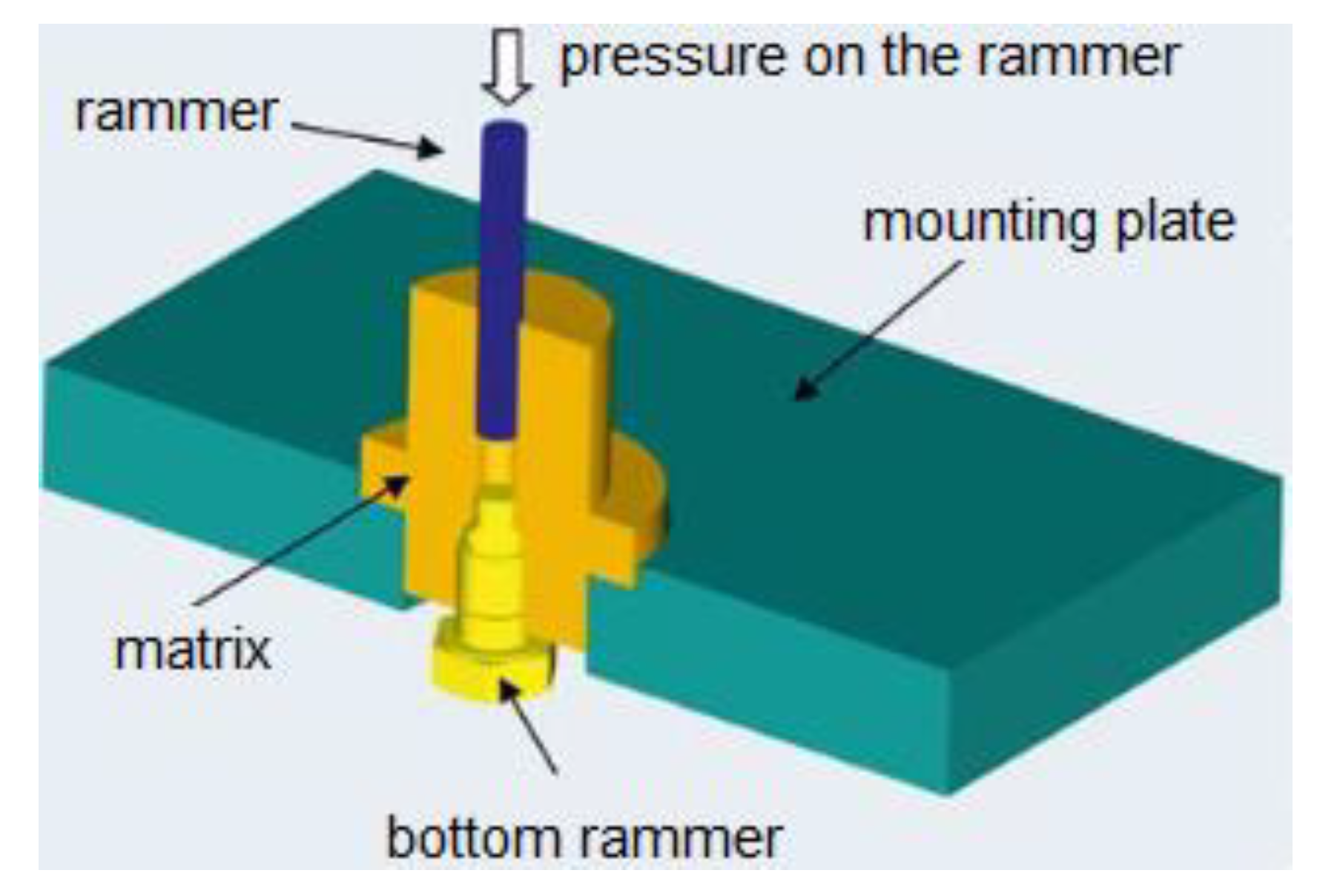
2.2. Investigation of Dissolution Rate of Oxide Coatings

In the second stage, the tests of the dissolution rate of rutile (titanium oxide) were performed in two simulated body fluids (SBF). The starting material was the titanium oxide powder (purity of 98.0%–100%) delivered by Acros Organics (Morris Plains, NJ, USA). Cylindrical samples, of dimensions 6 mm × 3 mm (diameter × length), were prepared using the classical powder metallurgy method without a filler. The material was formed in a single-axis pressing process using a force of 2 kN acting on the stamp for 60 s at the position shown in Figure 1.
The sintering process was carried out in a chamber oven (Type 22 MRT/1300, Conbest Ltd., Kraków, Poland) at 1300 °C for 2 h in an air atmosphere. The samples were heated at a rate 0.5 °C/min up to 100 °C and then at a rate 3 °C/min. Such two-stage heating significantly limited the appearance of cracks and delaminations were not observed.
Two simulated body fluids were used. Ringer’s solution was prepared based on commercial tablets (Ringer’s tablet, Merck, Germany) and artificial saliva according to the composition shown in Table 1. Hydrochloric acid (4M) (HCl) was used to prepare solutions of the appropriate pH values of 3, 5, and 7. The Elmetron CPI-505 pH meter was used to measure the pH values.
The samples were cleaned in an ultrasonic washer and immersed in the prepared solutions for 3 and 12 months. After this time the test solution samples were analyzed for total titanium content at the Centre of Biological and Chemical Sciences, the University of Warsaw (Warsaw, Poland). The inductively coupled plasma mass spectrometry (ICP-MS, Perkin Elmer, Inc., Waltham, MA, USA) was carried out by the norm E2371-13 to determine the titanium content in the solutions. The ions were separated using a special mass analyzer, distributing the ions according to the value of their mass-to-charge ratio. The ICP-MS was calibrated using an external calibration curve, which was prepared using 1% nitric acid and a titanium pattern. The quadrupole mass spectrometer, Elan 9000 Perkin Elmer ICP-MS, with conjugated plasma induction excitation was used for the study.
The solutions were mineralized before measurements in a closed microwave system. Approximately 1 g of the solution and 1 mL of 30% nitric acid were used for mineralization. The samples were then diluted to 15 mL.

2.3. Calculations of Diffusion of Titanium Ions through Oxide Coatings

The last stage was the calculations of the theoretical diffusion rate of titanium atoms through the rutile crystalline structure. The calculations were based on Arrhenius and Fick’s laws [38] and the earlier high-temperature measurements [39,40].

3. Results

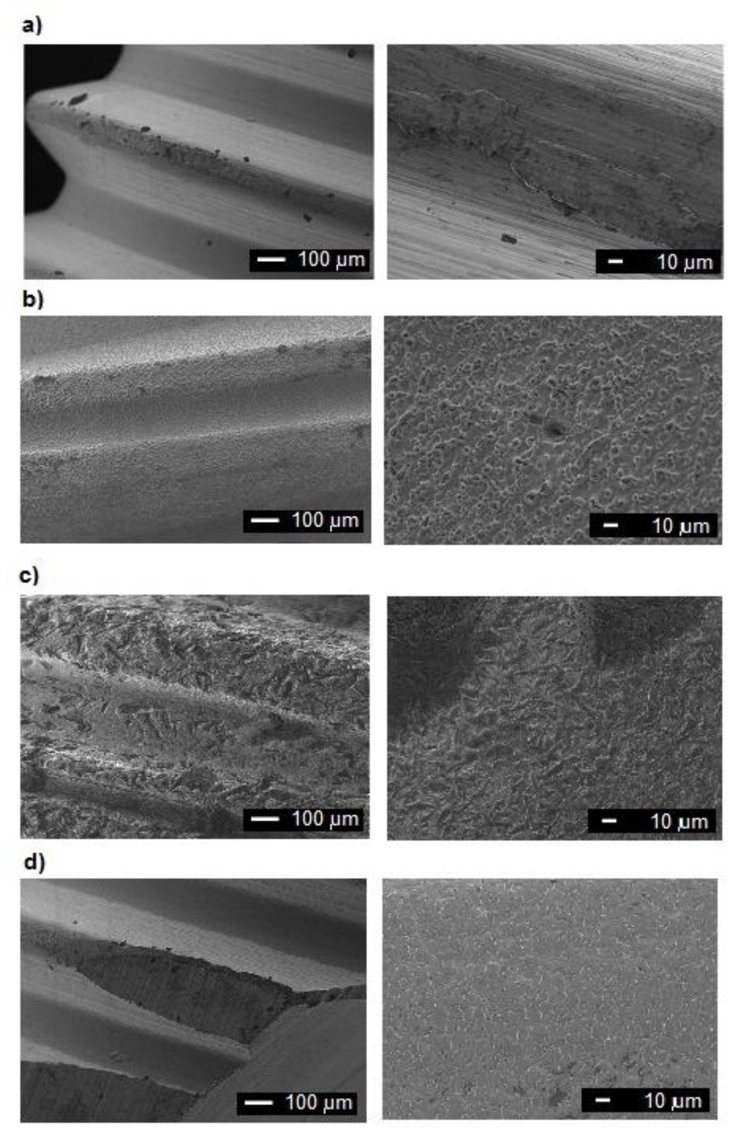
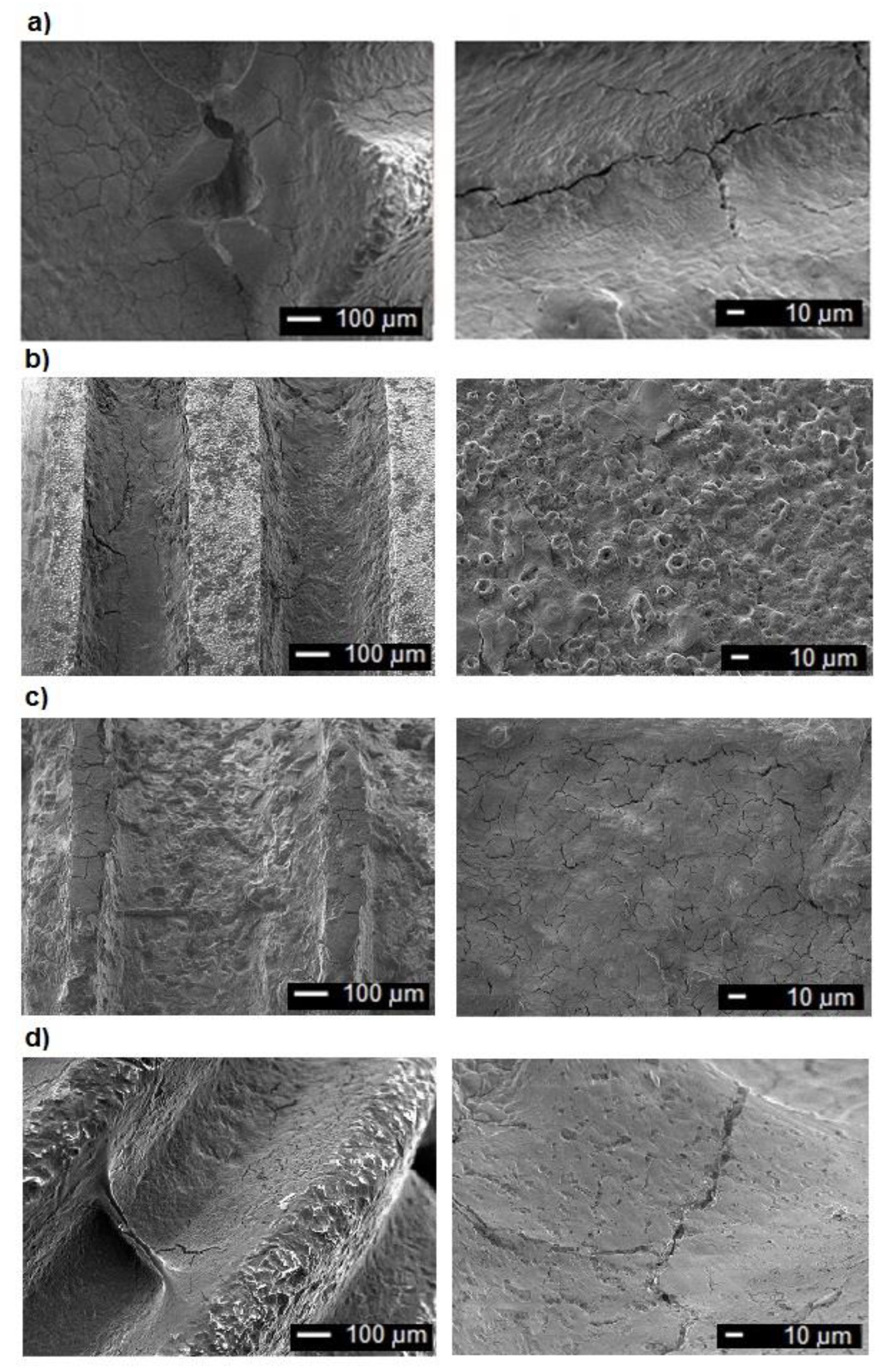
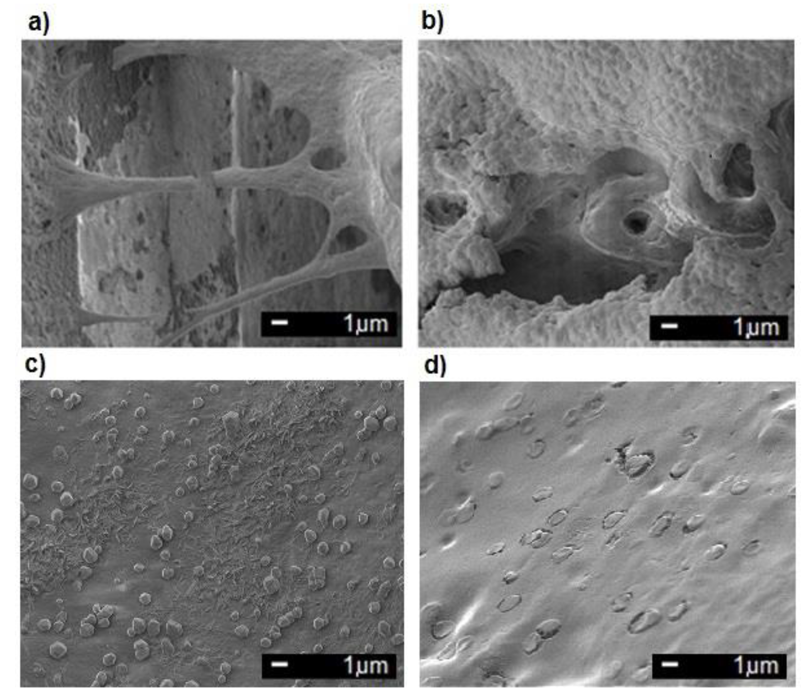
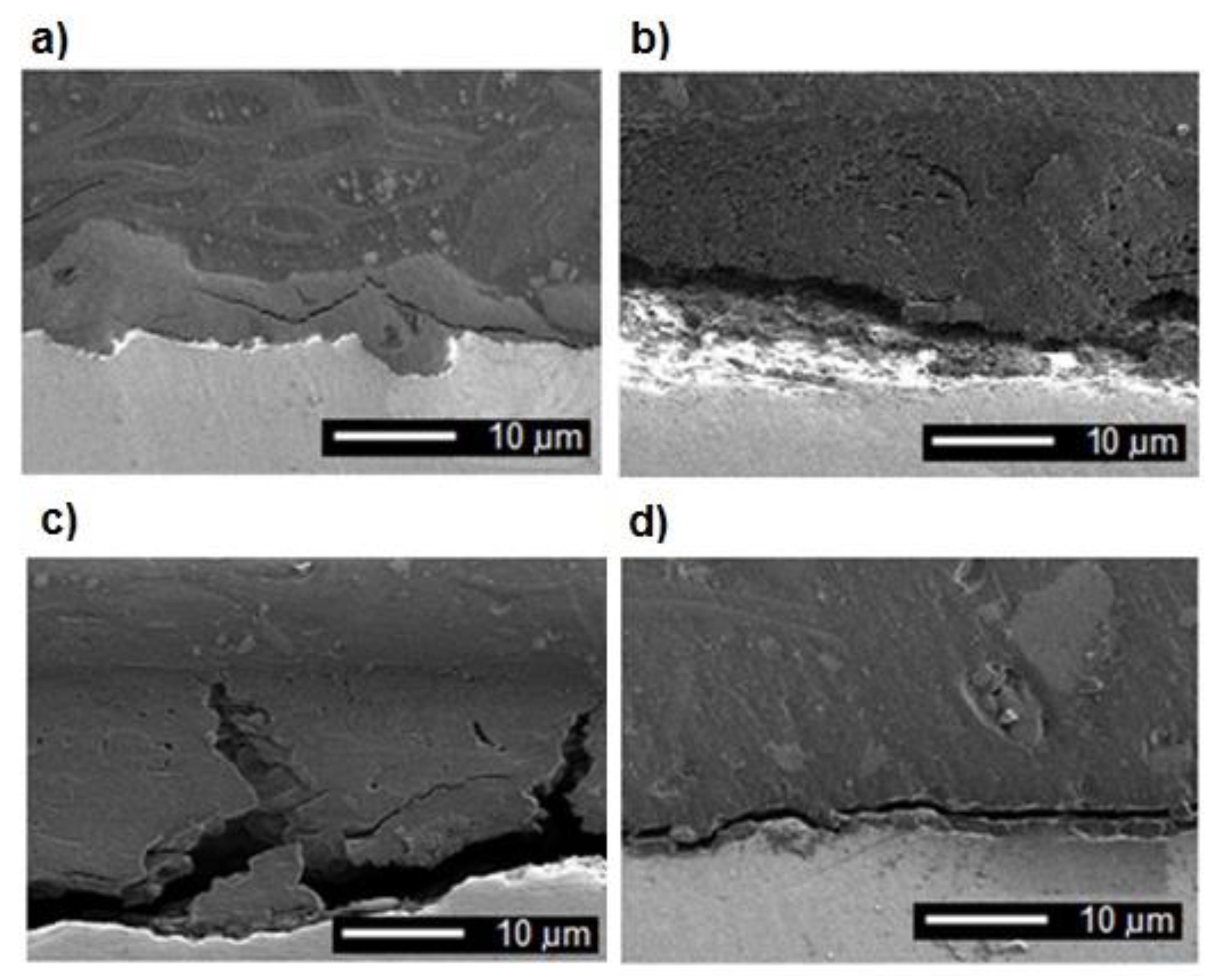
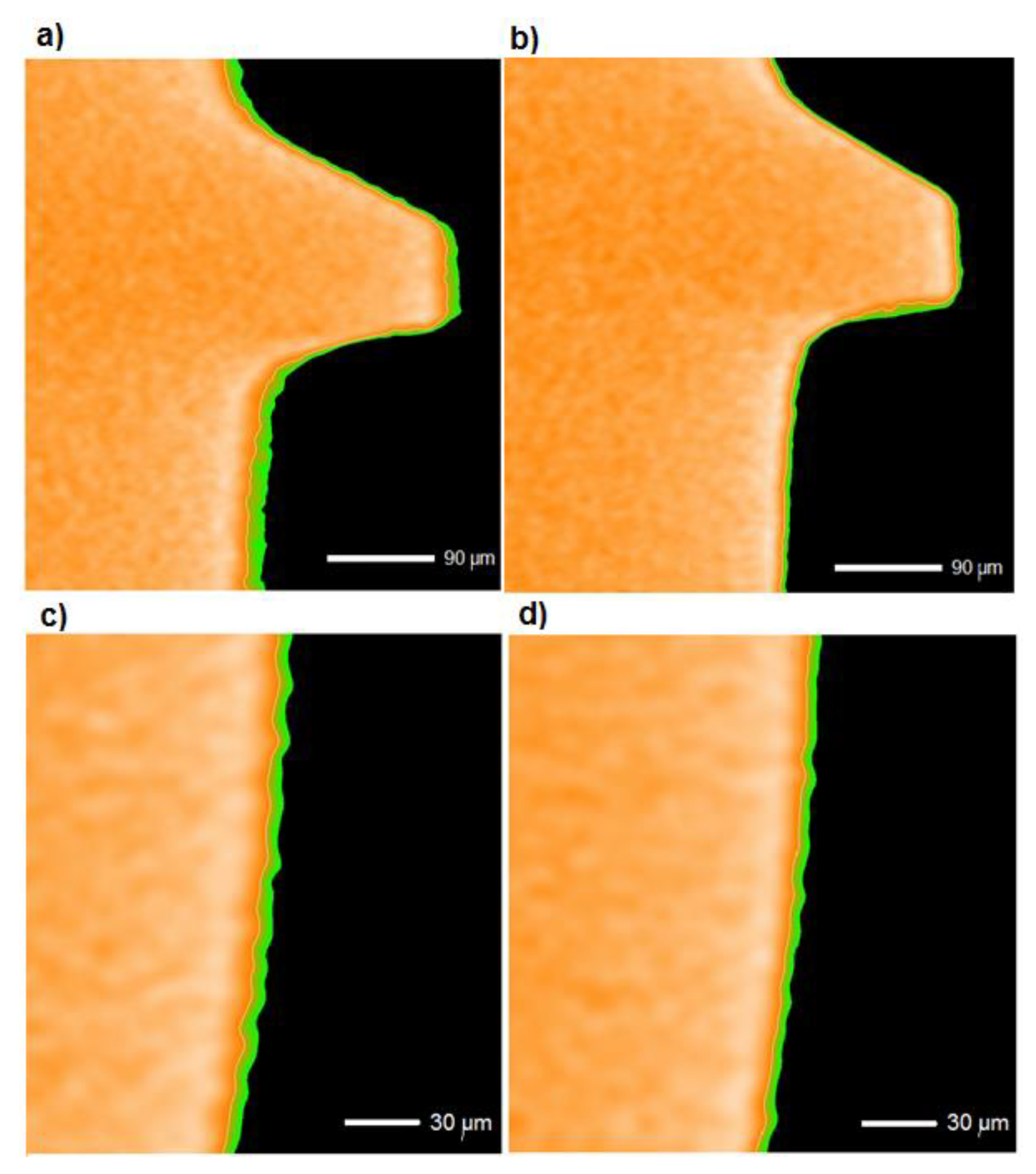
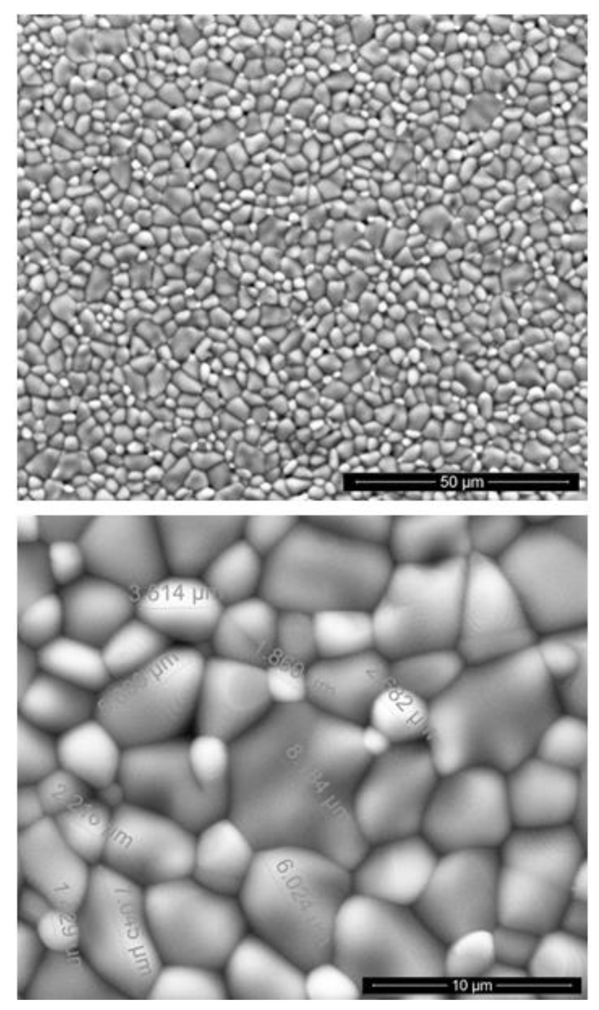
3.1. Examinations of Implants

Figure 2 presents the surfaces of different three new implants, produced by two companies, A and B. The layer discontinuities, material defects appearing in the layer, unevenness, numerous rolling scratches, and material allowances resulting from the surface treatment processes are visible before implantation.
The used implants were obtained from surgeons from the Warsaw Medical University. Only removed implants, among all, which demonstrated clear signs of damage, were selected. When examinations of the used implants (Figure 3) were made, two areas of surfaces could be distinguished. The first was the top of the threads, with characteristic flattened, rubbed bumps, with clearly visible pits on the surface. The second type was the bottom of the threads, in which there is a detachment of the material of the layer from the ground, and numerous deep cracks are visible likely arisen as a result of stress concentration, which are potential places of corrosion initiation and development of corrosion processes in the environment of body fluids and particularly aggressive saliva.
The surfaces of the used implants were subjected to purification or sterilization processes, thanks to which we can observe traces of organic residues on the surface—bacteria and tissues. Surprisingly, the largest clusters of bacterial colonies are located between the tip and the bottom of the thread, on the lateral surfaces. Perhaps this phenomenon is caused by adverse conditions at the tops and bottoms of the threads. Pits are visible on the surface of the thread tops, numerous and deep cracks in the thread cavities, which may indicate a significant impact of the environment and continuous operation of the implant—the influence of tensile forces and friction forces that affect the implant placed in the bone. On the implant surfaces, discontinuities of bone formation (Figure 4a,c) and bacterial colony residues (Figure 4b,d) can be observed, which tightly cover the material.
The implant cross-sections (Figure 5) illustrate surface unevenness, numerous discontinuities characterizing the layers produced, material stratification, a significant number of cracks of varying lengths, and arrangement occurring in the coating. They can be ideal for corrosion progress.
Using computed tomography, the layer thickness distribution on dental implants was depicted (Figure 6). The analysis shows that the thickness of the layers formed on the surface of the implants is diverse and does not evenly distribute. The surfaces of the vertices and thread cavities are characterized by a larger thickness of the coating. The images of the layer thickness distributions on the surface of the dental implants were very similar.

3.2. The Dissolution of Rutile

Knowing the characteristics and defects occurring in the layers covering dental implants, in the second stage of research, the determination of the rate of penetration of titanium ions into the solution was undertaken. Titanium oxide powder samples were prepared using powder metallurgy processes, which were immersed in Ringer’s solution and artificial saliva for a period of 3 and 12 months.
The surface of the samples produced, featuring a slight degree of porosity, is shown in Figure 7. The samples vary in grain size from 1.429 to 8.184 μm.
The analyses of the titanium ions carried out three months after their exposure to both SBFs at pH 3, 5, and 7 (Table 2), showed negligible solubility of the titanium dioxide, below the 0.080 mg/kg limit of determination. In solution adjusted to pH 3, titanium dissolution was distinctly higher, but only after 12 months, 0.093 mg/kg.
Slightly different results were achieved for the artificial saliva solution. The contents of titanium ions for artificial saliva of different pH were higher than those for Ringer’s solution and about 0.107 mg/kg for pH 5 and 7, while for pH 3 the titanium ion level exceeded 0.137 mg/kg.
These experiments show the importance of the pH value of body fluids and the possible dissolution and penetration of titanium ions into the human body. The increasing dissolutions of ferrous oxides [41] and copper oxides [42] with decreasing pH, and rutenium–titanium oxide coating at pH 2 [43] are in accordance with obtained results showing the important effect at the lowest pH value. The more distinct dissolution of rutile in artificial saliva is likely due to the higher chloride concentration in saliva than in blood, and the susceptibility of the oxides to the pitting. It is worth noting that the solutions used only simulated natural human body fluids and did not contain various biological substances such as enzymes, which can create an even more aggressive environment. The important conclusion, however, is that a titanium dental implant in the mouth is more susceptible to dissolution than in other tissues. The drastic lowering of the pH value of the solution accelerates the process of removal of titanium ions from rutile, always present on the titanium surface. Thus, changes in the pH value, occurring during some inflammatory reactions in the living organism, may significantly influence the condition of the oxide layer, and consequently the status of the implant.
In these long-term, expensive tests, we have analyzed the trends: the effect of decreasing pH and test solution on the dissolution rate. Taking into account the extremely low values of dissolubility, the precision of the spectrometric measurements, and the use of slightly porous materials, we have concluded that a significant number of specimens should be applied to obtain the homogenous sample and low standard deviations. Taking this into account, our purpose has been only to recognize at least the row of the magnitude of dissolubility and how the pH effects what has been reached. The results clearly show that the rutile ceramics dissolubility is extremely low and it decreases with decreasing pH, as it is for metallic substances.

3.3. Diffusion of Titanium Ions in The Oxide Layer

To calculate the distance of the diffusion of titanium ions in rutile lattice, data of two references were taken into account. In [40], the random tetravalent titanium atoms were assumed to be the predominant defects evident from self-diffusion. The enthalpy of motion was determined as ΔHm = 57.03% ± 4.9% kcal/mole. In another report [39], for diffusing the radio-isotope titanium-44 into single crystal rutile at temperatures in the range of 900 to 1300 °C, the activation energy was found to be 61,400 calories per mole and the frequency factor was calculated to be 6.4 × 10⁻⁴ m2/s. Assuming the diffusion enthalpy at 59.2 kcal/mol and D0 at the above value, the titanium diffusion coefficient at room temperature (293 K) was calculated at about 10−49 m2/s. That following, the diffusion distance at this temperature in one year is about 3 × 10⁻³⁴ nm.

4. Discussion

All new implants were made of the Ti6Al4V alloy by casting and milling (likely CNC). As a rule, such implants are assumed to have a perfect surface, at designed roughness achieved by mechanical treatment or chemical acidic (SLA implants) or alkaline treatment. Some of the commercial implants have deposited coatings (Osseotite and Nanotite implants). The detailed surface treatment is not disclosed. The majority of dental implants are likely subjected to micro-arc oxidation in phosphate solutions.
So far, the imperfections visible on new implants are attributed to the forces acting during implantation surgery. For example, the grooves and abraded facets, and loose titanium particles at the interface were reported for dental implants and attributed to the surgical procedure [44].
It is a damage that certainly locally destroys the titanium coatings. However, our investigations showed that several implants before any implantation possessed already imperfect surfaces with such damage forms of the oxide coating as the layer discontinuities (holes/pits), large unevenness, and rolling scratches. Such defects may initiate the local degradation of an implant, in particular the cracking and pitting corrosion.
The examinations of implants used for a relatively short time confirmed the above assumption, even indirectly. The pits are visible on the tops of the threads, and the detachment of the coating on the bottoms. The numerous deep cracks are likely arisen as a result of stress concentration, and can serve as potential places of corrosion initiation and development in the environment of body fluids and particularly aggressive saliva.
The traces of organic residues on the surface were between the tops and the bottoms of the threads. Perhaps this phenomenon is caused by adverse conditions at the tops and bottoms of the threads due to a significant impact of the environment and continuous influence of tensile and friction forces that affect the implant placed in the bone. The detailed mechanism of this phenomenon cannot, however, be proposed at the moment.
Three possible mechanisms of the release of titanium ions can be proposed as already shown. These results demonstrate that the most significant is corrosion initiation and propagation of corrosion in a presence of local damage of oxide coating and, on the other hand, complex stresses imposed on the screw implants. However, it is necessary to consider whether two other mechanisms can also operate and be comparable.
The dissolution of rutile may occur, but at an extremely low rate and only in strongly acidic environments. Such conditions may occur only in inflammation conditions at which pH may reach highly acidic values. Even if so, the dissolution rate achieves 0.136 mg/kg in 12 months, such results means that the oxide coating even 100 μm thick (after anodic oxidation) decreases less than 1 nm. Such a mechanism is then impossible and must be rejected.
The third mechanism, which can be considered, is diffusion of titanium ions through the rutile lattice. The diffusion of titanium at the temperature of a human body in the rutile crystalline structure seems unlikely. There is no such data even for high temperatures so that it seems desirable to consider the titanium diffusion in other structures. The performed calculations showed the titanium diffusion coefficient as extremely low, even below that for the diffusion of titanium in yttria-stabilized zirconia, the diffusion coefficient at room temperature is below 10−30 m2/s [45]. It means that the time necessary to diffuse through a 10 nm thick oxide layer would be as high as 1035 s.
Summarizing, it can be said that the only origin of the degradation processes resulting in, among other causes, in a necessity of removal of the dental implant, is the damage of the oxide coating. Such degradation may be attributed to the forces during implantation surgery, but they are likely initiated by the cracks, crevices, and discontinuities already appearing at the manufacturing stage.

5. Conclusions

The titanium dissolution occurs only by the corrosion tunnels in the oxide layer. The tunnels may be formed by cracks or discontinuities. Such potential corrosion initiation and development sites are already present in new implants and they become operative in applied implants during their use.
The present results demonstrate that among three possible mechanisms such as (i) diffusion of the liquid environment into the cracks and crevices in oxide coating; (ii) chemical dissolution of the titanium oxide layer; and (iii) diffusion of titanium atoms through the oxide layer, the two last processes are very unlikely to cause the damage of dental implants.
The both dissolution of rutile and titanium diffusion through the perfect oxide structure are negligible at the temperature of the human body. However, when the pH value at the implant surface and in the environment of saliva falls locally, the oxide layer starts to dissolve, but even at pH = 3, only a small fraction, 10−8 of the rutile oxide, may dissolve during 12 months.

Author Contributions

Methodology, A.O.; validation, A.O.; investigation, A.O.; resources, A.O.; original draft preparation, A.O.; formal analysis, A.Z.; writing—conceptualization, A.O. and A.Z.; writing—review & editing, A.O. and A.Z. All authors have read and agreed to the published version of the manuscript.

Funding

This research received no external funding.

Acknowledgments

We are grateful to Grzegorz Gajowiec (GUT) for his examinations of oxidized surfaces with the SEM, M.Eng. Gabriel Strugała for his examinations of oxidized surfaces with the CT, Eliza Kurek from Biological and Chemical Research Center University of Warsaw for the ICP-MS studies, and Andrzej Wojtowicz from the Department of Dental Surgery, the Medical University of Warsaw for delivery of the implants.

Conflicts of Interest

The authors declare no conflict of interest.

References

  1. Huang, H.H. Effects of fluoride concentration and elastic tensile strain on the corrosion resistance of commercially pure titanium. Biomaterials 2002, 23, 59–63. [Google Scholar] [CrossRef]
  2. Toumelin-Chemla, F.; Rouellet, F.; Burdairon, G. Corrosive properties of fluoride containing odontologic gels against titanium. J. Dent. 1996, 24, 109–115. [Google Scholar] [CrossRef]
  3. Vargas, E.; Baier, R.; Meyer, A. Reduce corrosion of cp Ti and Ti–6Al–4V alloy endosseous dental implant after glow discharge treatment: A preliminary report. Int. J. Oral Maxillofac. Implants 1992, 7, 338–344. [Google Scholar] [PubMed]
  4. Agarwal, A.; Tyagi, A.; Ahuja, A.; Kumar, N.; De, N.; Bhutani, H. Corrosion aspect of dental implants—An overview and literature review. Open J. Stomatol. 2014, 4, 56–60. [Google Scholar] [CrossRef] [Green Version]
  5. Lavos-Valereto, I.C.; Wolynec, S.; Ramires, I.; Guastaldi, A.C.; Costa, I. Electrochemical impedance spectroscopy characterization of passive film formed on implant Ti–6Al–7Nb alloy in Hank’s solution. J. Mater. Sci. Mater. Med. 2004, 15, 55–59. [Google Scholar] [CrossRef]
  6. González, J.E.G.; Mirza-Rosca, J.C. Study of the corrosion behavior of titanium alloys for biomedical and dental implants applications. J. Electroanal. Chem. 1999, 471, 109–115. [Google Scholar] [CrossRef]
  7. Cigada, A.; Cabrini, M.; Pedeferri, P. Increasing of the corrosion resistance of the Ti6Al4V alloy by high thickness anodic oxidation. J. Mat. Sci. Mat. Med. 1992, 3, 408–412. [Google Scholar] [CrossRef]
  8. Fathi, M.H.; Salehi, M.; Saatchi, A.; Mortazavi, V.; Moosavi, S.B. In vitro corrosion behavior of bioceramic, metallic, and bioceramic-metallic coated stainless steel dental implants. Dent. Mat. 2003, 19, 188–198. [Google Scholar] [CrossRef]
  9. Kasemo, B.; Lausmaa, J. Biomaterial and implant surfaces: A surface science approach. Int. J. Oral Maxillofac. Implants 1988, 3, 247–259. [Google Scholar]
  10. Aziz-Kerrzo, M.; Conroy, R.G.; Fenelon, A.M.; Farrell, S.T.; Breslin, C.B. Electrochemical studies on the stability and corrosion resistance of titanium-based implants materials. Biomaterials 2001, 22, 1531–1539. [Google Scholar] [CrossRef] [Green Version]
  11. Hanawa, T. Metals ions released from metal implants. Mat. Sci. Eng. C 2004, 24, 745–752. [Google Scholar] [CrossRef]
  12. Uo, M.; Watari, F.; Yokoyama, A.; Matsuno, H.; Kawasaki, T. Visualization and detectability of rarely contained elements in soft tissue by X-ray scanning analytical microscopy and electron probe micro analysis. Biomaterials 2001, 22, 1787–1794. [Google Scholar] [CrossRef]
  13. Przybyszewska-Doroś, I.; Okrój, W.; Walkowiak, B. Surface modifications of metallic implants. Eng. Biomater. 2005, 43–44, 52–62. [Google Scholar]
  14. De Sena, L.A.; Rocha, N.C.C.; Andrade, M.C.; Soares, G.A. Bioactivity assessment of titanium sheets electrochemically coated with thick oxide film. Surf. Coat. Technol. 2003, 166, 254–258. [Google Scholar] [CrossRef]
  15. Lavos-Valereto, I.C.; König, B.; Rossa, C., Jr.; Marcantonio, E.; Zavaglia, A.C. A study of historical responses from Ti–6Al–7Nb alloy dental implants with and without plasma-sprayed hydroxyapatite coatings in dogs. J. Mat. Sci. Mat. Med. 2001, 12, 273–276. [Google Scholar] [CrossRef] [PubMed]
  16. Yang, B.; Uchida, M.; Kim, H.-M.; Zhang, X.; Kokubo, T. Preparation of bioactive titanium metal via anodic oxidation treatment. Biomaterials 2004, 25, 1003–1010. [Google Scholar] [CrossRef]
  17. Zieliński, A.; Sobieszczyk, S. Corrosion of titanium biomaterials, mechanisms, effects and modelisation. Corros. Rev. 2008, 26, 1–22. [Google Scholar] [CrossRef]
  18. Long, M.; Rack, H.J. Titanium alloys in total joint replacement—A materials science perspective. Biomaterials 1998, 19, 1621–1639. [Google Scholar] [CrossRef]
  19. Veys-Renaux, D.; Ait El Haj, Z.; Rocca, E. Corrosion resistance in artificial saliva of titanium anodized by plasma electrolytic oxidation in Na3PO4. Surf. Coat. Techn. 2016, 285, 214–219. [Google Scholar] [CrossRef]
  20. Revathi, A.; Borrás, A.D.; Muñoz, A.I.; Richard, C.; Manivasagam, G. Degradation mechanisms and future challenges of titanium and its alloys for dental implant applications in oral environment. Mat. Sci. Eng. C 2017, 76, 1354–1368. [Google Scholar] [CrossRef]
  21. Strietzel, R.; Hösch, A.; Kalbfleisch, H.; Buch, D. In vitro corrosion of titanium. Biomaterials 1998, 19, 1495–1499. [Google Scholar] [CrossRef]
  22. Koike, M.; Fujii, H. The corrosion resistance of pure titanium in organic acids. Biomaterials 2001, 22, 2931–2936. [Google Scholar] [CrossRef]
  23. Wierzchoń, T.; Czarnowska, E.; Krupa, D. Surface Engineering in the Production of Titanium Biomaterials; Printing House PW: Warsaw, Poland, 2004. [Google Scholar]
  24. Haynes, D.R.; Rogers, S.D.; Hay, S. The differences in toxicity and release of bone-resorbing mediators induced by titanium and cobalt-chromium-alloy wear particles. J. Bone Jt. Surg. Am. 1993, 75, 825–834. [Google Scholar] [CrossRef] [Green Version]
  25. Wu, H.H.; Trinkle, D.R. Direct diffusion through interpenetrating networks: Oxygen in titanium. Phys. Rev. Lett. 2011, 107, 045504. [Google Scholar] [CrossRef] [Green Version]
  26. Sefer, B. Oxidation and Alpha-Case Phenomena in Titanium Alloys Used in Aerospace Industy: Ti-6Al-2Sn-4Zr-2Mo and Ti-6Al-4V. Bachelor’s Thesis, Luleå University of Technology, Luleå, Sweden, September 2014. [Google Scholar]
  27. Aerospace Series Test Method Titanium and Titanium Alloys Part 009-Determinantion of Surface Contaminantion; Swedish Standards Institute: Stockholm, Sweden, 2007.
  28. Hanawa, T.; Asami, K.; Asaoka, K. Repassivation of titanium and surface oxide film regenerated in simulated bioliquid. J. Biomed. Mat. Res. 1998, 40, 530–538. [Google Scholar] [CrossRef]
  29. Grant, D.M.; Lo, W.J.; Parker, K.H.; Parker, T.L. Biocompatible and mechanical properties of low temperature deposited quaternary (Ti, Al, V) N coatings on Ti6Al4V titanium alloy substrates. J. Mat. Sci. Mat. Med. 1996, 7, 576–584. [Google Scholar] [CrossRef]
  30. Li, J.; Jansen, J.A.; Walboomers, X.F.; van den Beucken, J.J.P. Mechanical aspects of dental implants and osseointegration: A narrative review. J. Mech. Behav. Biomed. Mat. 2020, 103, 103574. [Google Scholar] [CrossRef]
  31. Cheung, K.H.; Pabbruwe, M.B.; Chen, W.-F.; Koshy, P.; Sorrell, C.C. Effects of substrate preparation on TiO2 morphology and topography during anodization of biomedical Ti6Al4V. Mater. Chem. Phys. 2020, 252, 123224. [Google Scholar] [CrossRef]
  32. Kurup, A.; Dhatrak, P.; Khasnis, N. Surface modification techniques of titanium and titanium alloys for biomedical dental applications: A review. Mat. Today Proc. 2020. [Google Scholar] [CrossRef]
  33. Alves, A.C.; Wenger, F.; Ponthiaux, P.; Celis, J.-P.; Pinto, A.M.; Rocha, L.A.; Fernandes, J.C.S. Corrosion mechanisms in titanium oxide-based films produced by anodic treatment. Electrochim. Acta 2017, 234, 16–27. [Google Scholar] [CrossRef]
  34. Cui, W.F.; Jin, L.; Zhou, L. Surface characteristics and electrochemical corrosion behavior of a pre-anodized microarc oxidation coating on titanium alloy. Mater. Sci. Eng. C 2013, 33, 3775–3779. [Google Scholar] [CrossRef]
  35. Shokouhfar, M.; Allahkaram, S.R. Effect of incorporation of nanoparticles with different composition on wear and corrosion behavior of ceramic coatings developed on pure titanium by micro arc oxidation. Surf. Coat. Techn. 2017, 309, 767–778. [Google Scholar] [CrossRef]
  36. Sharma, A.; Mcquillan, A.J.; Sharma, L.A.; Waddell, J.; Shibata, Y.; Duncan, W. Spark anodization of titanium–zirconium alloy: Surface characterization and 216 bioactivity assessment. J. Mater. Sci. Mater. Med. 2015, 26, 1–11. [Google Scholar] [CrossRef]
  37. Ossowska, A.; Zieliński, A.; Supernak, M. Electrochemical oxidation and corrosion resistance of the Ti13Nb13Zr alloy. Eng. Biomat. 2013, XVI, 4–6. [Google Scholar]
  38. Callister, W.D. Materials Science and Engineering—An Introduction; Wiley: New York, NY, USA, 2007. [Google Scholar]
  39. Venkatu, D.A.; Poteat, L.E. Diffusion of titanium of single crystal rutile. Mater. Sci. Eng. 1970, 5, 258–262. [Google Scholar] [CrossRef]
  40. Akse, J.R.; Whitehurst, H.B. Diffusion of titanium in slightly reduced rutile. J. Phys. Chem. Solids 1978, 39, 457–465. [Google Scholar] [CrossRef]
  41. Schwertmann, U. Solubility and dissolution of iron oxides. Plant Soil 1991, 130, 1–25. [Google Scholar] [CrossRef]
  42. Ko, C.K.; Lee, W.G. Effects of pH variation in aqueous solutions on dissolution of copper oxide. Surf. Interface Anal. 2010, 42, 1128–1130. [Google Scholar] [CrossRef]
  43. Uzbekov, A.A.; Klement’eva, V.S. Radiochemical investigation of the selectivity of dissolution of components of ruthenium-titanium oxide anodes (ORTA) in chloride solutions. Sov. Electrochem. 1985, 21, 758–763. [Google Scholar]
  44. Streckbein, P.; Wilbrand, J.-F.; Kähling, C.; Pons-Kīühnemann, J.; Rehmann, P.; Wöstmann, B.; Howaldt, H.-P.; Möhlhenrich, S.C. Evaluation of the surface damage of dental implants caused by different surgical protocols: An in vitro study. Int. J. Oral Maxillofac. Surg. 2019, 48, 971–981. [Google Scholar] [CrossRef]
  45. Kowalski, K.; Bernasik, A.; Sadowski, A. Bulk and grain boundary diffusion of titanium in yttria-stabilized zirconia. J. Eur. Ceram. Soc. 2000, 20, 951–958. [Google Scholar] [CrossRef]
Figure 1. The cold pressing scheme for manufacturing the titanium dioxide specimens by powder metallurgy.
Coatings 10 00836 g001
Figure 2. Surfaces of new dental implants: (a) A company; (b) B company; (c) C company; (d) D company. SEM.
Coatings 10 00836 g002
Figure 3. Surfaces of used dental implants: (a) A company; (b) B company; (c) C company; (d) D company. SEM.
Coatings 10 00836 g003
Figure 4. Organic remains on the surfaces of used dental implants: (a) A company; (b) B company; (c) C company; (d) D company. SEM.
Coatings 10 00836 g004
Figure 5. Cross-sections of used dental implants: (a) A company; (b) B company; (c) C company; (d) D company. SEM.
Coatings 10 00836 g005
Figure 6. Images obtained by the computed tomography study of the sections: —longitudinal: (a) D company; (b) A company; —transverse dental implants: (c) D company; (d) A company.
Coatings 10 00836 g006
Figure 7. SEM microstructures of the sample surface after pressing with a force of 2 kN.
Coatings 10 00836 g007
Table 1. Chemical composition of the artificial saliva.
CompoundContent (g/L)
(NH2)2CO0.13
NaCl0.7
NaHCO31.5
Na3HPO40.26
K2HPO40.2
KSCN0.33
KCl1.2
Table 2. Titanium content according to ICP-MS analysis at different pH (mg/kg).
TimepHRinger’s SolutionArtificial Saliva
3 months3<GO *<GO *
5<GO *<GO *
7<GO *<GO *
12 months30.0930.136
5<GO *0.107
7<GO *0.107
* GO—Limit of identification of titanium 0.080 mg/kg.

