Review

Ibrexafungerp: A Novel Oral Triterpenoid Antifungal in Development for the Treatment of Candida auris Infections

1
Department of Dermatology, Case Western Reserve University, Cleveland, OH 44106, USA
2
Unit of Mycology, Statens Serum Institut, DK-2300 Copenhagen, Denmark
3
Department of Clinical Microbiology, Rigshospitalet, DK-2100 Copenhagen, Denmark
4
Department of Clinical Medicine, University of Copenhagen, DK-2200 Copenhagen, Denmark
5
New York State Department of Health Wadsworth Center, Albany, NY 12201, USA
6
Centers for Disease Control and Prevention, Atlanta, GA 30333, USA
7
Max Super Specialty Hospital, 110017 New Delhi, India
8
Scynexis Inc., 1 Evertrust Plaza, 13th Floor, Jersey City, NJ 07302, USA
9
Transplantation-Oncology Infectious Diseases Program, Weill Cornell Medicine of Cornell University, New York, NY 10065, USA
*
Author to whom correspondence should be addressed.
Antibiotics 2020, 9(9), 539; https://doi.org/10.3390/antibiotics9090539
Submission received: 31 July 2020 / Revised: 17 August 2020 / Accepted: 22 August 2020 / Published: 25 August 2020
(This article belongs to the Special Issue Diagnosis,Resistance and Treatment of Infections by Candida auris)

Abstract

:
Candida auris is an emerging multidrug-resistant fungal pathogen reported worldwide. Infections due to C. auris are usually nosocomial and associated with high rates of fluconazole resistance and mortality. Echinocandins are utilized as the first-line treatment. However, echinocandins are only available intravenously and are associated with increasingly higher rates of resistance by C. auris. Thus, a need exists for novel treatments that demonstrate potent activity against C. auris. Ibrexafungerp is a first-in-class triterpenoid antifungal agent. Similar to echinocandins, ibrexafungerp inhibits (1→3)-β-D-glucan synthase, a key component of the fungal cell wall, resulting in fungicidal activity against Candida spp. Ibrexafungerp demonstrates broad in vitro activity against various Candida spp. including C. auris and C. auris isolates with fks mutations. Minimum inhibitory concentration (MIC50 and MIC90) values in >400 C. auris isolates were 0.5 μg/mL and 1.0 μg/mL, respectively. Clinical results were reported for two patients with invasive candidiasis or candidemia due to C. auris treated during the CARES (Candidiasis Caused by Candida Auris) trial, an ongoing open-label study. These patients experienced a complete response after treatment with ibrexafungerp. Thus, ibrexafungerp represents a promising new antifungal agent for treating C. auris infections.

1. Introduction

Candida auris is an emerging fungal pathogen reported on all continents except Antarctica, in at least 39 countries worldwide [1], as well as in 20 states of the United States [2,3,4,5]. Five distinct clades of C. auris were identified with well-defined geographic distributions (South America, Africa, South Asia, East Asia, and West Asia), as well as antifungal resistance patterns and mechanisms that are both distinct and unique [1,6,7,8]. Infections due to C. auris are most often nosocomial, with easy transmission from patient-to-environment and environment-to-patient [6,9]. Patients heavily colonized with C. auris on the skin or mucosal surfaces can contaminate their surroundings, thereby contributing to transmission of C. auris in healthcare facilities. An additional challenge with C. auris is that the organism is exceedingly difficult to eradicate from the environment because of resistance to some standard disinfectants [6,9,10].
Timely and accurate diagnosis of invasive candidiasis are important for early initiation of antifungal therapy, while species identification is critical to ensure implementation of infection control measures [1,6,10,11]. Acceptable standard diagnostic methods for C. auris identification include matrix-assisted laser desorption ionization–time of flight (MALDI-TOF) and VITEK2™ with the appropriate updated databases and DNA sequencing [7,12].
Infections caused by C. auris are associated with severe illness, most often in hospitalized patients. Risk factors for C. auris infection include prior exposure to antibiotics or antifungal agents, diabetes mellitus, abdominal or vascular surgery, central venous or urinary catheters, chronic kidney disease, chemotherapy, blood transfusions, immunosuppression, and intensive care unit admission [6,7,10,11]. Invasive infections due to C. auris are associated with high mortality rates (30–78%), and multidrug resistance may play a role in mortality [6,7,10,13].
An outbreak of infections due to C. auris was identified in New York healthcare facilities with high rates of mortality [13]. Since 2016, more than 1000 C. auris isolates were tested at the New York State Department of Health where rates of resistance were >99% with fluconazole, approximately 60% with amphotericin B, and >80% with voriconazole [14,15]. The in vitro efficacy of antifungal drug combinations was evaluated against these resistant C. auris isolates, where combinations of flucytosine with echinocandins or amphotericin B were most active [14].
The epidemiology of C. auris was examined from isolates obtained from 54 patients collected from six countries outside the US [16]. Patient information was available for 41 (76%) isolates. At the time of C. auris detection, 41% of patients had diabetes mellitus, 51% had undergone recent surgery, 73% had a central venous catheter, and 41% were receiving systemic antifungal therapy. From hospital admission to confirmed infection with C. auris the median time was 19 days, 61% of patients had bloodstream infection, and 59% of patients died. Although breakpoints are not established for C. auris against any antifungal drugs, tentative breakpoints were established by the CDC (Centers for Disease Control) based on minimum inhibitory concentration (MIC) distribution, molecular mechanisms of resistance, and PK/PD (Pharmacokinetic/Pharmacodynamic) values in a mouse model of infection. Based on these preliminary breakpoints, the authors determined that 93% of the isolates were resistant to fluconazole, 35% to amphotericin B, and 7% to echinocandins; 22 (41%) isolates were resistant to at least two antifungal classes, including two (4%) that were resistant to fluconazole, voriconazole, echinocandins, and amphotericin B.
As suggested in the IDSA (Infectious Diseases Society of America) guidelines for the treatment of Candida infections and given the high frequency of resistance to fluconazole and polyenes, echinocandins are typically utilized as the first-line treatment for patients with C. auris infections. Although resistance is clone and clade specific, recent reports indicate that global resistance of C. auris to fluconazole may approach 90%; this is increasing with other antifungal agents, including echinocandins [7,9,10,11,17]. Antifungal resistance with C. auris is acquired rather than intrinsic, and the primary mechanisms of resistance were characterized for echinocandins and azoles [8]. Multidrug-resistant and pan-resistant isolates of C. auris were also identified from clinical isolates [15,16,18]. Thus, a need exists for novel antifungal agents that demonstrate high levels of activity against C. auris and address these treatment gaps. There is also need for effective infection control practices as well as treatment strategies that minimize the risk of nosocomial transmission associated with persistent C. auris colonization to control its spread.

2. Ibrexafungerp

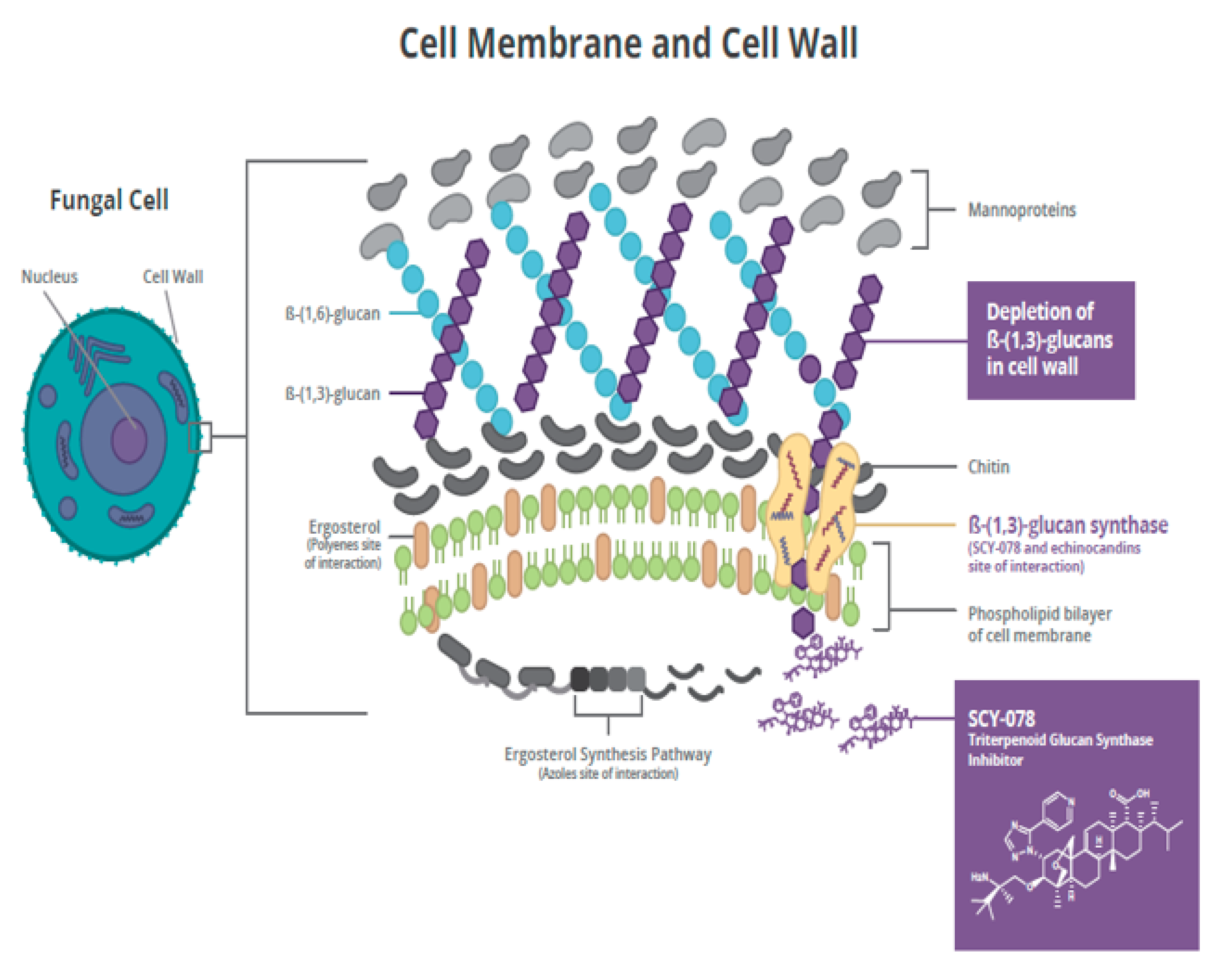
2.1. Mechanism of Action

Ibrexafungerp (formerly SCY-078) is the first compound of the enfumafungin-derived triterpenoid class of (1→3)-β-D-glucan synthase inhibitors (GSIs) (Figure 1). Glucan synthase inhibitors were first introduced for the treatment of invasive Candida infections in 2001, with caspofungin the first echinocandin to be approved [19]. This mechanism of action, i.e., blockade of the biosynthesis of ß-(1,3)-D-glucan in the fungal cell wall, was associated with potent and broad-spectrum antifungal activity and clinical efficacy for the treatment of fungal infections. Two additional echinocandins were later introduced, micafungin and anidulafungin. However, echinocandins lack clinically meaningful oral bioavailability, triggering the search for new molecules that shared the glucan synthase inhibition mechanism of action with echinocandins and could also be administered orally. Natural screening efforts led to the identification of enfumafungin derivatives as candidates, and subsequent synthetic modifications to these molecules resulted in increased oral bioavailability, potency, and stability, thereby leading to the discovery of ibrexafungerp.
Ibrexafungerp is being developed as the first oral and IV GSI (Intravenous glucan synthase inhibitor) for the treatment and prevention of fungal infections, including serious and life-threatening infections due to Candida spp., Aspergillus spp., and Pneumocystis jirovecii, with the potential to provide the therapeutic advantages of both IV and oral formulations [20]. Ibrexafungerp causes a decrease in (1→3)-β-D-glucan polymers and a weakening of the fungal cell wall [21]. Ibrexafungerp is structurally distinct from echinocandins and interacts differently with the target enzyme (Figure 2) [22]. Although the binding site on (1→3)-β-D-glucan synthase for ibrexafungerp partially overlaps with a binding site for echinocandins, it appears to be nonidentical, resulting in a lower rate of resistance to ibrexafungerp [22]. In in vitro studies, ibrexafungerp activity against wild-type and echinocandin-resistant strains of Candida spp. in the presence of fks mutations was minimally affected [23]. Thus, ibrexafungerp has limited potential for cross-resistance with echinocandins.

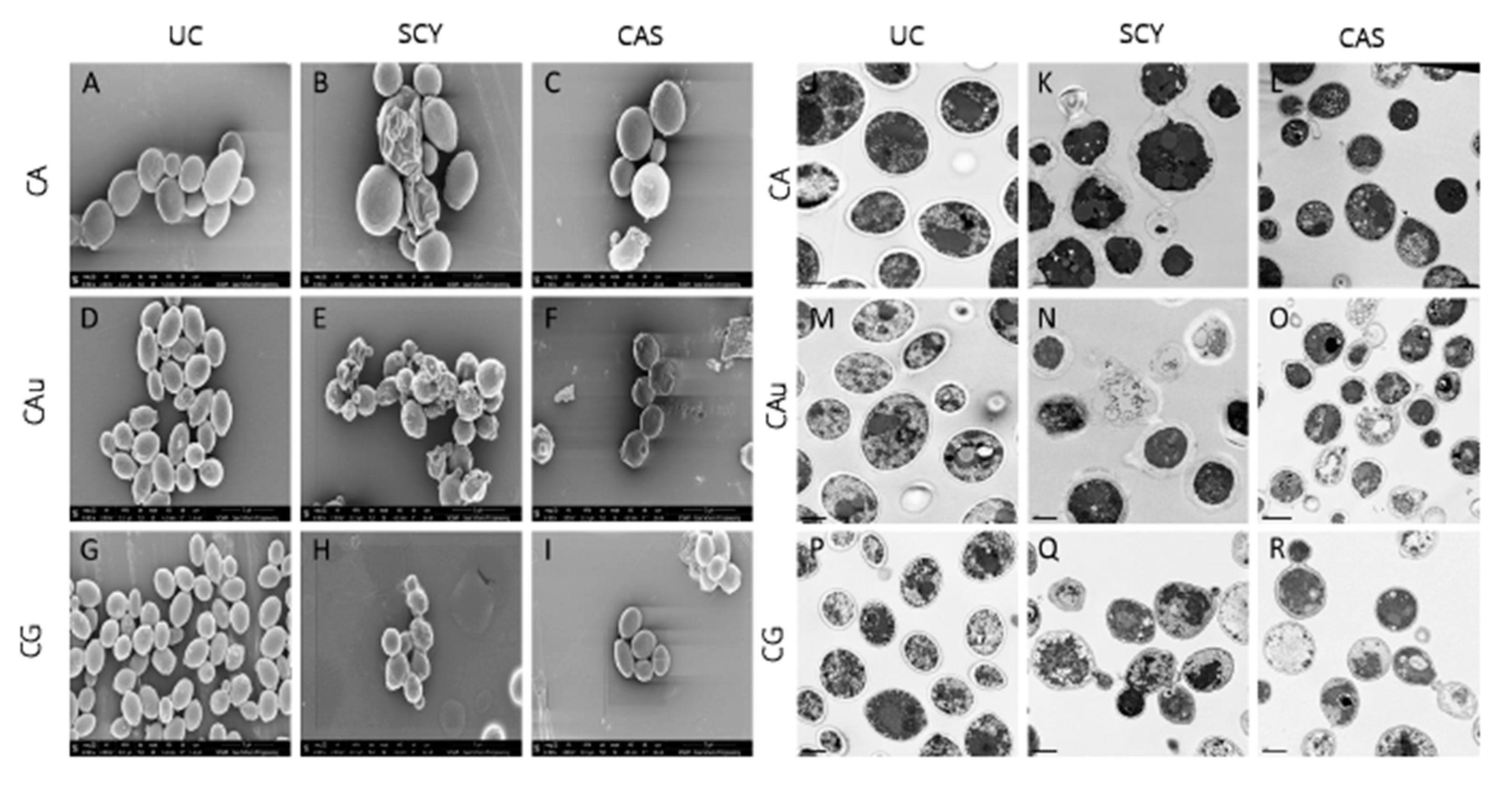
2.2. In Vitro and In Vivo Activity

Ibrexafungerp demonstrates broad in vitro activity against a range of Aspergillus spp. isolates and Candida isolates, including C. glabrata and C. auris, which exhibit fks1 and fks2 point mutations associated with resistance to echinocandin antifungals [23,24,25,26,27,28,29,30,31]. Among Candida species with reduced fluconazole susceptibility, including C. glabrata, C. krusei, C. tropicalis, and C. parapsilosis, MIC50 ranges with ibrexafungerp were 0.125–1 μg/mL, 0.5–1 μg/mL, <0.03–1 μg/mL, and 0.25–1 μg/mL, respectively. Additionally, as reported by Zhu [32] using isolates obtained from New York patients, the in vitro activity against C. auris of ibrexafungerp (ranging from 0.05 to 0.5 μg/mL) was superior to that of fluconazole (ranging from 2 to >256 μg/mL), and comparable or superior to that of echinocandins (ranging from 0.015 to 16 μg/mL). This observation was confirmed by other studies using global strains [28,33,34]. Ibrexafungerp showed a wild-type MIC distribution against ~80% of echinocandin-resistant Candida spp. isolates tested, suggesting that fks mutations have less of an effect on the in vitro activity of ibrexafungerp [23,35].
Ibrexafungerp demonstrates potent fungicidal activity against Candida spp. [31,36,37,38]. In a time-kill study, ibrexafungerp demonstrated a ≥3-log reduction in colony forming units (CFUs)/mL) at 24 h. Caspofungin demonstrated fungicidal activity similar to ibrexafungerp, but fluconazole and voriconazole were fungistatic [31]. Ibrexafungerp was shown to have activity against biofilms from different Candida species [28]. Consistent with clinical trials in treatment of vulvovaginal candidiasis, ibrexafungerp showed potent in vitro activity in the lower pH environment of vulvovaginitis [39].

2.3. Pharmacokinetics

After oral administration in dog, mouse, and rat, ibrexafungerp is absorbed rapidly from the gastrointestinal (GI) tract, with bioavailability of approximately 35–50% [40]. In animals, ibrexafungerp is widely distributed in tissues with a steady-state volume of distribution (Vdss) of >5 L/kg [41], which is several-fold greater than fluconazole and echinocandins [38]. Higher Vdss results in higher tissue-to-plasma concentration ratios could be beneficial for specific fungal infections, such as those located in the skin, liver, spleen, mucosa, bone, and lung, to mention some. Ibrexafungerp undergoes extensive metabolism by cytochrome P450 3A4 isoenzymes in the liver, and <2% of a dose is recovered unchanged in urine [42]. After single oral doses of 10 mg to 1600 mg in healthy subjects, peak ibrexafungerp plasma concentrations were reached after four to six hours, with a mean terminal half-life of approximately 20 to 30 h [43]. Mean area under the concentration-time curve (AUC0-∞) and peak concentration (Cmax) were dose-proportional across this dosage range. The proarrhythmic potential of ibrexafungerp was evaluated in animal models and healthy volunteers using cardiodynamic assessments [44]. Ibrexafungerp exhibited no clinically relevant effects on heart rate or PR and QRS intervals. No clinically meaningful effect of ibrexafungerp on the QTcF interval was observed at plasma concentrations up to 4000 ng/mL after IV administration in healthy subjects.
Results from in vitro studies indicated that ibrexafungerp is a substrate of CYP3A4 and a potential inhibitor of cytochrome (CYP) 2C8 [42,45]. In healthy subjects, the interaction potentials of ketoconazole and diltiazem (CYP3A4 inhibitors), rosiglitazone (CYP2C8 substrate), and tacrolimus (CYP3A4 substrate) were evaluated after single or multiple doses co-administered with ibrexafungerp [46]. No clinically relevant effects of ibrexafungerp on CYP2C8 inhibition or CYP3A4 substrates were observed, although a dosage adjustment for ibrexafungerp may be needed when co-administering with potent CYP3A4 inhibitors. Thus, ibrexafungerp shows low potential for CYP-mediated drug interactions at therapeutic exposures.

2.4. Clinical Development

Nineteen Phase 1, three Phase 2, and two Phase 3 clinical studies were completed with ibrexafungerp, the latter two (VANISH-303 and VANISH-306), in women with vulvovaginal candidiasis (VVC) (https://clinicaltrials.gov: NCT03734991 and NCT03987620). In both Phase 3 studies, the rate of clinical cure (complete resolution of all vaginal signs and symptoms at Day 10) and mycological eradication were significantly greater with ibrexafungerp than placebo. The most common adverse events reported with ibrexafungerp were mild gastrointestinal disturbances when administered to >1200 patients and healthy subjects.
Currently, ibrexafungerp is undergoing further clinical development in ongoing studies, including CANDLE-304 (clinicaltrials.gov: NCT04029116), a Phase 3 study in women with recurrent VVC, SCYNERGIA, a Phase 2 study of ibrexafungerp combined with voriconazole in patients with invasive aspergillosis (clinicaltrials.gov: NCT03672292), FURI, an open-label study in patients with refractory invasive fungal infections (clinicaltrials.gov: NCT02244606), and CARES (clinicaltrials.gov: NCT03363841), an open-label, emergency protocol study of patients with invasive infections due to C. auris.

3. Ibrexafungerp for Candida auris

3.1. In Vitro Activity

The in vitro activity of ibrexafungerp was tested against 16 C. auris clinical isolates obtained from Germany, Japan, India, and South Korea [27]. The MIC90 for ibrexafungerp was 1 μg/mL. Fluconazole and amphotericin B exhibited less in vitro activity against C. auris with MIC90 values of >64 and 4 μg/mL, respectively, while the MIC90 values for anidulafungin, caspofungin, and micafungin were 0.25 μg/mL, 1 μg/mL, and 1 μg/mL, respectively.
The in vitro activity of ibrexafungerp was evaluated against a global collection of 100 isolates of C. auris representing each of the four clades of C. auris known at that time [24]. MICs for ibrexafungerp ranged from 0.0625 to 2 μg/mL, with an MIC50 of 0.5 μg/mL and MIC90 1 μg/mL. MIC values for anidulafungin, caspofungin, and micafungin ranged from 0.03 up to >16 μg/mL. Among seven C. auris isolates exhibiting elevated MIC values for echinocandins, the ibrexafungerp MIC ranged from 0.5 to 1.0 μg/mL.
Ibrexafungerp and six comparator antifungal agents were evaluated against 122 C. auris isolates [33]. The MIC range for ibrexafungerp was 0.06 to 2.0 μg/mL. A wide distribution of MIC values was reported for anidulafungin and micafungin, ranging from 0.016 to >32 and 0.03 to >32 μg/mL, respectively (Table 1). All but one C. auris isolate were resistant to fluconazole. Out of 122 isolates, 8 displayed high MIC values for echinocandins associated with fks mutations (S639F Fks1 alteration). The MIC for ibrexafungerp for these eight resistant isolates ranged from 0.25 to 0.5 μg/mL.
Among 102 C. auris isolates with variable resistance to amphotericin B, flucytosine, azoles, and echinocandins, the ibrexafungerp MIC50 for 97 isolates ranged from 0.06–0.5 μg/mL, and the median and mode MIC were both 0.5 μg/mL [32]. Ibrexafungerp also showed activity against five C. auris isolates considered to be pan-resistant, with a low MIC50 range of 0.12 to 1 μg/mL.
Data were compiled from four studies reporting the in vitro activity of ibrexafungerp against 445 C. auris clinical isolates [47]. Most isolates were obtained from the United States and India and included 32 isolates with increased MIC values to echinocandins. The MIC50 and MIC90 for ibrexafungerp across all isolates tested were 0.5 μg/mL and 1.0 μg/mL, respectively (Table 2). Among 32 C. auris isolates with echinocandin resistance, MIC values for ibrexafungerp ranged from 0.5 μg/mL to 1.0 μg/mL. One isolate displayed high MIC values for echinocandins and showed reduced sensitivity (>2 dilutions vs. the mode) to ibrexafungerp, and this isolate exhibited elevated MIC values to anidulafungin, caspofungin, and micafungin (MIC = 1 μg/mL), luconazole (MIC > 256 μg/mL), and amphotericin B (MIC = 1 μg/mL). Thus, ibrexafungerp exhibits in vitro activity against a broad collection of C. auris isolates, including most echinocandin-resistant isolates.
The ability of Candida species to form biofilms is associated with catheter and device-related infections and may play a role in C. auris infections considering that many affected individuals are in intensive care units with intravascular lines. In this regard, 97% of patients infected with C. auris had central venous catheters (Sayeed et al., 2019) [48], and a retrospective analysis demonstrated significantly higher use of central venous catheters in patients infected with this multidrug- resistant Candida [49]. The activity of ibrexafungerp against C. auris biofilms was evaluated [27]. Following 48 h of incubation, metabolic activities of biofilms were measured. Images and thicknesses of biofilms growing in the presence or absence of a drug were captured using confocal scanning laser microscopy. Quantitation of the metabolic activity of C. auris biofilms was performed using a biochemical assay, the 2,3-bis(2-methoxy-4-nitro-5-sulfophenyl)-5-[(phenylamino) carbonyl]-2H-tetrazolium hydroxide (XTT) reduction assay, as described previously [27,50]. Ibrexafungerp demonstrated activity against C. auris biofilms by reducing biofilm thickness and metabolic activity.
The effects of ibrexafungerp and caspofungin on the morphology of C. albicans, C. auris, and C. glabrata were studied using scanning and transmission electron microscopy [51]. When evaluated at respective MIC50 levels, ibrexafungerp exhibited a profound effect on cellular morphology in caspofungin-resistant organisms, possibly indicative of a difference in target engagement between ibrexafungerp and echinocandins (Figure 3). Untreated control C. auris cells showed well-defined, oval-shaped yeast morphology, as well as several budding yeasts. In contrast, cells exposed to ibrexafungerp (at a concentration of 1 µL MIC) exhibited a severely distorted yeast cell topography, including cell collapse, deformed cellular appearance, irregular budding, and cells that were fused together and unable to undergo cell division [27].

3.2. In Vivo Activity

The in vivo efficacy of ibrexafungerp for C. auris was evaluated in a disseminated murine mouse model [37]. Immunocompromised mice were randomized to ibrexafungerp 10, 20, or 30 mg/kg twice daily (BID) vs. a vehicle given by oral gavage. At Day 7, the fungal burden in kidney tissue was reduced by all doses of ibrexafungerp, with a significant difference for the 30 mg/kg dose vs. vehicle. At Day 14, survival rates were 60–70% with ibrexafungerp vs. 20% with vehicle control. Exposures in mice dosed with ibrexafungerp 10, 20, or 30 mg/kg BID were consistent with steady-state plasma exposure (AUC0–24) of 8.4, 24.3, and 40.2 ug*h/mL, respectively. These results demonstrate potent antifungal activity of ibrexafungerp against C. auris.
C. auris colonization is a major problem in hospitals and long-term care facilities. In order to understand the ability of ibrexafungerp to potentially decolonize the skin of C. auris, a study was performed looking at the in vivo efficacy of ibrexafungerp in a cutaneous infection model in Guinea pigs [36]. Animals were treated with ibrexafungerp 10, 20, or 30 mg/kg BID by oral gavage, micafungin 5 mg/kg once daily IP, or vehicle by oral gavage, and prednisone 30 mg/kg SC was given one day before and three days after infection. Tissue burden at Day 7 was lower with all active treatments vs. vehicle. Animals dosed with ibrexafungerp 10, 20, or 30 mg/kg BID showed systemic exposures (AUC0–24) of 2.8, 5.6, and 15 ug*h/mL. Examination of Periodic Acid-Schiff (PAS)-stained skin sections revealed that sections obtained from untreated control animals showed yeast cells, demonstrating that the skin was infected with C. auris. In contrast, examination of multiple skin sections obtained from animals treated with either ibrexafungerp or micafungin did not reveal yeast cells at any of the dose levels tested, indicating that the C. auris infection was cleared. There was no significant difference in clinical scores between the treatment groups [36]. Thus, no fungal elements were observed with ibrexafungerp or micafungin from histological examination.

3.3. Clinical Experience

CARES is an open-label study of oral ibrexafungerp in patients with documented candidiasis or candidemia due to C. auris who were treatment naïve or refractory to or intolerant of standard-of-care antifungal agents (clinicaltrials.gov: NCT03363841). Patients were treated with oral ibrexafungerp 750 mg twice daily for two days, then 750 mg once daily for up to 90 days.
In the first two patients from CARES with candidemia due to C. auris, a complete response after 17 and 22 days of treatment was reported with ibrexafungerp [52]. The first patient was a 58-year-old male admitted to the ICU with pneumonia and septic shock. Antibiotics were given together with empiric IV fluconazole. When C. auris was isolated from blood cultures, antifungal therapy was switched to IV micafungin. However, blood cultures remained positive for C. auris after five days, and the patient was switched to ibrexafungerp for 17 days. Subsequent blood cultures at Day 3 of ibrexafungerp therapy were negative for C. auris, and the patient was considered to have a complete response at the end of therapy. Ibrexafungerp-related adverse events were mild loose stools from days two through four of therapy.
The second patient was a 64-year-old female admitted to the hospital with pneumonia, fever, and hypotension. When C. auris was isolated from blood cultures, ibrexafungerp was initiated. A blood culture collected on Day 3 of ibrexafungerp therapy remained positive for C. auris and subsequent cultures at Days 9 and 21 were reported negative. The patient improved clinically, received ibrexafungerp for 22 days and was considered a complete response at the end of therapy. No ibrexafungerp-related adverse events were reported.

3.4. Echinocandin Resistance and C. auris

For echinocandins, the primary mechanism of resistance in C. auris species comprises the fks1 and fks2 genes, where mutations of the S639F, S639P, and S639Y amino acid sequences were identified as the cause of elevated MICs to echinocandins [9]. Among 350 C. auris isolates from India, 2% were echinocandin-resistant due to the fks1 mutation expressing the S639F sequence [53]. A similar finding was reported from Kuwait, where 3 (1.0%) of 314 C. auris isolates were echinocandin-resistant due to the fks1 mutation expressing the S639F sequence [54]. Four additional C. auris isolates from a total of 106 isolates were resistant to all tested echinocandins (MIC ≥ 4 μg/mL) and contained an S639F mutation in fks1 [55].
Biagi et al. [56] reported a patient with recurrent candidemia due to C. auris that was echinocandin-resistant but azole-sensitive, who expressed the fks1 mutation for the S639P sequence. A single C. auris isolate was identified in the UK that displayed 5-flucytosine and echinocandin resistance; echinocandin resistance was due to fks1 mutation for the S639Y sequence [57].
Among C. auris isolates from India, 8 of 122 with the S639F sequence were echinocandin-resistant with MICs of 4–32 μg/mL; the ibrexafungerp MIC values for these same isolates ranged from 0.25–0.5 μg/mL [33].
Ostrowski and colleagues [18] described three cases of C. auris among 801 patients in New York state with confirmed C. auris that were pan-resistant, i.e., resistant to fluconazole, amphotericin B, and echinocandins. All three patients with pan-resistance were on mechanical ventilators; two died within two to four weeks from isolation of a pan-resistant C. auris, and the third died at 10 months. MICs for fluconazole were >256 μg/mL, 2 μg/mL for amphotericin B, and 2–16 μg/mL for echinocandins. From the same laboratory, an analysis of the susceptibility of five pan-resistant C. auris isolates, defined as in vitro resistance to more than two azoles, all echinocandins, and amphotericin B, reported MIC values to fluconazole (>256 μg/mL), amphotericin B (2 μg/mL), and echinocandins (ranging from 2 to >16 μg/mL) [15]. However, all these pan-resistant isolates exhibited MICs for ibrexafungerp ranging from 0.12 to 1 μg/mL, which were within the wild-type MIC range reported for C. auris.

4. Summary and Conclusions

In the past decade, C. auris has emerged as a critical public health concern because of its persistence in the environment, contagious nature, and high morbidity and mortality. Unfortunately, rates of resistance to antifungal drugs among C. auris isolates continue to increase with almost universal resistance to fluconazole and growing resistance to other azoles and antifungals, including echinocandins. New antifungal agents in clinical development may provide more effective treatment options to address the growing impact of C. auris. Ibrexafungerp offers the advantages of oral administration, a favorable PK profile, a well-characterized safety/tolerability profile in >1200 patients/subjects, a low risk for cross-resistance to echinocandins, and consistently potent in vitro activity against C. auris, including echinocandin-resistant isolates. Animal studies looking at the tissue distribution of ibrexafungerp achieved high concentrations in the skin, an attribute that may be of importance to limit C. auris skin colonization, with a potential impact of limiting transmission. Ibrexafungerp demonstrates activity across a range of invasive fungal diseases as a monotherapy but also in combination [58,59]. Early clinical evidence from an emergency-use Phase 3 study of ibrexafungerp for invasive candidiasis due to C. auris is promising. Additional preclinical and clinical evidence are awaited to confirm the role of ibrexafungerp in treating infections due to C. auris, as well as other causes of invasive fungal disease.

Author Contributions

All authors were involved in the conception, review, and revision for intellectual content. All authors have read and agreed to the published version of the manuscript.

Funding

Support for this work was provided by Scynexis, Inc., Jersey City, NJ, USA.

Acknowledgments

Richard Perry and Tom Chen provided editorial assistance in the finalization and submission of this manuscript funded by Scynexis, Inc., Jersey City, NJ. This manuscript represents a contribution from New York City Candida auris Research and Support (NYC CARES).

Conflicts of Interest

N.A. and D.A. are employees of and may hold stock in Scynexis, Inc., Jersey City, NJ. T.J.W. received grants for experimental and clinical antimicrobial pharmacology and therapeutics to his institution from Allergan, Amplyx, Astellas, Lediant, Medicines Company, Merck, Scynexis, Tetraphase, and Viosera and has served as consultant to Amplyx, Astellas, Allergan, ContraFect, Gilead, Lediant, Medicines Company, Merck, Methylgene, Pfizer, and Scynexis. V.P.C. received service fees from SCYNEXIS, Inc.; there are no other disclosures. M.C.A., over the past five years, received research grants/contract work (paid to the SSI) from Amplyx, Basilea, Cidara, F2G, Gilead, Novabiotics, Scynexis, and T2Biosystems, and speaker honoraria (personal fee) from Astellas, Gilead, MSD, and SEGES. She is the current chairman of the EUCAST-AFST.

Disclaimer

The findings and conclusions in this manuscript are those of the authors and do not necessarily represent the official position of the Centers for Disease Control and Prevention.

References

  1. Wickes, B.L. Analysis of a Candida auris Outbreak Provides New Insights into an Emerging Pathogen. J. Clin. Microbiol. 2020, 58. [Google Scholar] [CrossRef] [PubMed] [Green Version]
  2. Centers for Disease Control and Prevention. Tracking Candida Auris. Available online: https://www.cdc.gov/fungal/candida-auris/tracking-c-auris.html (accessed on 30 June 2020).
  3. Clancy, C.J.; Nguyen, M.H. Emergence of Candida auris: An International Call to Arms. Clin. Infect. Dis. 2016, 64, 141–143. [Google Scholar] [CrossRef] [Green Version]
  4. Chowdhary, A.; Sharma, C.; Meis, J.F. Candida auris: A rapidly emerging cause of hospital-acquired multidrug-resistant fungal infections globally. PLOS Pathog. 2017, 13, e1006290. [Google Scholar] [CrossRef] [PubMed]
  5. Zhu, Y.; O’Brien, B.; Leach, L.; Clarke, A.; Bates, M.; Adams, E.; Ostrowsky, B.; Quinn, M.; Dufort, E.; Southwick, K.; et al. Laboratory Analysis of an Outbreak of Candida auris in New York from 2016 to 2018: Impact and Lessons Learned. J. Clin. Microbiol. 2019, 58. [Google Scholar] [CrossRef] [PubMed] [Green Version]
  6. Sabino, R.F.P.; Veríssimo, C.; Pereira, Á.A.; Antunes, F. Candida auris, An Agent of Hospital-Associated Outbreaks: Which Challenging Issues Do We Need to Have in Mind? Microorganisms 2020, 8, 181. [Google Scholar] [CrossRef] [Green Version]
  7. Ademe, M.; Girma, F. Candida auris: From Multidrug Resistance to Pan-Resistant Strains. Infect. Drug Resist. 2020, 13, 1287–1294. [Google Scholar] [CrossRef]
  8. Lockhart, S.R. Candida auris and multidrug resistance: Defining the new normal. Fungal Genet. Boil. 2019, 131, 103243. [Google Scholar] [CrossRef]
  9. Chaabane, F.; Graf, A.; Jequier, L.; Coste, A.T. Review on Antifungal Resistance Mechanisms in the Emerging Pathogen Candida auris. Front. Microbiol. 2019, 10, 2788. [Google Scholar] [CrossRef]
  10. Forsberg, K.; Woodworth, K.; Walters, M.; Berkow, E.L.; Jackson, B.; Chiller, T.; Vallabhaneni, S. Candida auris: The recent emergence of a multidrug-resistant fungal pathogen. Med. Mycol. 2018, 57, 1–12. [Google Scholar] [CrossRef] [Green Version]
  11. Cortegiani, A.; Misseri, G.; Giarratano, A.; Bassetti, M.; Eyre, D. The global challenge of Candida auris in the intensive care unit. Crit. Care 2019, 23, 150. [Google Scholar] [CrossRef] [Green Version]
  12. Caceres, D.H.; Forsberg, K.; Welsh, R.M.; Sexton, D.J.; Lockhart, S.R.; Jackson, B.R.; Chiller, T. Candida auris: A Review of Recommendations for Detection and Control in Healthcare Settings. J. Fungi 2019, 5, 111. [Google Scholar] [CrossRef] [PubMed] [Green Version]
  13. Adams, E.; Quinn, M.; Tsay, S.; Poirot, E.; Chaturvedi, S.; Southwick, K.; Greenko, J.; Fernandez, R.; Kallen, A.; Vallabhaneni, S.; et al. Candida auris in Healthcare Facilities, New York, NY, USA, 2013–2017. Emerg. Infect. Dis. 2018, 24, 1816–1824. [Google Scholar] [CrossRef] [PubMed] [Green Version]
  14. O’Brien, B.; Chaturvedi, S.; Chaturvedi, V. In Vitro Evaluation of Antifungal Drug Combinations against Multidrug-Resistant Candida auris Isolates from New York Outbreak. Antimicrob. Agents Chemother. 2020, 64, e02195-19. [Google Scholar] [CrossRef] [PubMed]
  15. Zhu, Y.C.; Barat, S.A.; Borroto-Esoda, K.; Angulo, D.; Chaturvedi, S.; Chaturvedi, V. Pan-resistant Candida auris isolates from the outbreak in New York are susceptible to ibrexafungerp (a glucan synthase inhibitor). Int. J. Antimicrob. Agents 2020, 55, 105922. [Google Scholar] [CrossRef]
  16. Lockhart, S.R.; Etienne, K.A.; Vallabhaneni, S.; Farooqi, J.; Chowdhary, A.; Govender, N.P.; Colombo, A.L.; Calvo, B.; Cuomo, C.; Desjardins, C.A.; et al. Simultaneous Emergence of Multidrug-Resistant Candida auris on 3 Continents Confirmed by Whole-Genome Sequencing and Epidemiological Analyses. Clin. Infect. Dis. 2017, 64, 134–140. [Google Scholar] [CrossRef] [Green Version]
  17. Arensman, K.; Miller, J.L.; Chiang, A.; Mai, N.; Levato, J.; Lachance, E.; Anderson, M.; Beganovic, M.; Dela-Pena, J. Clinical Outcomes of Patients Treated for Candida auris Infections in a Multisite Health System, IL, USA. Emerg. Infect. Dis. 2020, 26, 876–880. [Google Scholar] [CrossRef]
  18. Ostrowsky, B.; Greenko, J.; Adams, E.; Quinn, M.; O’Brien, B.; Chaturvedi, V.; Berkow, E.; Vallabhaneni, S.; Forsberg, K.; Chaturvedi, S.; et al. Candida auris Isolates Resistant to Three Classes of Antifungal Medications—New York. MMWR Morb. Mortal. Wkly. Rep. 2020, 69, 6–9. [Google Scholar] [CrossRef] [Green Version]
  19. Heasley, B.; Pacofsky, G.J.; Mamai, A.; Liu, H.; Nelson, K.; Coti, G.; Peel, M.R.; Balkovec, J.M.; Greenlee, M.L.; Liberator, P.; et al. Synthesis and biological evaluation of antifungal derivatives of enfumafungin as orally bioavailable inhibitors of β-1,3-glucan synthase. Bioorg. Med. Chem. Lett. 2012, 22, 6811–6816. [Google Scholar] [CrossRef]
  20. Davis, M.R.; Donnelley, M.A.; Thompson, G.R. Ibrexafungerp: A novel oral glucan synthase inhibitor. Med. Mycol. 2020, 58, 579–592. [Google Scholar] [CrossRef]
  21. Pfaller, M.A.; Messer, S.A.; Motyl, M.R.; Jones, R.N.; Castanheira, M. In Vitro Activity of a New Oral Glucan Synthase Inhibitor (MK-3118) Tested against Aspergillus spp. by CLSI and EUCAST Broth Microdilution Methods. Antimicrob. Agents Chemother. 2013, 57, 1065–1068. [Google Scholar] [CrossRef] [Green Version]
  22. Jiménez-Ortigosa, C.; Perez, W.B.; Angulo, D.; Borroto-Esoda, K.; Perlin, D.S. De Novo Acquisition of Resistance to SCY-078 in Candida glabrata Involves FKS Mutations That both Overlap and Are Distinct from Those Conferring Echinocandin Resistance. Antimicrob. Agents Chemother. 2017, 61, e00833-17. [Google Scholar] [CrossRef] [PubMed] [Green Version]
  23. Pfaller, M.A.; Messer, S.A.; Rhomberg, P.R.; Borroto-Esoda, K.; Castanheira, M. Differential Activity of the Oral Glucan Synthase Inhibitor SCY-078 against Wild-Type and Echinocandin-Resistant Strains of Candida Species. Antimicrob. Agents Chemother. 2017, 61, e00161-17. [Google Scholar] [CrossRef] [PubMed] [Green Version]
  24. Berkow, E.L.; Angulo, D.; Lockhart, S.R. In Vitro Activity of a Novel Glucan Synthase Inhibitor, SCY-078, against Clinical Isolates of Candida auris. Antimicrob. Agents Chemother. 2017, 61, e00435-17. [Google Scholar] [CrossRef] [PubMed] [Green Version]
  25. Ghannoum, M.; Long, L.; Isham, N.; Hager, C.; Wilson, R.; Borroto-Esoda, K.; Barat, S.; Angulo, D. Activity of a Novel 1,3-Beta-D-Glucan Synthase Inhibitor, Ibrexafungerp (Formerly SCY-078), against Candida glabrata. Antimicrob. Agents Chemother. 2019, 63, AAC.01510-19. [Google Scholar] [CrossRef]
  26. Ghannoum, M.; Long, L.; Larkin, E.L.; Isham, N.; Sherif, R.; Borroto-Esoda, K.; Barat, S.; Angulo, D. Evaluation of the Antifungal Activity of the Novel Oral Glucan Synthase Inhibitor SCY-078, Singly and in Combination, for the Treatment of Invasive Aspergillosis. Antimicrob. Agents Chemother. 2018, 62, e00244-18. [Google Scholar] [CrossRef] [Green Version]
  27. Larkin, E.L.; Hager, C.; Chandra, J.; Mukherjee, P.K.; Retuerto, M.; Salem, I.; Long, L.; Isham, N.; Kovanda, L.; Borroto-Esoda, K.; et al. The Emerging Pathogen Candida auris: Growth Phenotype, Virulence Factors, Activity of Antifungals, and Effect of SCY-078, a Novel Glucan Synthesis Inhibitor, on Growth Morphology and Biofilm Formation. Antimicrob. Agents Chemother. 2017, 61, e02396-16. [Google Scholar] [CrossRef] [Green Version]
  28. Marcos-Zambrano, L.J.; Gómez-Perosanz, M.; Escribano, P.; Bouza, E.; Guinea, J. The novel oral glucan synthase inhibitor SCY-078 shows in vitro activity against sessile and planktonic Candida spp. J. Antimicrob. Chemother. 2017, 72, 1969–1976. [Google Scholar] [CrossRef] [Green Version]
  29. Nunnally, N.S.; Etienne, K.A.; Angulo, D.; Lockhart, S.R.; Berkow, E.L. In Vitro Activity of Ibrexafungerp, a Novel Glucan Synthase Inhibitor against Candida glabrata Isolates with FKS Mutations. Antimicrob. Agents Chemother. 2019, 63, E01692-19. [Google Scholar] [CrossRef] [Green Version]
  30. Rautermaa-Richardson, R.; Moore, C.B.; Rawson, K. Aspergillus isolates from patients with chronic pulmonary aspergillosis mycologically and clinically resistant to azole antifungals are sensitive to ibrexafungerp (SCY-078). Presented at the 9th Congress on Trends in Medical Mycology, Nice, France, 11–14 October 2019. [Google Scholar]
  31. Scorneaux, B.; Angulo, D.; Borroto-Esoda, K.; Ghannoum, M.; Peel, M.; Wring, S.A. SCY-078 Is Fungicidal against Candida Species in Time-Kill Studies. Antimicrob. Agents Chemother. 2017, 61, e01961-16. [Google Scholar] [CrossRef] [Green Version]
  32. Zhu, Y.C.; Barat, S.A.; Borroto-Esoda, K.; Angulo, D.; Chaturvedi, S.; Chaturvedi, V. In vitro Efficacy of Novel Glucan Synthase Inhibitor, Ibrexafungerp (SCY-078), Against Multidrug- and Pan-resistant Candida auris Isolates from the Outbreak in New York. BioRxiv 2019. [Google Scholar] [CrossRef] [Green Version]
  33. Arendrup, M.; Jørgensen, K.M.; Hare, R.K.; Chowdhary, A. In Vitro Activity of Ibrexafungerp (SCY-078) against Candida auris Isolates as Determined by EUCAST Methodology and Comparison with Activity against C. albicans and C. glabrata and with the Activities of Six Comparator Agents. Antimicrob. Agents Chemother. 2020, 64, e02136-19. [Google Scholar] [CrossRef] [PubMed]
  34. Barat, S.; Borroto-Esoda, A.D. Ibrexafungerp (SCY-078): A first-in-class, orally-bioavailable, glucan synthase inhibitor has fungicidal activity against C. auris, an emerging multidrug-resistant pathogen. Presented at the 2018 Mycoses Study Group Education and Research Consortium, Big Sky, MT, USA, 25–28 September 2018. [Google Scholar]
  35. Wiederhold, N.P.; Najvar, L.K.; Jaramillo, R.; Olivo, M.; Pizzini, J.; Catano, G.; Patterson, T.F. Oral glucan synthase inhibitor SCY-078 is effective in an experimental murine model of invasive candidiasis caused by WT and echinocandin-resistant Candida glabrata. J. Antimicrob. Chemother. 2018, 73, 448–451. [Google Scholar] [CrossRef] [PubMed]
  36. Ghannoum, M.; Isham, N.; Angulo, D.; Borroto-Esoda, K.; Barat, S.; Long, L. Efficacy of Ibrexafungerp (SCY-078) Against Candida auris in an In Vivo Guinea Pig Cutaneous Infection Model. Antimicrob. Agents Chemother. 2020, AAC.00854-20. [Google Scholar] [CrossRef] [PubMed]
  37. Ghannoum, M.; Long, L.; Hager, C.; Borroto-Esoda, K.; Barat, S.; Angulo, D. Efficacy of oral ibrexafungerp (IBX, Formerly SCY-078) in the treatment of Candida auris infection in a murine disseminated model. ASM Microbe 2019. [Google Scholar] [CrossRef]
  38. Lepak, A.J.; Marchillo, K.; Andes, D.R. Pharmacodynamic Target Evaluation of a Novel Oral Glucan Synthase Inhibitor, SCY-078 (MK-3118), Using anIn VivoMurine Invasive Candidiasis Model. Antimicrob. Agents Chemother. 2015, 59, 1265–1272. [Google Scholar] [CrossRef] [PubMed] [Green Version]
  39. Larkin, E.L.; Long, L.; Isham, N.; Borroto-Esoda, K.; Barat, S.; Angulo, D.; Wring, S.; Ghannoum, M. A Novel 1,3-Beta-D-Glucan Inhibitor, Ibrexafungerp (Formerly SCY-078), Shows Potent Activity in the Lower pH Environment of Vulvovaginitis. Antimicrob. Agents Chemother. 2019, 63, e02611-18. [Google Scholar] [CrossRef] [Green Version]
  40. Wring, S.A.; Randolph, R.; Park, S.; Abruzzo, G.; Chen, Q.; Flattery, A.; Garrett, G.; Peel, M.; Outcalt, R.; Powell, K.; et al. Preclinical Pharmacokinetics and Pharmacodynamic Target of SCY-078, a First-in-Class Orally Active Antifungal Glucan Synthesis Inhibitor, in Murine Models of Disseminated Candidiasis. Antimicrob. Agents Chemother. 2017, 61, e02068-16. [Google Scholar] [CrossRef] [Green Version]
  41. Wring, S.; Borroto-Esoda, K.; Solon, E.; Angulo, D. SCY-078, a Novel Fungicidal Agent, Demonstrates Distribution to Tissues Associated with Fungal Infections during Mass Balance Studies with Intravenous and Oral [14C]SCY-078 in Albino and Pigmented Rats. Antimicrob. Agents Chemother. 2019, 63, e02119-18. [Google Scholar] [CrossRef] [Green Version]
  42. Wring, S.; Murphy, G.; Atiee, G.; Corr, C.; Hyman, M.; Willett, M.; Angulo, D. Clinical Pharmacokinetics and Drug-Drug Interaction Potential for Coadministered SCY-078, an Oral Fungicidal Glucan Synthase Inhibitor, and Tacrolimus. Clin. Pharmacol. Drug Dev. 2018, 8, 60–69. [Google Scholar] [CrossRef] [Green Version]
  43. Trucksis, M.; Garrett, G.; Heirman, I. A phase I single rising dose study evaluating the safety, tolerability and pharmacokinetics of an oral glucan synthase inhibitor in healthy male volunteers. In Proceedings of the 50th Interscience Conference on Antimicrobial Agents and Chemotherapy (ICAAC), Boston, MA, USA, 12–15 September 2010. [Google Scholar]
  44. Murphy, G.; Darpo, B.; Marbury, T. Lack of an effect of SCY-078 a novel antifungal agent on QTc interval in healthy subjects. In Abstracts of ASM Microbe; ASM Press: Washington, DC, USA, 2017; Abstract 172. [Google Scholar]
  45. Wring, S.; Murphy, G.; Atiee, G.; Corr, C.; Hyman, M.; Willett, M.; Angulo, D. Lack of Impact by SCY-078, a First-in-Class Oral Fungicidal Glucan Synthase Inhibitor, on the Pharmacokinetics of Rosiglitazone, a Substrate for CYP450 2C8, Supports the Low Risk for Clinically Relevant Metabolic Drug-Drug Interactions. J. Clin. Pharmacol. 2018, 58, 1305–1313. [Google Scholar] [CrossRef]
  46. Murphy, G.; Hyman, M.; Willett, M. CYP-mediated drug interaction profile of SCY-078, a novel glucan synthase inhibitor (GSI). In Abstracts of ASM Microbe; ASM Press: Washington, DC, USA, 2017; Abstract 173. [Google Scholar]
  47. Barat, S.; Borroto-Esoda, A.D. Ibrexafungerp demonstrates potent and consistent in vitro activity against >400 global Candida auris isolates, including isolates with elevated MIC’s to echinocandins. In Proceedings of the ECCMID, Paris, France, 18–21 April 2020. [Google Scholar]
  48. Sayeed, M.A.; Farooqi, J.; Jabeen, K.; Mahmood, S.F. Comparison of risk factors and outcomes of Candida auris candidemia with non-Candida auris candidemia: A retrospective study from Pakistan. Med Mycol. 2019, 58, 721–729. [Google Scholar] [CrossRef] [PubMed]
  49. Akrabarti, A. Candida auris candidaemia in an intensive care ujit-prospective observational study to evaluate epidemiology, risk factors, and outcome. J. Crit. Care 2020, 57, 42–48. [Google Scholar] [CrossRef]
  50. Chandra, J.; Mukherjee, P.K.; Ghannoum, M.A. In vitro growth and analysis of Candida biofilms. Nat. Protoc. 2008, 3, 1909–1924. [Google Scholar] [CrossRef]
  51. Hager, C.L.; Sun, P.-L.; Fujioka, H. Morphological effect of SCY-078 and caspofungin on different caspofungin-resistant Candida species. Presented at ASM Microbe, Atlanta, GA, USA, 8–10 June 2018. [Google Scholar]
  52. Juneja, D.; Singh, O.; Tarai, B.; Angulo, D. Sucessful Treatment of Two patienTs with Candida Auris Candidemia with the Investigational Agent, Oral Ibrexafungerp (formerly SCY-078), from the CARES Study; European Congress of Clinical Microbiology and Infectious Diseases (ECCMID): Amsterdam, The Netherlands, 2019; Poster no. L0028. [Google Scholar]
  53. Chowdhary, A.; Prakash, A.; Sharma, C.; Kordalewska, M.; Kumar, A.; Sarma, S.; Tarai, B.; Singh, A.; Upadhyaya, G.; Upadhyay, S.; et al. A multicentre study of antifungal susceptibility patterns among 350 Candida auris isolates (2009-17) in India: Role of the ERG11 and FKS1 genes in azole and echinocandin resistance. J. Antimicrob. Chemother. 2018, 73, 891–899. [Google Scholar] [CrossRef] [PubMed]
  54. Ahmad, S.; Khan, Z.; Al-Sweih, N.; Alfouzan, W.; Joseph, L. Candida auris in various hospitals across Kuwait and their susceptibility and molecular basis of resistance to antifungal drugs. Mycoses 2020, 63, 104–112. [Google Scholar] [CrossRef]
  55. Kordalewska, M.; Lee, A.; Park, S.; Berrio, I.; Chowdhary, A.; Zhao, Y.; Perlin, D.S. Understanding Echinocandin Resistance in the Emerging Pathogen Candida auris. Antimicrob. Agents Chemother. 2018, 62, e00238-18. [Google Scholar] [CrossRef] [PubMed] [Green Version]
  56. Biagi, M.J.; Wiederhold, N.P.; Gibas, C.; Wickes, B.L.; Lozano, V.; Bleasdale, S.C.; Danziger, L. Development of High-Level Echinocandin Resistance in a Patient with Recurrent Candida auris Candidemia Secondary to Chronic Candiduria. Open Forum Infect. Dis. 2019, 6. [Google Scholar] [CrossRef]
  57. Rhodes, J.; Abdolrasouli, A.; Farrer, R.A. Genomic epidemiology of the UK outbreak of the emerging human fungal pathogen Candida auris. Emerg. Microbes Infect. 2018, 7, 43. [Google Scholar] [CrossRef] [PubMed] [Green Version]
  58. Petraitis, V.; Petraitiene, R.; Katragkou, A.; Maung, B.B.W.; Naing, E.; Kavaliauskas, P.; Barat, S.; Borroto-Esoda, K.; Azie, N.; Angulo, D.; et al. Combination Therapy with Ibrexafungerp (Formerly SCY-078), a First-in-Class Triterpenoid Inhibitor of (1→3)-β-D-Glucan Synthesis, and Isavuconazole for Treatment of Experimental Invasive Pulmonary Aspergillosis. Antimicrob. Agents Chemother. 2020, 64. [Google Scholar] [CrossRef]
  59. Spec, A.; Pullman, J.; Thompson, G.R., III; Powderly, W.G.; Tobin, E.H.; Vazquez, J.; Wring, S.A.; Angulo, D.; Helou, S.; Mycoses Study Group; et al. MSG-10: A Phase 2 study of oral ibrexafungerp (SCY-078) following initial echinocandin therapy in non-neutropenic patients with invasive candidiasis. J. Antimicrob. Chemother. 2019, 74, 3056–3062. [Google Scholar] [CrossRef] [Green Version]
Figure 1. Structure of ibrexafungerp.
Antibiotics 09 00539 g001
Figure 2. Mechanism of action for ibrexafungerp.
Antibiotics 09 00539 g002
Figure 3. SEM of CA UC (A), SCY treated CA (B), CAS treated CA (C), CAu UC (D), SCY treated CAu (E), CAS treated CAu (F), CG UC (G), SCY treated CG (H), CAS treated CG (I), and TEM of CA UC (J), SCY treated CA (K), CAS treated CA (L), CAu UC (M), SCY treated CAu (N), CAS treated CAu (O), CG UC (P), SCY treated CG (Q), and CAS treated CG (R). (Hager et al., 2018) [51].
Antibiotics 09 00539 g003
Table 1. In vitro activity of ibrexafungerp and comparators against C. auris isolates [33].
Drug (No. of Isolates)MIC50 aModal MICMIC Range
Ibrexafungerp (n = 122)0.50.50.06–2
Anidulafungin0.1250.060.016–>32
Micafungin0.1250.1250.03–>32
Amphotericin B110.5–1
Fluconazole≥64≥640.5–≥64
Voriconazole0.5Bimodal≤0.004–4
Isavuconazole0.125Trimodal≤0.004–2
a μg/mL; Ibrexafungerp minimum inhibitory concentration (MIC) values for eight isolates with S639F fks1 mutations ranged from 0.25 to 0.5 μg/mL.
Table 2. In vitro activity of ibrexafungerp against a compilation of 445 C. auris isolates [47].
ReferenceNo. of IsolatesMIC, μg/mL
MIC50MIC90ModeMIC Range
Berkow et al., 2017 [24]1071110.0625–2
Larkin et al., 2017 [27]161110.5–1
Zhu et al., 2020 [32]2000.510.50.0625–8
Arendrup et al., 2020 [33]1220.510.50.0625–2
Overall4450.510.50.625–8

